Recognition and identification of external branch of superior
laryngeal nerve and its implication IN THYROID SURGERY.
Dissertation submitted to THE TAMILNADU
DR. M.G.R. MEDICAL UNIVERSITY CHENNAI–600032
With fulfillment of the Regulations For the Award of the Degree of
M.S. GENERAL SURGERY (BRANCH - I) APRIL–2015
DEPARTMENT OF GENERAL SURGERY
CERTIFICATE
This is to certify that this Dissertation titled"
Recognition and
identification of external branch of superior laryngeal nerve and its
implication in thyroid surgery
”
at Government Rajaji Hospital, Madurai
submitted by DR.V.MURALIDHARAN, to the faculty of General Surgery, The
Tamilnadu Dr. M.G.R. Medical University, Chennai in partial fulfillment of
the requirement for the award of MS degree (Branch I) General Surgery, is a
bonafide research work carried out by him under my direct supervision and
guidance from November 2013 to September 2014.
Prof. Dr. A. SANKARAMAHALINGAM, M.S., PROF.DR.D.MARUTHUPANDIAN
Professor and Head of the Department, M.S., FICS., FAIS
Department of General Surgery, Professor and Unit Chief, Madurai Medical College, Department of General Surgery,
Madurai. Madurai Medical College,
Madurai.
Place: Madurai
CERTIFICATE
This is to certify that this Dissertation titled " Recognition and
identification of external branch of superior laryngeal nerve and its
implication in thyroid surgery”
at Government Rajaji Hospital, Madurai
submitted by DR. V.MURALIDHARAN, to the faculty of General Surgery, The
Tamilnadu Dr. M.G.R. Medical University, Chennai in partial fulfillment of
the requirement for the award of MS degree (Branch I) General Surgery, is a
bonafide research work carried out by him under my direct supervision and
guidance from September 2013 to September 2014.
I have great pleasure in forwarding it to The Tamilnadu Dr. M.G.R.
Medical University, Chennai.
Captain. Prof. Dr. B. SANTHAKUMAR M.Sc., M.D.,
Dean,
Madurai Medical College,
Madurai.
Place: Madurai
DECLARATION BY THE CANDIDATE
I hereby declare that this dissertation entitled " Recognition and
identification of external branch of superior laryngeal nerve and its
implication in thyroid surgery
”
is a bonafide and genuine research work carried
out by me in the Department of General Surgery, Madurai Medical College, during
the period of
November 2013 to September 2014 .This is submitted to The
Tamilnadu Dr. M.G.R. Medical University, Chennai, in partial fulfillment of
the regulations for the award of MS degree (Branch I) General Surgery course on
April 2015.
Dr. V.MURALIDHARAN,
Post Graduate Student,
Department of General Surgery,
Madurai Medical College,
Madurai.
Submission author: Assignment title: Submission title: File name: File size: Page count: Word count: Character count: Submission date: Submission ID:
Digital Receipt
This receipt acknowledges that Turnitin received your paper. Below you will find the receipt information regarding your submission.
The first page of your submissions is displayed below.
221211109.ms General Surgery MU… TNMGRMU EXAMINATIONS
“RECOGNITION AND IDENTIFICATI… final.docx
2.76M 102
13,755 75,499
01-Oct-2014 12:13PM 454604093
ACKNOWLEDGEMENT
First I would like to give thanks to the God almighty whose blessing made
this study possible.
I have absolutely no words to express my deep sense of gratitude and sincere
thanks to my guide Prof. Dr. D.MARUTHUPANDIAN M.S., F.I.C.S.,F.A.I.S
for his constant encouragement, help, invaluable guidance, moral support and
blessings throughout my Post Graduation course, I thank both of them for helping
me in selecting and preparing this dissertation.
I am greatly indebted and thankful to my unit Assistant Professors DR. K.
KARUNAKARAN M.S, DR.D. LATHA M.S.,D.A., DR.C.SARAVANAN
M.S., D.ortho., who have put in countless hours in guiding me in many aspects of
this study and also in tuning my surgical skills
I express my deep sense of gratitude and heartfelt thanks to
Prof.Dr.
A.Sankaramahalingam M.S., Head of The Department of General Surgery, for
his invaluable guidance and helpful suggestions throughout my study.
I thank the Dean of Madurai Medical College and Govt Rajaji Hospital,
Captain. Prof. Dr. B. SANTHAKUMAR M.Sc., M.D., for permitting me to
conduct this study in the Department of General Surgery of the Govt Rajaji
Last but not the least I am thankful to all my Patients without whom this
LIST OF ABBREVIATIONS
CT Computed Tomography
DVT Deep Vein Thrombosis
EBSNL External Branch of Superior Laryngeal Nerve
EMG Electro Myography
ESU Electro Surgical Unit
FDG Fluorodeoxyglucose
FNAC Fine Needle Aspiration Cytology
IBSNL Internal Branch of Superior Laryngeal Nerve
IDL Indirect Laryngoscopy
IRMA Immuno Radiometric Assay
ITA Inferior Thyroid Artery
KTP Potasium Titanium Phosphate
MIT Mono Iodo Thyronine
MNG Multi Nodular Goitre
MRI Magnetic Resonance Imaging
NIS Sodium Iodide Symport
PET Positron Emission Tomography
PTH Parathormone
RAI Radio Active Iodine
RLN Recurrent Laryngeal Nerve
RLND Recurrent Laryngeal Nerve Dysfunction
SLN Superior Laryngeal Nerve
SNG Solitary Nodular Goitre
SSKI Saturated Solution of Potassium Iodide
T3 Triiodothyronine
T4 Tetraiodothyronine
TBG Thyroid Binding Globulin
TE Tracheoesophageal
Tg Thyroglobulin
TPO Thyroid Peroxidase
TR Thyroid Receptors
TSH Thyroid Stimulating Hormone
TSHR Thyroid Stimulating Hormone Receptor
TABLE OF CONTENTS
S.NO CONTENTS Page No.
1 Introduction 1
2 Abstract 3
3 Aims and Objectives 5
4 Study Criteria 6
5 Review Of Literature 8
6 Materials and Methods 93
7 Results 95
8 Discussion 99
9 Conclusion 101
10 Bibliography 104
Annexure
Proforma
Key To Master Chart 102
Master Chart 103
1
INTRODUCTION INTRODUCTIONINTRODUCTION INTRODUCTION
Thyroid surgery is one of the most commonly performed surgeries
for benign and malignant conditions of the thyroid gland worldwide. The
thyroid gland is closely related to many vital structures and hence poses a
unique challenge to the surgeon.
Kocher and Billroth developed the method of knowledge to
understand the physiology of the gland; both revolutionized the
understanding of treatment of thyroid disease.Postoperative voice change
after a partial or complete thyroidectomy is generally due to injury to the
recurrent laryngeal nerve.(1–3) The external laryngeal branch of the
superior laryngeal nerve, however, is also at risk due to its proximity to
the superior pole of the thyroid gland. The external laryngeal nerve
(ELN) innervates the cricothyroid muscle, which acts as the tensor of the
vocal cord. Injury to the ELN will result in voice changes ranging from
slight huskiness, poor volume and tired voice to inability to reach a high
pitch that significantly has its effect on those who are professional voice
users. The external branch of superior laryngeal nerve has a course close
to the superior thyroid vessels and thus has a potential risk of injury
during ligation of the vessel during thyroid surgery. Careful and
meticulous dissection is imperative to avoid injury to the nerve(4,5). It is
2
of the course of the nerve.I, IV and VI. Although most surgeons tend to
avoid rather than to expose the nerve, injury of the low-lying ELN
intertwining with the superior thyroid artery would be inevitable. The
presence of a potential avascular space (space of Reeve) between the
upper pole of the thyroid and cricothyroid muscle has been re-emphasized
because it helps in making the external laryngeal nerve visible that’s
necessary for careful dissection(6). The aim of the present study was to
analyse the frequency and types of ELN crossing this avascular space in
relation to the structures of the upper pole of the thyroid and the related
thyroid pathology.
In rapid succession, the understanding of altered physiology,
advances in imaging, minimally invasive diagnostic and surgical
techniques have taken place.
3
ABSTRACT ABSTRACT ABSTRACT ABSTRACT
Background and Objectives
Thyroid surgery is known to its complications post operatively. In
recent times these complications are reduced due to expertise in
techniques and technologies.Injury to the external branch of the superior
laryngeal nerve during thyroidectomy is common. Most of the surgeons
do not have enough confidence to dissect and expose the nerve.
The aim of this study is to analyse the frequency and types of
External branch of superior laryngeal nerve coursing through the space of
Reeve in relationship to the upper pole of the thyroid and related
structures.
.
Methods
From November 2013 to September 2014 our study has been
conducted which includes 30 successive patients undergoing
(Hemi/Total) Thyroidectomy procedures and meeting the inclusion
4
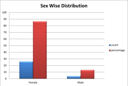
Results : A total of 30 thyroidectomies were done during the study period
of which18 were total thyroidectomy and the remaining 10 were hemi
thyroidectomy (8 left hemi thyroidectomy and 4 right hemi
thyroidectomy). The frequency of the ELN documented crossing the
reeve’s avascular space were: TYPE 1 nerve were 12 (24%) TYPE 2A
nerve 28 (56%) and TYPE 2B 5 (10%). 5(10%) ELN were not seen
despite an extensive search.
Conclusion : The preservation of the external branch of laryngeal nerve
has various technical difficulties should be considered. Exposure and
preservation of the nerve would be aided by the recognition of the
potential avascular space of Reeve.in order to reduce the morbidity
related to the thyroid surgery, every attempt should be made to ensure
safe dissection.There is a considerable variation in the anatomical course
of the nerve and its relation to various structures and are influenced by
various factors. However every attempt should be made to identify the
nerve through meticulous dissection by the surgeons in order to avoid the
nerve injury.
Key words: Cricothyroid space, External laryngeal nerve, Thyroid
surgery.
5
AIMS AND OBJECTIVES AIMS AND OBJECTIVESAIMS AND OBJECTIVES AIMS AND OBJECTIVES
To study the anatomy of external branch of superior laryngeal nerve in
6
STUDY CRITERIA STUDY CRITERIASTUDY CRITERIA STUDY CRITERIA
INCLUSION CRITERIA:
All patients admitted in General surgical wards of Govt Rajaji Hospital
with thyroid swellings being taking up for surgery are included in the
study.
EXCLUSION CRITERIA :
Patients with
• proven thyroid malignancy
• recurrence
7
METHODOLOGY :
All patients admitted in General surgical wards of Govt Rajaji Hospital,
from the time period from November 2013– September 2014 will be
studied. Clinical features, symptomatology, investigations, operative
findings, post- operative complications, morbidity and mortality will be
studied according to the proforma.
ETHICAL CLEARANCE:
Approved by the Institute of Ethical Committee, Govt Rajaji Hospital.
ANALYSIS:
8
REVI REVI REVI
REVI EW OF LITERTUREEW OF LITERTUREEW OF LITERTUREEW OF LITERTURE
“The extirpation of the thyroid gland for goiter typifies perhaps, better
than any operation the supreme triumph of the surgeon’s arm”-William
Halsted.
Thyroid Embryology and Developmental Abnormalities
The thyroid gland has a double origin form the primitive pharynx
and the neural crest. The main body of the thyroid glands is derived from
epithelial cells of the endoderm of the primitive pharynx(7,8). These
cells will form the greater portion of the follicular elements of the thyroid
tissue. They arise as a diverticulum from the midline of the pharyngeal
floor. It soon develops as a bi-lobed, encapsulated structure that descends
in the midline of the neck. With further development, this diverticulum
remains attached to the buccal cavity by a narrow tract - the
Thyroglosasal duct. Its distal end may become the pyramidal lobe.The
most commonly encountered congenital cervical anomalies are
Thyroglossal duct cysts. The thyroglossal duct lumen starts to obliterate
from fifth week of gestation, and at the end of eighth week of gestation,
duct disappears fully
The neural crest is the source of the parafollicular cells, or C cells,
which secrete calcitonin. These C cells migrate form the neural crest of
9
fifth branchial pouch. The incorporation of the fifth pouch with the P IV
leads to the formation of the caudal-pharyngeal complex, which includes
not only the ultimobranchial bodies, (lateral thyroids) but also the P IV.
Eventually, C cells populate the thyroid tissue by way of its lateral lobes.
which join the main body on each side
Midline ectopic thyroid rests are the result of the failure of or
incomplete descent of the thyroglossal duct and of abnormal development
of its epithelium(9,10). The most common example is the pyramidal
lobe, which extends upward from the isthmus or forms either lateral lobe
10
Surgical Anatomy of the Thyroid
The normal adult thyroid gland weighs about 17 g. It is
wrapped around the anterolateral portion of the upper trachea and larynx.
Either lobe occupies a space between the trachea and the esophagus
medially; the carotid sheath posteriorly; and the sternocleidomastoid, the
sternohyoid, and the sternothyriod muscles laterally and anteriorly.
If the sternothyroid and sternohyroid muscles are to be
divided, they are transected high at the level of thyroid, to preserve their
motor nerve, the ansa hypoglossi. Section of the strap muscles has no
clinical functional consequence.
The normal thyroid is soft, dark wine-red in color, and covered by
a thin capsule. It is loosely attached to neighboring structures. The
variations in fixation of the gland may arouse suspicion of pathologic
change, particularly when the history suggests acute thyroiditis or cancer.
Normally the gland adheres only to the cricoids cartilage and the upper
tracheal rings. This is the posterior suspensory ligament, or Berry’s
11
The superior thyroid artery arises from external carotid
artery and inferior thyroid artery originates from thyrocervical trunk.
Occasionally a branch from innominate artery or aorta, the arteria
thyroidea ima artery. The external carotid artery gives of superior thyroid
artery as its first branch. It gives off superior laryngeal artery and
descends on the surface of inferior constrictor muscle.
It gives off anterior and posterior branch over the superior
aspect of the thyroid gland. The anterior branch of the superior thyroid
artery descends over the anterior border of the thyroid lobe and continues
along the upper border of the isthmus to anastomose with its fellow of the
opposite side. The posterior branch descends on the posterior border of
the lobe and gets anastamosed with the ascending branch of the inferior
thyroid artery(11,12).
The inferior thyroid artery, a branch of the thyrocervical
trunk runs upwards, medially, and then downwards and reaches the lower
pole of the gland. During its course, it passes behind the carotid sheath
and the middle cervical sympathetic ganglion; and in front of the
vertebral vessels; and gives off branches to adjacent structures. Its
12
artery divides into 4 or 5 glandular branches which pierce the fascia
separately to reach the lower part of the gland. One ascending branch
anastomoses with the posterior branch of the superior thyroid artery, and
supplies the parathyroid gland.
It is often said that the superior thyroid artery supplies the upper
one third of the lobe and the upper half of the isthmus; and that the
inferior thyroid artery supplies the lower two thirds of the lobe and the
lower half of the isthmus. However, the inferior and superior thyroid
arteries anastomose freely both on the surface of the gland as well as in
its substance; and the territories supplied by the two arteries overlap
considerably.
Sometimes (in 3% of individuals) the thyroid is also supplied by
the lowest artery (thyroidea ima artery) which arises from the
branchiocephalic trunk or directly from the arch of the aorta. It enters the
lower part of the isthmus. Accessory thyroid arteries arising from tracheal
13
VENOUS DRAINAGE
The thyroid is drained by three veins namely superior thyroid vein,
middle thyroid vein and inferior thyroid vein. The superior thyroid vein
emerges at the upper pole and accompanies the superior thyroid artery it
ends either in the common facial vein or internal jugular vein. The middle
thyroid vein is a short wide channel which emerges at the middle of the
lobe and soon enters the internal jugular vein. The inferior thyroid vein
emerges at the lower border of the isthmus. They form a plexus in front if
the trachea, and empties into the left brachiocephalic vein. A fourth
thyroid vein (of Kocher) may emerge between the middle and inferior
veins, and drain into the internal jugular vein.
LYMPHATIC DRAINAGE:
The relationship of the thyroid gland to its lymphatic drainage is
most important when considering surgical management of carcinoma of
thyroid. The thyroid gland and its neighboring structures have rich
lymphatics that drain the thyroid in almost every direction. Within the
gland, lymphatic channels are present immediately beneath the capsule
and communicate between lobes through the isthmus. This drainage
connects to structures directly adjacent to the thyroid, with numerous
14
Clinically it is useful to divide the lymph nodes between the central
and lateral neck; the boundary between them is marked by the carotid
sheath. The lateral neck zones are further subdivided. Most thyroid
cancers drain directly to central nodal basins except in superior third of
15
The right recurrent laryngeal nerve.
The right recurrent laryngeal nerve arises from the cranial nerve
X, the vagus in the front of the right subclavian artery and winds
backwards below the artery, and they runs upwards and medially behind
the subclavian and common carotid arteries to reach the trachea
oesophageal groove. In the upper part of the groove it is related to the
inferior thyroid artery. It may be superficial or deep to the artery.
Occasionally, some branches are in front of the nerve, and some are
behind it. The nerve then passes deep to inferior constrictor lower border,
and enters the larynx behind the cricothyroid joint. It supplies: a) intrinsic
16
larynx below the level of the vocal cords c) cardiac branches to the deep
cardiac plexus, d) branches to the trachea and branches to oesophagus,
and e) to the inferior constrictor muscle. The cardiac branches are
superior and inferior. Out of the four cardiac branches of the vagi (two on
each side) the left inferior branch goes to the superficial cardiac plexus.
The other three cardiac nerves go to the deep cardiac plexus(13,14).
17
SIMON’S TRIANGLE
THE LEFT RECURRENT LARYNGEAL NERVE
The recurrent laryngeal nerve on the left arises from the vagus in
the thorax, as the latter crosses the left side of the arch of the aorta. It
loops around the ligamentum arteriosum and reaches the trachea
oesophageal groove. Its distribution is similar to that of the right nerve. It
does not have to pass behind the subclavian and carotid arteries, and
usually it is posterior to the inferior thyroid artery(15,16). The anatomical
landmark to identify the right recurrent laryngeal nerve is the triangle by
name Simon triangle. The boundary of this triangle is bounded medially
by the esophagus, laterally by the carotid artery and the base is formed by
the middle thyroid vessels.
esophagus
RLN
Middle thyroid A
18
PARATHYROID GLAND
The parathyroid glands develop from Branchial Pouches III and
IV. The superior parathyroid glands develop from Pouch IV, travel a
shorter distance than the inferior glands, and are typically located along
the posterior border of the thyroid gland at approximately 1 cm superior
to the entrance of the inferior thyroid artery. Because of this location,
when the superior glands descend further, they almost always remain
posterior, in the tracheoesophageal groove or retroesophageal space(17).
The location of the inferior gland can range from being high, anterior to
the carotid artery to being in the anterior mediastinum within the thymus
Inferior glands associated with the thyroid gland usually remain anterior
to the recurrent laryngeal nerve, whereas the superior glands are found
19
There is an extremely close relationship between superior thyroid artery
and external laryngeal nerve. The nerve is the sole motor supply to
cricothyroid muscle which is a tensor of the vocal cord. This nerve has
high risk of injury during superior pole ligation(18,19).
To avoid nerve injury while ligating the superior thyroid vascular
pedicle, it is recommended not to ligate the main trunk as a whole but to
identify the branches and then dissecting it .The superior thyroid arteries
should be ligated as low as possible. Secondly it is advisable to dissect
the superior thyroid vessels. The dissection requires strong downward and
outward traction of the thyroid upper pole. The dissection must be
20
The inferior thyroid artery and its end branches intimately associated
with recurrent laryngeal nerve at above the level of junctions of lower and
middle third of thyroid gland. The left recurrent laryngeal nerve ascends
at the depth of tracheo-oesophageal groove. The right recurrent laryngeal
nerve courses more obliquely somewhat more lateral in position caudally.
The recurrent laryngeal nerve continues upward and medially and at the
postero lateral aspect of middle third of thyroid gland and is extremely
close to capsule of thyroid. The recurrent laryngeal nerve is accompanied
by inferior laryngeal artery.(20,21) The site near berry’s ligament, this
artery is usually just posterior to recurrent laryngeal nerve and divides to
give multiple small branches that cross the nerve to enter the thyroid
gland. The recurrent laryngeal nerve is motor nerve to intrinsic muscles
of the larynx. Injury to motor trunk causes paralysis of vocal cord on the
ipsilateral side. The other extra laryngeal branches are sensory.
Non recurrence of the inferior laryngeal nerve is due to vascular
anomaly of aortic arches during the embryonic development- no
innominate artery but an aberrant subclavian artery. Nerve anomaly on
the left side requires in addition a right aortic arch associated with situs
inversus. A non recurrent laryngeal nerve has been also reported in
21
The most critical structure that is to be kept in mind when dividing
the vessels of superior pole is the external laryngeal nerve. This nerve has
been referred as neglected nerve of thyroid surgery. Injury to it may
easily be overlooked because they are difficult to diagnose at
laryngoscopy and because the initial symptoms are minimal and regarded
as natural post operative voice disturbance without injury to recurrent
laryngeal nerve.
THYROID RESTS
Thyroid rests are isolated rests of normal thyroid tissue which may
lies below the lower pole of thyroid within the upper anterior
mediastinum or within the line of thyrothymic tract(22,23)
.
Grade I : thyroid rests consists of thyroid tissue protrusion arising from
thyroid gland the inferior aspect in the region of thyrothymic ligament.
Grade II: thyroid rests include thyroid tissue lying within the
thyrothymic tract and attached to thyroid proper.
Grade III : thyroid rests are similar to grade II but are attached to thyroid
gland by a fibrovascular core.
22
SUPERIOR POLE AND SLN
Keeping the superior pole in the final phase of lobectomy allows
the superior pole to be managed after the superior parathyroid has been
reflected on a good vascular pedicle. Given its dorsal location, dissection
of the superior parathyroid gland is necessarily kept until a final phase of
surgery. Once this gland is reflected posterolaterally and preserved, the
superior pole can be aggressively mobilized and downwardly retracted
and the superior pole vessels can be dissected with optimal exposure
afforded for the external laryngeal nerve.
As the superior pole vessels are dissected, downward mobilization
of the gland is facilitated by Mayo clamp on the superior pole
parenchyma facilitating downward retraction. If the superior pole is
enlarged significantly or when the dissection is difficult, the following
two steps can be taken to improve superior pole exposure. The first is
transection of sternothyroid muscle. This muscle tends to hood the
superior pole as it extends to insert on oblique line of thyroid cartilage
superiorly. Lateral retraction of sternohyoid muscle and medial retraction
on thyroid laryngotracheal complex allows visualization of discrete
muscle band as it extends medially to its laryngeal insertion. The
transected muscle need not be re-approximated. The laryngeal head of the
23
position of external laryngeal nerve as it runs down posteriorly on the
inferior constrictor muscle. Patient does not experience additional pain,
edema or drainage from this mini strap division(24).
The second manoeuvre for superior pole dissection involves
divison of the isthmus and ligament of berry dissection allowing the
thyroid to be supported only by superior pole attachment. The vessels of
the superior pole should be taken individually to control optimally and
avoid risk to external branch of laryngeal nerve injury(25).
The superior laryngeal nerve branches from the vagus high in the
neck running deep to external and internal carotid arteries. It can be
visually identified in 80% cases and 100% through electrical stimulation
virtually. It innervates both vertical and oblique portions of cricothyroid
muscle which is a primary tensor. A 20% to 20% rule can be considered
for the superior laryngeal nerve. 20% of the time it is deep to deep fascia
of inferior constrictor muscle and 20% of time it extends caudally
interacting with superior pole vessels, placing it at risk as the superior
24
In anatomy textbooks, the EBSLN is described as passing just
superficial to inferior pharyngeal constrictor muscle and piercing it to
supply cricothyroid muscle. Various landmarks have been described to
identify the EBSLN in thyroid surgery. As described by Stell and Maran,
25
Joll is formed by the superior pole of the thyroid gland laterally and the
thyroid vessels in the superior pole, superiorly by the attachment of the
strap muscles to the thyroid cartilage and the midline medially. On the
floor of the triangle is formed by cricothyroid muscle which is supplied
by the EBSLN.
Different identification rates of the EBSLN have been quoted by
authors with few not performing routine identifications The techniques
used for the identification of the EBSLN mentioned in literature include
the use of nerve stimulator, the inspection of the distal part of the inferior
constrictor muscle and individual ligation of the superior thyroid vessels
The principle of any surgery is the identification of any structure in order
to preserve it. This applies to identification of the EBSLN as well.
SURGICAL ANATOMY OF EXTERNAL LARYNGEAL NERVE
Little attention was paid initially to the surgical anatomy of
external branch of superior laryngeal nerve during beginning of 20th
century. In fact Kocher did not specifically mention this nerve in his book
the importance of the preservation of the external branch of superior
laryngeal nerve was made clear as a result of thyroidectomy performed in
1935. At that time Amelita galli curci was the most famous soprano of the
26
that her recurrent laryngeal nerve was not damaged. However her vocal
registry postoperatively lowered since the time, the EBSLN has been
known as the nerve of Amelita galli curc(i28,29,30).
The superior laryngeal nerve is a branch of vagus, the tenth cranial
nerve.it separates at level of no dose ganglion from vagus, about 4cm
cranially to carotid artery bifurcation. The superior laryngeal nerve
divides into external branch and internal branch about 1.5cm inferiorly.
27
them medially, extending to the larynx .the nerve is 0.8mm wide and
length varies from 8.0 to 8.9cm.
Joll’s triangle or the sterno thyro laryngeal triangle is described as
the identification for the external laryngeal nerve. The boundaries of the
triangle are superior pole of the thyroid and the superior thyroid vessels
laterally, attachment of the strap muscles to the thyroid cartilage
superiorly and midline as the medial boundary.
JOLL’S TRIANGLE
The surgical importance of this nerve relates to its proximity to the
superior vessels of the thyroid. The external branch of superior laryngeal
nerve crosses the superior thyroid vein and artery well above the superior
border of thyroid. Only 80% of the external branch of superior laryngeal
nerve were identified during the procedure of thyroidectomy and in
remaining 20% the nerve could not be identified due to its location within
28
There is a relation between the nerve, superior thyroid vessels and
superior thyroid pole which classifies the surgical anatomy of the nerve
into three types(31)
Type I: the nerve crosses the superior thyroid vessels 1 or more cm
above the horizontal line that passes through the upper border of the
superior pole of thyroid.
Type II a: in this type the crossing of the nerves over the vesses is less
than 1 cm above the horizontal plane.
Type II b: the crossing of the nerve over the vessels is less than 1 cm
29
Of the above three types type IIb nerves has the highest risk of iatrogenic
damage.
Kierner et al published a similar of external branch of superior
laryngeal nerve classification adding a fourth category in which the nerve
runs quite dorsally to the superior thyroid pedicle making the
identification of the nerve more difficult(32).
Kierner classification
Type I Crosses STA > 1 cm above upper pole of thyroid
Type II Crosses STA < 1 cm above upper pole of thyroid
Type III Crosses STA under cover of upper pole of thyroid
Type IV
Descends dorsal to artery and crosses STA branches
30
Friedman et al proposed a different type of external branch of superior
laryngeal nerve anatomy classification focusing on the relationship
between the nerve and the inferior constrictor muscle at its junction to
the cricothyroid(33).
Type 1: The nerve is superficial to the inferior pharyngeal constrictor
muscle.
Type 2: The nerve penetrates the lower part of the inferior
constrictor muscle.
Type 3: The nerve runs deep to the inferior constrictor muscle.
The Type 3 variant is the fact that many surgeons state that the reason for
not identifying the nerve in the zone of the upper pole of
thyroid gland during thyroid surgical procedure.
PHYSIOLOGY AND PATHOPHYSIOLOGY
External branch of superior laryngeal nerve is the only motor
supply to cricothyroid muscle. The cricothyroid muscle has two bellies:
pars recta and pars oblique. The work action of the two subunits is not
fully understood but combined contraction of these two components is
vital in adjusting the vocal fold tension and length. The frequency of
vocal fold vibration is determined by the vocal fold tension which in turn
31
thyroarytenoid muscle which tends to shorten the fold length and the
cricothyroid muscle. The cricothyroid muscle promotes elevation of the
cricoid cartilage which shortens the distance with the thyroid cartilage.
This motion of the cricoid cartilage increases the tension of vocal fold
and its length. This cricothyroid muscle induced vocal cord tension is
termed as external tension of vocal cord, compared with the more refined
increase in internal tension which occurs due to the action of
thyroarytenoid muscle. The cricothyroid induced vocal cord tension is
thought to be of highest importance in production of high frequency
sounds during phonation. Besides it has some role in respiration mainly
32
The injury of external branch of superior laryngeal nerve causes
complete cricothyroid muscle paralysis which can be identified by
electrical silence at electromyography. Functionally the fundamental
frequency of the voice is lowered and the voice performance gets
worsened markedly, especially in high frequency sounds.
SURGICAL TECHNIQUE
The surgical approach of the superior pole of thyroid is most
necessary in operations involving in the thyroid gland. The dissection of
the superior thyroid pole should start with mobilization of thyroid lobe as
a whole. The middle thyroid vein ligation is advisable which helps in
facilitation of initial mobilization.
It is necessary to completely expose the sterno-thyroid-laryngeal
triangle before any superior pole suture is placed. In most cases, if there
is normal thyroid lobe of normal size or minimally enlarged, there is no
necessitation of complete section of strap muscles. However, partial
incision on sternothyroid with cautery will improve the access to superior
thyroid pedicle(36).
33
The superior thyroid vessels usually divide into three branches that
embrace superior thyroid pole: two branches are located anteriorly and
34 .
It is necessary to dissect and ligate these two branches individually
and placing the sutures as caudally as possible. Sometimes, gentle
traction of the thyroid lobe caudally helps preserving the integrity of the
nerve. Generally this nerve is located cranially to the superior border of
35
The surgeon should have a low threshold use of nerve stimulator
while dissecting this area. When the nerve is stimulated electrically, quick
and powerful contractions of the cricothyroid is obtained. Once the
external nerve is visualized, it should be kept at constant direct vision
36
Advantage in using the nerve stimulator in external branch of
superior laryngeal nerve management is its ability to stimulate the nerve
superiorly and even if the nerve is subfascial within the fibers of inferior
constrictor, we can get a positive signal.
The positive response results with cricothyroid twitch as well as
small response on endotracheal monitoring systems which can be seen
37
Dissection of superior thyroid pole is more difficult in dealing with
a large goiter. In this instance the upper pole border is markedly elevated
which puts the upper pole in close contact with the nerve. Additionally
there will be superior thyroid vessel enlargement which parallels the
dimensions of the goiter and demanding more careful dissection. The
sectioning of the strap muscles offers a better and safer exposure.
38
DIAGNOSIS OF SUPERIOR LARYNGEAL NERVE PARALYSIS
The diagnosis of external branch of superior laryngeal nerve palsy is
not easy to confirm based on endoscoping and clinical findings. The
notable changes are subtle voice especially in the male patients. However,
in women and voice professionally, some symptoms are common.
1. Lowering of fundamental frequency
2. Inability to produce high tone sounds
39
4. Shortening of the phonic time of consonants
5. Lowering of high tones
6. Contraction of vocal range
Video laryngeo stroboscopy can be used in diagnosis of external
laryngeal nerve paralysis after thyroidectomy. The paralysis of the nerve
results in
1. the vocal cord bowing
2. rotation of the posterior glottis towards the paralytic side
3. displacement of affected vocal cord inferiorly
4. vocal fold mucosal wave asymmetry
Most objective method for external laryngeal nerve injury detection is the
electromyography of the cricothryoid muscle using the thyroid cartilage
inferior border and superior aspect of cricoid cartilage as anatomical
landmarks externally. Electrodes are placed per cutaneously over the
muscle. The patient is asked to produce high tone. If there is no nerve
injury, increase in electrical activity of cricothryoid muscle is noted. If
there is injury to the nerve, no background electrical activity is observed.
Contralateral muscle can be used as control. Electromyography is an
40
INCIDENCE OF EBSLN INJURY
Lore et al reported 0.9% injury in 111 patients employing only
indirect laryngoscopy. Smith et al found 2.6% injury in 13 patients
prospectively. Randolff clinical trial 12-28% of injury were not
identified intra operatively. Some of these were permanent as confirmed
by electro myographic evidence(39,40).
TREATMENT OF EBSLN INJURY
Unfortunately once the nerve gets injured, no true elective
treatment is available. Intensive phono therapy is highly recommended.
The paralysis is permanent. The consequence in the career of a voice
professional might be significant. Laryngoplasty might be useful in such
situations(41).
Results in other studies
The external branch of superior laryngeal nerve has a close
anatomical relationship with the superior thyroid pedicle and in 15 – 20%
of cases the nerve may be type IIb in which the nerve crosses the superior
thyroid vessels below the upper border of superior pole of thyroid. In
these instances it may be at risk during thyroidectomy. So, it is always
41
possible. And in case any nerve is found in this area, a positive
identification with nerve stimulator is extremely useful.
The EBSLN is another argument to use nerve monitoring during
thyroidectomy, especially on a enlarged thyroid lobe. The only effective
way of avoiding paralysis of the nerve which can be extremely un
favourable for a voice professional is comprehensive anatomic
knowledge about the nerve and gentle handling of the superior thyroid
pole.
RLN-SLN CONNECTIONS
To understand the functional significance of extra laryngeal branching
and for interpreting intra operative RLN monitoring information, the
surgeon needs to understand not only the anatomy of the RLN and SLN
systems but also the interconnections. The SLN internal branch chiefly
innervates the afferent part of the hypo pharynx, base of tongue, supra
glottis and vocal cords. The external branch of the superior laryngeal
nerve provides motor function to cricothyroid muscle and innervates
sensory part of the anterior sub glottis. The efferent activity is important
in regulating the laryngeal protective mechanism which results primarily
in SLN internal branch. Many regard the RLN SLN connection as a
42
superior laryngeal nerve and recurrent laryngeal nerve. Some believe
there may be a possible motor component within SLN branches. Galen
and Martin believed that return of the voice that sometimes occurs after
transection of RLN may result from regrowth from SLN branches. More
recently, it has been suggested that a method of vocal cord recovery after
an RLN injury should involve re innervation through supplemental motor
branches of SLN(42). Functionally the SLN and RLN system
interconnection can be divided into four groups:
1. Galen’ s anastomosis
2. SLN external branch/ distal RLN anastomosis known as human
communicating nerve
3. Inter arytenoid anastomosis
4. SLN internal branch - RLN thyro arytenoid regional anastomosis
SLN MONITORING
Damage to external branch of superior laryngeal nerve results in voice
changes significantly which are typically described as pitch reduction,
inability to attain higher registers, decreased voice projection. Most time,
glottic examination of external branch paralysis or paresis can be virtually
43
electromyographic study documented SLN paralysis or paresis found
significant decrease in
• maximum phonation time,
• frequency range
• increased, jitter, shimmer , noise to harmonic ratio and increased
flow rate.
In 20% cases the external branch of the nerve is hidden beneath the
inferior constrictor muscle and therefore not seen directly. Despite this
surgical anatomy, a nerve stimulator can be passed along the inferior
constrictor and discrete pitch of cricothyroid muscle can be elicited as the
stimulator passes the external branch. An exact landmark for linear
oblique part of external laryngeal nerve as it courses down along the
inferior pharyngeal constrictor towards the cricothyroid muscle is the
laryngeal head of sternothyroid muscle. Within 1-3 mm of this obliquely
oriented laryngeal line, the external branch of superior laryngeal nerve
can be found with a high degree of certainty. Blind stimulation of the
nerve in this area with a nerve stimulator uniformly shows identification
of the linear path. EMG monitoring with endotracheal tube recording
electrode, EMG data may be less critical for SLN than for RLN because
the cricothyroid pitch is usually apparent. The average SLN wave form
44
mA. There was no significant difference in waveform characteristics
suggesting surgical dissection and current stimulating did not adversely
affect electrophysiology of neural function
There was no difference between stimulation of 1 or 2 mA and no
difference in stimulation characteristics of men and women. Lower
thyroartyenoid amplitude with external branch stimulation is consistent
with canine data.Two recent studies have demonstrated positive voice
outcomes with neural monitoring of external branch. In a small study of
thyroidectomy under local anaesthesia, external branch monitoring
resulted in improved visualization of the nerve and improvement in voice
according to post operative surveys(43,44,45).
PHYSIOLOGY OF THYROID
Thyroid hormone synthesis begins in the fetus at 11 weeks of
gestation. TSH is the important stimulator of thyroid gland and also a
marker for thyroid dysfunction. TSH works by negative endocrine
feedback system. TSH production occurs by pulsatile manner and it
reaches peak value at night. Thyroid hormone acts by its nuclear receptor.
Ultimobronchial bodies gives to thyroid medullary C cells that
produce calcitonin. C cells aggregated at upper 2/3 lower 1/3 of thyroid
45
THYROID HORMONE SYNTHESIS
Spectrum of thyroid disease
Functional abnormality of the thyroid is grossly classified in to Hypo
thyroid and hyper thyroid.
In both conditions medical management in the mainstay of treatment
Inflammatory conditions of the thyroid are called as thyroiditis
Hypothalamus Pituitary Thyroid TS
T
4T
3 TRHTSH-R
Tg
CAMP
I
-NIS
Trapping
TPO
Tg + I
-Coupling
Iodination
DIT MIT
46
a. Acute thyroiditis is due to Bacterial infection and fungal
infection, radiation thyroiditis and drug induced (amiodarone).
b. Sub acute thyroiditis includes viral, that is otherwise called as
granulomatous thyroiditis, silent thyroiditis, TB thyroiditis.
c. Chronic thyroiditis includes Hashimoto’s, Reidel’s Thyroiditis
Goiter and Nodular thyroid Disease are common clinical thyroid
problems.
Pathology of nodule formation
Persistent growth stimulation can cause diffuse hyperplasia which
is reversible. Then fluctuating stimulation leads to areas of active and
inactive lobules. The active lobules are more vascular leading to
haemorrhage into follicle will cause necrosis leaving surrounding active
follicles. Necrotic lobules unite to form nodules filled colloid or mass of
inactive follicles. This process continues will cause nodule formation.
1. Diffuse non toxic simple goiter (colloid goiter)
- cause : iodine deficiency (Endemic goiter)
- treatment is medical
2. Single solitary nodular goiter
47
Complications
a. Toxicity
b. Malignancy
c. Retrosternal extension
d. Pressure effects
e. calcification
4. Thyroid malignancy
- differentiated thyroid cancer includes papillary and follicular
variety and Hurthle cell carcinoma.
- Undifferentiated thyroid cancer includes anaplastic and
medullary carcinoma of thyroid.
Clinical features of thyroid disorders
In thyroid disorders, age of the patient should be significantly
considered. Simple goiter is observed in pubertal girls. MNG, SNG and
colloid goiters are seen in females of 20s and 30s. Papillary carcinoma is
seen in young girls and follicular carcinoma seen in middle aged women.
Anaplastic carcinoma is the disease of old age. Primary toxic goiter is
seen in young females whereas Hashimoto’s thyroiditis is seen in middle
48
Areas where there is iodine deficiency there will be the occurrence
of endemic goiter. Most of the thyroid disorders present with swelling
infront of the neck. The inflammatory conditions of thyroid are painful.
The malignant conditions of thyroid gland are initially painless to start
with and then later on become painful.
Pressure effects
Thyroid swelling may compress on the trachea to produce
dyspnoea or it can compress on oesophagus to produce dysphagia.
Hoarseness or change in voice is commonly due to infiltration of the
recurrent laryngeal nerve by the malignant thyroid.
Symptoms of primary hyperthyroidism
The most notorious symptom of primary thyrotoxicosis is loss of
weight inspite of good appetite. Cold preference, heat intolerance and
excessive sweating are the next prominent symptoms. Neurological
symptoms such as nervous excitability, irritability, insomnia, tremors in
hand, muscle weakness are more pronounced in primary thyrotoxicosis.
49
closing the eyes, chemosis are usually associated with this condition.
Some females have amenorrhea.
Symptoms of secondary thyrotoxicosis
When a longstanding colloid goitre, SNG or MNG shows
manifestations of thyrotoxicosis this condition is called as secondary
thyrotoxicosis. The brunt of attack falls more on cardiovascular system
than nervous system. Palpitations, ectopic beat, cardiac arrhythmia,
dyspnoea on exertion and chest pain are prominent symptoms. Even
patient may have congestive cardiac failure at later stages. Ophthalmic
symptoms and nervous symptoms are mild or absent.
Symptoms of hypothyroidism
Increase of weight, inspite of poor appetite is the significant
symptom. Cold intolerance, preference for warmth, dry skin puffiness of
face, pouting lips, dull expressions, constipation , muscle fatigue,
lethargy, failing memory, loss of hair,hoarseness of voice, and
50
Regarding past history patient should be questioned about anti
thyroid drugs, any previous history of neck swelling, neck surgery.
Patient should be asked about the intake of goitrogens such as cabbage,
kale and rape. And family history of any thyroid disorders should also be
sought for.
Physical examination
1. General survey
2. Build and state of nutrition
In thyrotoxicosis the patient is usually thin and underweight. The patient
sweats a lot with wasting of muscles and in hypothyroidism the patient is
obese and overweight. In case of carcinoma of thyroid there will be signs
of anaemia and cachexia.
3. Facies
In thyrotoxicosis one can see the facial expression of excitement, tension,
nervousness or agitation with or without variable degree of
exophthalmos. In hypothyroidism one can see puffy face without any
51
4.Mental state and intelligence - Hypothyroid patients are naturally dull
with low intelligence. This is more obvious in cretins.
Not only the pulse rate becomes rapid, but it becomes irregular in
thyrotoxicosis. Irregularity is more of a feature of secondary
thyrotoxicosis. Particularly sleeping pulse rate is a very useful index to
determine the degree of thyrotoxicosis. In case of mild thyrotoxicosis, it
should be below 90, where as in case of moderate or serve thyrotoxicosis
it should be between 90 to 110 and above 110 respectively. In
hypothyroidism the pulse becomes slow.
Skin
The skin is most particularly the hands in case of primary
thyrotoxicosis. The clinician while feeling for the pulse should take the
opportunity to touch the hand as well. Hot and moist palm is came across
in primary thyrotoxicosis. Skin is dry and inelastic in myxoedema.
Local examination
Inspection
Normal thyroid gland is not obvious on inspection. It can be seen
52
can follow Pizzillo’s method in which the hands of the patient are placed
behind the head and the patient is asked to push her head backwards
against her clasped hands on the back of his/her head. The thyroid
swelling may be uniform or isolated nodules of different sizes. A thyroid
swelling moves upwards on deglutition. This is due to the fact that the
thyroid gland is fixed to the larynx. Such movement of the thyroid
becomes greatly limited when it is fixed by inflammation or malignant
infiltration.
In retrosternal goiter patient should be asked to raise both the arms
of the patient over his head until they touch the ears. This position is to be
maintained for sometime. Distress and congestion of face become evident
in retrosternal goiter due to obstruction at the level of thoracic inlet of the
great veins.
A thyroglossal cyst also moves upwards on deglutition. But the
pathognomonic feature is that it moves upwards with protrusion of the
tongue.
Palpation
The thyroid gland should always be palpated with the patient’s
53
the front. Careful assessment of the margins of the thyroid gland is
important, particularly the lower margin.
Palpation of each lobe is best carried out by Lahey’s method. The
examiner stands in front of the patient. To palpate the left lobe properly,
the thyroid gland is pushed to the left from the right side by the left hand
of the examiner. This makes the left lobe more prominent so that the
examiner can palpate it thoroughly with his right hand.
Slight enlargement of the thyroid gland or presence of nodules in
its substance can be appreciated by simply placing the thumb on the
thyroid gland while the patient swallows. (Crile’s method)
Following points should be noted during palpation:
- the whole thyroid gland enlargement is whole or not.
- When a swelling is localized
- Mobility
To rule out the possibility of the retrosternal extension of the gland,
getting below the thyroid examination should be done.
Pressure effect
Pressure may be on the trachea or larynx, which may lead to stridor
54
lead to dysphagia. Pressure may be on the recurrent laryngeal nerve,
which may lead to hoarseness of voice. If pressure on trachea is
suspected, slight push on the lateral lobes will produce stridor(Kocher’s
test). This test, if positive, indicates an obstructed trachea.
Narrowing of the trachea i.e. scabbard trachea becomes quite
obvious in skiagram. A malignant thyroid may engulf the carotid sheath
completely and so the pulsation of the artery could not be made out.
Sympathetic trunk may also be affected by thyroid swelling. This will
lead to Horner’s syndrome.
- Enophthalmos
- Pseudoptosis
- Miosis
- Anhidrosis
- Palpation of cervical lymph nodes
This is extremely important particularly in malignancy of thyroid.
Papillary carcinoma of thyroid is notorious for early lymphatic metastasis
when the primary tumour remains quite small.
Percussion
This is done to rule out the retro sternal extension of the
55
Auscultation
A systolic bruit can be heard over the enlarged thyroid gland in
case of primary toxic goiter.
General examination
Eye signs
1. Lid retraction – This sign is caused by over-activity of the
involuntary part of the levator palpebrae superioris muscle. When
the upper eye lid is higher than normal and the lower eyelid is in its
normal position this condition is called lid retraction.
2. Exophthalmos – when eyeball is pushed forwards due to increase
in fat or oedema or cellular infiltration in the retro-orbital space the
eyelid are retracted and sclera becomes visible below the lower
edge of the iris first followed by above the upper edge of the iris.
i. Von Graefe’s sign – The upper eyelid lags behind the eyeball as
the patient is asked to look downwards.
ii. Joffroy’s sign – Absence of wrinkling on the forehead when the
56
iii.Stellwag’s sign – This is staring look and infrequent blinking of
eyes with widening of palpebral fissure.
iv.Moebius’s sign – This means inability or failure to converge the
eyeballs.
v. Dalrympte’s sign – This means the upper sclera is visible due to
retraction of upper eyelid.
3. Ophthalmoplegia – there may be weakness of the ocular muscles
due to oedema and cellular infiltration of these muscles.
4. Chemosis is caused by obstruction of the venous and lymphatic
drainage of the conjunctiva by the increased retro-orbital pressure.
Tachycardia or increased pulse rate without rise of temperature is
constantly present in primary toxic goiter. Sleeping pulse rate is more
confirmatory in thyrotoxicosis.
Tremor of the hands is almost always present in a primary thyrotoxic
case.
Moist skin particularly of the hands and feet are quite common in
primary thyrotoxic cases.
Thyroid bruit is also quite characteristic in Graves’ disease (primary
57
Secondary thyrotoxicosis
Here atrial fibrillation is quite common. Signs of cardiac failure
such as oedema of the ankles, orthopnoea, dyspnoea while walking up the
stairs may be observed. Exophthalmos and tremor are usually absent.
INVESTIGATIONS OF THYROID
DISORDERS(46,47,48,49,50,51,52)
The various investigations for diagnosing thyroid diseases can be
divided as follows
1.Tests of thyroid function
2.Thyroid autoantibodies
3.Thyroid imaging
4.Cytology
Tests of Thyroid function:
The improved sensitivity and specificity of TSH assays have
greatly improved laboratory measurement of thyroid function. A rational
58
decreased or normal. A normal TSH level excludes a primary
abnormality of thyroid function in rare occasions. Immune radiometric
assays one of the tools used to determine the thyroid function and they
are very sensitive. The widespread use of TSH IRMA has rendered the
TRH stimulation test outdated. The finding of an atypical TSH level
should be followed by measurement of circulating thyroid hormone levels
to prove the diagnosis of hyperthyroidism (suppressed TSH) or
hypothyroidism (elevated TSH).T3 and T4 are highly protein bound and
numerous factors can manipulate protein binding. It is useful to measure
free or unbound levels of hormone.
For most patients the T4 level which is unbound is sufficient to
confirm the state of thyrotoxicosis but elevated T3 levels(T3
thyrotoxicosis) seen in 2% of individuals .Thus unbound T3 levels be
required to be measured in those with suppressed TSH levels with normal
unbound T4 levels.
In thyroid cancer patients, Serum thyroglobulin levels are used in
the follow up. After total thyroidectomy and radio-ablation it should be
undetectable. Levels greater than 1-2ng/ml suggest inadequate ablation or
59
Thyroid auto-antibodies
Autoimmune thyroid disease is detected most easily by detecting
circulating antibodies against thyroid peroxidase (TPO) and
Thyroglobulin(Tg).Almost all patients with autoimmune hypothyroidism
and upto 80% of those with Graves Disease have TPO antibodies at high
levels.
Thyroid Imaging:
1. Chest and Thoracic Inlet Radiography
Simple Radiographs of the chest and neck will demonstrate if there
is any retrosternal extension of goiter or significant tracheal compression.
Pulmonary metastasis might also be detectable.
2. Radiography of Neck
Plain radiographs of neck in both antero-posterior and lateral views are
taken to look for-
• Position of Trachea
• Pre tracheal soft tissue shadow
• Evidence of retrosternal extension
• Any compression of trachea
60
• Status of cervical spine
2. Indirect Laryngoscopy(IDL)
This is done preoperatively to look for vocal cord movements.
Some patients will have asymptomatic paralysis of recurrent laryngeal
nerves. So this examination is very important from medico-legal aspect.
4 Ultrasound Scanning [USG]
Now USG is considered as extension of the clinical examination. It is one
of the basic investigation for thyroid swelling.
a. Differentiate benign from malignant nodule
b. The ultrasonography can demonstrate sub clinical nodularity and
identify deep non-palpable thyroid nodules.
c. Size of the nodule can be measured
d. It can also differentiate solid from cystic swellings
e. Sono guided FNAC can be done
f. Identify cervical lymph nodes
g. Identify multicentricity.
Features of Benign Lesion in USG
The following features are indicative of a benign nature
61 2. Significant cystic component
3. Peripheral egg shell like calcification
4. A sonolucent rim (halo) around the nodule
5. Well – defined nodule margin
Feature of Malignant Lesion in USG
The following features should arouse a strong suspicion of malignancy
1. Hypo echoic
2. Cystic component need not be there
3. Micro calcifications
4. No halo
5. Poorly defined margin
6. Taller than wide lesion
62
USG Showing Normal Thyroid
USG Guided placement of the needle during a thyroid FNAC biopsy
Ultrasound uses a high frequency probe in the 7.5-12MHz range.
Ultrasound devices have become portable enough to allow use in the
63
USG Showing Solid and cystic Components
5.Radioisotope Scanning
Radionuclide imaging serves to confirm the presence of the nodule
within the thyroid, identifies the functional characteristics of the nodule.
The only absolute indication in thyrotoxicosis for isotope scanning
is for the diagnosis Autonomous Toxic Nodules
Toxicity with nodularity is an indication. It can identify hypo
functioning nodules (cold). Cold nodule in Graves’ is likely to be
64
It is the only method by which one can definitely differentiate
primary, secondary and toxic nodules.
Isotope scan can also differentiate hyper thyroidism from toxicosis
due to other causes. ( to differentiate hyperthyroid thyrotoxicosis from
non- hyperthyroid thyrotoxicosis). The radioactive iodine uptake (RAIU)
is increased in hyperthyroidism whereas toxicosis because of
extrathyroidal causes the RAIU is decreased (eg. thyroiditis)
Other indications for isotope scan are:
1. To identify ectopic thyroid tissue.
2. To identify recurrence and metastases in thyroid carcinoma.
3. 99mTc is the isotope of choice for diagnostic purposes. It is
cheap and the radiation is less than radioiodine. Twenty minutes
after intravenous injection of 99mTc, scanning is done over the
thyroid.
4. If radioactive iodine is used 123I is the isotope of choice for
diagnostic purposes.
5. Carcinoma concentrates technetium and therefore a hot nodule
65
6. A nodule which is warm on technetium scanning and cold on
radioiodine scanning is called discordant scan. This is
suggestive of malignancy.
Surgery is generally required to provide a definitive diagnosis,
although needle biopsies have been used in some major medical centers.
Another indication for radioisotope thyroid scanning was in the
treatment of thyroglossal cyst before excision to rule out the possibility
of an ectopic thyroid. Pertechnetate ion is selectively concentrated in the
thyroid gland, salivary glands and stomach. In normal subjects, up to 2 %
of the radioactivity from intravenously injected pertechnetate-99m is
accumulated by the ion-concentrating mechanism of the thyroid at 1 hr.
Thirty min after administering 1 me of pertechnetate-99m intravenously,
good scans of the thyroid are obtainable. Iodine 123 and Iodine 131
scintigraphy is also used to evaluate the functional status of the thyroid
gland. Both are trapped by active follicles and organified. 123I has a
shorter half-life (12-13hrs) and gives a quicker image and low dose of
radiation (30 mrad). It is a good choice for suspecting lingual thyroid or
<