AGE ESTIMATION FROM FUSION OF ECTO CRANIAL
(Coronal, Sagittal, Lambdoid, Temporal) SUTURES IN HUMANS
– A STUDY IN BODIES DURING AUTOPSY TO PROVE /
DISPROVE AGES OF SUTURAL CLOSURE USED IN ROUTINE
PRACTICE.
Dissertation submitted for partial fulfilment of the requirements for the degree
M.D. (Forensic Medicine)
BRANCH – XIV
DEPARTMENT OF FORENSIC MEDICINE, TIRUNELVELI MEDICAL COLLEGE,
TIRUNELVELI - 620711.
THE TAMILNADU DR.M.G.R.MEDICAL UNIVERSITY, CHENNAI.
BONAFIDE CERTIFICATE
This is to certify that the work undertaken in this dissertation entitled “Age estimation from fusion of ecto cranial (Coronal, Sagittal, Lambdoid, Temporal) sutures in humans – A study in bodies during autopsy to prove / disprove ages of sutural closure used in routine practice” has been carried out by
Dr.Manivasagam.M, M.B.B.S., a Post Graduate student under my supervision and guidance for his study leading to Branch XIV M.D. Degree in Forensic Medicine during the period of June 2012 to May 2015.
DEAN HEAD OF THE DEPARTMENT, Tirunelveli Medical College, Department of Forensic Medicine, Tirunelveli. Tirunelveli Medical College,
Tirunelveli.
DECLARATION
I, Dr.Manivasagam.M, M.B.B.S., solemnly declare that this dissertation
titled “Age estimation from fusion of ecto cranial (Coronal, Sagittal, Lambdoid,
Temporal) sutures in humans – A study in bodies during autopsy to prove /
disprove ages of sutural closure used in routine practice” is a bonafide work done
by me, under the expert guidance and supervision of Dr.A.Selvamurugan.,MD., DNB., MNAMS., Associate Professor & Head of the Department of Forensic Medicine, Tirunelveli Medical College, Tirunelveli – 11. This dissertation is
submitted to The Tamil Nadu Dr. M. G. R. Medical University towards partial
fulfilment of requirement for the award of M.D. Degree (Branch XIV) in Forensic
Medicine.
Place:
ACKNOWLEDGEMENT
I take this chance to thank from my heart, a person of high caliber
Dr.A.Selvamurugan., MD., D.N.B., MNAMS, Associate Professor and Head of the Department of Forensic Medicine, Tirunelveli Medical College, Tirunelveli –
11, who took the burden of guiding me into the realms of Forensic Medicine and
has been the centre of significance since the inception of this dissertation till its
completion.
I offer thanks to Dr.R.Sudalaimuthu, M.D., D.N.B., MNAMS, Associate Professor, Department of Forensic Medicine, Tirunelveli Medical College,
Tirunelveli – 11, for his valuable advices as to this work.
I am extremely thankful to Dean, Tirunelveli Medical College, Tirunelveli for his grateful permission on this study.
I thank Dr. Shanthosh Priyan, a friend and well-wisher for his statistical support,
and also my Colleagues, Staffs and Technicians of the Department of Forensic
Medicine, Tirunelveli Medical College for their contributions during my study
CONTENTS PAGE NO
1. INTRODUCTION 01
2. AIM OF THE STUDY 07
3. REVIEW OF LITERATURE 11
4. MATERIALS AND METHODS 33
5. RESULTS AND ANALYSIS 49
6. DISCUSSION 82
7. CONCLUSION 99
8. RECOMMENDATIONS 102
REFERENCES
ANNEXURES
ABBREVATIONS
& - And
< - Lesser than
> - Greater than
% - Percentage.
C - Coronal Suture
CF - Completely fused
Cr. No - Crime Number
C/o - Care of
D/o - Daughter of
Ed. - Edition
F - Female
F/o - Father of
H/o - Husband of
L - Left (at suture staging)
L - Lambdoid Suture
Ltd - Limited
IBM - International Business Machines
i.e. - That is
M/o - Mother of
n - Number of subjects
NF - Not fused
No - Number of cases
M - Male
p - Probability significance
PASW - Predictive and analysis software
PM No. - Post Mortem Number
P. S - Police Station
R - Right
S - Sagittal Suture
SB - Sutural Bones
S.D - Standard Deviation
S.E - Standard Error
SPSS - Statistical Package for Social Sciences
Sl. No - Serial number
S/o - Son of
T - Temporal Suture
Age estimation from fusion of ecto cranial sutures (Coronal, Sagittal,
Lambdoid and Temporal) in humans – A study in bodies during autopsy to prove / disprove ages of sutural closure used in routine practice.
Abstract:
Estimation of age of an individual is one of the interesting areas in Forensic Medicine. There are many age estimation techniques in practice. But, most of the techniques can be applied only in a narrow age group. Age from suture closure is a technique of age estimation that can be applied to wide age groups between 18 to 80 years. This study is about estimating age at death of a person from the ecto cranial suture closure status in local population between 30 to 80 years. Individuals selected in this study are with known age (identity proofs). Result obtained is analyzed and compared with accepted standard.
INTRODUCTION:
Identification is one of the foremost and interesting sectors in Forensic field.
If individuality of a person is conclusively established, it becomes complete or
definitive identification. If the points of individuality are pointing towards but not
concluding, it becomes an incomplete or partial identification. Age, sex and
ancestry are called primary characters of identification; in this age at death of a
person or skeletal remains becomes a matter of issue as in civil or criminal
responsibilities of a person; validity of contract, marriage and consent; kidnapping,
prostitution; old age benefits etc. Age at death finds its importance as corpus
delicti, particularly with an increasing trend towards effacement of identity or in
identifying an unknown person.
With increasing tendency of crime perpetrators towards effacement of
identity, either by acts of commission (dismembering, decapitation, burning post
mortem, dumping in water sources) or by intentional neglect of killed on less
habited dwellings end up with partly skeletonized remains of deceased reaching us
for our expertise. To listen to the bones for answers, Skull is the most widely
studied human skeletal structure because of its relatively higher resistance to
on skull for age estimation are widely studied till date.
02
From Forensic point of view these properties of durability of skull with its
resistance, hard to get destroyed by animal disinterment are of immense value in
cases of advanced decomposition, fire, mass disasters as an identification tool.
There are many techniques for estimating age at death of a deceased person.
This may include hair color and complexion, physical development like primary
and secondary sexual characters, tooth eruption pattern and even the number of
carpal bones (between the age of 2 to 7 years) on simpler means. Cranial sutural
closure pattern, tooth examination by Gustafson`s technique modified by Johanson,
lines of increment on teeth as devised by Boyde`s, Stack`s method of age
estimation, epiphyseal appearances and their fusion with their primary centers or
bone shafts, diaphyseal length, thyroid cartilage ossification by Cerny, morphology
of sternal end of 4th rib, changes in symphysis pubis, recent trends of relating acetabular sharpness and porosity as age indicator in adults and racemization of
aspartic acid in bones and cartilage are also helpful in estimating the approximate
or probable age at death of a deceased.
Age related changes are coherent and occur in a defined age limit that is
as there are number of factors like wear and tear to pose difficulty and thus less
precision in age estimation.
03
This error can be better solved by combining several of these age estimation
techniques. Further, physiological adulthood ranges from the third decade through
the tenth; this extent between the decades adds another bias to our techniques. This
issue of overestimating the age of young adults and under estimating the aged
crops up. Bias tends to shift on overestimation of age in case of young or shifts on
underestimating the age of aged persons. But in middle years, this bias is said to be
overcome or with equal probability of positive and negative biases on either side.
In skull, regarding dentition as an age estimating tool, eruption pattern
applies to newborn (temporary dentition) and to early years of life (permanent
dentition). Gustafson’s method may be used in later parts of life but with
increasing errors with increasing age. Other morphological traits such as sutural
closure pattern, surface appearance of vault (granular & muscle markings),
Pacchionian depressions on either side of Sagittal sinus and radiological methods
to visualize vascularity of diploe, skull thickness are the possible age estimation
Of these methods using skull, most would not give an age related
progressive change to be applied to group persons into various age divisions. For
example, smooth ivorine skull of young is replaced by a skull with prominent
muscle markings that of an adult.
04
It is not possible for many of these techniques to define age groups but only
above or below limits can be said. To conclude, all these techniques would only
have application in estimating a narrow spectrum of age group.
Sutural closure pattern is one of these age estimating techniques that is
commonly adopted in our day to day medico legal practices. It is by far one of the
common age estimation studies from bones that are being undertaken. It has the
benefit of involving a wide range of age groups. This method can involve
recording of sutural obliteration as a whole or at specific points as studied by
Meindl and Lovejoy (1) or by devising a regression equation as done by Nawrocki
(2)
.
Studies are being undertaken not only on vault sutures but also to include
Advent of computer technologies has revolutionized our field of forensic
studies by exploring every detail of individual sutures and whole bone by X rays,
CT imaging, laser scanning and processing (5).
Further, sutural closure pattern is the only possible age estimation technique
applicable with extensive studies in age group from 30 years to 80 years.
05
Skull sutures are easier to identify and record even in decomposed bodies or
in skeletal remains. In skull, ecto cranial suture and endo cranial suture closure are
appreciated as two different entities. Endo cranial sutures are earlier to fuse than
ecto cranial sutures. They tend to close 5 to 10 years in advance to their ecto
cranial counter parts. Hence, fusion of sutures (endo cranial) is comparable and
correlated to symphysis pubis age estimation techniques (between 18 to 60 years of
age) (6).
To involve age limits between 30 and 80 years, age estimation technique
from ecto cranial suture closure by naked eye examination is selected. Of various
parameters that could affect closure of sutures like sex, climate and ethnicity, this
study is aimed at estimating age at death of persons with known age from their ecto
A cross sectional study with 500 cases from 30 to 80 years of age of both
sexes is selected from the bodies received for medico legal autopsies in Tirunelveli
region and their ecto cranial suture closure pattern is studied.
06
Aim & Objectives:
To find ages of ecto cranial suture fusion in different age groups in male and
female sexes of local population.
To compare the results obtained with that of various works on ages of ecto
cranial suture fusion.
To prove / disprove ages of ecto cranial suture closure used in routine
practice in correlation with local population.
Justification for study:
Present times, we are following same ages of ecto cranial suture closure
pattern in our medico legal works in many parts of India. Being a vast nation, with
closure age pattern, it is prudent to consider the biological and natural variations.
So, studies with respect to local populations can be carried out.
Hence, ages at which ecto cranial sutures fuse are studied and analyzed with
a sample of local population that reaches our Medical College Hospital and
subsequently to our College mortuary for autopsy.
08
Methodology:
Assess ages of ecto cranial suture closure pattern during autopsy on bodies
with known age from 30 to 80 years at death.
Risks:
No risk of mutilation of bodies.
No risk to the examiner.
Has institutional ethical committee clearance obtained for this study?
Outcome:
May prove or disprove the practicing standards of ages of fusion of ecto
cranial sutures in our medico legal works.
09
Benefits:
To carry out medico legal works with more efficacy and certainty.
National Significance:
If the ages of ecto cranial suture closure is proved, we shall continue using
the same standards that are routinely practiced in our medico legal works.
If it is disproved, extensive work shall be further undertaken in varied conditions
so as to formulate population specific or region specific age standards in this
10
REVIEW
OF
Review of literature:
Dwight (1890) noted that suture closure begins endo cranially, and arrived at a
conclusion that “the time of closure of any particular part of a suture, and the
order in which the process advances, are very uncertain. And the process is later
in females”.
Parsons and Box (1905) stated that closure “is later in females and also simple
sutures (less serrated or denticulated) closed earlier. No differences between
Bolk (1915) suggests that physiologic obliteration of the sutures may be an
atavistic trait. Since the suture in the ape closes as soon as the skull is fully
grown.
Pittard and Kaufmann (1936) found no racial or right left differences in skull
sutural closure.
12
Montagu (1938) quotes that Vesalius and his student Fallopius first noted the
obliteration of sutures with age.
Glaister (1947) mentions that the absence of any sign of closure of any of the
sutures of the skull points to a strong probability that the age does not exceed
thirty years. The closure of Sagittal, Coronal and Lambdoid sutures has
unusually begun by the age of thirty. The Parieto Mastoid and squamous suture
commence at a later stage between thirty five and fifty years but do not show
great advancement until between fifty and sixty years.
BRASH, J. C. in Glaister and Brash’s Medico Legal Aspects of Ruxton Case
Sagittal, Coronal and Lambdoid sutures normally begin to close at from 20 to
30 years, followed about 5 years later, by the onset of closure of parieto mastoid
and squamous sutures, which, however, may remain open or only incompletely
closed at 60; the spheno parietal suture does not usually close until the age of
70’.
13
SIR SYDNEY SMITH AND FREDERICK SMITH FIDDES (1949) in their
treatise on Forensic Medicine write ‘the sutures of the skull become obliterated,
a condition first observed on the internal aspect. They further write about
beginning of closure as ‘usually beginning at the obelion (on Sagittal suture),
and closure may be quite marked by the age of forty (years of age). Closure of
the lambdoid suture is usually not complete before fifty years (of age), and the
masto – occipital, squamous and parieto - mastoid is at very old age.
Regarding its variations in respect to age and reliability they noted that the
range must lengthen; and after thirty years, when the mature skeleton
already begin to show signs of “ageing” – including the beginning of
progressive closure of the cranial sutures. It will be hardly safe to estimate
the sutures of the skull points to a strong probability that the age does not
exceed thirty years.
Cobb (1952) stated that age assessment from cranial sutures is not reliable
because it “probably approximates the true age of an individual within plus or
minus nine years”.
14
McKern and Stewart (1957) stated that Ecto cranial sutural closure is highly
variable and only broadly be correlated with age. In the Sagittal suture (25 % of
55 skulls) for example, closure had already begun in 17 – 18 years of age and
had become very common in 31 – 40 years of age (90 % of 43 skulls).
Francis Camps (1968) writes “From the forensic stand point this is very
different from saying, as formerly that the sagittal suture begins to closure at 22
years and completes its closure at 35 years. It is better, therefore, not to rely on
the sutures in skeletal age determinations”.
Ascadi and Nemeskeri (1970) on their study concluded sex was not found to be
In his book ESSENTIALS OF FORENSIC ANTHROPOLOGY, T.D.
STEWART (1979) Quotes Cobb ‘that there is some general or constitutional
influence which modifies progress in suture closure, commencing
approximately at thirty years, is evident since all sutural elements, whatever
15
their degree of union, show arrested activity at this age. While bone along a
suture edge is still in a state of active change it possess a granular texture
difficult to describe but easy to recognize. After activity has ceased the
granularity gives place to a waxy smoothness of texture. Heaped - up edges on
an unclosed suture are characteristic evidence of quiescence which absolutely
differentiates the suture from one still in a state of active closure. Todd called
this condition lapsed union’.
Shapiro (1981) writes that sutures begin to close on both the outer and inner
aspects of the calvarium at about the same time. Ecto cranial fusion proceeds
Zivanovic (1983) recorded that left and right sides of the coronal and lamdoid
sutures, both endocranially and ectocranially on East African Bantu skulls. Pars
Bregmatica and pars complicata of the coronal suture and pars intermedia of
Lambdoid suture were the most assymetrical. Asymmetry was common in both
population and sexes. He adds that if asymmetry is noted it should be
considered as pre mature synostosis not be discarded from age estimation.
16
It is common between 41 and 50 years.
Baker (1984) found that asymmetry is predominantly observed between 36 to
40 years and 61 to 65 years of age. In general symmetry is predominant over
asymmetry.
Perizonius (1984) analyzed both ectocranial and endocranial sutural closure in a
sample of 256 males and 20 females. He did not observe any sex difference in
Parson, Gee and Knight (1985) mentions that the last suture to close is the
spheno parietal suture which may still be open in old age, even after 70 years.
They add open sutures are often found and they are a menace in finding age
from suture closure pattern. Hence, suture closure has guarded applicability in
age estimation. It is much helpful in assessing age at death of skeletal remains
particularly if the remains are fragmentary.
17
Meindl RS, Lovejoy CO (1985) on their study on Ecto cranial suture, followed
a method by reading cranial suture closure at points of Cranial suture in mid
Lambdoid, lambda, obelion, anterior Sagittal, bregma, mid coronal, pterion,
spheno frontal, inferior spheno temporal, superior spheno temporal.
Mann et al. (1987) documented that males typically exhibit more suture
obliteration at all ages than do females, but that race has only a minor influence
on this parameter.
Snow (1982) says that in his case series all sutures of a 101 year old male have
year old man has been found to exhibit completely fused sutures to show its
erratic nature in closure.
Iscan M. Y. and Kennedy K. E. R. (1989) quote McKern and Stewart that the
progress of suture closure has only a general relationship with age in their work
Reconstruction of life from Skeleton.
18
Massler & Schour (1991) studied osteoblastic accretion along the suture lines as
an indicator of bone growth and suture closure in their work on ‘Maxillary
suture obliteration – A visual method for estimating skeletal age’.
Dr. Krishnan MKR (1992) on his work Medico legal Necropsy states that
fusion age for sutures are as follows, Posterior 1/3rd Sagittal: 30 – 40 years, middle 1/3rd Sagittal: 50 – 60 years, anterior 1/3rd Sagittal: 40 – 50 years, upper ½ of Coronal: 50 – 60 years, lower ½ Coronal: 40 – 50 years, upper ½ of
Lambdoid: 50 – 60 years, lower ½ of Lambdoid: 60 -70 years and Temporal: 80
MALLIK CC, (1993) writes suture closure as estimation tool can be used with
caution. The result obtained would place an individual in decades. There is no
male and female difference. Various environmental factors can modify the
suture closure. It starts to close on inner table of skull followed by outer table.
Ecto cranial sutures start 5 to 10 years after endo cranial sutures. It takes further
20 to 40 years to fuse completely.
19
In a book edited by Mehmet Yasar Iscan and Richard P. Palmer FORENSIC
ANTHROPOLOGY OF THE SKULL (1993) Novonty et al writes ‘Scores for
each site noted (in skull) were added by region and plotted against ages. Ages
calculated for composite scores from the five lateral anterior sutures which were
found to have the best correlation with age’.
Kanisius and Luke (1994) reported ancestral biases were found to be significant
between Europeans and Australian Aborigines, and research which reports
austosomal dominant and recessive patterns of premature suture closure
Key et al (1994) tested cranial suture closure on known age individuals from the
sixteenth century crypt of Christ Church, and reported great variability in suture
closure, so that only broad age categories could be identified. They found that
advanced suture closure of the inner table of skull was complete by 50 years of
age and there by useful only at ages younger than that. The ectocranial sutures
displayed pattern and age variability between males and females.
20
Moreover, they found out that open ecto cranial sutures could be found in all
age groups with about equal frequency and so could not be taken as evidence of
youth or younger individual.
Buikstra and Ubelaker (1994) on their text ‘Standards for Data Collection from
Human Skeletal Remains’ described suture closure scoring system as Score 0 –
Open, 1 – Minimal closure, 3 – Significant closure, 4 - Completely obliterated.
50 years of age, upper ½ of Coronal: 50 – 60 years of age, lower ½ Coronal: 40
– 50 years of age, upper ½ of Lambdoid: 50 – 60 years of age, lower ½ of
Lambdoid: 60 -70 years of age.
H1. Hershkowitz et al (1997) observed the following on closure of sutures:
(1) closed (TC);
2) partially closed (PC): less than 10% of the suture length was open;
3) totally open (TO): the suture line was clearly visible with almost no
21
interruptions along its entire length. Minor closure at the area of the parietal
foraminae was ignored;
4) partially open (PO): between 10% and 90% of the suture length was open;
and 5) premature suture closure (PMSC).
He concludes that suture obliteration patterns are independent permanent
phenomena; some are genetically inherited and the relative frequency of the
‘‘totally open’’ category is higher in females.
His study also introduces a new name ‘Double Y suture – where sagittal suture
SIMPSON’s Forensic Medicine by Bernard Knight edition (1997) “The cranial
sutures tend to fuse with age. But it is too variable to be of any practical use”.
DAS B.M. and DEKA RANJAN (1998) write ‘it is possible to suggest the
approximate age on the basis of obliteration of sutures. But, it is a fact that
many changes occur in the skeleton including the skull with age. For example,
with the advancement of age obliteration of the suture of cranial vault takes
place on the outer surface between forty and fifty’.
22
About the starting of sutural obliteration, ‘the lower part of the coronal
suture is the first to exhibit obliteration, next comes the posterior part of the
sagittal suture, to be followed by the lambdoid suture’.
Peggy Thomas in his book FORENSIC ANTHROPLOGY – The Grading
Science Of Taking Back (2000) writes ‘the squiggly lines on the skull where the
bones meet are called cranial sutures. To date the age of a person at death using
the skull, specific segments of the suture are assigned and given a score from 0
closed (less than 50 percent). A score of two means that it is more than 50
percent closed and a score of three indicates that the suture is fully closed. They
are added and compared to established standards. For example, a score of 11
correlates with an age range of 24 to 60 years with a mean age of 35.4.
The Sagittal suture on the top of the head, running from front to back, fuses by
age 35. The coronal suture at the front of the skull, from temple to temple, fuses
by age 40.
23
Schiel Rebecca quotes White and Folkens’ (2000) method of grading cranial
sutures which involves giving scores to the degree of suture closure at 17 points
on the skull. Each site is assigned a score between 0 – 3, and calculating a
composite score to assign a corresponding age range. Open
sutures are considered with a site score of 0. A score of 1 equates to minimal
sutural closure. Significant closure is given score 2, and complete fusion or
Thus this method gives two age ranges. Average age can be calculated using
both of these mean ages and the age range is the average of the standard
deviation for both estimated ages.
Taylor’s Principles and Practice of Medical Jurisprudence edited by Keith Mant
(2000) says “For many years, the closure of the skull suture was considered the
most accurate method. Recent works has shown that there is too much variation
in closure times to merit the use of this method in forensic work”.
Rogers and Allard (2004) observed the partial obliteration of sutures in older
adults in about 3.5% of the population.
24
Sahni et al (2005) worked on age assessment from cranial suture closure from
Northwestern India sample containing 538 males and 127 females. A total of
665 individuals were in the sample. Sutures were recorded only as “open” or
“closed”. Sahni and coworkers found earlier obliteration in males than in
females; obliteration is so erratic to be used for estimating age. Since previous
research (by author) concluded long bone epiphyses fuse earlier in Indians than
David Dolinak, Evan W. Matshes, Emma O. Lew (2005) write Age – dependant
differences in suture closure (ossification of the sutural ligaments) have been
used for years in age estimation. Studies of the ecto cranium have the potential
to under estimate age.
Nawrocki and Zambrano (2005) reported at the American Association of
Physical Anthropology that based on the results of an analysis of variance, there
are significant differences in the rate and/or pattern of the suture closure
between the sexes and among the ancestral groups.
25
Klepinger (2006) endorses Nawrocki’s idea of individual regression formula for
sagittal, coronal, squamous and palatine sutures to make the method more
reliable and as the method of choice for this purpose (Age estimation), pending
Rao Nagesh Kumar G (2007) writes in his Practical Forensic Medicine that
Posterior 1/3rd Sagittal: 30 – 40 years; anterior 1/3rd Sagittal, lower ½ Coronal: 40 – 50 years; middle 1/3rd Sagittal, upper ½ of Coronal: 50 – 60 years.
PV Guharaj and MR Chandran (2009) in their text on Forensic Medicine write
“it occurs (suture closure) externally in the following order: posterior third of
the Sagittal suture at about 30 – 40 years of age, anterior third of Sagittal and
lower half of the Coronal at about 40 – 50 years of age and middle Sagittal and
upper half of coronal at about 50 – 60 years of age. The Temporo parietal
sutures close much later”.
Further in a “diagram showing approximate ages of closure of sutures of cranial
vault, Lambdoid – 50 – 60 years, 60 – 70 years” is mentioned.
26
In the book, THE USE OF FORENSIC ANTHROPOLOGY, Robert Pickering
and David Bachman (2009) mentions age at death as one of the Ten Key
questions in skeletal survey. They add suture close or fuse in a relatively
consistent pattern and this pattern can give an age range. The problem is that
this range is quite broad and provides only an estimate rather than a definite
Shetty (2009) on his work on cranial sutures concludes ‘though, there is some
difference in suture closure in males and females, it is not significant
statistically. There is some statistically significant correlation suture closure up
to 40 years of age. Then, they appear to close independently of each other.
There is no significant variation in suture closure of right and left sides of
coronal and lambdoid sutures, both ectocranially and endocranially. Complete
closure (mean value > 3.5) never occurred in ectocranial sutures’.
Parikh (2010) in his work Medical Jurisprudence for Courtrooms and
classrooms writes “The closure starts with the Sagittal (30 – 35 years of age),
Coronal (35 – 40 years of age), and Lambdoid suture (45 – 50 years of age -
variable),
27
followed by parieto mastoid & squamous sutures (55 – 60 years of age), and
the spheno parietal suture which closes by about 70 years.
Pillay V. V., (2010) states that Age evaluation by this method (ecto cranial
suture) is generally not accurate, since it can only provide a range in decades.
The usual sequence is for the sagittal suture to close by 30 – 40 years of age
(posterior 1/3rd), 40 – 50 years of age (anterior 1/3rd) and 50 – 60 years of age (middle 1/3rd), followed by Coronal suture which fuses between 40 – 50 years of age (lower part) and 50 – 60 years of age (upper part), and temporal suture
(around 60 years of age).
Suture closure occurs a little later in females than in males.
Rao Nagesh Kumar G. (2010) writes “the scheme of ossification of skull by
suture closure as Sagittal (posterior 1/3rd: 40 – 50, middle 1/3rd: 50 – 60, anterior 1/3rd: - years), Coronal (upper half: - years, lower half: - years), Lambdoid (upper half: - years, lower half: - years), Temporal (years).
28
Alexandra B. Millard (2011) in his work concludes there are no sex and
ancestral variation in suture closure. He further emphasizes the importance of
Bardale Rajesh (2011) writes “No difference exists between right and left sides
of skull. No sex difference in fusion of suture. Sagittal fuses at 30 – 40 years,
Coronal 40 – 50 years, Lambdoid 40 to 50 years, Squamous >80 years.
Umadethan B (2011) in his book Forensic Medicine mentions “ anterior Sagittal
40 – 50 years, middle Sagittal 50 – 60 years, posterior Sagittal 30 – 40 years,
lower Coronal 40 – 50 years, upper Coronal 50 – 60 years, Lambdoid 50 – 70
years, Temporal – 80 years.
Vij Krishan (2012) writes “Fusion of sutures occurs comparatively early in
males. In many skulls, fusion of sutures at the ecto cranial surface may remain
incomplete. This is called lapsed union and occurs most often in the sagittal
suture.
29
On the outer side, fusion occurs in the following order,
- Posterior one third of Sagittal suture at about 30 – 40 years,
- Anterior one third of Sagittal and lower half of Coronal at about 40 – 50
- Middle Sagittal and upper half of Coronal at about 50 – 60 years,
- In the Lambdoid sutures, fusion activity starts late and the progress is also
slow. The closure starts around 25 – 30 years near the asterion, and the peak
of maximum closure occurs at about 55 years.
Estimation of age by suture closure is not reliable. It can be given only in the
range of a decade. The usual reliability falls in the order of Sagittal,
Lambdoid and then the Coronal”.
Chandrasekaran on his work on Cranial Sutures as an identification tool
comments that the skulls of monozygotic twins do contain similar pattern but
considerable variations are noticed in the details of the suture pattern.
It seems that the suture pattern in skulls like finger prints is controlled
through inheritance. That even the maximum possible loading by
inheritance is found to be insufficient to contract the random
30
production of minute variation in the details of suture pattern.
DEROBERT and FULLY based on study of 480 skulls established the
They considered it a criterion of value but the criteria is one which must be
interpreted... ‘Avec la plus grande prudence’ (with most prudence).
FRANCHINI examined 629 skulls and found that there was no closure of
sutures in four of them despite the fact that the subjects were aged, respectively,
45, 48, 55, and 74 years of age.
Singhal mentions that Sagittal suture (between two parietal bones) – obliterates
at 30 – 35 years. Coronoid suture (between frontal & two parietal bones)
obliterates at 35 to 40 years. Lambdoid suture (between occipital & two parietal
bones) obliterates at 45 – 50 years. Squamous suture (between frontal, parietal
& temporal bones) obliterates at about 60 years.
31
An approximate estimate of age of an adult can be obtained. The degree of
closure of the three main suture of vault of the skull is divided into 4/5 stages.
Dr. Akilesh Pathak reports a case of 12 – 14 year old boy with complete fusion
of ecto cranial and endo cranial sutures in his paper ‘Contradictory Skull and
Age Estimation’.
MATERIALS
AND
METHODS
MATERIALS AND METHODS
Subjects are selected at random from the bodies we received for medico
legal autopsies during the study period. This study comprises 500 subjects with
known age at death. Ages from 30 to 80 years of both sexes are included, as the
Subjects are grouped into decades starting from 30 to 40 years, 41 to 50
years, 51 to 60 years, 61 to 70 years and 71 to 80 years.
Age at death is taken from the post mortem requisition, Case history, Form:
86 details given by the Police / Magistrate depending upon the cases. Valid age
proof issued by the Government like Aadhar card, Driving license, Marriage
certificate, Passport, Ration card, Voter’s ID, Government employer ID cards or
any Government / Quasi Government recognized documents are taken for age at
death. Preference is given to the identity proofs with date of birth. Age at death
from the identity proofs are rounded off to the nearest year.
A careful scrutiny is done on the analyzing work period to look for age
mismatch with one document to the available other documents and also for cases
with change of crime numbers on subsequent dates due to typing errors or wrong
entries.
34
Though rare, instances of age, name and crime number changes pose
difficulty with data collection.
Study design:
Methods:
To read the cranial suture closure or fusion one can rely on naked eye
examination after reflecting the tight periosteal layer on outer table of skull. This
simpler technique can also be complimented by 2D & 3D CT techniques, X-rays
(5)
, laser instrumentation and Osteoblast accretion along suture lines (3). Skeletal age by radiological methods is advanced than direct anatomical methods in age
estimation. Hence, radiological examination of skeletal systems would yield an
early detection and accurate finding. Even though, radiological methods are more
accurate and reliable, direct anatomical observation is preferred in many cases
owing to the following reasons: Radiological examination is not preferred in large
sample studies, living individuals face the danger of radiation exposure, cost –
benefit analysis from the receiver’s side, and radiographs plays the role of an
intermediary object on which opinions are given while direct examination has no
35
such drawback where opinions are based on direct examination facts. Further, in
our country post mortem radiographs for research and study purposes is less
practicable.
Researchers on the other hand support direct anatomical studies than
the bones rather than radiographs which are shadows of what you want to see’’. In
case of dead, direct examination of ecto cranial sutures is easier and more preferred
when compared to radiographs.
Naked eye examination methodology is selected for this study to assess
suture closure pattern on ecto cranial sutures before opening the skull. This
methodology has advantages of being easy with simpler modus operandi and
noninvasiveness. Direct observation of suture status is possible with this
methodology.
A proforma is prepared to collect all relevant information like name, age,
sex, post mortem number, police station with crime number, general physical
examination findings such as built, height in cm, address, occupation and marriage
status of the subjects. Age stated is accepted from any of Government issued or
Government authorized certificates. Most age proofs are given with driving
licenses, aadhar card and PAN cards.
36
Some cases are provided with Marriage registration certificate, Baptism
certificates, and Service register certificate of uniformed services staffs as identity
and age proofs. Marriage status is included as an indicator of a reliable partner or
A judgment given by our Honorable Supreme court of India recognized the
inclusion of Third sex in gender columns (7). As the judgment is passed during the study period of this study, third sex column is added to the proforma in a
retrospective manner. Though, the study is aimed at finding any sexual differences
in suture closure pattern, male and female sexes are only encountered in the
studies. Third sex subjects are not encountered in the study.
Ecto cranial suture closure pattern is studied during the Autopsy procedures
on bodies at our Department Mortuary hall. On opening the scalp, with the behind
ear mastoid to mastoid incision, soft tissue layer of scalp is retracted on front and
back of incision. Retraction to the anterior of incision extends to the level just
above super ciliary arch and to the posterior to make the full view of lambdoid
suture. This procedure needs further separation of the tightly attached periosteal
layer from outer table of skull after reflecting the scalp through the loose areolar
37
layer from outer table of skull after reflecting the scalp through the loose areolar
issue layer of scalp.
For this, sand papers used for rubbing off paint are tried. Wetness is the
Next methodology is with periosteal elevator. An effective method of periosteal
removal can be done with this methodology.
Knives are also used for scrapping and peeling the periosteal layer instead or
in addition to this methodology. Periosteal layer is scrapped with sharp edge of
knife or point scrapped with its tip. Tip scratching is used for finer removal of
periosteal layer after gross scrapping.
Periosteal layer is removed with ease in cases of decomposition or treated
burns, as this layer is highly vascular, the increased fluid content separates the
periosteal layer from underlying outer table of skull. Loosening of tissues in
decomposition makes the removal of outer periosteum elegant and complete. In
treated burns, soft tissue edema causes loosening of outer periosteal layer from the
outer table of skull. But these circumstances are only a few out of total 500 cases.
Two cases in the study are exhumed bodies with known identity. Skulls of
those cases are brought to our mortuary, examined and findings recorded.
38
Decomposition changes causing detachment of the periosteum is very well
recognized in these two cases.
Periosteal layer is removed on the vault from frontal eminence to the point
traced down on either side till its Pars pterica to expose it full. Squamous part of
temporal suture on both sides is made visible by clean slicing and separating the
temporalis muscle on that area. These three sutures namely, Sagittal Coronal &
Squamous can be visualized on the body in its lying down position, viewed from
Norma verticalis or Norma lateralis view.
But for exploring Lambdoid suture, the head has to be lifted and made
upright as much as possible because rigor mortis of neck muscles would prevent
flexion of neck on chest. Or the block placed under the shoulders for autopsy
procedure can be raised in such a way that graded height blocks under the body to
make more exposure to the region of Lambdoid suture. However, making the neck
forcefully flex over the chest is relatively easier to read Lambdoid suture.
Lambdoid suture is traced infero laterally to make visible its Right and Left
wings up to the termination of suture at mastoid process. At instances, even after
careful and meticulous removal, tight periosteal layer at certain points on a suture
would be present.
39
This might indicate an open suture or fusing suture where periosteal layer
dips into it. In those instances, suture closure status at remaining portion of the part
Observations are made prior to removal of skull by manual sawing, in view
that the force used in removing the skull might damage the suture lines or cause
sutures to separate if they are in fusing state. Also, recently fused sutures might
open up along their lines of fusion if sufficient force is applied on them. Careful
examinations on sutures while removing the skull cap by manual sawing and T
piece torsion removal would reveal extension of cut ends into sutural separation in
non-union sutures. But in cases of united sutures, the extension would take a
different line than that of the intervening suture.
Well-known scaling in skull suture status assessment are as follows:
1. Scaling by Ascadi and Nemeskeri,
2. Scaling by Meindl and Lovejoy and
3. Hershkowitz:
Scaling by Ascadi and Nemeskeri is widely followed because of its reliability.
Meindl and Lovejoy scaling is used in point examination of sutures. Hershkowitz
scaling is newer among these and widely used in excavations and archeology.
40
Stage 0 - Open suture. A clear space between adjoining bones is seen,
Stage 1- Incipient suture.
Suture is closed, but visible as continuous, zigzagging line,
Stage 2 - Closure in process.
Suture line becomes thinner, less zigzags and may be interrupted by areas of
complete suture,
Stage 3 - Advanced suture.
Only scattered pits remain on location of suture,
Stage 4 - Closed suture.
Suture completely obliterated; even its location cannot be recognized.
This system of scaling is Modified Perizonius phases (0 to 4) after Ascadi and Nemeskeri system of sutural closure pattern staging (9).
This scaling has 5 phases in total.
41
Stage 0: Open suture.
There is no evidence of any ectocranial closure at the site,
Stage 1: Minimal closure; some closure has occurred.
This score is given for any minimal to moderate closure, i.e., from a single bony
bridge across the suture to about 50 % synostosis,
Stage 2: Significant closure;
There is a marked degree of closure but some portion of the site is still not
completely fused,
Stage 3: Advanced closure.
Only pits indicate where the suture is located (1).
This is Meindl and Lovejoy scoring system of reading suture closure status at
specific points of skull. This scaling has only 4 phases (Ascadi & Nemeskeri – 5
phases)
42
(1) Closed (Total Closure);
(2) Partially Closed (PC): less than 10% of the suture length was open;
(3) Totally Open (TO): the suture line was clearly visible with almost no
interruptions along its entire length.
Minor closure at the area of the parietal foraminae was ignored;
(4) Partially Open (PO): between 10% and 90% of the suture length was open; and
(5) Pre mature suture closure (PMSC) (9).
Among this three systems of scaling, each system has its own advantage.
Observations in this study are made on the grounds of the system suggested by
Ascadi and Nemeskeri (Modified Perizonius phases 0 to 4) because it is widely
used and reliable.
Hence, sutures are read and scaled into Stage 0, Stage 1, Stage 2, Stage 3 or
Stage 4 as per this Ascadi and Nemeskeri system of scaling suture closure pattern
for all the four ecto cranial vault sutures analyzed in this study.
43
Stages in this notation would mean,
For Score 1 to 3, maximal score of suture closure in case of different phases with
in the observed part of the suture.
Score 4 is given only if there is complete fusion.
Suture closure status is done for,
Sagittal suture:
Anterior 1/3rd, Middle 1/3rd and Posterior 1/3rd,
Coronal Suture:
Right half and Left half of corona. Each half is further divided into Upper ½ and
Lower ½ segments,
Lambdoid suture:
Right wing and left wing of lambdoid suture, dividing it further into Upper ½ and
Lower ½ segments.
Squamous temporal suture: Right and Left (whole suture).
44
Suture closure finding is entered with notations used in studies by Ascadi
Coronal, L – Lambdoid, T – Temporal suture. Next alphabet is either R for right or
L for Left in Coronal, Lambdoid and Temporal sutures. Numbers following the
alphabet would indicate the suture fusion stage according to the segments of
suture. Sagittal suture will have three numbers, Coronal and Lambdoid will have
two numbers and Temporal suture will have only one number.
Example: The notation S 2 1 3 would mean Sagittal suture with its Anterior
one third at fusion status stage 2, Middle third at stage 1, Posterior third at stage 3.
In case of Coronal and Lambdoid sutures, C R 1 2 would mean Coronal suture –
Right half having suture closure stage 1 at upper ½ and stage 2 for lower ½.
L L 1 2 would mean Lambdoid suture left part with its upper half at stage 1, lower
half at stage 2. Notation for temporal suture is indicated by T R 1, T L 2 –
Temporal suture Right side with stage 1, temporal suture on left side with Stage 2.
In addition to this, incidental findings like colour of the skull, sutural bones
with its location, Metopism, healed depressed fractures, a variant of Parietal
foraman are also noted with interest.
Sutural closure pattern staging are analyzed statistically with SPSS and Epi
Info 7 software.
45
500 cases with known age at death.
Sampling design:
Purposive sampling.
Study design:
Descriptive Study.
Inclusion criteria:
Bodies with known age at death from 30 to 80 years.
Known age at death is obtained from Government issued or Government approved
or Quasi Government certificates with date of birth in it.
46
Deformed head (Traffic / assault injuries, charred remains),
Congenital anomalies (bony defects),
Nutritional deficiencies (if any demonstrable at autopsy),
Fracture of skull leading onto sutural separation,
Crush injury head or heavy cut injuries to head involving the sutures,
Fracture line extending / crossing sutural line and
Migratory populations (different ethnicity) are excluded. Industrial abundance,
harbor vicinity are main attraction to the migratory population. Other issue of
different ethnicity interfering with the study is that of tourists who are
unfortunately lose their lives while here. Such cases of interference by population
other than local population is carefully excluded to maximum possibility.
Study period:
One and a half year
47
Temple of Surprise - Autopsy Hall,
Department of Forensic Medicine & Toxicology,
Tirunelveli Medical College, Tirunelveli.
Statistical methods involved:
The data collected in this study are analyzed statistically using IBM – SPSS
and Epi info 7 software. Variables like mean, standard deviation of fusion age and
percentages of cases with suture closure in each age groups are calculated.
48
Ecto cranial suture closure pattern in bodies at autopsy
(Known age from 30 to 80 y of age at death)
Sl. No. / Case no: Date:
PM No:
Station & Crime No:
1. Name : Mr/Mrs/Ms ---, S/O, D/O, H/O, W/O,
F/O, M/O, C/O
2. Age at death from (Corrections at cutoff date: )
Driving license :
Ration card :
Others, if any :
3. Sex : M F Third sex
4. Address :
5. Occupation :
6. Married / Unmarried :
7. General Physical Examination :
a. Built & Nourishment (Poor / Moderated / Well)
b. Height in Cm
8. Suture closure status
Coronal Suture
Right half Left half
Lambdoid Suture
Right half Left half
Temporal Suture
Right Left
Other notable points, if any:
10. Consent: I ____________________________ S/o, D/o ___________________
have no objection for using these findings, photographs for presenting / publishing
for study / research purposes.
Signature of the legal Heir. Signature of the examiner.
(Ecto cranial suture closure pattern in bodies at autopsy
with known age at death from 30 to 80 years)
Serial
no.
PM
no.
Station &
Crime no
Age
& Sex
Coronal
suture
Sagittal
suture
Lambdoid
suture
Temporal
suture
Stage 0: Open suture (NF),
Stage 1: Continuous line,
Stage 2: Interrupted by complete fusion,
Stage 3: Only pits and
RESULTS
AND
RESULTS AND ANALYSIS:
After, completing cranial suture closure status assessment of 500 cases with
known age at death, data entered in separate forms are transferred into a master
chart. Master chart containing 500 cases is fed into IBM SPSS and Epi Info 7
software for analyzing data. Frequency distribution for data analysis is made as
[image:63.612.68.547.371.632.2]follows:
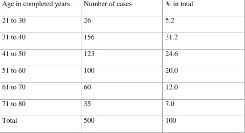
Table 1: Frequency distribution of cases with respect to age.
Age in completed years Number of cases % in total
21 to 30 26 5.2
31 to 40 156 31.2
41 to 50 123 24.6
51 to 60 100 20.0
61 to 70 60 12.0
71 to 80 35 7.0
Total 500 100
During statistical analysis of case details Age group 21 to 30 years
constitutes 26 cases out of total 500, this 26 cases comes to 5.2 % of total 500
cases. Similarly, Age group 31 to 40 years constitutes 156 out of 500 cases or 31.2
% of total cases. Age group 41 to 50 years constitutes 123 cases or 24.6 % of total,
Age group 51 to 60 years constitutes 100 cases or 20.0 % of total, Age group 61 to
70 years constitutes 60 cases or 12.0% of total 500 cases, Age group 71 to 80 years
constitutes 35 cases or 7.0 % out of total 500 cases (Chart 1). Sex wise distribution
of cases in each age group is also analyzed.
Chart 1: % distribution of cases in various age groups.
51
5.2
31.2
24.6
20
12
7
21 TO 30 Y 31 TO 40 Y 41 TO 50 Y 51 TO 60 Y 61 TO 70 Y 71 TO 80 Y
% in total
In age group 21 to 30 years, 13 0f 26 (50 %) are male, 13 0f 26 (50 %) are female;
age group 31 to 40 years has 119 of 156 (76.3 %) male, 37 of 156 (23.7 %) female;
age group 41 to 50 years has 85 of 123 (69.1 %) male, 38 of 123 (30.9 %) female;
age group 51 to 60 years has 72 of 100 (72.0 %) male, 28 0f 100 (28. 0 %) female;
age group 61 to 70 years has 46 of 60 (76.7 %) male, 14 of 60 (23.3 %) female and
age group 71 to 80 years has 23 0f 35 (65.7 %) male, 12 of 35 (34.3 %) are female.
This forms a total of 358 of 500 (71.6 %) male and 142 of 500 (28.4 %) female
subjects in this study.
Chart 2: Sex distribution in Age groups (in %)
52
50
76.3
69.1 72
76.7
65.7
50
23.7
30.9
28
23.3
34.3
21 TO 30 Y 31 TO 40 Y 41 TO 50 Y 51 TO 60 Y 61 TO 70 Y 71 TO 80 Y Sex distribution in Age groups
In this analysis, Age group 21 to 30 years is included for analysis purpose.
To get parallel and equal Frequency distribution, this type of grouping cases into
age groups is adopted. Though the Age group 21 to 30 years contain cases only
with age of 30 years, it is named and grouped as Age group 21 to 30 years to
achieve Statistical correction and convenience as mentioned above.
Percentage and other statistical values for expected age group 30 to 40 years
can be obtained by combining the values in calculated age group 21 to 30 and age
group 31 to 40 years. Other age groups are to be read as such.
Analysis are done for individual sutures with their divisions. Statistical
variables for Sutures are analyzed under following divisions:
Right wing of Coronal suture’s Upper ½ , Right wing of Coronal suture’s lower ½,
Left wing of Coronal suture’s Upper ½, Left wing of Coronal suture’s lower ½,
Sagittal Posterior 1/3rd, Sagittal Middle 1/3rd, Sagittal Anterior 1/3rd,
Right wing of Lambdoid suture’s Upper ½, Right wing of Lambdoid suture’s
Lower ½, Left wing of Lambdoid suture’s Upper ½, Left wing of Lambdoid
suture’s Lower ½,
Right Squamous temporal suture and Left Squamous temporal suture.
Right wing of Coronal suture’s Upper half:
In the statistical analysis of Upper half of Right wing of Coronal suture,
Age group 21 to 30 years has 26 cases in total by 13 male and 13 female. In male
population all 13 are at Stage 0 or in other words all the 13 male in this age group
is found to have open suture. Remaining 13 by female population is also at Stage 0
or their suture is open.
Age group 31 to 40 years that constitutes 156 cases out of total 500, has male 119
and female 37 subjects. In 119 male, 106 are at Stage 0 and the remaining 13 are at
Stage 1 to 4. In 37 female, 36 are at Stage 0 and only 1 female individual is at
Stage 1 to 4. In these analyses Stage 1, Stage 2, Stage 3 and Stage 4 are assigned as
Stage 1 for statistical convenience (S6). So, a total of 142 cases at Stage 0 and 14 cases are at Stage 1 when male and female statistics are combined in this age group
of 31 to 40 years is analyzed.
In this age group of 41 to 50 years, total 123 cases contain 85 male and 38 female.
Of 85 male, 67 are at Stage 0, 18 are at Stage 1 to 4. Of 38 female, 25 are at Stage
0, remaining 13 are at Stage 1 to 4. Combining male and female statistics, we get
92 cases at Stage 0, 31 cases at Stage 1 to 4.
With age group 51 to 60 years having100 cases, 72 are male and 28 are female. 72
[image:68.612.68.512.179.674.2]male cases has 10 cases at Stage 0, 62 cases with Stage 1 to 4.
Table 2: Right wing of Coronal suture’s Upper half
Age group in Years (Sex specific)
CS Upper half – Right side
Total
0 1
21 to 30
Male 13 0 13
Female 13 0 13
Total 26 0 26
31 to 40
Male 106 13 119
Female 36 1 37
Total 142 14 156
41 to 50
Male 67 18 85
Female 25 13 38
Total 92 31 123
51 to 60
Male 10 62 72
Female 5 23 28
Total 15 85 100
61 to 70
Male 1 45 46
Female 0 14 14
Total 1 59 60
71 to 80
Male 1 22 23
Female 1 11 12
Total 2 33 35
278 222 500
28 female cases has 5 cases at Stage 0, 23 cases at Stage 1 or above. So, 15 cases
are at Stage 0, 85 cases are at Stage 1 to 4 on male and female combined statistics
in this age group.
Age group 61 to 70 years has 60 cases, of which male are 46 and female are 14. Of
46 male, only 1 case is at Stage 0 and the rest of cases (45) are at Stage 1 to 4. Of
14 female, 0 case (no case) is at Stage 0 and so all the 14 cases are at Stage 1 to 4.
1 case at Stage 0 and 59 cases at Stage 1 to 4 is the combined value.
Chart 3: Progression of Stages of fusion with age (CS R Upper 1/2)
Last age group with 71 to 80 years has got 35 cases. Out of this 23 are male and 12
are female. 23 male has 1 individual at Stage 0 and other 22 at Stage 1 to 4.
56
21 to 30 31 to 40 41 to 50 51 to 60 61 to 70 71 to 80
% at 0 100 91 74.8 15 1.7 5.7
% at 1 0 9 25.2 85 98.3 94.3
100
91
74.8
15
1.7 5.7 0
9
25.2
85
98.3
94.3
0 20 40 60 80 100 120
Progression of stages of fusion with age
Among 12 female only 1 is at Stage 0 and other 11 are at Stage 1 to 4. Combining
the values we get 2 cases at Stage 0 and 33 cases at Stage 1 to 4.
To summarize, Right side of Coronal suture’s Upper half is examined in 358 male
and 142 female skulls. Out of 358 male skulls, 198 are at Stage 0 and 160 are at
Stage 1 to 4. Out of remaining 142 female skulls, 80 skulls are at Stage 0 and 62
are at Stage 1 to 4. Combining male and female skull statistics we arrive at 278
skulls at Stage 0 and 222 skulls are at Stage 1 to 4 (Table 2). In the Chart 3, we can
see the percentage of Stage 0 skull decrease as age increases (blue dotted line) and
[image:70.612.67.509.424.525.2]rise in percentage of Stage 1 skulls as age increases (red dotted line).
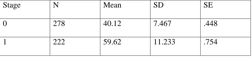
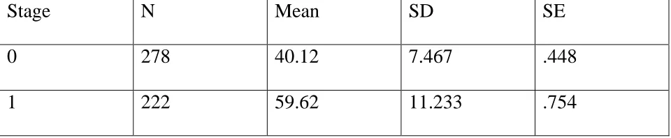
Table 3: Statistical variables for Right wing of Coronal suture’s Upper half
Stage N Mean SD SE
0 278 40.12 7.467 .448
1 222 59.62 11.233 .754
Mean obtained for Stage 0 (non-fusion) is 40.12 years, Stage 1 is 59.62 years.
Standard deviation thus calculated is 7.467 for Stage 0 and 11.233 for Stage 1.
Standard error Mean is .448 for Stage 0 and .754 for Stage 1 (Table 3).
Right wing of Coronal suture’s Lower half:
[image:71.612.76.538.186.685.2]In the statistical analysis of Lower half of Right wing of Coronal suture,
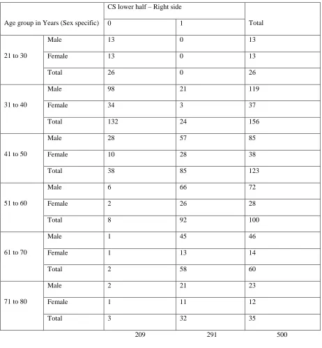
Table 4: Right wing of Coronal suture’s Lower half
Age group in Years (Sex specific)
CS lower half – Right side
Total
0 1
21 to 30
Male 13 0 13
Female 13 0 13
Total 26 0 26
31 to 40
Male 98 21 119
Female 34 3 37
Total 132 24 156
41 to 50
Male 28 57 85
Female 10 28 38
Total 38 85 123
51 to 60
Male 6 66 72
Female 2 26 28
Total 8 92 100
61 to 70
Male 1 45 46
Female 1 13 14
Total 2 58 60
71 to 80
Male 2 21 23
Female 1 11 12
Total 3 32 35
First Age group 21 to 30 years has 13 male and 13 female out of 26 cases. In male
population all 13 cases are at Stage 0 (open suture) and 13 cases by female
population are also at Stage 0 or their suture is open. So, the number of cases in
this age group towards Stage 1 becomes Zero.
Age group 31 to 40 years that constitutes 156 cases out of total sample of 500, has
male 119 and female 37 cases. In 119 male, 98 are at Stage 0 and the remaining 21
are at Stage 1 to 4. In 37 female, 34 are at Stage 0 and only 3 female individuals
are at Stage 1 to 4. A total of 132 cases at Stage 0 and 24 cases at Stage 1 to 4 is
obtained when male and female statistics are combined in this age group of 31 to
40 years.
In the age group of 41 to 50 years, a total of 123 cases contain 85 male and 38
female. Of 85 male, 28 are at Stage 0, 57 are at Stage 1 to 4. Of 38 female, 10 are
at Stage 0, remaining 28 are at Stage 1 to 4. Combining male and female statistics,
we get 38 cases at Stage 0, 85 cases at Stage 1 to 4.
With age group 51 to 60 years having100 cases, 72 are male and 28 are female. 72
male cases has 6 cases at Stage 0, 66 cases with Stage 1 to 4. 28 female cases has 2
cases at Stage 0 and 26 cases at Stage 1 to 4. So, 8 cases are at Stage 0 and 92
cases are at Stage 1 to 4on male and female combined statistics.
Age group 61 to 70 years has 60 cases, of which male are 46 and female are 14. Of
46 male, only 1 case is at Stage 0 and 45 cases are at Stage 1 to 4. Of 14 female, 1
case is at Stage 0 and 13 cases are at Stage 1 to 4. 2 cases at Stage 0 and 58 cases at
Stage 1 to 4 is the combined value.
Chart 4: Progression of Stages of fusion with age (CS R Lower 1/2)
Last age group with 71 to 80 years has got 35 cases. Out of this 23 are male and 12
are female. 23 male has only 2 subjects at Stage 0 and other 21 subjects at Stage 1
to 4. Among 12 female only 1 is at Stage 0 and other 11 are at Stage 1 to 4.
Combining the values we get 3 cases at Stage 0 and 32 cases at Stage 1 to 4.
60
21 to 30 y 31 to 40 y 41 to 50 y 51 to 60 y 61 to 70 y 71 to 80 y
% at 0 50 84.6 30.9 8 3.3 8.6
% at 1 50 17.6 69.1 92 96.7 91.4
50
84.6
30.9
8
3.3 8.6 50
17.6
69.1
92 96.7 91.4
0 20 40 60 80 100 120
Progression of stages of fusion with age
In summary, Right side of Coronal suture’s Lower half has 358 male and
142 female skulls assessed. Out of 358 male skulls, 148 are at Stage 0 and 210 are
at Stage 1 to 4. Out of remaining 142 female skulls, 61 skulls are at Stage 0 and 81
are at Stage 1 to 4. Combining male and female skull statistics we arrive at 209
skulls at Stage 0 and 291 skulls are at Stage 1 to 4 (Table 4). In the Chart 4, we can
see fall in non-closure status percentage as age advances (blue dotted line) and rise
in percentage of fusion Stage in skulls as age increases (red dotted line).
Table 5: Statistical variables for Right wing of Coronal suture’s Lower half
Stage N Mean SD SE
0 209 38.61 8.205 .568
1 291 56.08 11.620 .681
Mean obtained for Stage 0 is 38.61 years, Stage 1 is 56.08 years.
Standard deviation thus calculated is 8.205 for Stage 0 and 11.620 for Stage 1.
Standard error Mean is .568 for Stage 0 and .681 for Stage 1 (Table 5).
These are variables calculated for Right side of Coronal Suture’s Lower half.
Left wing of Coronal suture’s Upper half:
Left wing of Coronal suture’s Upper half is analyzed and found to have
same statistical values that of Right side of Coronal suture’s Upper half.
[image:75.612.67.547.264.363.2]Distribution of male and female in individual sutures is constant in the study.
Table 6: Statistical variables for Left wing of Coronal suture’s Upper half
Stage N Mean SD SE
0 278 40.12 7.467 .448
1 222 59.62 11.233 .754
Distribution of number and percentage of cases in Stage 0 and Stage 1 in age
groups divided at the starting of analysis is also the same between Right side of
Coronal suture’s Upper half and Left side of Coronal suture’s Upper half (Table 2).
Hence, mean age of fusion for Stage 0, Stage 1; standard deviation, standard error
of mean are all the same with Right side of Coronal suture’s Upper half (Table 6).
Here, the Chart 5: Statistical variables for Coronal suture Upper half, blue bar with
38.61 mean value belongs to mean age of non-fusion or mean age of Stage 0. A
black link line inside and above the blue bar is area for Standard deviation.
Likewise, blue bar with 56.08 mean value belongs to mean age of Stage 1
and the link line on it refers the area of Standard deviation.
Chart 5: Statistical variables (SD) for Coronal suture Upper half.
Standard deviation is indicated by black link line
Here, in this Chart 6: Statistical variables for Coronal suture Upper half, blue bar
with 38.61 mean value belongs to mean age of non-fusion or mean age of Stage 0.
A black link line inside and above the blue bar is for Standard error.
63
Stage 0 Stage 1
Mean 38.61 56.08
38.61
56.08
0 10 20 30 40 50 60 70
Statistical Variables for Coronal suture Upper half