This is a repository copy of
Molecular characterisation of Czech Clostridium difficile
isolates collected in 2013-2015
.
White Rose Research Online URL for this paper:
http://eprints.whiterose.ac.uk/104630/
Version: Accepted Version
Article:
Krutova, M, Nyc, O, Matejkova, J et al. (3 more authors) (2016) Molecular characterisation
of Czech Clostridium difficile isolates collected in 2013-2015. International Journal of
Medical Microbiology, 306 (7). pp. 479-485. ISSN 1438-4221
https://doi.org/10.1016/j.ijmm.2016.07.003
© 2016, Elsevier. Licensed under the Creative Commons
Attribution-NonCommercial-NoDerivatives 4.0 International
http://creativecommons.org/licenses/by-nc-nd/4.0/
[email protected] https://eprints.whiterose.ac.uk/ Reuse
Unless indicated otherwise, fulltext items are protected by copyright with all rights reserved. The copyright exception in section 29 of the Copyright, Designs and Patents Act 1988 allows the making of a single copy solely for the purpose of non-commercial research or private study within the limits of fair dealing. The publisher or other rights-holder may allow further reproduction and re-use of this version - refer to the White Rose Research Online record for this item. Where records identify the publisher as the copyright holder, users can verify any specific terms of use on the publisher’s website.
Takedown
If you consider content in White Rose Research Online to be in breach of UK law, please notify us by
1
Title: Molecular characterisation of Czech
Clostridium difficile
isolates collected in 2013-2015
Krutova M.1, 2, Nyc O.1, Matejkova J.1, Allerberger F.3, Wilcox M. H.4, Kuijper E. J.5
1. Department of Medical Microbiology, 2nd Faculty of Medicine, Charles University in Prague and
Motol University Hospital, Czech Republic
2. DNA laboratory, Department of Paediatric Neurology, 2nd Faculty of Medicine, Charles University in Prague and Motol University Hospital, Czech Republic
3. Austrian Agency for Health and Food Safety (AGES), Wien, Austria
4. Leeds Teaching Hospitals NHS Trust, Leeds, United Kingdom
5. Leiden University Medical Centre, Leiden, the Netherlands
Corresponding author:
MSc. Marcela Krutova
Department of Medical Microbiology
2nd Faculty of Medicine, Charles University in Prague and University Hospital in Motol V Uvalu 84
150 06 Prague 5
Czech Republic
Tel: +420 732 532 499
2
Molecular characterisation of Czech
Clostridium difficile
isolates
collected in 2013-2015
Abstract
Clostridium difficile is a leading nosocomial pathogen and molecular typing is a crucial part of
monitoring its occurrence and spread. Over a three-year period (2013-2015), clinical Clostridium
difficile isolates from 32 Czech hospitals were collected for molecular characterisation. Of 2,201 C.
difficile isolates, 177 (8%) were non-toxigenic, 2,024 (92%) were toxigenic (tcdA and tcdB) and of
these, 677 (33.5%) carried genes for binary toxin production (cdtA, cdtB). Capillary-electrophoresis
(CE) ribotyping of the 2,201 isolates yielded 166 different CE-ribotyping profiles, of which 53 were
represented by at least two isolates for each profile. Of these, 29 CE-ribotyping patterns were
common to the Leeds-Leiden C. difficile reference strain library and the WEBRIBO database (83.7%
isolates), and 24 patterns were recognized only by the WEBRIBO database (11.2% isolates). Isolates
belonging to these 53 CE-ribotyping profiles comprised 94.9% of all isolates. The ten most frequent
CE-ribotyping profiles were 176 (n=588, 26.7%), 001 (n=456, 20.7%), 014 (n=176, 8%), 012 (n=127,
5.8%), 017 (n=85, 3.9%), 020 (n=68, 3.1%), 596 (n=55, 2.5%), 002-like (n=45, 2.1%), 010 (n=35, 1.6%)
and 078 (n=34, 1.6%). Multi-locus sequence typing (MLST) of seven housekeeping genes performed
in one isolate of each of 53 different CE-ribotyping profiles revealed 40 different sequence types
(STs). We conclude that molecular characterisation of Czech C. difficile isolates revealed a high
diversity of CE-ribotyping profiles; the prevailing RTs were 001 (20.7%) and 176 (027-like, 26.7%).
Introduction
Clostridium difficile is a major causative agent of hospital-acquired diarrhoea. Molecular typing of
clinically significant C. difficile isolates is a crucial tool for surveillance and spread control of C. difficile
infections (CDI). The typing approaches are focused on conserved parts, repetitive regions or entire
genomes (Knetsch et al., 2013). They include PCR-ribotyping (Bidet et al., 1999, Stubbs et al., 1999;
Indra et al., 2008; Fawley et al., 2015), multi-locus sequence typing (MLST) (Griffiths et al., 2010), and
toxinotyping (Rupnik, 2010). Multi-locus variable tandem-repeats analysis (MLVA) (van den Berg et
al., 2007) and whole-genome sequencing (Eyre et al., 2013).
The Czech Republic participated in the European Clostridium difficile infection surveillance Network
(ECDIS-net), a European Centre for Disease Prevention and Control (ECDC) supported project that
started in 2011 and focused on building laboratory capacity for pan-European Clostridium difficile
3 Motol University Hospital introduced CE-ribotyping, and 2,201 Czech C. difficile isolates were sent
from 32 hospitals for molecular typing over a three-year period (2013-2015).
The aim of this study was to use molecular methods to characterise C. difficile isolates circulating in
the Czech Republic from 2013-2015.
Material and methods
C. difficile
strain collection
Microbiology laboratories in 32 Czech healthcare facilities (7 tertiary care hospitals, 24 secondary
care hospitals and 1 specialized care hospital), covering 39% of the hospital beds in the Czech
Republic, were invited to cooperate voluntarily in this three-year project (2013-2015). Information
about the participating hospitals, CDI testing algorithm used, and the number of submitted isolates is
shown in in the supplementary material: Characterisation of hospitals in the study. C. difficile isolates
were cultured from stool samples taken from hospitalised patients of all ages suspected of CDI,
including community-acquired and hospital-acquired CDI. The number of isolates sent for molecular
characterisation from each hospital was not strictly determined. A total of 2,201 C. difficile isolates
was received or cultured at the department of Medical Microbiology of Motol University Hospital
and characterised by molecular methods.
Molecular characterisation of
C. difficile
strain collection
Ribotyping (ECDIS-net protocol)
Amplification of 16S-23S intergenic spacer regions was performed using the ECDIS-net protocol,
using primers described by Stubbs et al. (Stubbs et al., 1999). Capillary electrophoresis was
performed using an ABI 3130 Genetic Analyser (Applied Biosystems), a 36 cm array length, default
fragment analysis, POP7 polymer and LIZ1200 (Applied Biosystems) as a size standard. The ribotypes
were determined using the freely available WEBRIBO database (https://webribo.ages.at/) (Indra et
al., 2008) after Gene Mapper® v4.0 (Applied Biosystems) software processing. Subsequently, the
CE-ribotyping profiles obtained were also compared with the Leeds-Leiden C. difficile reference strain
set of CE-ribotyping profiles (n=70) generated using Gene Mapper® v4.0 software (Applied
Biosystems) from *.fsa files used at the first stage of the CE-ribotyping validation study (Fawley et al.,
2015).
Presence of genes for toxin production
The presence of genes (tcdA, tcdB, cdtA and cdtB) for toxin production (A, B and binary) was
4 Leeds-Leiden reference strain (RT 027) as a positive control. The tcdA (due to their
) could not be identified because the location of the primers is upstream from the
repetitive region. These strains revealed positive tcdA fragment PCR amplification (Persson et al.,
2008, 2009).
Molecular characterisation of 53 selected CE-ribotyping profile
C. difficile
isolates
Ribotyping (new consensus protocol)
Selected isolates of 53 CE-ribotyping profiles were reinvestigated according to the recently published
consensus CE-ribotyping protocol (Fawley et al., 2015), which applies primers described by Bidet et
al. (Bidet et al., 1999). We carried out a cluster analysis of these CE-ribotyping profiles using the
Unweighted Pair Group Method, with Arithmetic Mean (UPGMA) distance analysis based on the
presence of CE-ribotyping peaks of defined molecular weight (Bionumerics v7.1 Applied Maths; the
UPGMA figure is in the supplementary material).
MLST
The MLST was performed by amplification and sequencing of seven housekeeping genes: adk1,
atpA1, dxr3, glyA1, recA2, sodA5 and tpi2 (Griffiths et al., 2010). The sequences obtained were
uploaded to the MLST database (http://pubmlst.org/cdifficile) to determine the appropriate alleles
of the genes. The sequence type was determined by the combination of identified alleles. A
maximum-likelihood tree was generated from the alignment of concatenated DNA sequences of
seven housekeeping loci using the MEGA5 software available at http://www.megasoftware.net/
(Tamura et al., 2011).
Presence of deletions in the tcdC gene
The tcdC gene was amplified with primers C1 and C2 (Spigaglia and Mastrantonio, 2002) and
sequenced in a reverse direction. The sequences obtained were compared with the NCBI reference
sequence Peptoclostridium difficile 630, NC_009089.1.
Results
A total of 2,201 C. difficile isolates was collected from 32 hospitals from 2013-2015. The geographical
distribution of participating hospitals and the number of C. difficile isolates available for molecular
characterization is shown in Figure 1. The mean age of patients was 65.7 years (range 30 days - 97
years). Of 2,201 isolates, 82 (3.7%), 103 (4.7%), 509 (23.1%) and 1507 (68.5%) were from patients
5
Ribotyping and the presence of toxin genes
Of 2,201 C. difficile isolates, CE-ribotyping revealed 53 profiles in 2,088 isolates (94.9%) when at least
two isolates per profile were identified. Of the 53 CE-ribotyping profiles, 29 were recognized both by
the Leeds-Leiden reference set and the WEBRIBO database and comprised 1,841 (83.7%) of all
isolates (n=2,201). The remaining 24 CE-ribotyping profiles (247, 11.2%, of all isolates) were only
identified by the WEBRIBO database and were designated as WEBRIBO types (WRTs). The remaining
113 (5.1%) isolates yielded unique single profiles. An overview of the RTs and WRTs identified is
shown in Table 1. The highest diversity was found among 1,507 isolates derived from patients of age
>65 years, from whom 28 RTs, 24 WRTs and 58 single profiles were identified. In the 19-64 years age
group, 509 isolates yielded 27 RTs, 17 WRTs and 38 single profiles; in the 3-18 years group, 103
isolates showed 24 RTs, 9 WRTs and 12 single profiles, and in the two years and younger group 11
RTs, 4 WRTs and 5 single profiles were found for 82 isolates.
Of 2,201 C. difficile isolates, 2,024 (92%) were toxigenic (tcdA and tcdB) and of these, 677 (33.5%)
isolates carried genes for binary toxin production (cdtA, cdtB) and the remaining 177 (8%) isolates
were non-toxigenic. The highest ratio of non-toxigenic to toxigenic isolates (64:18) was found for the
group of patients two years old and younger. By comparison, the non-toxigenic to toxigenic isolate
ratio was 27:76 for patients of age 3-18 years, 33:476 for those of age 19-64 years, and 53: 1,454 for
those >65 years.
The most frequently identified toxigenic CE-ribotyping profiles were RTs 176 (n=588, 29.1%), 001
(n=456, 22.5%), 014 (n=176, 8.7%), 012 (n=127, 6.3%), 017 (n=85, 4.2%), 020 (n=68, 3.4%), 078 (n=34,
1.7%), 005 (n=30, 1.5%) and WRT 002-like (n=45, 2.2%). The distribution of predominant RTs 001 and
176 differs distinctly within age groups of patients. Whereas in group of patient two years old and
younger is the presence of these RTs rare (1.2% of RT 001 only), in group of patients 3-18 years was
11.7% (10.7%, 1%), in group of patient 19-64 years was 35.5% (14.7%, 20.8%) and in group of
patients >65 years was 56.4% (24.5%,31.9%)
The most frequent non-toxigenic CE-profiles were WRT 596 (n=55, 31.1%) and RT 010 (n=35, 19.8%).
WRT 596 was identified in isolates derived from all patient age groups, but the majority (39/55) were
detected in isolates from paediatric patients aged <2 years. The presence of the 11 most common
CE-ribotyping profiles in individual hospitals is shown in Figure 1, and its distribution according to
patient age in Figure 2.
Of the 53 CE-ribotyping profiles, one isolate from each profile was reinvestigated by the new
6 a change in their CE-ribotyping profile due to an additional amplification of the 326 bp fragment.
WRT 203 changed to WRT 209 and RT 002 to WRT 002-like, whereas WRT AI-60 and WRT AI-75
retained the same designation in the WEBRIBO database. The additional amplification of a 326 bp
fragment, observed in RT 002, was not noticed in the Leeds-Leiden reference RT 002 strain,
suggesting that only a local Czech RT 002 variant showed this difference.
The UPGMA analysis of CE-ribotyping profiles and the CE-ribotyping profiles together with their band
sizes are shown in the supplementary material (UPGMA, Supplementary material Molecular data
on Czech C. difficile strain collection).
MLST and the presence of deletions in the
tcdC
gene
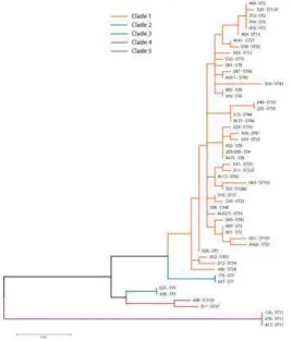
The application of the MLST of seven housekeeping genes in isolates from 53 ribotypes revealed 40
different STs clustering to 5 clades (Table 1, Figure 3). The isolates revealing similar ST but different
RT or WRT are listed in Table 2. Clade 1 was heterogeneous and consisted of 44 CE-ribotyping
profiles, 25 RTs and 19 WRTs, 37 toxigenic (tcdA+, tcdB+) and 7 non-toxigenic. Clade 2 included only
two RTs, both of ST1: RTs 027 and 176. Clade 3 contained two isolates with an identical ST5: RT 023
and WRT 438. Clade 4 consisted of two isolates belonging to RT 017 (ST37) and WRT 498 (ST170). In
Clade 5, three isolates of ST11 (RTs 078 and 126, WRT 413) were recognized.
RTs 027 and 176 had the one base pair deletion at nucleotide position 117,and the 18 bp deletion in
the tcdC gene. RT 023 and WRT 438 had the 54 bp deletion in the tcdC gene. RTs 078, 126 and WRT
413 showed the 39 bp deletion in the tcdC gene. The isolates harbouring 54 bp and 39 bp deletions
(except for WRT 413) revealed a nonsense mutation C184T. All isolates belonged to RT 023, 027, 126,
176 and WRTs 413 and 438 were also binary toxin gene positive.
Discussion
During a three-year period (2013-2015) a total of 32 hospitals voluntarily participated in this project,
but only 11 hospitals sent isolates for molecular characterisation in each year of the study. Eight
percent of C. difficile isolates were non-toxigenic although they were cultured from patients
suspected of CDI. These isolates were sent from hospitals with a suboptimal CDI diagnostic
algorithm, which means the absence of a confirmatory test for GDH-tested positive only samples
(Debast et al., 2014), or they were cultured in our laboratory, where all cultured C. difficile isolates
are ribotyped and tested for the presence of genes for toxin production. In our study, 12 hospitals
did not confirm the production of toxins by C. difficile isolates cultured from GDH-tested positive only
stool samples. Additionally, one laboratory did not test for the presence of toxins in stool samples
7 ribotyping of 2,201 Czech isolates revealed 166 different ribotyping profiles. Of these, 113
CE-ribotyping profiles (5.1%) were represented by only a single isolate, and its clinical and/or
epidemiological significance is unclear. Fifty-three different CE-ribotyping profiles contained at least
two isolates per profile. Of the 53 CE-ribotyping profiles, 29 profiles comprising 83.7% of all isolates
were recognized identically by two large, frequently used databases. The spectrum of the most
frequently found toxigenic RTs found in our study is similar to the most frequently found toxigenic
RTs in the European hospital-based survey (Bauer et al., 2011). The exception is RT 176, with its
specifically geographic-epidemiological occurrence in the Czech Republic (Krutova et al., 2014b)and
Poland (Pituch et al., 2015). RT 176 belongs to the RT 027 family (Valiente et al., 2012). Data on CDI
patients infected by RT 176 outcomes have recently been published in two single-centre studies,
including 30 and 111 patients, respectively. The results showed a higher rate of severe CDI (11/7 and
13/3) and mortality (5/2 and 16/8) in patients infected by RT 176 compared with patients infected by
non-176 ribotypes (Drabek et al., 2015; Polivkova et al., 2016). While RT 027 is distributed worldwide
(He et al., 2013), its occurrence is rare to date in the Czech Republic (Krutova et al., 2014b). We
identified only five isolates in four different hospitals over three years; however, hospitals from
border areas with Germany and Poland (Figure 1) did not participate in this study, and both countries
have high prevalence rates of RT 027 (Arvand et al., 2014, Pituch et al., 2015). The second most
common CE-ribotyping profile was RT 001 (n=456). In contrast with RT 176, RT 001 is frequently
found in many European countries (Bauer et al., 2011; Wiuf et al., 2011; Arvand et al., 2014; Taori et
al., 2014; Nyc et al., 2015; Freeman et al., 2015). In our study, the simultaneous presence of
ribotypes 001 and 176 was detected in 28 of the 32 hospitals.
Of 53 CE-ribotyping profiles, 24 were recognized only by the WEBRIBO database and these isolates
comprised 11.2% (n=247) of our collection. The occurrence of several WRTs identified in our study
(209, 220, 404, 416, 438, 500, 555, AI-12, AI-20, AI-21, AI-75, AI-9-1) has been reported as human
clinical isolates (Novak et al., 2015; Indra et al., 2015; Fang et al., 2014; Rafila et al., 2014; Hell et al.,
2011; Indra et al., 2008) or as animal isolates WRTs 203, 209, 413, 446, 596, AI-12, AI-60, AI-8/1,
AI-9-1 (Janezic et al., 2014; Schneeberg et a.l, 2013; Indra et al., 2009; Goldova et al., 2012; Indra et al.,
2008). Four of these WRTs (AI-82/1, AI-9-1, AI-60, AI-12) have recently been identified in the UK
Ribotyping Reference Laboratory (Leeds, UK) as RTs 103, 013, 097 and 150 respectively (Janezic et al.,
2014).
WRTs AI-82/1, AI-9-1 and AI-60 showed the same ST as was published by Dingle et al. in RTS 103, 013
and 097 (Dingle et al., 2011). WRTs 015 and 002 were assigned as WRTs 015-like and 002-like due to
slight changes in their CE-ribotyping profiles; however, the ST of WRT 002-like (ST8) was identical to
8 2011).The ST of WRT 015-like (ST44) corresponds with the findings of Dingle et al., who identified
two STs in RT 015 isolates: ST44-tcdC wild type and ST10, similarly to Knetsch et al. (Knetsch et al.,
2012), with the presence of 18 bp deletion in the tcdC gene (Dingle et al., 2011).
The distribution isolates depending on the age of the patients revealed the highest ratio of
non-toxigenic to non-toxigenic ribotypes (64:18) and low presence and absence of two predominant non-toxigenic
RTs 001 (1.2%) and 176 (0%) in patients two years old and younger. In other age groups (3-18 years,
19-64 years and >65 year), the non-toxigenic and toxigenic isolates ratio decreases (27:76, 33:476
and 53: 1,454), while the occurrence of RTs 001 and 176 increases (11.7%, 35.5%, to 56.4%),
respectively. The predominant occurrence of RTs 001 and 027 in older population was also found in
the study authors von Müller et al., where RT 027 was not present and RT 001 was present in 9.6 % in
group of patients 0-17 years and these ratios increased to 30.7% for RT 027 and 38.6% for RT001 in
oldest group of patients (>85 years). (von Müller et al., 2015).
The application of the new CE-ribotyping protocol (Fawley et al., 2015) changed the CE-ribotyping
profile in 7.5% of profiles (n=4) with a subsequent change of identification by the WEBRIBO database
in two profiles. The WEBRIBO database provides a broad spectrum of available CE-ribotyping profiles,
but the raw data are obtained by different protocols (primer design, polymer type) and some of the
CE-ribotyping profiles are designated only by a WEBRIBO number or by a combination of letter and
number. This stresses the importance of the use of a standardized protocol and also the
standardisation of an appropriate dataset of reference C. difficile strains uploaded to the WEBRIBO
database.
The MLST of seven housekeeping genes of 53 ribotypes revealed 40 different STs clustering to 5
clades. Although the MLST was performed only in one isolate of each identified CE-ribotyping profile,
we found the correlation with STs identified in ribotypes represented in the Leeds-Leiden C. difficile
reference strain collection published by Knetsch et al. (Table 1, marked with*) (Knetsch et al., 2012).
The most heterogeneous was MLST clade 1, which included 44 ribotyping profiles of 53
CE-ribotyping profiles. MLST clade 1 heterogeneity was also observed in the study by Stabler et al., who
found that this clade contained 106 STs of the 141 studied STs (Stabler et al., 2012). Knetsch et al.
typed 35 STs out of 56 as belonging to clade 1 (Knetsch et al., 2012), whereas Griffiths et al.
concluded that 31 STs out of 40 belonged to clade 1 (Griffiths et al., 2010). Similarly, Dingle et al.
found 60 STs out of 69 belonging to clade 1 (Dingle et al., 2011).
Several isolates belonging to a different RT or WTR revealed the same ST (clade) and the specific
deletion in tcdC gene that suggests their phylogenetic relationship. RTs 027 and 176 revealed ST1
9 base pair deletion at nucleotide position 117, which is a target site for commercial molecular systems
(Krutova et al., 2014a; Mentula et al., 2015),and 18 bp deletions in the tcdC gene. RT023 and WRT
438 revealed ST5 (clade 3) and had 54 bp deletions in the tcdC gene. RTs 078, 126 and WRT 413
showed ST11 and 39 bp deletions in the tcdC gene. Isolates harbouring 54 bp and 39 bp deletions
(except WRT 413) as previously described above revealed a nonsense mutation C184T (Spigaglia and
Mastrantonio, 2002; Curry et al., 2007). All these isolates (RT 023, 027, 126, 176 and WRTs 413 and
438) revealed the presence of binary toxin genes, another important C. difficile virulence factor
(Gerding et al., 2014).
The Czech Republic is a country with increasing CDI incidence (1.1 cases per 10,000 patient bed-days
in 2008 to 4.4 cases in 2011 2012 and 6.2 cases per 10,000 patient bed-days in 2012 2013) (Bauer
et al., 2008; Davies et al., 2014) and relatively high rates of antibiotic resistant C. difficile strains
(Freeman et al., 2015). Implementation of CDI surveillance based on the recently released CDI
surveillance protocol Control (ECDC, 2015) in the Czech Republic would fill the gap in Czech CDI
epidemiology with national CDI incidence data, including clinical case information and C. difficile
isolate antibiotic susceptibility results.
Conclusion
The molecular characterisation of 2,201 Czech clinical C. difficile isolates revealed 53 different
CE-ribotyping profiles and 40 multi-locus sequence types. Of 2,201 C. difficile isolates, 2,024 were
toxigenic (tcdA and tcdB), and of these, 677 isolates carried genes for binary toxin production (cdtA,
cdtB). The results of molecular characterisation showed a high diversity of C. difficile strains
circulating in the Czech Republic with prevailing representation of RTs 001 and 176 (027-like).
CE-ribotyping applied on a Czech C. difficile isolate collection demonstrates its high discrimination
capability and the results highlight the need to use a standardised protocol as well as a standardised
CE-ribotyping profile library to gain inter-laboratory comparable data on clinically and/or
epidemiologically significant C. difficile isolates.
Acknowledgement
We thank the ESCMID Study Group for Clostridium difficile (ESGCD) for their professional support.
We would like to thank Dr James Partridge for proofreading the text.
10 All procedures performed in studies involving human participants were in accordance with the
ethical standards of the institutional research committee and with the 1964 Helsinki declaration and
its later amendments or comparable ethical standards.
For this type of study, formal consent was not required.
Conflicts of interest
EJK, FA, ON, JM, MK declare no conflict of interests. MHW has received: consulting fees from
Actelion, Astellas, bioMerieux, MedImmune, Merck, Pfizer, Qiagen, Sanofi-Pasteur, Seres, Summit,
Synthetic Biologics and Valneva; lecture fees from Alere, Astellas, Merck & Pfizer; and grant support
from Actelion, Astellas, bioMerieux, Da Volterra, Merck, Sanofi-Pasteur, Seres and Summit. There is
no relationship between above mentioned companies and the study presented in this manuscript.
Funding
Supported by the MH CZ DRO, University Hospital Motol, Prague, Czech Republic 00064203.
References
Arvand M, Vollandt D, Bettge-Weller G, Harmanus C, Kuijper EJ; Clostridium difficile study group
Hesse., 2014. Increased incidence of Clostridium difficile PCR ribotype 027 in Hesse, Germany, 2011
to 2013. Euro Surveill. 19(10). pii: 20732.
Bauer, M.P., Notermans, D.W., van Benthem, B.H., Brazier, J.S., Wilcox, M.H., Rupnik, M., et al., 2011.
Clostridium difficile infection in Europe: a hospital-based survey. Lancet. 377: 63-73.
Bidet, P., Barbut , F., Lalande, V., Burghoffer, B., Petit J.C., 1999. Development of a new
PCR-ribotyping method for Clostridium difficile based on ribosomal RNA gene sequencing. FEMS Microbiol
Lett. 175: 261-6.
Curry, S.R., Marsh, J.W., Muto, C.A., O'Leary, M.M., Pasculle, A.W., Harrison, L.H., 2007. tcdC
genotypes associated with severe TcdC truncation in an epidemic clone and other strains of
Clostridium difficile. J. Clin. Microbiol. 45: 215-221.
Davies KA, Longshaw CM, Davis GL, Bouza E, Barbut F, Barna Z, Delmée M, Fitzpatrick F, Ivanova K,
Kuijper E, Macovei IS, Mentula S, Mastrantonio P, von Müller L, Oleastro M, Petinaki E, Pituch H,
N T N E N O Rupnik M, Schmid D, Wilcox MH., 2014. Underdiagnosis of Clostridium difficile across Europe: the European, multicentre, prospective, biannual, point-prevalence study of
Clostridium difficile infection in hospitalised patients with diarrhoea (EUCLID). Lancet Infect Dis.
11 Debast SB, Bauer MP, Kuijper EJ; European Society of Clinical Microbiology and Infectious Diseases.
2014. European Society of Clinical Microbiology and Infectious Diseases: update of the treatment
guidance document for Clostridium difficile infection. Clin Microbiol Infect. 20 Suppl 2:1-26.
Dingle, K.E., Griffiths, D., Didelot, X., Evans, J., Vaughan, A., Kachrimanidou M., et al., 2011. Clinical
Clostridium difficile: clonality and pathogenicity locus diversity. PLoS One. 6: e19993.
Drabek J, Nyc O, Krutova M, Stovicek J, Matejkova J, Keil R., 2015. Clinical features and characteristics
of Clostridium difficile PCR-ribotype 176 infection: results from a 1-year university hospital internal
ward study. Ann Clin Microbiol Antimicrob. 14:55.
European Centre for Disease Prevention and Control. 2015. European Surveillance of Clostridium
difficile infections. Surveillance protocol version 2.2 Stockholm: ECDC.
Eyre DW, Fawley WN, Best EL, Griffiths D, Stoesser NE, Crook DW, Peto TE, Walker AS, Wilcox MH.,
2013. Comparison of multilocus variable-number tandem-repeat analysis and whole-genome
sequencing for investigation of Clostridium difficile transmission. J Clin Microbiol. 51(12):4141-9.
Fang WJ, Jing DZ, Luo Y, Fu CY, Zhao P, Qian J, Tian BR, Chen XG, Zheng YL, Zheng Y, Deng J, Zou WH,
Feng XR, Liu FL, Mou XZ, Zheng SS., 2014. Clostridium difficile carriage in hospitalized cancer patients:
a prospective investigation in eastern China. BMC Infect Dis. 14: 523.
Fawley WN, Knetsch CW, MacCannell DR, Harmanus C, Du T, Mulvey MR, Paulick A, Anderson L,
Kuijper EJ, Wilcox MH., 2015. Development and validation of an internationally-standardized,
high-resolution capillary gel-based electrophoresis PCR-ribotyping protocol for Clostridium difficile. PLoS
One. 10: e0118150.
Freeman J, Vernon J, Morris K, Nicholson S, Todhunter S, Longshaw C, Wilcox MH; Pan-European
Longitudinal Surveillance of Antibiotic Resistance among Prevalent Clostridium difficile Ribotypes'
Study Group., 2015. Pan-European longitudinal surveillance of antibiotic resistance among prevalent
Clostridium difficile ribotypes. Clin Microbiol Infect. 21(3):248.e9-248.e16.
Gerding DN, Johnson S, Rupnik M, Aktories K., 2014. Clostridium difficile binary toxin CDT:
mechanism, epidemiology, and potential clinical importance. Gut Microbes. 5(1):15-27.
Griffiths D, Fawley W, Kachrimanidou M, Bowden R, Crook DW, Fung R, Golubchik T, Harding RM,
Jeffery KJ, Jolley KA, Kirton R, Peto TE, Rees G, Stoesser N, Vaughan A, Walker AS, Young BC, Wilcox
M, Dingle KE., 2010. Multilocus sequence typing of Clostridium difficile. J Clin Microbiol. 48: 770-8.
Goldová, J., Malinová, A., Indra, A., Vítek, L., Branny, P., Jirásková, A., 2012. Clostridium difficile in
12 He M, Miyajima F, Roberts P, Ellison L, Pickard DJ, Martin MJ, Connor TR, Harris SR, Fairley D,
Bamford KB, D'Arc S, Brazier J, Brown D, Coia JE, Douce G, Gerding D, Kim HJ, Koh TH, Kato H, Senoh
M, Louie T, Michell S, Butt E, Peacock SJ, Brown NM, Riley T, Songer G, Wilcox M, Pirmohamed M,
Kuijper E, Hawkey P, Wren BW, Dougan G, Parkhill J, Lawley TD., 2013. Emergence and global spread
of epidemic healthcare-associated Clostridium difficile.Nat Genet. 45(1):109-13.
Hell M, Permoser M, Chmelizek G, Kern JM, Maass M, Huhulescu S, Indra A, Allerberger F.., 2011.
Clostridium difficile infection: monoclonal or polyclonal genesis? Infection. 39: 461-5.
Indra A, Huhulescu S, Schneeweis M, Hasenberger P, Kernbichler S, Fiedler A, Wewalka G, Allerberger
F, Kuijper EJ., 2008. Characterization of Clostridium difficile isolates using capillary gel
electrophoresis-based PCR ribotyping. J Med Microbiol. 57: 1377-82.
Indra, A., Lassnig, H., Baliko, N., Much, P., Fiedler, A., Huhulescu, S., Allerberger, F., 2009. Clostridium
difficile: a new zoonotic agent? Wien Klin Wochenschr. 121: 91-5.
Indra A, Schmid D, Huhulescu S, Simons E, Hell M, Stickler K, Allerberger F; Austrian C. difficile Study
Group., 2015. Clostridium difficile ribotypes in Austria: a multicenter, hospital-based survey. Wien
Klin Wochenschr. 127: 587-93.
Janezic S, Zidaric V, Pardon B, Indra A, Kokotovic B, Blanco JL, Seyboldt C, Diaz CR, Poxton IR, Perreten
V, Drigo I, Jiraskova A, Ocepek M, Weese JS, Songer JG, Wilcox MH, Rupnik M., 2014. International
Clostridium difficile animal strain collection and large diversity of animal associated strains. BMC
Microbiol. 14: 173.
Knetsch CW, Terveer EM, Lauber C, Gorbalenya AE, Harmanus C, Kuijper EJ, Corver J, van Leeuwen
HC., 2012. Comparative analysis of an expanded Clostridium difficile reference strain collection
reveals genetic diversity and evolution through six lineages. Infect Genet Evol. 12: 1577-85.
Knetsch, C.W., Lawley, T.D., Hensgens, M.P., Corver, J., Wilcox, M.W., Kuijper, E.J., 2013. Current
application and future perspectives of molecular typing methods to study Clostridium difficile
infections. Euro Surveill. 18: 20381.
Krutova, M., Matejkova, J., Nyc, O., 2014a. C. difficile ribotype 027 or 176? Folia Microbiol. 59: 523-6.
Krutova, M., Nyc, O., Kuijper, E.J., Geigerova, L., Matejkova, J., Bergerova, T., Arvand, M., 2014b. A
case of imported Clostridium difficile PCR-ribotype 027 infection within the Czech Republic which has
a high prevalence of C. difficile ribotype 176. Anaerobe. 30: 153-5.
Mentula S, Laakso S, Lyytikäinen O, Kirveskari J., 2015. Differentiating virulent 027 and non-027
13 Novak, A., Rubic, Z., Dogas, V., Goic-Barisic, I., Radic, M., Tonkic, M., 2015. Antimicrobial susceptibility
of clinically isolated anaerobic bacteria in a University Hospital Centre Split, Croatia in 2013.
Anaerobe. 31: 31-6.
Nyc O, Krutova M, Liskova A, Matejkova J, Drabek J, Kuijper EJ., 2015. The emergence of Clostridium
difficile PCR-ribotype 001 in Slovakia. Eur J Clin Microbiol Infect Dis. 34(8):1701-8.
Persson, S., Torpdahl, M., Olsen, K.E., 2008 and 2009. New multiplex PCR method for the detection of
Clostridium difficile toxin A (tcdA) and toxin B (tcdB) and the binary toxin (cdtA/cdtB) genes applied
to a Danish strain collection. Clin Microbiol Infect. 14: 1057-64. Erratum in: Clin Microbiol Infect. 15:
296.
Pituch H, Obuch-W P L D W D K P M G D
SM, Kuijper EJ; Polish Clostridium difficile Study Group., 2015. Hospital-based Clostridium difficile
infection surveillance reveals high proportions of PCR ribotypes 027 and 176 in different areas of
Poland, 2011 to 2013. Euro Surveill. 20(38).
Polivkova S, Krutova M, Petrlova K, Benes J, Nyc O., 2016. Clostridium difficile ribotype 176 - A
predictor for high mortality and risk of nosocomial spread? Anaerobe. 40:35-40.
Rafila A, Indra A, Popescu GA, Wewalka G, Allerberger F, Benea S, Badicut I, Aschbacher R, Huhulescu
S., 2014. Occurrence of Clostridium difficile infections due to PCR ribotype 027 in Bucharest,
Romania. J Infect Dev Ctries. 8: 694-8.
Rupnik M., 2010. Clostridium difficile toxinotyping. Methods Mol Biol. 646:67-76.
Spigaglia, P., Mastrantonio, P., 2002. Molecular analysis of the pathogenicity locus and polymorphism
in the putative negative regulator of toxin production (TcdC) among Clostridium difficile clinical
isolates. J Clin Microbiol. 40: 3470-5.
Stabler RA, Dawson LF, Valiente E, Cairns MD, Martin MJ, Donahue EH, Riley TV, Songer JG, Kuijper
EJ, Dingle KE, Wren BW., 2012. Macro and micro diversity of Clostridium difficile isolates from diverse
sources and geographical locations. PLoS One. 7(3):e31559.
Stubbs, S.L., Brazier, J.S., O'Neill, G.L., Duerden, B.I., 1999. PCR targeted to the 16S-23S rRNA gene
intergenic spacer region of Clostridium difficile and construction of a library consisting of 116
different PCR ribotypes. J Clin Microbiol. 37: 461-3.
Schneeberg A, Neubauer H, Schmoock G, Baier S, Harlizius J, Nienhoff H, Brase K, Zimmermann S,
Seyboldt C., 2013. Clostridium difficile genotypes in piglet populations in Germany. J Clin Microbiol.
14 Tamura, K., Peterson, D., Peterson, N., Stecher, G., Nei, M., Kumar, S., 2011. MEGA5: molecular
evolutionary genetics analysis using maximum likelihood, evolutionary distance, and maximum
parsimony methods. Mol Biol Evol. 28: 2731-9.
Taori SK, Wroe A, Hardie A, Gibb AP, Poxton IR., 2014. A prospective study of community-associated
Clostridium difficile infections: the role of antibiotics and co-infections. J Infect. 69(2):134-44.
Valiente E, Dawson LF, Cairns MD, Stabler RA, Wren BW., 2012. Emergence of new PCR ribotypes
from the hypervirulent Clostridium difficile 027 lineage. J Med Microbiol. 61(Pt 1):49-56.
van den Berg RJ, Schaap I, Templeton KE, Klaassen CH, Kuijper EJ., 2007. Typing and subtyping of
Clostridium difficile isolates by using multiple-locus variable-number tandem-repeat analysis. J Clin
Microbiol. 45(3):1024-8.
von Müller L, Mock M, Halfmann A, Stahlmann J, Simon A, Herrmann M., 2015. Epidemiology of
Clostridium difficile in Germany based on a single center long-term surveillance and German-wide
genotyping of recent isolates provided to the advisory laboratory for diagnostic reasons. Int J Med
Microbiol. 305(7):807-13.
Wiuff C, Brown DJ, Mather H, Banks AL, Eastaway A, Coia JE., 2011. The epidemiology of Clostridium
15
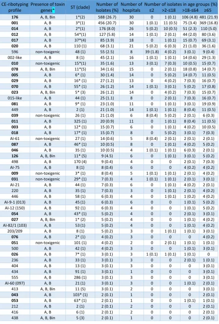
Table 1: Distribution of toxigenic and non-toxigenic ribotypes (bold) and WEBRIBO types of Czech C. difficile isolates as identified by Leeds-Leiden database and WEBRIBO database in a Czech C. difficile collection.
CE-ribotyping profile
Presence of toxin
genes* ST (clade)
Number of Isolates (%)
Number of hospitals
Number of isolates in age groups (%) >2- >18- 65
176 A, B, Bin 1*(2) 588 (26.7) 30 0 1 (0.1) 106 (4.8) 481 (21.9)
001 A, B 3*(1) 456 (20.7) 30 1 (0.1) 11 (0.5) 75 (3.4) 369 (16.8)
014 A, B 2*(1) 176 (8.0) 26 5 (0.2) 10 (0.5) 51 (2.3) 110 (5.0)
012 A, B 54*(1) 127 (5.8) 14 1 (0.1) 2 (0.1) 44 (2.0) 80 (3.6)
017 A, B 37*(4) 85 (3.9) 18 0 1 (0.1) 15 (0.7) 69 (3.1)
020 A, B 110 (1) 68 (3.1) 21 5 (0.2) 6 (0.3) 21 (1.0) 36 (1.6)
596 non-toxigenic 48 (1) 55 (2.5) 8 39 (1.8) 4 (0.2) 3 (0.1) 9 (0.4)
002-like A, B 8 (1) 45 (2.1) 16 1 (0.1) 1 (0.1) 14 (0.6) 29 (1.3)
010 non-toxigenic 15*(1) 35 (1.6) 13 3 (0.1) 7 (0.3) 10 (0.5) 15 (0.7)
078 A, B, Bin 11*(5) 34 (1.6) 15 0 2 (0.1) 18 (0.8) 14 (0.7)
005 A, B 6* (1) 30 (1.4) 14 0 5 (0.2) 14 (0.7) 11 (0.5)
029 A, B 16* (1) 27 (1.2) 13 0 4 (0.2) 7 (0.3) 16 (0.7)
070 A, B 55* (1) 26 (1.2) 14 1 (0.1) 3 (0.1) 5 (0.2) 17 (0.8)
023 A, B, Bin 5* (3) 26 (1.2) 14 0 4 (0.2) 7 (0.3) 15 (0.7)
015-like A, B 44 (1) 25 (1.1) 8 0 2 (0.1) 7 (0.3) 16 (0.7)
081 A, B 9* (1) 23 (1.0) 11 0 1 (0.1) 3 (0.1) 19 (0.9)
449 A, B 2 (1) 21 (1.0) 14 1 (0.1) 1 (0.1) 8 (0.4) 11 (0.5)
039 non-toxigenic 26 (1) 21 (1.0) 6 8 (0.4) 5 (0.2) 2 (0.1) 6 (0.3)
011 A, B 325 (1) 20 (0.9) 11 0 1 (0.1) 8 (0.4) 11 (0.5)
003 A, B 12* (1) 15 (0.7) 6 0 1 (0.1) 4 (0.2) 10 (0.5)
018 A, B 17* (1) 15 (0.7) 8 0 5 (0.2) 3 (0.1) 7 (0.3)
AI-61 non-toxigenic 27 (1) 14 (0.6) 5 8 (0.4) 2 (0.1) 2 (0.1) 2 (0.1)
087 A, B 46* (1) 10 (0.5) 8 0 1 (0.1) 4 (0.2) 5 (0.2)
046 A, B 35 (1) 10 (0.5) 4 1 (0.1) 1 (0.1) 6 (0.3) 2 (0.1)
126 A, B, Bin 11* (5) 9 (4.5) 6 0 1 (0.1) 3 (0.1) 5 (0.2)
498 A, B 170 (4) 9 (0.4) 4 0 0 2 (0.1) 7 (0.3)
AI-75 A, B 8 (1) 8 (0.4) 6 0 0 4 (0.2) 4 (0.2)
009 non-toxigenic 3* (1) 8 (0.4) 5 1 (0.1) 1 (0.1) 2 (0.1) 4 (0.2)
031 non-toxigenic 29* (1) 7 (0.3) 4 1 (0.1) 1 (0.1) 2 (0.1) 3 (0.1)
AI-21 A, B 44 (1) 7 (0.3) 6 0 1 (0.1) 4 (0.2) 2 (0.1)
220 A, B 35 (1) 7 (0.3) 3 0 1 (0.1) 2 (0.1) 4 (0.2)
446 A, B 58 (1) 6 (0.3) 4 0 1 (0.1) 1 (0.2) 4 (0.2)
AI-9-1 (013) A, B 45(1) 6 (0.3) 6 0 0 1 (0.1) 5 (0.2)
AI-12 (150) A, B 92 (1) 6 (0.3) 4 0 0 1 (0.1) 5 (0.2)
054 A, B 43* (1) 5 (0.2) 4 0 0 2 (0.1) 3 (0.1)
027 A, B, Bin 1* (2) 5 (0.2) 4 0 0 1 (0.1) 4 (0.2)
AI-82/1 (103) A, B 53(1) 5 (0.2) 4 0 0 1 (0.1) 4 (0.2)
203/209 A, B 8 (1) 5 (0.2) 3 0 1 (0.1) 1 (0.1) 3 (0.1)
076 A, B 2* (1) 4 (0.2) 3 0 0 0 4 (0.2)
051 non-toxigenic 101 (1) 4 (0.2) 2 0 2 (0.1) 1 (0.1) 1 (0.1)
500 A, B 42 (1) 4 (0.2) 3 0 0 1 (0.1) 3 (0.1)
026 A, B 7* (1) 3 (0.1) 3 1 (0.1) 1 (0.1) 1 (0.1) 0
236 A, B 33 (1) 3 (0.1) 3 0 0 2 (0.1) 1 (0.1)
404 A, B 13 (1) 3 (0.1) 3 0 0 0 3 (0.1)
434 A, B 91 (1) 3 (0.1) 1 0 0 0 3 (0.1)
555 A, B 286 (1) 3 (0.1) 3 0 0 0 3 (0.1)
AI-60 (097) A, B 21(1) 3 (0.1) 3 0 0 1 (0.1) 2 (0.1)
413 A, B, Bin 11 (5) 3 (0.1) 2 0 0 0 3 (0.1)
043 A, B 103* (1) 2 (0.1) 1 0 0 0 2 (0.1)
053 A, B 63* (1) 2 (0.1) 1 0 0 1 (0.1) 1 (0.1)
212 A, B 2 (1) 2 (0.1) 2 0 0 0 2 (0.1)
416 A, B 6 (1) 2 (0.1) 2 0 0 0 2 (0.1)
16
[image:17.595.66.280.180.301.2]Table 1 footnotes: MLST and tcdC sequencing were performed in representative isolates of each CE-ribotyping profile (n=53). Knetsch et al. identified STs-RTs marked with *. (ST: sequence type; tcdA/B: genes for toxin A/B production; cdtA/B: genes for binary toxin production; tcdC: toxin gene expression negative regulator). *Primers used to amplify tcdA are located upstream of the repetitive region in the 3´-end. The TcdA-negative strains due to 3´-end deletion revealed positive PCR amplification [Persoon et al., 2008, 2009].
Table 2: Ribotypes and WEBRIBO types (italics) revealing identical sequence type
ST Ribotype Clade
1 027, 176 2
2 014, 076, 212, 449 1
3 001, 009 1
5 023, 438 3
6 005, 416 1
8 002-like, 203/209, AI-75 1
11 078, 126, 413 5
35 046, 220 1
[image:17.595.76.570.388.706.2]44 015-like, AI-21 1
17
Figure 2: Distribution of commonest CE-ribotyping profiles depending on age of patients.
[image:18.595.76.345.398.712.2]