0022-538X/84/07057-06$02.00/0
Copyright C) 1984, American SocietyforMicrobiology
Isolation,
Characterization,
and
Physical
Mapping
of
a
Pseudorabies
Virus Mutant
Containing Antigenically
Altered
gp5O
MICHAEL W. WATHENl* AND LYNNE M. K.WATHEN2
NationalAnimalDisease Center, Agricultural Research Service, U.S. DepartmentofAgriculture,1 andDepartment of Animal
Science,
Iowa StateUniversity,2
Ames, Iowa50010Received6January 1984/Accepted26March1984
A pseudorabies virus variant (marl97-1) containing a mutation in a viral glycoprotein with a molecular weight of 50,000 (gp5O) was isolated by selecting for resistance to a neutralizing monoclonal antibody (MCA50-1)directedagainstgpSO.This mutantwascompletelyresistant to neutralization withMCA50-1inthe
presence or absence ofcomplement, and was therefore defined as a mar (monoclonal-antibody-resistant)
mutant. The mutation did not affect neutralization with polyvalent immune serum. The marl97-1 mutant synthesized and processed gp5O normally, but the mutation preventedthebindingandimmunoprecipitation of
gpSObyMCA50-1.Thus, the mutationwaswithinthe structuralportionofthegp5Ogeneaffectingtheepitope ofthe monoclonalantibody. The mutationwasmapped by markerrescuewithclonedpseudorabies restriction enzymefragmentstothe short unique region of the pseudorabiesgenomebetween 0.813 and 0.832mapunits. This isequivalent toa2.1-kilobase-pair region.
Pseudorabies virus (PRV) contains a double-stranded DNA genome withamolecularweight ofca. 90 x
106
(32). The genome can be divided into long and short unique regions, withtheshortunique regionsurroundedby inverted repeat sequences (4). Like otheralphaherpesviruses, PRV has ashortreplicativecycle in cell culture,resultingin mass destruction of susceptible cells (31). In swine (its natural host), PRV can causedeathinpigs ofallages andabortionof fetuses (19), thus making itaneconomically important virus.Sinceglycoproteinsarepresent on thesurface ofa herpes-virus envelope, they are the principal target antigens in-volved in virus neutralization (9, 15, 42). The envelope of herpes simplex virustype 1 (HSV-1) contains at least four antigenically distinct glycoproteins, designated gB, gC, gD, and gE(2, 20, 37). Glycoproteins gB,gC,andgDappear to be the
principal
immunogens involved in inducing boththe humoralandcellularhost immuneresponses (16,25,27,42). PRV also has been reported to have fourglycoproteins with molecular weights estimated to be 100,000 (100k) to 120k, 72k to 82k, 60k to 65k, and 45k to 50k (3, 23; L. K. Wathen, K. B. Platt, M. W. Wathen, R. A. Van Deusen, and C.A. Whetstone, manuscript inpreparation). A monoclonal antibody directedagainst the glycoprotein with amolecular weight of 50,000(gpSO)is a strong neutralizer of PRV with or without the aid of complement (Wathen et al., in prepara-tion). Furthermore, polyvalent immune serum is highly reactiveagainstgpSO.
Itthereforeappears thatgpSO
is oneof the importantPRV immunogens.Neutralizingmonoclonalantibodies directedagainst HSV-1 glycoproteins have been used to select viral antigenic variants which are resistant to neutralization (21). These antigenic variants usuallyexpress the mutated glycoprotein but donotbind the selectingmonoclonalantibodydue to an alteration in the epitope against which the monoclonal antibodyisdirected(21). Such mutants are distinctfromthe normalconditional lethal mutants and have beendesignated
mar(monoclonal-antibody-resistant) mutants(21).
Wereport here on theisolationandanalysisof an antigen-ic variant resistant to a neutralizing monoclonal antibody
*Corresponding author.
directed against the PRV gp5O. We used this mar mutantto
mapthegp5Ogene to an areawithin theshort uniqueregion of the PRV genome. The similarities between this PRV glycoprotein and glycoproteingDof HSV-1 are discussed.
MATERIALS AND METHODS
Virus and cell culture. Plaque-purified PRV strain Ind-FH (34) wasroutinelypassed at amultiplicityofinfection of0.01 PFU/ml inporcine MVPK-1 cells (41). Cells were propagat-ed in Eagle minimal essential medium (MEM) containing 10% fetal calf serum (FCS). Mutagenized stocks of PRV were prepared by adding 5-bromo-2'-deoxyuridine (BUdR; 10 ,ug/ml in MEM-10% FCS) at 1 h postinfection (p.i.). BUdR remained in the medium until the extracellular virus was harvested.
Production of monoclonalantibody stocks. Production and characterization of the hybridoma cell line MCA50-1, as well asthepreparation of ascites fluid used in this paper, will be described elsewhere (Wathen et al., in preparation). Clari-fied ascites fluid from mice injected intraperitoneally with lMlCA50-1 hybridomacells wasusedwithoutfurther purifica-tion.
Virus neutralization assay. Titers of monoclonal ascites fluid orporcine immune serum to PRV were determinedby serialtwofold dilutions in 96-well tissue cultureplates. The assays were performed by mixing 50 pu1 of serially diluted antibody which had beenincubatedat56°Cfor30min with ca. 100PFU (50
RI)
of virus. Complement-mediated neutral-ization was assayed by the addition of 0.5% normal rabbit serum. After a 3-h incubation at 37°C, 104 MVPK-1 cells were added to each well, and the assay was incubated at 37°Cfor48 h.Antibodytiters wereexpressedasthe recipro-cal ofthehighest dilution which reducedtheviralcytopathic effect (CPE)by 50% relative to unneutralized controls.Isolation of mar mutants. Antigenic variants of PRV were selectedon thebasisoftheir resistance toneutralizationwith themonoclonalantibodyMCA50-1. Approximately2 x
105
PFU of mutagenized virus was incubated with 0.25 ml of MCA50-1 ascites fluid for 3 h at 37°C. Survivingvirus was
allowed to adsorb to MVPK-1 cells at37°Cfor1h. The cell monolayer wasthen washed twice and overlaid with 0.8% agarcontaining MEM-5% FCS. At 48 h p.i.,plaques were
57
on November 10, 2019 by guest
http://jvi.asm.org/
58 WATHEN AND WATHEN
picked and dispensed into 24-well tissue culture plates containing confluent monolayers of MVPK-1 cells. When theinfection in these cells reached 100%CPE, the progeny virus was tested for resistance to MCA50-1 by using the neutralization assay described above. Suspected mar mu-tants were plaquepurified twiceand retested forresistance to neutralization. One mar mutant (marl97-1) was chosen for further experimentation.
Radiolabeling of cells. MVPK-1 cells (5 x 10⁶) were infected withPRV at amultiplicityof infectionof50 PFU per cell. After a 1-h adsorption period, the cells were washed twice, and fresh MEM-10% FCS was added at 37°C. At 16 h p.i., 2 ,uCi of
[14C]glucosamine
per ml (54 mCi/mmol; Amersham Corp., Arlington Heights, Ill.) was added, and the cells wereincubatedat37°C. The cells were harvested at 20 hp.i. by washing twiceand thenscrapinginto phosphate-buffered saline(PBS) (pH 7.4). The cellswere pelleted and resuspended in 0.5 mlof extractionbuffer(1%Nonidet P-40, 0.1 mM phenylmethylsulfonyl fluoride in PBS, pH 8.0). Afterabriefsonication,thedisrupted cellswere solubilized for 15 min at4°C. Extracts wereclarifiedbycentrifugationat 12,000 x gfor 15min.Immunoprecipitations. Immunoprecipitationswerecarried out as previously described (21). Briefly, radiolabeled cell extracts (100pAl)were incubated with5,ulofascitesfluidor 20 ,ul ofporcine immune serum overnight at 4°C. Antigen-antibody complexeswere harvestedbytheaddition of50,ul of protein A-Sepharose CL-4B beads (40% [vol/vol] in extraction buffer; Pharmacia Fine Chemicals, Piscataway, N.J.). The beads that were added to monoclonal antibody immunoprecipitates were presensitized with rabbit anti-mouseimmunoglobulin (Cappel Laboratories, Cochranville, Pa.). Afterincubating for1h at4°C, the beadswere washed six times with extraction bufferandresuspended in25
,u1
of 2x electrophoresis sample buffer (2% sodium dodecyl sul-fate, 5% 2-mercaptoethanol, 10% sucrose, 2 mg of brom-phenol blueperml, 10 mMTris-hydrochloride, pH8.0).Polyacrylamide gel electrophoresis. Immunoprecipitates and total cell extracts (5
,u1
in 2x electrophoresis sample buffer)wereboiledfor3minbefore sodiumdodecyl sulfate-polyacrylamide gel electrophoresis. Sepharose beads from theimmunoprecipitations were removed by centrifugation. The polypeptides were then fractionated in a resolving gelcontaining
10% acrylamide and 1.3 mg of bisacrylamide (Bio-RadLaboratories, Richmond, Calif.)per ml.The stack-ing gel contained 5% acrylamide and1.3 mgofbisacrylamide per ml. Electrophoresis was carriedout at75 V throughthe stacking gel and 150 V through the resolving gel by the method ofLaemmli (24). Molecular weight standards (Bio-RadLaboratories)werecoelectrophoresedin eachgel. Afterelectrophoresis,
gels werestained, destained,
and dried as previously described (38). Dried gelswereexposedtoKodak X-OmatARfilmatroomtemperature.Purification of viral DNA. Viral DNA used forcloningthe PRV genome was purified from cytoplasmic virions by a
modificationofthesodiumiodide-ethidiumbromidegradient centrifugation procedure ofWalboomers and Schegget (43) as previously described (28). Viral DNA used for marker rescueexperimentswaspurified fromPRVnucleocapsidsas previously described(40).
CloningDNArestrictionenzymefragmentsofPRV.BamHI restriction enzyme fragments of PRV DNA were inserted into the plasmid pACYC184 (8) by using standard cloning procedures.Subcloning of BamHIfragmentHwithSallwas
carriedoutin pBR325 (7).The
plasmids
pY165D, pY104H, and pY109Kcontaining
the PRV BamHI fragments DKN,H, and K,respectively, that were cloned into pBR322 were kindly provided by Prem S. Paul.
Ligated recombinant plasmid DNA was transformed into Escherichia coli HB101 by the calcium shock method of Cohen et al. (11). Cells were plated onto LB agar plates containing25
jig
ofchloramphenicolperml (pACYC184) or 200 Fig of ampicillin per ml (pBR325).Recombinant plasmid-containing colonies were initially screened by insertional inactivation of an antibiotic resist-ancemarker. Plasmid DNA was purifiedby using asodium hydroxide-sodiumdodecyl sulfate extraction procedure (6), followed by cesiumchloride-ethidium bromide equilibrium centrifugation (18). The identity of the viral insert was preliminarily determined by digestion of recombinant plas-mid DNA with various restriction enzymes (New England Biolabs,Beverly, Mass.) and electrophoresis in agarosegels by the method of Bachi and Arber (1). The identity of the DNA insert was verified byhybridizing 32P-labeled plasmid DNA to KpnI-digested PRV DNA fixed to nitrocellulose filters by the method ofSouthern (36). Plasmid DNA was radiolabeled in vitro by thenick translation method of Rigby etal. (30).
Excision of DNA fragments from agarose gels. DNA restric-tion enzyme fragments were recovered from horizontal agarose gelsby bandinterception onto NA-45 DEAE mem-branes (Schleicher and Schuell, Keene, N.H.) by using a modification of the method of Dretzen et al. (13). The NA-45 membranes were washed for 10 min in 10 mM EDTA (pH 7.5)and 5 min in 0.5 M NaOH and then wererinsedseveral times in deionized water. The activated membranes were thenplaced in slits in the agarose gel directly below the DNA bands ofinterest, and the DNA was electrophoresed onto the NA-45 membranes. After residual agarose was rinsed from themembranes with NET buffer (0.15 M NaCl, 0.1 mM EDTA, 20 mMTris-hydrochloride, pH 8.0), the DNA was' elutedfrom the membranes byincubationinhighsalt (1.0 M NaCI) NET buffer at 56°C for45 min. The DNA was then extractedthreetimes with anequal volumeof water-saturat-edn-butanol and wasethanol precipitated.
Marker rescue of marmutant with recombinantplasmids. The marker rescue technique used was essentially that of Stow et al. (39). Briefly, MVPK-1 cells at ca. 90% con-fluency were cotransfected with DNA from intact PRV (marl97-1) mutantvirus and PRVrecombinantplasmids by thecalciumphosphate infectivity technique ofGraham and van der Eb (17). Plasmid DNA had been digested with BamHI orBamHI-SalI, phenol extracted threetimes, chlo-roform extractedtwice,andethanolprecipitated.At 4hp.i., the cells were subjected to a 3-min polyethylene glycol-sucrose shock as previously described (35). Extracellular viruswas harvested when the infection reached 100% CPE (3 to4days p.i.).
Assay for marker rescue. Recombinant virus which had regained a wild-type gpSO was detected with an in situ enzyme immunoassay described by Holland et al. (22). Briefly, progeny virus from the transfections were plated onto60-mmpetridishes containingamonolayerof MVPK-1 cells at 100 to 1,000 PFU per petri dish. After a 1-h adsorption period, the cells were washed andoverlaid with 0.8%agarose containing MEM-5% FCS. At 2
days p.i.,
the overlay was removed, and the cells were fixed with glutar-aldehyde (0.25%in PBS, pH 7.4) for5 min. The cellswerewashed three times with PBS(pH 7.4)containing 3%bovine serumalbumin(BSA) (PBS-BSA)and then incubated for 2 h
at room temperature with a 1:1,000 dilution of MCA50-1 ascites fluid in PBS-BSA. Afterunbound monoclonal
anti-J. VIROL.
on November 10, 2019 by guest
http://jvi.asm.org/
body was removedbythree washeswithPBS-BSA,the cells were incubated for 2 h at room temperature with 2 ml of horseradish peroxidase-conjugated goat anti-mouse immunoglobulin (CappelLaboratories) diluted1:250in PBS-BSA. After this incubation, the cells were washed three times with PBS-BSA and incubated in 2 ml of substrate containing 4-chloro-1-naphthol and H202. Plaques with a wild-type morphology stained black in10to 20min,whereas nonrescued virus remained unstained.
RESULTS
Characterization of PRV (marl97-1) mutant. Antigenic variants of PRV (Ind-FH)containinga mutation ingpSOwere selectedonthebasis ofresistanceto aneutralizing monoclo-nal antibody (MCA50-1) directed against gpSO. Approxi-mately4% of theBUdR-treatedstock virus survived neutral-ization with MCA50-1, and ofthe survivors approximately 1% were mutants. Although an exactmutation rate forthe gpSO gene could not be determined because of the low number of mutantsisolated,otherexperiments with BUdR-treatedPRVyieldedamutationfrequency forthegp5Ogene of3 x
10-5
(unpublished data).The mutantmarl97-1waschosen for further experimenta-tion because of its complete resistance to MCA50-1. This antigenic variant was resistantto MCA50-1 in the presence or absence of complement at the lowest dilution tested (Table 1). Neutralization with immune serumfrom a PRV-infected pig wasunaffected by themutation (Table 1).
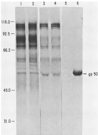
The inability of MCA50-1 to bind the mutated gpSO was demonstrated by immunoprecipitation of
[14C]glucosamine-labeled infectedcell extracts(Fig. 1). Comparison oflabeled total cell extractfrommutant (Fig. 1, lane 1) and wild-type (Fig. 1, lane 2) infectedcells indicated thatgp5O was being synthesized in the proper amounts and processed to the correctsizebymarl97-1. This was substantiated by immuno-precipitation ofthe cell extractswith porcine immuneserum (Fig. 1, lanes 3 and 4). However, MCA50-1, which readily immunoprecipitated wild-type gp5O (Fig. 1, lane 6), did not
immunoprecipitate
mutantgp5O
in detectableamounts (Fig. 1, lane 5).Cloning PRV BamHI restriction enzyme fragments. To obtainadequateamountsofuncontaminatedPRVrestriction enzyme fragments for use as probes in marker rescue
experiments,
PRV DNA was cleaved with BamHI and cloned into the plasmid vectors pACYC184 or pBR322 as described above.The twoplasmidvectors can be differenti-ated onagarosegels by the slower electrophoretic migration ofpBR322 (Fig. 2, plasmids DKN, H, and K). Two of theTABLE 1. Neutralization ofmar mutantandwild-type PRV'
Neutralizationtiter'for:
Antibody Complement
marl97-1 Ind-FH
MCA50-1C - <200d 205,000
MCA50-1 + <200 640,000
Immuneserume - 51,000 51,000
Immune serum + 102,000 102,000
a The two PRV isolates tested were the mutant
marl97-1,
which has antigenically alteredgp5O,andthe wild-type strain Ind-FH.bThe neutralization titer is expressed as the reciprocal of the highest dilution ofantibodywhich reduced the viral CPE by50%.
cClarified ascites fluid of monoclonalantibody directed againstgp5O.
dDilutionsbelow 1:200 were not tested because oftoxicitytocellsfrom the
ascitesfluid.
eMultivalent serum from a pig which had been naturally infected with a field strain of PRV.
2 3 4 5 6
116.0-92.
5fl-
66.2-,. .gpso
45.0
-
31.0-FIG. 1. Autoradiograph of 10% sodium dodecyl sulfate-poly-acrylamide gel containing [14C]glucosamine-labeled glycoproteins. Clarified cell extractsfromthemutantmarl97-1(lane 1) and
wild-type Ind-FH (lane 2) were eitherdirectly compared or
immuno-precipitated with porcine immuneserum(lane 3, marl97-1;lane 4, Ind-FH)ormonoclonalantibody MCA50-1(lane 5, marl97-1; lane
6,Ind-FH). Themigration of molecular weightmarkers (x103) and gpSOareindicated.
P D
R C K
V BPN E F G H I J K L M N P
BA"
C
-0
--EFG _ H IJ
-- K-L M
---N
p R V
__,PBR322
"PACYCl84
0 _
p ..
FIG. 2. Ethidium bromide-stained agarose gel containing PRV recombinant clones. PRV DNA was digested with BamHI and
electrophoresedin theouterlanesas areference.Theletterdenoting each PRVBamHI restriction enzyme fragment is shownontheleft. BqmHI-digested recombinant clones containing PRV BamHI re-striction enzyme fragments were arranged in the gel accordingto insert size. ThePRVinsert(s) in each clone is indicated above its lane. The migration of the two cloning vectors pBR322 and
pACYC184is markedontheright.
on November 10, 2019 by guest
http://jvi.asm.org/
[image:3.612.331.528.60.332.2] [image:3.612.313.553.435.631.2] [image:3.612.53.293.580.647.2]60 WATHEN AND WATHEN
MAP
UNITS
COMPONENTS
Bam
HI
Sal
I
Pvu I
0 0.1 0.2 0.3 0.4 0.5 0.6
0?
0.8 0.9 1.0I I I I I I I I I I I
UL
-
IRUs
-
TRQ F A B KM 0 C 6PJ I E L H N E 10
I I 1iIg 1 1 i 11iH
B A
iB
C AI ~~~ I -I
MAP
UNITS
rrKILOBASE
82PAIRS
I I I I I I
t0 0.830 0840 0.850 0.860 0.870 0.875
I I I I , 1
1.0 2.0 3.0 4.0 5.0 6.0 6.8
FIG. 3. Physical map of theBamHlcleavage sites for PRV (Ind-FH) and of theSallandPmlulcleavage sites for BamHI-H. TheBamHI restrictionenzyme map was originally described for the Shope strain of PRV (14) and was verified to be correct for strain Ind-FH by Southern blothybridization (unpublisheddata). BamHI-H lies within the short unique sectionofthe PRVgenome between 0.813 and 0.875 map units and is6.8kilobase pairslong. Restriction enzyme fragments within the BamHI-H region werealphabeticallylabeledaccording to size for each enzyme.
recombinant plasmids (CP and DKN) contained multiple PRVrestriction enzymefragments. However, the contribu-tion ofeachfragmentin these clones toward the ability of the clonetorescueamutationcould bedeterminedby compari-son withthesinglefragmentclones K, N, and P (Fig. 2). As expected,theend fragments0andQ (notdetectableinFig. 2) were not cloned. Since the sequences represented by BamHI-O are within the terminal repeat region (Fig. 3), these sequencesarealso present in theportion oftheinternal repeat represented by clone J (Fig. 2 and 3). The PRV sequences represented by restriction enzymeBamHI frag-mentsAand Qwere not present in thislibrary of clones.
Marker rescueofmarl97-1 with PRV BamHIclones. The genomic maplocation ofthemarmutationin the gpSOgene ofPRV (marl97-1) was determined by marker rescue with the PRV recombinant clones described above. Rescued virus wasdetectedbyusingan in situ enzyme immunoassay described above. This assay is based on the fact that only wild-type plaques derived from rescued viruswill bind the monoclonal antibody MCA50-1. Such plaques stain black underthe assay conditions, whereasmutantplaquesremain unstained. A petri dishcontaining virus from atransfection in which a fraction ofthe marl97-1 virus was rescued by
recombination
with aPRVclone is shown inFig.
4. The results of cotransfections with DNA from marl97-1and
PRV clonescontainingBamHI restrictionenzyme frag-ments arepresented in Table 2. OnlyBamHI-H wasableto rescue the mutation in the gpSO gene. This fragment is containedwithin
theshortuniqueregionof the PRV genome between 0.813 and 0.875 map units (Fig. 3). Background levels of marker rescue with the other BamHI clones orreversion of marl97-1 were belowdetectability (Table 2). Marker rescue ofmarl97-1 with restriction enzyme
frag-ments from the BamHI-H region. A map of the BamHI-H regionisshown inFig.3. This mapwasderivedbysingleand doublerestriction enzyme digestionsof theBamHI2H clone with BamHI, Sall, and PvuI. The Sall fragments were
subcloned by digesting the BamHI-H clone with BamHI-Sall andinsertingtheresultingfragmentsintopBR325which
had been digested with BamHI-SalI. This double digestion removed the viral DNA from the original pBR322 plasmid vector. ThePvuIviralfragmentswereisolatedfromagarose gels afterelectrophoresis of PvuI-digested BamHI-H clone as described above.
The results of marker rescue experiments with the Sall subclones orPvuI fragmentsfrom the BamHI-H region are presentedinTable 3. These results indicate that the mutation in the
gp5O
gene of marl97-1 is located at the left end of BamHI-H between 0.813 and 0.832 map units (Fig. 3, Sall-B).DISCUSSION
We used a neutralizing monoclonal antibody
(MCA50-1)
directed against PRV
gpSO
to select anantigenic
variant(marl97-1)
which is resistanttoneutralizationwithMCA50-FIG. 4. Comparison ofinsituenzymeimmunoassay andcrystal
violet staining. Progeny virus from transfections with mutant
marl97-1 DNA and cloned fragments of PRV were assayed for markerrescueasdescribedin the text. (A)Wild-typeplaquesfrom rescued virus stained black under theimmunoassayconditions. (B) After the number of wild-type plaques were counted, the total number of plaques were counted by staining with 0.1% crystal violet.
J. VIROL.
on November 10, 2019 by guest
http://jvi.asm.org/
[image:4.612.161.455.73.271.2] [image:4.612.323.561.522.652.2]1.This antigenic variantwascompletely resistantto neutral-ization in the presence or absence of complement. The marl97-1 gpSO could not be differentiated from wild-type gp50byits sizeorabundance in infectedcells, indicatingthat themutation did notresult in improper processing andwas
notin the control region of the gp50 gene. The mutation in marl97-1 did prevent the immunoprecipitation of gpSO by MCA50-1. These results prove that the mutation is in the structuralportion ofgp50and that the resistanceto neutral-ization is due to the inability of MCA50-1 to bind the antigenically altered gp50. Since BUdR is a base analog
mutagen, the mutation in marl97-1 is probably a point mutation. So the inability ofMCA50-1 to bind the antigeni-cally alteredgpSOis probably duetothe changeofoneamino acid within the otherwise complementary epitope.
Because of its inabilityto bind MCA50-1, marl97-1 could be used in markerrescueexperiments tomapthegpSOgene. Rescued virus was detected by using an in situ enzyme immunoassay which stained black that virus capable of binding MCA50-1. Using this procedure, we mapped the mutation within thegp50 genetothe short unique region of the PRVgenomebetween0.813 and 0.832mapunits.This is equivalent to a 2.1-kilobase-pair region. The coordinates provided above only demarcate the location ofa mutation within thegp50 gene. The mRNA coding for this glycopro-teinmay extend beyond these limits.
The importance of glycoproteins to the host immune response elicited against a herpesvirus infection has been well documented (15, 25, 27, 42). The glycoprotein gD of HSV-1 has been shownto beatype-common antigen capa-ble of inducing ahost immune response which can protect
[image:5.612.321.563.95.185.2]against both HSV-1 and HSV-2 (10). For these reasons,the efforttoproducearecombinant HSV vaccine by expression ofaviralantigen in E. coli has centeredongD (44, 45).The PRVgp50has severalsimilaritiestoHSV-1 gD. The estimat-ed molecular weight of both glycoproteins isca. 50k (3, 29;
TABLE 2. Markerrescueof marl97-1 with BamHI restriction enzyme fragmentsa
Restriction Total of No.ofblackRescue
enzymeplqeplqe'%Rsu fragment
>2,000 0 <0.05
B >2,000 0 <0.05
CPC >2,000 0 <0.05
DKNc 652 0 <0.2
E >2,000 0 <0.05
F 160 0 <0.6
G 458 0 <0.2
H(expt 1) 1,037 280 27
H(expt 2) 6,754 388 5.7
I >2,000 0 <0.05
J >2,000 0 <0.05
K 268 0 <0.3
L >2,000 0 <0.05
M 1,925 0 <0.05
N >2,000 0 <0.05
P >2,000 0 <0.05
aIntact DNA (0.5 ,ug) from the PRVmutantmarl97-1 wascotransfected with 5 p.gofa recombinant plasmid containing aPRV BamHI restriction enzymefragment into MVPK-1 cellsasdescribed in thetext. Extracellular
virus washarvested when the CPEwasgeneralized. This virus wasplated
onto60-mmpetri dishes, and the resultant plaqueswereassayed forrescueof mutatedgpSO byanin situenzymeimmunoassay.
bA black plaque indicates that the virus was able to bind monoclonal antibody MCA5O-1 and therefore had regained wild-type gpSO.
[image:5.612.58.301.478.655.2]c The clone contained multiple PRV BamHI restrictionenzymefragments.
TABLE 3. Markerrescueof marl97-1with fragments from the BamHI-Hregiona
Restriction Totalno. of No.of black %Rescue
enzymefragment plaques plaquesb
PvuI-A' 1,669 0 <0.06
PvuI-B 6,732 8 0.1
PvuI-C 1,969 0 <0.05
Sall-A (expt 1)d 3,320 0 <0.03
Sall-A(expt 2) 14,421 0 <0.007
Sall-B (expt 1) 1,836 11 0.6
Sall-B(expt 2) 9,218 28 0.3
aMarker rescue ofthe PRV mutant marl97-1 containing antigenically altered gp5O wascarriedout asdescribed inTable 2,footnotea,and in the
text.
bA black plaque indicates that the virus was ableto bind monoclonal antibody MCA50-1 and therefore had regained wild-type gp5O.
c PRV Pvul fragments from the BamHI-H clone were recovered from
agarose gels by bandinterceptiononto NA45 DEAEmembranes. Approxi-mately5-p.g equivalents of each fragmentwereusedfor markerrescue.
d SaIl subclones of theBamHI-H clonewereobtained bydigestion with BamHI-Sallandinsertion into pBR325which had beendigested with BamHI-SalI.Before transfection,5p.g of eachclone wasdigested with BamHI and SalItorelease the viralDNAfrom theplasmid.
Wathen et al., in preparation), and they both map in the shortunique region of their respective genomes (26, 33, 44). Furthermore, some monoclonal antibodies directed against gD and gpSO can neutralize their respective viruses without the aid ofcomplement (21; Wathen et al., in preparation). Despite these similarities, no DNA sequence homology between HSV and PRV has been detected in the regions coding for these proteins (5, 12). Further studies will be required to determine if HSV-1 gD and PRV gpSO are
functionally related.
ACKNOWLEDGMENTS
Wethank PaulaDiaco for technical assistance andTomGlasson, Wayne Romp, andGene Hedberg for photographic and art assist-ance.
LITERATURE CITED
1. Bachi, B., and W. Arber. 1977. Physical mapping of BglIl, BamHI, EcoRI, Hindlll, and Pstl restriction fragments of bacteriophageP1 DNA. Mol.Gen. Genet. 153:311-324. 2. Baucke, R. B., and P. G. Spear. 1979. Membrane proteins
specified by herpes simplexviruses. V. Identification ofan Fc-binding glycoprotein. J. Virol.32:779-789.
3. Ben-Porat,T., and A.S. Kaplan. 1970.Synthesis ofproteinsin cellsinfected withherpesvirus.V.Viralglycoproteins.Virology
41:265-273.
4. Ben-Porat, T., F. J. Rixon, and M. L. Blankenship. 1979. Analysis of the structureof thegenomeof pseudorabies virus. Virology95:285-294.
5. Ben-Porat,T., R. A. Veach, and S. Ihara.1983. Localization of theregions of homology between thegenomesof herpessimplex virustype 1andpseudorabies virus. Virology 127:194-204. 6. Berman, M. L., L. W. Enquist, and T. J. Silhavy. 1981.
Ad-vancedbacterialgenetics, p. 84-85.ColdSpring Harbor Labo-ratory, ColdSpringHarbor, New York.
7. Bolivar, F. 1978. Construction and characterization of new cloning vehicles. III. Derivatives of plasmid pBR322 carrying unique EcoRl sites forselection ofEcoRI generated
recombi-nantDNAmolecules.Gene 4:121-136.
8. Chang, A. C. Y., and S. N. Cohen. 1978. Construction and characterizationofamplifiable multicopyDNAcloningvehicles derived from the P15A cryptic miniplasmid. J. Bacteriol. 134:1141-1156.
9. Choppin,P.W.,and A.Scheid. 1980. Therole of viral glycopro-teins inadsorption, penetration, and pathogenicity of viruses.
on November 10, 2019 by guest
http://jvi.asm.org/
62 WATHEN AND WATHEN
Rev. Infect. Dis. 2:40-61.
10. Cohen, G. H., M. Katze, C.Hydrean-Stern, and R. J. Eisenberg. 1978. Type-common CP-1 antigen of herpes simplex virus is associated with a59,000-molecular-weight envelope glycopro-tein. J. Virol. 27:172-181.
11. Cohen, S. N., A. C. Y. Chang, and L. Hsu. 1973.Construction of biologically functional bacterial plasmids in vitro. Proc. Natl. Acad. Sci. U.S.A.70:3240-3244.
12. Davidson,A.J., and N.M.Wilkie. 1983. Location and orienta-tion of homologous sequences in the genomes of five herpesvi-ruses. J. Gen. Virol.64:1927-1942.
13. Dretzen, G., M. Bellard, P. Sassone-Corsi, and P. Chambon. 1981. A reliable method for the recovery of DNA fragments fromagaroseandacrylamide gels. Anal.Biochem. 112:295-298. 14. Feldman, L., F. J. Rixon, J. H. Jean, T.Ben-Porat, and A. S. Kaplan. 1979. Transcription of the genome of pseudorabies virus (a herpesvirus) is strictly controlled.Virology 97:316-327. 15. Glorioso, J. C., and J. W. Smith.1977.Immuneinteraction with cellsinfected withherpes simplex virus: antibodiesto radioio-dinated surface antigens. J.Immunol. 118:114-121.
16. Glorioso,J. C., M. Levine, T. C. Holland, and M. S. Szczesiul. 1980. Mutant analysis of herpes simplex virus-induced cell surface antigens: resistance to complement-mediated immune cytolysis.J. Virol.35:672-681.
17. Graham,E.L.,and A.J. vander Eb.1973.Anewtechnique for theassay ofinfectivity ofhumanadenovirus5DNA.Virology 52:456-467.
18. Guerry, P., D. J. LeBlanc, and S. Falkow. 1973. General method for the isolation ofplasmiddeoxyribonucleicacid.J. Bacteriol. 116:1064-1066.
19. Gustafson, D. P.1975.Pseudorabiesvirus, p. 391-410.In H. W. Dunne and A. D. Leman (ed.), Diseases of swine. The Iowa StateUniversity Press, Ames.
20. Heine, J. W., R.W.Honess,E. Cassai,and B. Roizman. 1974. Proteins specified by herpes simplex virus. XII. The virion polypeptides of type1 strains. J. Virol. 14:640-651.
21. Holland,T.C., S. D.Marlin,M.Levine,andJ. Glorioso. 1983. Antigenic variants of herpes simplex virus selected with glyco-protein-specific monoclonal antibodies.J. Virol. 45:672-682. 22. Holland, T. C., R. M. Sandri-Goldin, L. E. Holland, S.D.
Marlin, M.Levine, andJ.C. Glorioso. 1983.Physicalmapping ofthemutation in anantigenic variant ofherpes simplex virus type 1 by use ofan immunoreactive plaque assay. J. Virol. 46:649-652.
23. Killington, R. A., J. Yeo, R. W. Honess, D. H. Watson, B. E. Duncan, I.W.Halliburton,andJ.Mumford. 1977.Comparative
analyses of the proteins and antigens of five herpesviruses. J. Gen. Virol. 37:297-310.
24. Laemmli,U. K. 1970.Cleavage of structural proteinsduringthe
assembly of the head ofbacteriophage T4. Nature (London)
227:680-685.
25. Lawman, M.J. P., R.J. Courtney, R. Eberle, P.A. Schaffer, M. K.O'Hara,and B. T.Rouse. 1980.Cell-mediatedimmunity
toherpessimplex virus: specificity of cytotoxicTcells.Infect. Immun. 30:451-461.
26. Lee, G.T.-Y., M. F.Para, and P.G.Spear. 1982. Location of thestructuralgenes forglycoproteins gD and gEandforother polypeptidesinthe S componentofherpessimplexvirus type1 DNA.J. Virol. 43:41-49.
27. Norrild, B., S. L. Shore, and A.J. Nahmias. 1979. Herpes simplex virus glycoproteins: participation ofindividualherpes
simplex virus glycoprotein antigens in immunocytolysis and
their correlation with previously identified glycoproteins. J. Virol. 32:741-748.
28. Paul,P.S.,W.L.Mengeling,and E. C. Pirtle.1982. Differentia-tion of pseudorabies (Aujeszky's disease) virus strains by
restrictionendonucleaseanalysis. Arch. Virol. 73:193-198. 29. Pizer, L.I.,G. H.Cohen,and R.J.Eisenberg. 1980. Effectof
tunicamysin on herpes simplex virusglycoproteins and infec-tious virusproduction. J. Virol. 34:142-153.
30. Rigby,P.W.J.,M.Dieckman,C.Rhoades,and P. Berg.1977. Labeling deoxyribonucleic acidtohigh specificactivityinvitro
by nick translation with DNA polymerase I. J. Mol. Biol. 113:237-251.
31. Roizman, B. 1982. The family herpesviridae: general
descrip-tion, taxonomy, and classificadescrip-tion, p. 1-23.In B.Roizman(ed.), Theherpesviruses. Plenum Press, New York.
32. Rubenstein,A.S.,and A.S.Kaplan. 1975.Electronmicroscopic
studies of the DNA ofdefective and standard pseudorabies
virions. Virology 66:385-392.
33. Ruyechan, W.T., L. S.Morse,D. M. Knipe, and B.Roizman. 1979.Moleculargenetics of herpes simplex virus. II. Mapping of themajor viral glycoproteins and of the genetic loci specifying thesocial behavior of infected cells. J. Virol.29:677-697. 34. Scherba, G.,D. P.Gustafson,C. L.Kanitz, and I. L. Sun. 1978.
Delayed hypersensitivity reaction to pseudorabies virus as a
field diagnostic test in swine. J. Am. Vet. Med. Assoc. 173:1490-1493.
35. Shen, Y.-M., R. R. Hischhorn, W. E. Mercer, E. Surmacz, Y. Tsutsui, K.J. Soprano, and R. Baserga. 1982. Gene transfer: DNA microinjection compared with DNA transfection with a very highefficiency. Mol. Cell. Biol. 2:1145-1154.
36. Southern,E. M. 1975. Detection ofspecific sequences among DNAfragments separated by gelelectrophoresis. J. Mol. Biol. 98:503-517.
37. Spear, P. G. 1976. Membrane proteins specified by herpes simplex viruses. I. Identification of fourglycoprotein precursors andtheirproducts in type 1-infected cells. J. Virol. 17:991-1008. 38. Stinski, M. F. 1978. Sequence of protein synthesis in cells infected by human cytomegalovirus: early and late virus-in-ducedpolypeptides. J. Virol. 26:686-701.
39. Stow, N. D., J. H. Subak-Sharpe, and N. M. Wilkie. 1978. Physical mapping of herpes simplex virus type 1 mutationsby markerrescue.J. Virol. 28:182-192.
40. Straus, S. E., H. S. Aulakh, W.T. Ruyechan, J. Hay, T. A. Casey, G. F. Vande Woude, J. Owens, and H. A. Smith. 1981. Structure of varicella-zoster virus DNA. J. Virol.40:516-525. 41. Swaney, L. M. 1976.Susceptibility of anewfetalpigkidney cell
line (MVPK-1) to foot-and-mouth disease virus. Am. J. Vet. Res. 37:1319-1322.
42. Vestergaard, B. F. 1980. Herpes simplex virus antigens and antibodies: a survey of studies based onquantitative immuno-electrophoresis. Rev. Infect. Dis.2:899-913.
43. Walboomers,J. M. M., and J. T.Schegget.1976.Anewmethod for the isolation ofherpes simplexvirustype 2 DNA.Virology 74:256-258.
44. Watson, R. J., J. H. Weis, J. S.Salstrom,and L. W. Enquist. 1982.Herpes simplexvirus type1glycoproteinDgene: nucleo-tide sequence and expression in Escherichia coli. Science 218:381-384.
45. Weis, J. H., L.W. Enquist, J.S.Salstrom, and R.J. Watson. 1983. Animmunologically active chimaeric proteincontaining herpes simplex virus type 1 glycoprotein D. Nature (London) 302:72-74.
J. VIROL.