Copyright © 1990, American Society forMicrobiology
Identification and Characterization of the Herpes
Simplex
Virus
Type 2
Gene Encoding the Essential Capsid Protein
ICP32/VP19c
SOONPIN YEI,1SHAFIQUL I. CHOWDHURY,2 BHEEM M. BHAT,3 ANTHONY J. CONLEY,4
WILLIAM S. M. WOLD,' ANDWILLIAM BATTERSON2*
Department ofMicrobiology, University ofTexasDental Branch, University ofTexasHealth Science CenteratHouston, Houston, Texas 770302; TheInstituteof Molecular Virology, St. Louis University School ofMedicine, St. Louis,
Missouri 64303'; Division of Microbiology andBiotechnology, Wyeth-Ayerst Laboratories, Inc., Philadelphia, Pennsylvania 191013; and DepartmentofVirus and CellBiology, Merck Sharp &
Dohme Research Laboratories, West Point, Pennsylvania 194864 Received 21 August 1989/Accepted 14 November 1989
We describe the characterization ofthe herpes simplex virus type 2 (HSV-2) gene encoding infected cell
protein 32(ICP32) andvirion protein 19c(VP19c). We also demonstrate that the HSV-1UL38/ORF.553open
reading frame (ORF), which has been shown to specify a viral protein essential for capsid formation (B.
Pertuiset, M. Boccara, J. Cebrian, N. Berthelot, S. Chousterman, F. Puvian-Dutilleul, J. Sisman, and P. Sheldrick, J. Virol. 63:2169-2179, 1989),mustencode thecognateHSV type 1(HSV-1) ICP32/VP19c protein. The region of the HSV-2 genome deduced to contain the gene specifying ICP32/VP19c was isolated and
subcloned, and the nucleotidesequenceof2,158 base pairs of HSV-2 DNA mapping immediately upstreamof thegeneencoding the large subunit of the viral ribonucleotide reductase wasdetermined. This region of the
HSV-2 genomecontains a large ORF capable of encodingtwo related 50,538- and 49,472-molecular-weight polypeptides. Direct evidence that this ORF encodes HSV-2 ICP32/VP19c was provided byimmunoblotting experiments that utilized antisera directed against synthetic oligopeptidescorrespondingtointernalportions of thepredicted polypeptides encoded by the HSV-2 ORForantisera directedagainstaTrpE/HSV-2 ORF fusion
protein. The type-common immunoreactivityof thetwo antisera and comparison of theprimary amino acid
sequencesof the predicted products of the HSV-2 ORF and the equivalent genomic region of HSV-1 provided evidence that the HSV-1 UL38 ORFencodes the HSV-1ICP32/VP19c. Analysis of the expressionof theHSV-1 and HSV-2ICP32/VP19ccognateproteins indicated that theremaybe differencesintheir modesofsynthesis. Comparison of the predicted structure ofthe HSV-2 ICP32/VP19c protein with the structures of related proteinsencoded by otherherpesviruses suggested that the internal capsid architecture of the herpes family of virusesvariessubstantially.
The herpes simplex viruses type 1 (HSV-1) and type 2
(HSV-2) arecloselyrelated with respect tovirus structure,
biochemistry, and biology (62). The virion morphologies of
the viruses are virtually indistinguishable (70). The genetic arrangement of the two viral genomes is colinear for the most part, and the viruses display significant nucleotide sequence identity (>50%) (40, 62). Comparisons of the
primary amino acid sequences of analogous HSV-1 and
HSV-2 proteins predicted from DNA sequence analyses
have demonstrated that the structures of many cognate
proteinsareconservedtoagreatextent(11, 26, 37,74). The HSVsshare some similarbiological properties including the
ability to establish and reactivate from latent infection in neural tissues of the infected host, a broad host range in
culturedcells,andanumberof features of viralpathogenesis
(62). However, theviruses differ with respect tothe
partic-ulars of viral gene regulation (32, 56), the structures of specific genes andproteins (47, 48, 52, 63), theirabilities to
transformcells inculture(60), and their pathogenic potential
in certainhosts and tissues (16).
The complete nucleotide (nt) sequence of one HSV-1
isolate(strain 17) hasbeen determined(51). Thisvirus hasa
genome of152,260 nt which contains at least 72 probable
protein-encoding open reading frames (ORFs). Additional
ORFs have been identified in other HSV-1 strains (12, 78), andmore arelikelytobe discovered. Still, it isprobable that
* Corresponding author.
the total number ofORFs discerned will remain in general
agreement with reported estimates of less than 100 viral proteins specified in infected cells (62). The viral proteins
encodedbymany ofthe HSV-1 ORFshave beenidentified,
and their basic roles in the coordinately regulated ot, 3, -y
cascade of viral gene expression have been determined (33-35, 51). This is particularly true of the viral ot
trans-acting proteinsand of the virala proteinsthatareinvolvedin
viral DNAreplicationandntpool synthesis.However,many of the viral-y structuralproteins have yetto be assigned to
specificgenes,and their roles in virionassemblyorstructure remain tobeelucidated (62).
One such protein, infected cell protein 32 (ICP32), has
been showntocorrespondtovirionprotein 19c(VP19c) (8).
This identificationwasbasedoncharacteristic differences in
electrophoretic mobilities between the HSV-1 and HSV-2
ICP32/VP19c proteins and on the extremely tight genetic
cosegregation of ICP32 and VP19c in studies of HSV-1/HSV-2 intertypic recombinants (4, 41). These genetic
studies mapped the HSV-1 and HSV-2 ICP32/VP19c genes
to a region of the viral genome very close to the gene encoding ICP6, thelarge subunit of the viralribonucleotide reductase.
VP19c is acomponent oftype Acapsids, but the role of the protein in capsid structure or assembly is currently
unclear(25,28, 66). TypeAcapsidsarefoundpredominantly
in the nucleus and have also been termed empty capsids
because their centers are electron translucent and thus 1124
on November 10, 2019 by guest
http://jvi.asm.org/
HSV-2 GENE ENCODING ICP32/VP19c 1125
A)
L
B)
BAM i0.533
E (7
S
i
ba'a'c,,2E
7 4 kb)
0.583
a0
I SWAIN ANDGALLOWAY, 1986
D)
HSV-2 OPEN ICP-32
READING FRAMES: RIBONUCLEOTIDE (ICP-6)
REDUCTASE(39k)
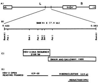
FIG. 1. (A) Positiononthe HSV-2(G)genomeof the DNA sequenced in this study. (B) Map locations of relevant restriction endonuclease
sites. The BamHI Efragment whichmapsfrom approximately 0.533to0.583 HSV-2mapunitswassubclonedasthree SstI fragments and
twoPstIfragments. (C)Mapposition of the 2,104-base-pair(bp)region of HSV-2 DNA sequenced in these studies and its relationtopublished
HSV-2 DNAsequences(74). (D) Location of the HSV-2 large ORF shown in this studytoencode ICP32/VP19c is depicted relativetothe
geneencoding the large subunit of the viral ribonucleotide reductase (74).
appear to lack any internal core structure (25, 67). In addition to VP19c, type A capsids also contain four other proteins, VP5, VP23, VP24, andasmall 12-kilodalton (kDa)
protein (15, 17, 25, 27). VP5 composes the hexameric
capsomers andappearstobe linkedto VP19cbyadisulfide
bond(19, 22, 73, 83). VP23isexposedtothe exterior of the virion and probably serves to connect the hexameric cap-somers to one another (9). VP24 and the small 12-kDa
proteinhave been assigned to thecapsid interior, but their structural roles in the capsid remain obscure (9, 66). One
report suggested that VP19c composes the 12 pentameric
capsomers of the viralcapsid (77). However, otherreports
have adduced VP19c tobe an internal capsid protein (8, 9,
25, 67). One clue to the function of VP19c may lie in the
observation that the HSV-1 and HSV-2 ICP32/VP19c
pro-teins (after denaturation and partial renaturation) have a strong affinity for DNA (8). Whether this property of the
ICP32/VP19c molecule(s) reflects the structural role of the
protein(s) in the capsid or is functionally important to the
processes ofcapsid assembly or viral DNA packaging
re-mainstobe determined.
Recently, Pertuiset et al. (58) reported on the physical mapping and nt sequence ofan HSV-1 gene essential for
capsid assembly. Theseinvestigators observed thata
muta-tion in an ORF mapping from 0.553 to 0.565 HSV-1 map units (ORF.553), corresponding to the HSV-1 (strain 17)
UL38ORF, affectscapsid assemblyin a
temperature-sensi-tivemanner. However,theproteinencodedbyORF.553was notidentified in these studies,which made it difficult for the authorstopostulate arole for the ORF.553 geneproductin
capsid morphogenesis.
Inthisaccount, wereportthentsequenceofalarge ORF presentinthe HSV-2genomethatcorrespondstothe HSV-1 ORF.553 and UL38ORFs. We demonstrate that this HSV-2
ORFencodes ICP32/VP19c, and we provide evidence that the cognate HSV-1 ICP32/VP19c molecule is the protein
essential forcapsidformation that is encodedby the UL38/
ORF.533 HSV-1 ORF. In addition, the data demonstrate that there are type-specific differences in the regulation or
processing or both of the HSV-1 and HSV-2 ICP32/VP19c
molecule(s).The resultsof these studies resolve the location of thegeneencoding ICP32/VP19c onthe viralgenome and
provide detailed molecular information on which to base future studies of the structural and functional roles of
ICP32/VP19c incapsid assemblyandvirionmorphogenesis.
MATERIALS AND METHODS
Viruses and cells. Theoriginandproperties of theHSV-1
(Justin) and HSV-2 (G) isolates used in these studies have been described previously (20, 21). Virus stocks were
pre-paredand their titerswere determined onHEp-2cells (64).
Crude cell lysates were prepared by infecting HEp-2 cells with 5 to 10 PFU per cell, and cultures were incubated at
37-C (5, 56).
DNA sequencing. The 7.4-kilobase (kb) BamHI E
frag-ment, map coordinates 0.553 to 0.583, ofHSV-2 (G) DNA isolated from virions was cloned into pUC19 for DNA
sequencing (57) (Fig. 1).ThreeSstIfragmentsof2, 1,and 0.3 kb andtwoPstIfragmentsof2.4and1.4 kbwere subcloned into the polylinker regionofM13mp2OandM13mp2l
(Inter-z E 0
a a
C)
Uw 6.
CO o.U Oa
HSV-2DNA SEOUENCE 2158 bp
E
m VOL. 64, 1990
I
on November 10, 2019 by guest
http://jvi.asm.org/
[image:2.612.137.478.65.351.2]nationalBiotechnologies, Inc., New Haven,Conn.). Single-stranded and replicative-form DNAs were prepared as
de-scribed previously (54).
Both strands of the HSV-2 DNAinserts weresequenced
by the dideoxy-chain termination method (65). The first sequencing reactions were done with the M13 universal
primer (International Biotechnologies). Subsequent
reac-tions were done with custom primers (17 nt) derived from
sequence generated in previous sequencing experiments.
The oligonucleotide primers, all purified by high-pressure liquidchromatography, werepurchased from Operon
Tech-nologies, Inc. (Alamita, Calif.). Both strands of all HSV-2
DNA inserts were completely sequenced, and computer
methodswere used to compile and analyze the data.
In initial experiments, considerable difficulty was
experi-encedin obtaining readable sequence. Thiswasbecause the
secondary structures which formed in the G+C-rich DNA
resulted in blocks to the Klenow polymerase as well as in
compressions in the gel. Improved results were obtained
with deoxy-7-deaza-GTP (Boehringer Mannheim
Biochemi-cals, Indianapolis, Ind.) in place of dGTP (55) and strongly denaturing gels (7 M urea, 40% formamide) (49). The best
results wereobtained by using dIPT in place of dGTP (75),
Escherichia coli single-stranded-DNA-binding protein (10),
and Sequenase (modified bacteriophage T4 DNA
polymer-ase) (United States Biochemical Corp., Cleveland, Ohio). It
was not necessary to use formamide in the gels with the
latter method. [35S]dATP (Dupont, NEN Research Prod-ucts, Boston, Mass.) wasused asthe radioactive label. The
gelswerefixed in 12%aceticacid-10%methanol, dried, and
autoradiographed for approximately 15 h (7).
Generation of antipeptide antisera.Thechoice of
oligopep-tideswasbased onregions of aminoacid identity conserved
between the predicted primary amino acid sequence
en-coded by the HSV-2ICP32/VP19c ORF and the predicted
polypeptide product of the varicella-zoster virus type 20
(VZV-20) ORF (18). Predicted regional hydropathicity and
structuralmobilitywerealso considered (13, 14, 36, 45, 61).
Oligopeptides(12to15amino acids long)wereobtainedfrom
Biosearch (Palo Alto, Calif.). Peptides were coupled to
keyhole limpet hemocyanin, and antisera were prepared
essentially as described previously (1). Three rabbits were
immunized for each peptide, and hyperimmune antisera were obtained (three bleedings perrabbit).
Preparation of 58,700-kDa TrpE fusion protein and
gener-ationof antiserum. The pATH10plasmidwasobtained from
T. J. Koerner. This plasmid makes a
37,000-molecular-weightfragmentof theTrpEprotein under control of thetrp
promoter. The 1.4-kbPstIfragment of HSV-2 DNA (Fig. 1) was ligated intothePstI site atthe 3' end of the trpgenein
pATH10 to produce a fusion gene and cloned by
transfor-mation ofE. coliRR1 cells. Thefusion protein synthesized
by this recombinant clone should have 197 amino acids of theHSV-2protein (amino acids260to466) andamolecular
weightof58,700. The fusion proteinwasinduced with20
,ug
of 3-,-indoleacrylic acid (Sigma Chemical Co., St. Louis,
Mo.) per ml and separated by sodium dodecyl
sulfate-polyacrylamidegel electrophoresis(SDS-PAGE). Thefusion
proteinband wasexcisedfromthe SDS-polyacrylamide gel,
and 25 to 50 ,ug of purified protein was used to immunize
rabbits (76).
PAA inhibitionof viral DNA synthesis. Cells were treated
with phosphonoacetic acid (PAA)at aconcentration of 300 ,ug/mlof culturemedium4hbefore infection, andthislevel
was maintained throughoutinfection (82).
Radiolabeling of proteins and SDS-PAGE. Mock- and
vi-rus-infected cultures wereradiolabeled with
[35S]methionine
(>1,000 Ci/mmol; Dupont) at 20,uCi/ml
in methionine-freemedium. Polypeptides in cell lysates were
electrophoreti-cally separated on a9.25%
N,N'-diallyltartardiamide-cross-linked SDS-polyacrylamide gel (56).
Immunoblotting. Polypeptides separated by SDS-PAGE
were electrophoretically transferred to nitrocellulose mem-branes (8). For autoradiography, blots were dried at
37°C
for several hours before being exposed to Kodak XRP1 X-ray film at-70°C.
For immunoblotting, membrane filters wereblocked by incubation for 2 h in phosphate-buffered saline containing 3% nonfat milk powder and then reacted with rabbit antipeptide serum diluted inphosphate-bufferedsaline
containing 1% gelatin for 2 to 12 h at room temperature. After being washed with phosphate-buffered saline contain-ing 0.05% Tween 20, blots were reacted with biotin-conju-gated goat anti-rabbitimmunoglobulin G for 1 to 2 h at
'room
temperature, followed by incubation for 30
min
at room temperature with avidin-peroxidase complex (VectaStain; Vector Laboratories). Bound antibody was visualized by developing in horseradish peroxidase color development substrate solution (Bio-Rad Laboratories, Richmond, Cal-if.).Computer analysis. Alignment of the predicted primary amino acid sequence of the HSV-2
ICP32/VP19c
polypeptide employed several protein alignment programs (46, 72, 81), and minor adjustments were done byhand. The alignment of the predicted product of the Epstein-Barr virus (EBV)BORFi
ORF in regions that showed little homology with theother predicted proteins was arbitrarily biased toward max-imizing conservative substitutions with little consideration of gap penalties. Protein hydropathicity indexes were pre-dicted by the algorithms of Hopp and Woods (36) and Kyte and Doolittle (45).
RESULTS
DNA sequence analysis. To determine the nucleotide se-quence of the portion of HSV-2 DNA deduced to encode the
ICP32/VP19c protein, we sequenced the region of the viral genome mapping from approximately 0.553 to 0.565.
SstI
andPstI
subfragments of theBamHI
E fragment were cloned into appropriate M13 vectors, and primer walking techniques were used to determine the DNA sequence. The organization of the region of the viral genome sequenced, including locations of pertinent restriction endonuclease sites, orientation with respect topreviously
published HSV-2 DNA sequences, and the provenance of the DNA se-quences encoding theICP32/VP19c
and ICP6 (large subunit of ribonucleotide reductase) proteins is illustrated in Fig. 1. The nucleotide sequence of the noncoding strand of the HSV-2ICP32/VP19c
gene is shown in Fig. 2. Only one 1,222-base-pair ORF (from nt 541 to 1938) large enough to encodeICP32/VP19c
was identified in the sequenced region. This ORF contains two ATG codons located near the 5' end, at nt 541 and 571, which could serve as translation start sites (42-44), and the ORF extends to the TGA stop codon beginning at nt 1939. Translation initiation and termination at these codons would result in polypeptides of 50,538 and 49,472 Da, both of which are in close agreement to previous size estimates for HSV-2ICP32/VP19c
(56). The predicted sequence of the larger polypeptide is shown in Fig. 2. The DNA sequences 5' to the ORF contain several possible transcriptional regulatory elements. These are elucidated in the Discussion and are illustrated in Fig. 5. The consensus polyadenylation signals AATAAA and TGTGTTG (53, 59) appear in the sequence 121 and 150 nt 3' to the ORF.on November 10, 2019 by guest
http://jvi.asm.org/
HSV-2 GENE ENCODING ICP32/VP19c 1127 TTTAAACATTTGCGTATGCACCGGCCCAGCCAGTCGGACACCGGAACCCACCAGAGGCGGAAGCCGCCTTCGCCCGTGAGGGTGCGTGT
GTTTTCTGGTGGCGTGTTTTTCCTTTCCGCCCTCCTCCCTCCCCACCQCCACCACCCCCCACTCGCCCGTTGGCGATCGGCGGGAAAACCQTGAAAACCAAGCCQCTCCCGACAGCCCCG MetLysThrLysProLeuProThrAlaPro
*Sst I
ATGGCGTGGGCCGAGAGTGCCGTGGAAACCACCCCGGCCCGCGCGAGCTCGCGGGCCQACGCCCCGCTCCGGCGCGTCCTGCGCCCGCCCQATCGCTCGCCGCGACGGCCCGGTGCTTTTG
MetAlaTrpAlaGluSerAlavalGluThrThrThrGlyProArgGluLeuAlaGlyHisAlaProLeuArgArgValLeuArgProProIleAlaArgArgAspGlyProValLeuLeu
GGGGACAGGGCCCCCQGGAGGACGGCCQGTACGATGTGGCTGCTGGGGATCGACCCCGCGGAGTCGTCTCCGGGAACGCGCGCTACCCGAGACGATACCGAGCAGGCCGTGGACAAGATC
GlyAspArgAlaProArgArgThrAlaSerThrMetTrpLeuLeuGlyIleAspProAlaGluSerSerProGlyThrArgAlaThrArgAspAspThrGluGlnAlaValAspLysIle
CTCAGGGGAGCCCGGCGCGCGGGAGGGCTGACCGTCCCCGGCGCCCCCCGCTATCACCTGACCCGCCQGGTAACCCTGACGGATCTCTGCCAACCAAACGCGGAGCGGGCCGGGGCGCTC
ATCACGACCAACTACGGCGGGACGCGGGCCGGGGCGCGGCTGGACCGGTTTTCCGAATGCCTGCGCGCCQTGGTCCQQCGCACGTGTTTCCCCACGAGGTCATGCGGTTTTTCGGGGGG
IleThrThrAsnTyrGlyGlyThrArgAlaGlyAlaArgLeuAspArgPh S-rGluCyaLeuArgAlaMetValHisThrHisValPheProHisGIuValMetArgPhePheGlyGly *Pat I
CTAGTGTCGTGGGTCACACGGACGAGCTGGCTAGCGTCACCGCCGTCTGCAGCGGACCCCAGGAGGCCQQQQCCCGGCCQCCCGGGCAGGCCCCGTTCGGCCGTTACCQTCCCGGCC
LeuValSerTrpValThrGlnAspGluLouAlaSerValThrAlaValCyaSerGlyProGlnGluAlaThrHisThrGlyHisProGlyArgProArgSerAlaValThrIleProAla
* Sat I
TGCGCCTTCGTGGACCTGGACGCCGAGCTGTGCCTGGGGGGCCCTGGGGCGGCGTTCCTGTACTTGGTCTTQACCTACCGACAGTGCCGGGACCQGGAGCTCTGTTGCGTGTACGTGGTC
CysAlaPheValAspLeuAspAlaGluLeuCyaLeuGlyGlyProGlyAlaAlaPh ILeuTyrLeuValPheThrTyrArgGlnCysArgAspGlnGluLeuCysCysValTyrValVal
AAGAGCCAGCTCCCCCCGCGCGGACTGGAGGCGGCCCTCGAGCGGCTGTTCGGGCGCCTCCGGATAACCAACACGATTQACGGGGCCGAGGACATGACGCCCCCTCCCCCGAACCGAAAC
LysSerGlnLeuProProArgGlyLeuGluAlaAlaLeuGluArgLeuPheGlyArgLeuArgIleThrAsnThrIleHiaGlyAlaGluA3pMetThrProProProProAsnArgAsn
*Sst I
GTTGACTTTCCGCTCGCCGTCCTGGCCGCGAGCTCGCAATCCCCGCGGTGCTCGGCGAGCCAAGTCACGAACCCCCAGTTTGTCGACAGGCTGTACCGCTGGCAGCCGGATCTGCGGGGG
ATCCTGGAGGGCGTGGTGTGGCGCCCCGGCGGGTGGCGGGCCTGCGCGTGATCGTCTATTGACGACGGCCGCCCAACCCGAGCGACCTTCTCCTCCCACTTTCCCCCCCCCTCCTACACA IleLeuGluGlyValValTrpArgProGlyGlyTrpArgAlaCysAla
CCAACTCCGCCCTCGCCGTCTTGGCCGTGCGCGGCCCCGTGCGTCCGTCTCAATAAAGCCAGGTTAAATCCGTGACGTGGTGTGTTTGGCGTCTCTCTCTGAAATGSCGGAAACCGACAT
I---> Swfain and Galloway --->
GCAAATGGGATTCATGGACACGTTACAC
FIG. 2. Nucleotidesequenceof the HSV-2ICP32/VP19cgene.Thenucleotidesequenceof the ORF is depicted. The amino acidsequence ofthe predicted polypeptide is presented startingatthefirst ATG, whichrepresentsthefirst oftwopossible translation initiation sites (see text). The DNA sequence presented in this figure begins with the probable TATA box whichoccurs at nucleotide position 361 of the 2,158-base-pair sequence determined in this study. HSV-2 DNA sequences 5' to this position are presented in Fig. 5. The consensus polyadenylation signals AATAAA and TGTGTTG associated with the ORFareunderlined. The locations of the SstI and PstI restriction sites areindicated by asterisks (*).
Identification ofICP32/VP19cas the productoftheHSV-2
ORF.(i) Immunoblotting experiments.Thepolypeptide
prod-uct of the HSV-2 ORF was identified as ICP32/VP19c by
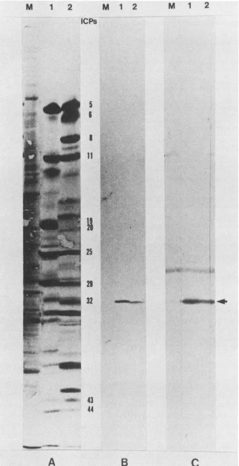
immunoblotting experiments. These experiments (Fig. 3 and 4) utilizedtwo typesof antisera: (i) antisera directed against synthetic oligopeptides corresponding to portions of the predicted 50,538-molecular-weight polypeptide encoded by
the ORF, and (ii) antisera directed against a TrpE/HSV-2
ORFfusionprotein overexpressedand isolated from E. coli.
Synthetic oligopeptides corresponding to regions of the
predicted polypeptide encoded by the HSV-2 ORF that share sequence or structural identity with the predicted
product oftheVZV 20 ORFwere synthesizedand used to raiseantisera inrabbits. The peptides synthesized and their locations in the predicted molecule were as follows:
LTRQVTLTDLCQPNA (number 1), residues 110 to 124; ERAGALLLALRHPTD (number 2), residues 125 to 139;
and TAVCSGPQEATHT (number 3), residues 264 to 276. All the antisera reacted with both the HSV-1 and HSV-2
ICP32/VP19c proteins, although antiseratopeptides1and2 showed somenonspecific bindingto both cellularandother
viral proteins (datanot shown). The results (Fig. 3B)
dem-onstratethat thepeptide2 antiserum reactsspecificallywith both the HSV-1 and HSV-2 ICP32/VP19c molecules in
immunoblotting experiments. Antiserum was also prepared
from rabbits inoculated with a TrpE/HSV-2 ORF fusion
protein specifying the carboxy-terminal 197 amino acids
encoded by the ORF. The immunoblotting experiments in
Fig. 3C indicate that this antiserum directed against the fusion protein also displayed a high affinity for both the
HSV-1 and HSV-2 ICP32/VP19c molecules. These results
strongly reinforce the conclusionsof theantipeptide
immu-noblotting experimentsandagaindemonstrate that the
prod-uctof the HSV-2ORFmust beICP32/VP19c.
(ii) Kinetic class of ICP32/VP19c. The HSV-1 and HSV-2
ICP32 proteins have been classified as _Y2 proteins and as
suchdependonviral DNAsynthesisforexpression (29, 81).
To ascertain the kinetic class of the HSV-1 and HSV-2
proteins reacting with theantisera, weperformed immuno-blot analyses on HSV-1 and HSV-2 ICPs produced in
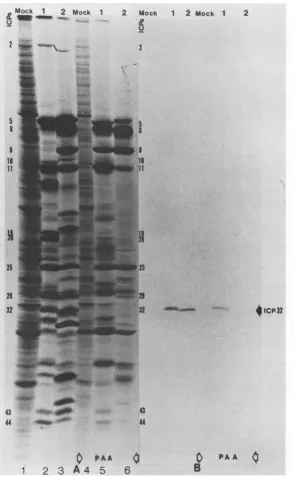
infected cultures treated with PAA. Figure4A shows
SDS-PAGE-separated 35S-labeled polypeptides (18 to 20 h
post-infection) extracted from untreated or PAA-treated, mock-infectedHSV-1-infected, or HSV-2-infectedHEp-2cells. A
comparisonof theprotein profilesof the untreated(lanes1to
3) and PAA-treated (lanes 4to6) infected cultures
demon-stratesthat PAA-mediatedinhibition of viral DNAsynthesis
dramaticallydecreased the amounts of both theHSV-1 and HSV-2 ICP32 molecules synthesized in infected cultures. The results ofimmunoblotting of the proteins produced in untreated and PAA-treated infected cultures(Fig. 4B) dem-onstrated that the HSV-1 and HSV-2polypeptidesthat are
immunoreactive with the antipeptide sera depend on viral DNA synthesisforexpression.
Taken together, the results of these immunoblotting ex-perimentsdemonstrate thatICP32/VP19cis thepolypeptide
450
570
690
810
930
1050
1170
1290
1410
1530
1650
1770
1890
2010
2130 CTTTTGGCCCTGCGGCACCCCACCGACCTCCCCCACCTGGCCCGCCATCGGGCTCCGCCCGGCCGGCAGACCGAGCGACTGGCCGAGGCCTGGGGCCAGCTCCTGGAGGCCTCCGCCCTG
GGGTCCGGGCGGGCCGAGAGCGGCTGCGCGCGCGCGGGCCTTGTGTCGTTTAACTTTCTGGTGGCCGCGTGCGCCGCCGCCTACGATGCGCGCGACGCCGCCGAGGCGGTCCGGGCCCAC
GlySerGlyArgAlaGluSerGlyCysAlaArgAlaGlyLeuValSerPheAsnPhoLeuValAlaAlaCysAlaAlaAlaTyrAspAlaArgAspAlaAlaGluAlaValArgAlaHis
CGCCCTACCGCACGCACCTGCACATACGCCGCCTTCGCAGAGCTGGGTGTCATGCCAGACGACAGCCCCCGCTGTCTGCACCGCACCGAGCGGTTTGGGGCGGTCGGCGTTCCGGTTGTC
VOL.64,1990
on November 10, 2019 by guest
http://jvi.asm.org/
M 1 2 M 1 2 ICPs
5
IS::
25
M 1 2
M-m
21
32
., 43
-4 44
A
B
FIG. 3. Western immunoblots of HSV-1- and
cell polypeptides.(A)I'S-labeled(18to20h postini tides producedinmock-infected (lane M), HSV-1-i and HSV-2-infected (lane 2) cells, separated by
through a 9.25%N,N'-diallyltartardiamide-cross-li
acrylamide gel, andtransferredto nitrocellulosepa
als and Methods). (B) Western blot of polypepti
mock-infected (lane M), HSV-1-infected (lane 1)
fected (lane2)cells with antiserum directedagain
LLALRHPTD oligopeptide. (C) Western immunc
produced inmock-infected (lane M), HSV-1-infeci HSV-2-infected (lane 2) cells with antiserum ag
HSV-2ICP32/VP19c fusion protein. The arrowin
tion of the HSV-1 and HSV-2ICP32/VP19cbands
product of the HSV-2 large ORF mappin
upstream ofthegene encodingthe large subu
ribonucleotide reductase. These studies also
evidence thattheHSV-1 UL38/ORF.553 OR] encode thecognate HSV-1 ICP32NVP19c.
DISCUSSION
In this report, wecharacterized the HSV-2 gene encoding infected cell protein 32 (ICP32) and virion protein 19c (VP19c). We also demonstrated that the HSV-1 UL38 and
ORF.553 ORFs,which have been showntospecifyaprotein
essentialfor capsidformation(58),mustencode the cognate HSV-1 ICP32/VP19c protein.
Comparison of ORFs encoding HSV-2 andHSV-1 1CP32/ VP19c proteins. Comparisonof the HSV-1 and HSV-2 DNA sequencesencoding their respectiveICP32/VP19c molecules indicates that the overall organization of the two genes is quite similar. However, there are a number ofdifferences between them. The two DNA sequencesdisplay66% (413 of 622 nt) overall identity, with 164 mismatched and 45
un-matched bases. Conserved features include the putative translation start sites for the genes encoding ICP32/VP19c andICP10 (51), two TATA box-like structures whichoccur
close to the transcription initiation sites for the 3.6-kb (ICP10) and 1.9-kb (ICP32) mRNAs previously mapped in this region of the HSV-1 genome (58), and a conserved SP1 transcription factor-binding site (HSV-2 position 344). A major difference is the presence in the HSV-2 sequence of several smalloligonucleotide stretchesthat haveno
counter-parts in the HSV-1 sequence. These occur in the region between the conserved TATA boxes that probably repre-sentstruly intergenic DNA sequences. Other dissimilarities include the presence in the HSV-2 ICP32/VP19c ORF ofa
second potential translation initiation site (ATG) and the
occurrence of possible internal transcription factor-binding sites within the HSV-1 ORFs predicted to encode ICP32/ VP19c andICP10 (Fig. 5).
Todate, most of the studies of HSV late gene expression have focused on the regulation of three HSV-1 Y2 genes, US11(39), glycoprotein C (2, 30, 31), and a geneencoding a 70-kDa regulatory protein (50, 69). These studies have shown that Y2 gene expression depends on viral DNA
replication and can be authentically examined only in the contextof the viral genome (2, 69).Within the virus genome,
the promoter-regulatory sequences important to the
expres-sion oftheglycoprotein C gene mapbetween -34 and-19nt
relative to the transcription start site (30, 31). In addition, analyses of glycoprotein C genes resident in cells have shown that sequences downstream of +22 are needed for -y2-specific regulation (2), suggesting that acomplex arrayof
cis-acting elements is involved in
Y2
gene regulation. TheC HSV-1 and HSV-2 DNA sequences display approximately
HSV-2-infected 76% nucleotide
identity
in the 5'regions
ofthe ORFs.fection) polypep- Nucleotide sequence variability in the promoter-regula-infected (lane 1), tory elements of the HSV-1 and HSV-2 genes may be electrophoresis reflected in the fact that the synthesis of the HSV-1 and inked SDS-poly- HSV-2 proteins is affected differently by PAA. After PAA ?per(see Materi- treatment of infected cells, a small amount of HSV-1ICP32
des
produced
in was detected on immunoblots with the antipeptide sera,and HSV-2-in- while noHSV-2
ICP32
wasdetected under the samecondi-;st
the ERAGAL tions (Fig. 4). While it is possible that these viruses vary in ted (lane 1), and their sensitivity to DNA synthesis inhibitors, studies of lateainst the TrpE/ gene expression in other virus systems (80) suggestthat the dicates the posi- sequence variation observed in the promoter-regulatory
re-gions of the HSV-1 and HSV-2 ICP32/VP19c genes is sufficient to generate minor differences in the levels of
g immediately expression of the two genes.
initofthe viral The occurrence of a second possible translation initiation provide strong site in the HSV-2 ICP32/VP19c ORF may account for the F (51,58) must observed differences in the synthesis or processing or both of the proteins. Thus, the HSV-1 and HSV-2ICP32/VP19c 4010
'i
.-4w
"Ok.
iv;,. .,: ..--am
inob
on November 10, 2019 by guest
http://jvi.asm.org/
[image:5.612.60.297.75.537.2]HSV-2 GENE ENCODING ICP32/VP19c 1129
Mock 1 2 Mock 1 2 a
2
5 S
IS
is
11
11
25
32
.,
..-:al.-;
43 -W _
44
4mw.401
0
PAA1 2
3
A4
5
6
Mock 1 2 Mock 1
2
FIG. 4. Effect of PAAonWestern immunoblots of HSV-1- andHSV-2-infected cell polypeptides. (A) Polypeptide profiles obtained after
SDS-PAGE (9.5% N,N'-diallyltartardiamide) of 35S-labeled (18 to 20 h postinfection) proteins synthesized in mock-infected (lane M), HSV-1-infected(lane 1), and HSV-2-infected (lane 2) cultures (lanes 1to3)orin identically processed cultures treated with 300,ug of PAA permlthroughout infection (lanes 4to6).(B) Western blot of polypeptides produced in untreated and PAA-treatedmock-infected (lane M), HSV-1-infected(lane 1), and HSV-2-infected (lane 2) cultures after reaction with rabbit antiserum directed against the ERA oligopeptide.
proteins differ with respect to electrophoretic mobilities (Fig. 3 and 4) even though the HSV-1 UL38 ORF is
predictedto encodea465-amino-acid polypeptide of 50,269
Daand the HSV-2 ORFis predicted to encode a similarly
sized466-amino-acid polypeptide of50,538Da. These elec-trophoretic differences were previously noted by Morse et
al.(56),whoestimated the sizeofHSV-1ICP32at51,500Da and that of HSV-2 ICP32 at 50,500 Da. A straightforward
mannerofreconcilingthepredicted sizesof the HSV-1 and
HSV-2ICP32 proteins withtheirexperimentally determined sizes would be to assume that the HSV-1 protein is more
extensively modified than the HSV-2protein by posttrans-lational phosphorylationorotheradditions. However, early
in infection, the HSV-2 protein displays anelectrophoretic
mobility equivalenttothat of HSV-1ICP32,butasinfection
progresses, the mobility of the HSV-2 protein appears to
increase while that of the HSV-1 protein remains constant
(data not shown). In addition, two to three radiolabeled bands were detected in infected cell extracts after SDS-PAGE thatseemtobe intermediates between the fasterana slower forms of HSV-2 ICP32. It isimportantthatonly the smallest of these seemingly related forms of the HSV-2
2
$ICP32
4
PAA
0
VOL.64, 1990
on November 10, 2019 by guest
http://jvi.asm.org/
[image:6.612.167.461.76.553.2]1 TGCAACGGTCCGTTCGGGGGTGGAGCGGACGGGGGGGTCATGCCGGCGGGCGCCGGGACC
MIii 1111111 11 III III III 11 1 1111 11 11 III III 1 84005 TGCATAGGTCCGTCCGAGGGCGGACCGGCGGGTGAGGTCGTGACGACGGGGGCCTCGGAC
SPi > < CREB/ATF
61 TGGAGCGCGCTGTCCGACATGGCGACCGGCGTGCG-CGCTCGGCGAC-GCGGCGCGGAGA
MI III I II Il II 11111 III I 11 III III I 1
84065 GGGAGACCGCGGTCTGCCATGACGCCCGGCTCGCGTGGGTGGGGGACAGCGTAGACCA-A
< Spi
109 CCGCGGGCCCAAACGGGAATGACTGCCGCCGCCCTATACGGAGGGGCTAAGTATCG---C 11 1 11 111111111111II11111111I11111111111111i 1
84124 CGACGAGACCGGGCGGGAATGACTGTCG-TGCGCTGTAGGGAGCGGCGAATTATCGATCC
Spi >
166 CCGGGGACCCTTC-GAAACCCCGGGCGTGTCGCAAGTACGCCGCGAAGGCGCGGCGTGTT 11 1111 11111 11 11 11111 11111 11111 11111
84183 CCCGCGGCCCTCCAGGACCCCCGCAGGCGTTGCGAGTACCCCGCGTCTTCGCGGGGTGTT
215
84243
275 84291
1111111 III IIIIIIIIII IIIIIII 1111111
ATACGGCCACTTAAGTCCCGGCATCCCGTTCGCGGACCCAGGCCCGG---G spi >
GGAGTGTGTGTGTGGGGGGGGGGCGGCGCGACGGCGGCCCGGACCAAGTGTATCGCGGCC
111111 11 1III II 11111111 III 1 1111 1
GGATTGT---CCGGATGTGCG---GGCAGCCCGGACGGCGTGGGTTGCGGAC
Spi >
335 GTTCCGTGGGGCGGCCCAACAGGCCCTTTAAACATTTGCGTATGCACCGGCCCAGCCAGT III 111111111111 111111111111 I 11 11 1 11 III 111111 84337 TTTCTGCGGGGCGGCCCAAATGGCCCTTTAAACGTGTGTATACGGACGCGCCGGGCCAGT
Spi > >>>
395 CGGACACCGGAACCCACCAGAGGCGGAAGCCGCCTTCGCCCGTGAGGGTGCGTGTGTTTT 111 11 I 11111111 1111111111 1111III 11 11 111 84397 CGGCCAACACAACCCACCGGAGGCGGTAGCCGCGTTTGGCTGTG-GGGT--GGGTGGTTC
< Spi
455 CTGGTGGCGTGTTTTTCCTTTCCGCCCTCCTCCCTCCCCACCACCACCACCCCCCACTCG 1 H1 1 11H1 11 I 11II llIllI 11 11 111 III 84454 CGCCTTGCGTGAGTGTCCTTT-CGAC--CCCCCCCCCCCTC--CCTCC-CCCGGGTCTTG
515 CCCGTTGGCGATCGGCGGGAAAACCATGAAAACCAAGCCACTCCCGACAGCCCCGATGGC I I I HI M1 III I IIIII Hi 1 11 11 11 11 lill 1 84508 CTAGGTCGCGATC--TGGGGTCGCAATGAAGACCAATCCGCTACCCGCAACCCCTTCCGT
575
84564
GTGGGCCGAGAGTGCCGTGGAA 11111 11 1111 11111111 GTGGGGCGGGAGTACCGTGGAA
spi >
FIG. 5. Comparison oftheHSV-2DNAsequences upstreamofthe1CP32/VP19cgenewith theHSV-1(strain 17) UL37-UL38intragenic
region (51). The HSV-2DNAsequences upstreamofthe geneencodingICP32/VP19carealignedwithnt84005to84585ofthe HSV-1(strain
17)DNAsequence. Thisregionof HSV-1DNAcontainsportions ofthe UL37 and UL38 ORFstogetherwithintergenicDNAsequences. Translational start sitesfortheHSV-1andHSV-2 ICP32- andICP10-encodingORFs areindicatedbytheunderscoredandoverscored ATGs andCATs, respectively.Asecondpossible translationstartsiteoccurringin the HSV-2ICP32 ORFis alsoindicated.Transcriptioninitiation sites and orientationoftranscriptsmapped fortheHSV-1 1.9-kb(ICP32)and3.6-kb(ICP10)mRNAs(58)aredenotedbytriple-headedarrows.
The locations of potentialtranscription factor-bindingsitesidentifiedbyacomputersearch for44such sitesaredenotedbythetriple-headed
arrows.
ICP32/VP19cprotein has been identified asaconstituent of thecapsid (8). There are a number of possible mechanisms that could generate multiple forms of HSV-2 ICP32 at different times postinfection. The most plausible would appear to be a switch to an alternate site of translational
initiation late in virus infection (24). Posttranslational addi-tions to the resultant smaller polypeptide might then give rise
totheintermediate-sizebands. Insupportof this
interpreta-tion, the HSV-2 ICP32/VP19c ORFdoes contain a second ATG codon 30nt downstream of the first, within a Kozak
milieu(42-44), whichmightfunctionas analternate
transla-tioninitiation site. The HSV-1ICP32/VP19c ORF lacks such
a second ATG. Alternatively, the different-sized HSV-2
ICP32/VP19c proteins mightarise from
proteolytic
process-ing ofalarger precursor into progressivelysmaller formsof the protein. However, the cognate HSV-1 proteindoes notappear to be subject to such processing, and computer
analysis of the predicted HSV-1 and HSV-2 proteins with respecttoknown proteasecleavage sites failedtoreveal any substantial differences in expected protease sensitivities
between the two molecules. Also, the predicted HSV-2
polypeptide does not contain cleavage sites for known proteases that might generate cleavage products similar in sizetothe observed HSV-2ICP32/VP19c molecules.
The HSV-2 DNA andpredicted protein sequences deter-mined in thisstudywereanalyzedwith respecttosignificant
on November 10, 2019 by guest
http://jvi.asm.org/
[image:7.612.145.470.64.488.2]HSV-2 GENE ENCODING ICP32/VP19c 1131
A
HSV2 HSV1 UL38
VZV 20 EBV BORF1 CONSENSUS
HSV2
HSV1 UL38
VZV 20 EBV BORF1 CONSENSUS
HSV2 HSV1 UL38
VZV 20
EBV BORF1 CONSENSUS
HSV2 HSV1 UL38
VZV 20
EBV BORF1
CONSENSUS
HSV2 HSV1 UL38
VZV 20
EBV BORF1
CONSENSUS
HSV2 HSV1 UL38
VZV 20
EBV BORF1
CONSENSUS
B
HSV-2 ICP32
10 20 30 40 50 60 70 80 90 100
MK----TKPLPTAPMAWAESAVE----TTTGPRELAG---HAPLRRV---LRPP-IARRD---GPVLLGDRA-PRRT--ASTMWLLGIDPAESSPGTRAT MK----TNPLPATPSVWGGSTVE-L-PPTT--RDTAGQ---GLLRRV---LRPP-ISRRD---GPGLPRGSG-PRRA--ASTLWLLGLDGTDAPPGALTP
MGSQP-TNSHFTLNEQTLCGTNISLLGNN---RFI--QIGNGLHMTYAPGFFGNWS--RDLTIGPRFGGLNKQPIHVPPKRTETASIQVTPRSIVINRMN
MKVQGSV
110 120 130 140 150 160 170 180 190 200
LDDTEQAVDK-ILRG-ARRAGGLTVPGAPRY---HLTRQVT-LTDLCQPNAERAGALLLALRHPTDLPHLAR--H- --RAPPGRQTERLAEA-WG
NDDTEQALDK-ILRG-TMR-GGALI-GSPRH---HLTRQVI-LTDLCQPNADRAGTLLLALRHPADLPHLA---HQ--RAPPGRQTERLGEA-WG
NI---QINPTSIGNP-Q--VTIRLPLNNFKS---TTQLIQQVS-LTDFFRPDIEHAGSIVLILRHPSDMIGEA---NTLTQAGRDPDV-LLEGLRNL-FN
DRRRLQRRIAGLLPPPARRLNISRGSEFTRDVRGLVEEHAQASSLSAAAVW---RAG--LLAPGEVAVAGGGSGGGSFSWSGWR-PPVFGDFLIHASSFN
Q * * Q L* *AG *L * * R P* *
210 220 230 240 250 260 270 280 290 300
QLLEASALGSGRAESGCARAGLVSFNFLVAAC-AAAYDARD-AA--EAVRAHI--TTN---YGG---TRAGARLDR-FSECLRAMVHTHVFPHEV
QLMEATALGSGRAESGCTRAGLVSFNFLVAAC-AASYDARD-AA--DAVRAHV--TAN---YRG---TRVGARLDR-FSECLRAMVHTHVFPHEV
-AC--TAPWTV-GEGGGLRAYVTSLSF-IAACRAEEYTDKQ-AA--DANR---TAIVSAYGC---SRMETRLIR-FSECLRAMVQCHVFPHRF NA-EATGTPLFQFKQSDPFSG-VDAVFTPLSLFILMNHGRGVAARVEAGGGLTRM-ANLL-YDSPATLADLVPDFG-RL---VADRRF-HNF
** * F ** * AA *A * Y RL V * F H
310 320 330 340 350 360 370 380 390 400
MRFFGGLVSWVTQDELASVTAVCSGPQEATHTGHPGRPRSAV--TIPACAFVDLDAELCL-GGPGAA---FLYLVFTYR----QCREDQ-ELCCVYVVKS
MRFFGGLVSWVTQDELASVTAVCAGPQEAAHTGHPGRPRSAV--ILPACAFVDLDAELGL-GGPGAA---FLYLVFTYR----QRR-DQ-ELCCVYVIKS ISFFGSLLEYTIQDNLCNLTAVAKGPQEAARTDKTSTRRVTA--NIPACVFWDVDKDLHLSAD-GL-KHVFL--VFVYTCF--QRR--QREGVRLHLALS
ITPVGPLVENIKSTYLNKITT-VVHGPVVS----KAI-PRSTVKVTVPQEAFVDLDA--WLSGGAGGGGGV----CFVGGLGLGQPCPAD--A-RLYVAL-* G L* *T*V GP * PR *P F D*D L *G G * Q **
410 420 430 440 450 460 470 480 490 500
QLPPRGLEAALERLFGRLRITNTIHGAEDMT-PPPPNRNVDFPLAVLARASSQSPR--CSASQVTNPQF-V--DRLYRWQPDLRGRPTARTCTYAAFAEL QLPPRGLEPALERLFGRLRITNTIHGTEDMT-PPAPNRNPDFPLAGLA-ANPQTPR--CSAGQVTNPQF-A--DRLYRWQPDLRGRPTARTCTYAAFAEL
QLNEQCFGRGIGFLLGRIRAENAAWGTEGVANTHQPYNTRALPLVQLS-NDPTSPR--CSIGEITGVNWNLARQRLYQWTGDFRGLPTQLSCMYAAYTLI
----TYEEAGPRFTFF---QSSRGHCQIMNI---LRIYYS-PSI---MHRYAVV---* * R C * R*Y YA
510 520 530 540 550 560
GVMPDDSPRCLHRTE-RFGAVGVPVVI-LEGVVW--RPGGWRACA
GMMPEDSPRCLHRTE-RFGAVSVPVWI-LEGVVW--RPGEWRACA GTIPSESVRYTRRME-RFGGYNVPT-IWLEGVVWGGTNT-WNECYY
---QPLHIEELTFGAVACLGT-FSATDGWRRSAFNYRGSSLPVVEIDSFYSNVSDWEVIL
* E FG* * * W *
E
t
o4h
lA
A
cL ,. A sn: Oi%
...
K v wV.F t-\ r %
wV
V -'V
-Vw-A% AAr-f A
A ~ O~ ' &A 4 MoU -A-A .I
p-,--toyvtviAi".,~1,,4JA
un.0
~V--INv~w
M0a
IVV;"XA
AIWA.4
-fP\
AW0
V
hA
-
VV
IJ -~ 'lfwt/J V V% W NIV \v
'4 ' , -wIy
Nj~
VT;
EBV
BORf1 _
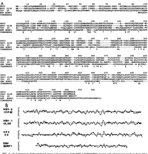
FIG. 6. (A)Comparisonofthe predicted amino acid sequence of the HSV-2ICP32/VP19cprotein with related geneproducts from other herpesviruses.The predicted amino acid sequence of the HSV-2ICP32/VP19cmolecule is aligned with the products of theHSV-1 UL38 (51), VZV20 (18), and EBV BORF1 (3) ORFs. Residue assignments are given in the single-letter amino acid code. The consensus sequence indicatespositions in which all four predicted proteins display amino acid identity or conservative substitution (*). (B)Comparisonof the hydropathicity plots ofthe VP19c-related herpesvirus proteins. This comparative alignment of hydropathicity plots of the predicted products of theORFsencoding therelated herpesvirus proteinsis based on the results ofprimarysequence alignment presented inpanel A.
homologies to known oncogenes or tumor suppressor
pro-tein-binding motifs because this ORF, which encodes a
DNA-bindingprotein, maps to a region of the viral genome (mtr-III) implicated in cellular transformation (6, 23, 38, 68). Nosignificant homologies were found.
Structure and functions ofICP32/VP19c-related
herpesvi-rusproteins. Several reports have noted that the sequences
of the predicted proteinsof the HSV-1 UL38/ORF.553 (51,
58), VZV 20 (18), and EBV BORFi (3) ORFs are signifi-cantly related. Computer analysis of the predicted HSV-2 ICP32/VP19c sequence indicated that it also isamember of this family of viral proteins (Fig. 6A). The HSV-2 protein displays significant amino acid identity with the HSV-1
(78%, 466-amino-acid overlap), VZV (29%, 362-amino-acid HSV-i
UL38
vzv 20
VOL.64,1990
-A pa.
-Vkv %-,141-Yvk a% A .Aoa, mC3 A M P. A ., ON iN-w %rnmIN Wa-W%Ilr w -wN,'-- V %4 V,4^V k.-r -vu.
on November 10, 2019 by guest
http://jvi.asm.org/
[image:8.612.61.561.67.587.2]overlap), and EBV (20.2%, 119-amino-acid overlap) pro-teins. Much of the sequence variation in this group of proteins occurs in the region equivalent to the amino-terminal 100 to 120 amino acids of the HSV-2 ICP32protein.
Such amino-terminal variation has been noted before in othercomparisonsof related groups ofherpesvirus proteins
(11, 26, 74). However, the observation that the predicted
EBV BORF1 product appears to becompletely missing an
equivalentprotein domain, coupled with the hypothesis that theHSV-2 protein may exhibitalterationsin thisregion due to alternative translational initiation late in infection, sug-gests that this region of the molecules is not essential for
protein function. Analysis of the primary amino acid
se-quences ofthis familyof viral proteins failed toreveal any
obvious structural motifs that mightexplain the previously
reported DNA-bindingproperties of the HSV-1 and HSV-2 VP19c molecules (9).
Comparative
hydropathicity
analysis (45)(Fig.
6B)of theICP32/VP19c family ofherpesvirus proteins illustrates two
important points. First, with respect tohydropathicity, the
HSV-1, HSV-2, and VZVproteins appearremarkably simi-larbeyondthefirst 100 or soamino acids.Mostof the amino acid substitutions that have occurred among these three viral proteins have been very conservative. The physical and
biochemical properties of these proteins might also be
ex-pected to be conserved. Second, while the EBV BORF1 protein sequence is significantly related to the other
pro-teins, it has diverged considerably with respectto size and
predicted physicalproperties. Thisvarianceis not untoward
giventhepostulatedevolutionary relationshipsbetween the
alpha Herpesviridae (HSV-1, HSV-2, and VZV) and the gammaHerpesviridae (EBV) (32). However, the extent of
variation is somewhat surprising in agenethat presumably
encodes the EBV counterpart ofthe essential capsid
struc-tural protein VP19c. It will be of interest to examine the DNA sequences of the members of the related beta
Herpes-viridae to determine whether they also encode a protein relatedto VP19c.
The role ofthe VP19c molecule in capsid structure and
virion assemblyis currently unclear.Theavailable evidence is asfollows. (i)VP19c isanintegral structural partof type Acapsids(25, 66) and is essential forcapsidformation (59).
(ii) It may form a disulfide bond to VP5, the majorcapsid protein that makes up the hexameric capsomeres (19, 22, 83). (iii) Both HSV-1 and HSV-2 VP19cs after denaturation and partial renaturation display affinity for DNA (8). (iv)
VP19c has been reported to be both a component of pen-tameric capsomeres(78) andan internalvirionprotein (8, 9,
25). If VP19c does compose the HSV pentameric caps-omeres, it obviously has an essential role in capsid
assem-bly,anditsjuxtapositiontothehexameric capsomeresmight allow for disulfide bonding to VP5. However, the
DNA-binding properties of the HSV-1 and HSV-2 VP19c mole-culesseemdifficulttoreconcile withapentamer assignment.
Also, if thepredictedproduct of theEBV BORF1 gene has
ananalogous role in thestructureof the EBVcapsid, then it would appear that a rather different structural solution to pentamerassemblymust occurin theEBVcapsid compared with the HSV capsids. If, on the other hand, a significant
portionof VP19c is located in thecapsidinterior,then it may
contributeto theband ofmassdensity shown by Schrag et
al. (66) to lie directly beneath the hexameric capsomeres. VP19c might thus form part of an essential matrix holding the hexamers together, and it might easily form disulfide
bonds to VP5 (66). In this position, VP19c might also be
oriented in such a manner as to present a DNA-binding
domaintothe interior of thecapsid,thusconferringastrong
affinityforDNA totheinterior surface of the empty type A
capsid. Such a molecular arrangement could be important
for the processes of viral DNA packaging or virion core formation or both. In addition, if all the VP19c-related
herpesvirus proteins function similarly, then the
divergent
structure of the product of the EBV BORF1 gene, in
particular, suggeststhattheinternalcapsid structuresof the herpesfamilyof viruses differsubstantially. Such variations in internal capsid architecture may reflect or influence the size of the viral genomes packaged by each of the
herpesvi-ruses.
ACKNOWLEDGMENTS
This work was supported by Public Health Service grant NS-23949fromtheNational Institute ofNeurological and Communica-tive Disorders and Stroke to W.B. andby Public Health Service grants CA 33101 and CA 24710 from the National Institutes of Health to W.S.M.W.
LITERATURECITED
1. Ackermann,M., M.Sarmiento,and B.Roizman.1985. Applica-tion ofantibody to syntheticpeptidesfor the characterization of the intactand truncated a 22 protein specified by herpessimplex virus 1 and the R325 a 22- deletion mutation. J. Virol. 56: 207-215.
2. Arsenakis, M., G. Campadelli-Fiume, M. T. Lombardo, and B. Roizman. 1988. The glycoprotein C gene of herpessimplexvirus 1 resident in clonal L cell lines manifests two regulatory domains conferring a dominant i and a subordinate_Y2 regula-tion. Virology 162:300-310.
3. Baer, R., A. T. Bankier, M. D. Biggin, P. L. Deininger, P. J. Farrell, T. G.Gibson, G. Hatfull, G. S.Hudson, S. C.Satchwell, C. Seguin, P. S. Tuffnell, and B. G. Barrell. 1984. DNA sequence and expression of the B95-8 Epstein-Barr virus ge-nome. Nature(London) 310:207-211.
4. Batterson, W., D. Furlong, and B. Roizman. 1983. Molecular geneticsofherpes simplexvirus. VIII. Furthercharacterization of a temperature-sensitive mutant defective in release of viral DNA and in other stages of the viral reproductive cycle. J. Virol. 45:397-407.
5. Batterson, W., and B. Roizman. 1983. Characterization ofthe herpesvirion-associatedfactor responsible for the induction of the a genes. J. Virol. 46:371-377.
6. Bejcek,B., and A. J. Conley. 1986. Atransformingplasmidfrom HSV-2 transformed cells contains rat DNA homologous to the HSV-1 and HSV-2 genomes. Virology 154:41-55.
7. Biggin, M. D., T. J. Gibson, and G. F. Hong. 1983. Buffer gradient gels and 35S label as an aid to rapid DNA sequence determination. Proc. Natl. Acad. Sci. USA 80:3963-3965. 8. Braun, D. K., W. Batterson, and B. Roizman. 1984.
Identifica-tion and genetic mapping of a herpes simplex virus capsid protein whichbinds DNA. J. Virol. 50:645-648.
9. Braun, D. K., B. Roizman, and L. Pereira. 1984. Characteriza-tion ofthe post-translational products of herpes simplex virus gene 35 proteins binding to the surface of full but not empty capsids. J. Virol. 49:142-153.
10. Chase, J. W., and K. R. Williams. 1986. Single-stranded DNA binding proteins required for DNA replication. Annu. Rev. Biochem. 55:103-136.
11. Chee, M., S.-A.Rudolph, B. Plachter, B. Barrell, and G. Jahn. 1989.Identification of themajor capsid gene of human cytomeg-alovirus. J. Virol. 63:1345-1353.
12. Chou, J., and B. Roizman. 1986. The terminal a sequence of the herpes simplex virus genome contains the promoter of a gene located in the repeat sequences of the L component. J. Virol. 57:629-637.
13. Chou, P. Y., and G. D. Fassman. 1974. Prediction of protein conformation. Biochemistry 13:222-245.
14. Cohen,F. E., R. M. Abarbanel, I. D. Kuntz, and R. J. Fletterick. 1983. Secondary structure assignment for al/ proteins by a
on November 10, 2019 by guest
http://jvi.asm.org/
HSV-2 GENE ENCODING ICP32/VP19c 1133 combinatorialapproach. Biochemistry22:4894-4904.
15. Cohen, G. H., M. Ponce de Leon, H. Diggleman, W. C. Lawrence, S.K.Vernon, and R.J. Eisenberg. 1980. Structural
analysis of the capsid polypeptides of herpes simplex virus types 1 and 2. J. Virol.34:521-531.
16. Corey, L., R. J. Whitley, E. F. Stone, and K. Mohan. 1988. Difference between herpes simplex virus type 1 and type 2 neonatalencephalitis inneurologicaloutcome. Lancet i:1-4. 17. Dargen, D. J. 1986. The structure and assembly of
herpesvi-ruses, p. 359-437. In J. R. Harris and R. W. Home (ed.), Electron microscopy ofproteins, vol. 5. Virus structure. Aca-demicPress, Inc. (London), Ltd.,London.
18. Davison, A.J., andJ.E. Scott. 1986. The complete nucleotide sequence ofvaricella-zoster virus. J. Gen. Virol. 67:1759-1816. 19. Davison, A. J., andJ. E. Scott. 1986. DNA sequence ofthe
major capsid protein gene ofherpes simplex virus type 1. J. Gen. Virol.67:2279-2286.
20. Ejercito,P. M.,E. D.Kieff,and B.Roizman. 1968. Characteri-zation ofherpessimplexvirus strainsdifferingintheir effecton
social behavior of infected cells. J.Gen. Virol. 3:357-364. 21. Frenkel, N., R. J. Jacob, R. W. Honess, G. S. Hayward, H.
Locker,and B. Roizman. 1975. The anatomyofherpes simplex
virusDNA. III. Characterization of defective DNAmolecules andbiologic propertiesof viruspopulations containingthem.J. Virol. 16:153-167.
22. Furlong, D. 1978. Direct evidence for 6-fold symmetry of the
herpes virus hexon capsomers. Proc. Natl. Acad. Sci. USA 75:2764-2766.
23. Galloway,D.A., J.A.Nelson,andJ.K.McDougall.1984.Small
fragmentsofherpesvirusDNAwithtransformingactivity con-tain insertionsequence-like structures. Proc. Natl. Acad. Sci. USA81:4736-4740.
24. Geballe,A.P.,F.S.Leach,and E.S. Mocarski.1986.Regulation ofcytomegaloviruslategeneexpression:-y genesarecontrolled
by posttranscriptional events.J. Virol.57:864-874.
25. Gibson,W.,andB. Roizman. 1972.Proteinsspecifiedby herpes
simplex virus. VIII.Characterization and compositionof
mul-tiple capsidforms ofsubtypes1 and 2. J.Virol. 10:1044-1052. 26. Gompels, U. A., M. A. Craxton, and R. W. Honess. 1988.
Conservation ofglycoprotein H (gH)in herpesviruses: nucleo-tidesequenceof thegHgenefromherpesvirus saimiri.J. Gen. Virol. 69:2819-2829.
27. Hay, J.,C. R.Roberts,W. T.Ruyechan,and A.C. Steven. 1987.
Herpesviridae,p. 391-401. In M. V. NermutandA.C. Steven
(ed.), Perspectives in medical virology, vol. 3. Animal virus structure. Elsevier/North-HollandPublishing Co.,NewYork. 28. Heine, J. W.,R. W. Honess, E. Cassai,and B. Roizman. 1974.
Proteins specified by herpes simplex virus. XII. The virion
polypeptidesoftype 1 strains.J. Virol. 14:640-651.
29. Holland,L.E.,K.Anderson,C. Shipman, Jr.,and E.Wagner. 1980. Viral DNA synthesisisrequiredfortheefficient expres-sion ofspecificmRNAspecies. Virology 101:10-24.
30. Homa, F. L., J. C. Glorioso, and M. Levine. 1988. Aspecific 15-bpTATAboxpromoter elementisrequiredforexpressionof
herpes simplexvirus lategene. Genes Dev.2:40-53.
31. Homa,F. L.,T. M.Otal, J. C. Glorioso,and M. Levine. 1986. Transcriptional controlsignalsofaherpessimplexvirus type 1 late (y2) gene lie within bases -34 to +124relative to the 5' terminus ofthe mRNA. Mol. Cell. Biol. 6:3652-3666. 32. Honess,R.W.1984. Herpessimplexand 'theherpes complex':
diverseobservationsandaunifying hypothesis.The 8th Flem-ingLecture.J. Gen. Virol. 65:2077-2107.
33. Honess, R. W., and B. Roizman. 1973. Proteins specified by herpes simplexvirus. XI.Identificationandrelative molarrates
ofsynthesis ofstructural and non-structural herpesvirus poly-peptidesininfectedcells. J.Virol. 12:1346-1365.
34. Honess,R.W.,and B. Roizman. 1974.Regulationofherpesvirus
macromolecularsynthesis. I. Cascaderegulationof the
synthe-sis of three groups of viralproteins.J.Virol. 14:8-19. 35. Honess,R.W.,andB. Roizman.1975.Regulationofherpesvirus
macromolecularsynthesis: sequentialtransition ofpolypeptide synthesis requires functional viral polypeptides. Proc. Natl. Acad. Sci. USA72:1276-1280.
36. Hopp, T. P., and K. R. Woods. 1981. Prediction of protein antigenicdeterminants from amino acidsequences. Proc. Natl. Acad. Sci. USA78:3824-3828.
37. Jacobson, J. G., S. L. Martin, and D. M. Coen. 1989. A conservedopenreadingframe thatoverlapstheherpessimplex virus thymidine kinase gene is important for viral growth in cell culture. J. Virol. 63:1839-1843.
38. Jariwalla,R.J., B. Tanczos,C.Jones, J. Ortiz, and S. Salimi-Lopez. 1986. DNA amplification and neoplastic transformation mediated bya herpes simplex DNA fragment containing cell-relatedsequences. Proc. Natl. Acad. Sci. USA 83:1738-1742. 39. Johnson, P.A.,and R. D. Everett. 1986. The control ofherpes
simplex virus type 1 late gene transcription: a TATA-box/ capsite region is sufficient for fully regulated activity. Nucleic Acids Res. 14:8247-8264.
40. Kieff, E. D., B. Hoyer, S. L. Bachenheimer, and B. Roizman. 1972. Genetic relatedness oftype 1 and type 2 herpes simplex virus. J. Virol. 9:738-745.
41. Knipe, D. M., W. Batterson, C. Nosal, B. Roizman, and A. Buchan. 1981. Moleculargeneticsofherpes simplexvirus. VI. Characterization of atemperature sensitive mutant defective in the expression of all early viral gene products. J. Virol. 38: 539-547.
42. Kozak,M.1983.Comparisonofinitiation ofprotein synthesisin procaryotes, eucaryotes, and organelles. Microbiol. Rev. 47: 1-45.
43. Kozak, M. 1984. Point mutations close to the AUG initiator codon affect theefficiency oftranslation ofratpreproinsulinin vivo. Nature(London)308:241-246.
44. Kozak, M. 1986. Point mutationsdefineasequenceflankingthe AUG initiator codon that modulates translation byeukaryotic ribosomes. Cell 44:283-292.
45. Kyte, J., and R. F. Doolittle. 1982. A simple method for displayingthehydropathiccharacterofaprotein.J. Mol.Biol. 157:105-132.
46. Lipman, D., and W. R. Pearson. 1985. Rapid and sensitive protein similaritysearches. Science 227:1435-1441.
47. Longnecker, R., S.Chatterjee,R.J. Whitley,and B.Roizman. 1987. Identification of a herpes simplex virus 1 glycoprotein gene withinageneclusterdispensableforgrowthin cell culture. Proc.Natl. Acad.Sci. USA 84:4303-4307.
48. Longnecker, R., and B. Roizman. 1987. Clustering of genes
dispensable forgrowth in culture in the S component ofthe HSV-1 genome. Science 236:573-576.
49. Martin,R. 1987. OvercomingDNAsequencingartifacts: stops andcompressions. Focus9:8-9.
50. Mavromara-Nazos, P., and B. Roizman. 1987. Activation of herpes simplex virus 1 _Y2 genes by viral DNA replication. Virology161:593-598.
51. McGeoch, D. J., M. A. Dalrymple, A. J. Davison, A. Dolan, M.C.Frame,D.McNab,L.J. Perry, J.E.Scott,and P.Taylor. 1988. The complete nucleotide sequence of the long unique
region in the genomeofherpes simplex virus type 1. J. Gen. Virol. 69:1531-1574.
52. McGeoch, D. J., H. W. Moss, D. McNab, and M. C. Frame. 1987. DNA sequenceandgeneticcontentoftheHindIII region
in the shortuniquecomponentoftheherpessimplex virustype 2genome: identification ofthegeneencodingaglycoprotein G, andevolutionary comparisons.J. Gen.Virol. 68:19-38. 53. McLauchlan, J.,D.Gaffney, J.L.Whitton,andJ.B.Clements.
1985.Theconsensussequence YGTGTTY located downstream fromthe AATAAAsignal is required for efficient formationof mRNA3' termini. Nucleic Acids Res. 13:1347-1368.
54. Messing, J. 1983. New M13 vectors for cloning. Methods Enzymol. 101:20-78.
55. Mizusawa,S.,S.Nishimura,and F.Seela. 1986.Improvementof thedideoxychain termination methodof DNA sequencing by
use ofdeoxy-7-deazaguanosine triphosphate in place of GTP. Nucleic Acids Res. 14:1319-1324.
56. Morse,L.S.,L.Pereira,B.Roizman,andP. A.Schaeffer.1978. Anatomyof HSV DNA. XI.Mappingof viral genesbyanalysis
of polypeptides and functions specified by HSV-1 x HSV-2 recombinants. J. Virol.26:389-410.
VOL.64,1990
on November 10, 2019 by guest
http://jvi.asm.org/
57. Pearson, R. E., B. Bejcek, and A. J. Conley. 1985. A physical domain of herpes simplex virus ICP8 is expressed and active in Escherichia coli. J. Virol. 53:360-365.
58. Pertuiset, B., M. Boccara, J. Cebrian, N.Berthelot, S. Choust-erman, F. Puvian-Dutilleul, J. Sisman, and P. Sheldrick. 1989. Physical mapping and nucleotide sequence of a herpes simplex virus type 1 gene required for capsid assembly. J. Virol. 63:2169-2179.
59. Proudfoot, N. J., and G. G. Brownlee. 1976. 3'noncoding region sequences in eucaryotic mRNA. Nature(London) 263:211-214. 60. Reyes, G. R., R. LaFemina, S. D. Hayward, andG. S. Hayward. 1980. Morphological transformation by DNAfragments of hu-man herpesviruses: evidence for two distinct transforming re-gions in herpes simplex virus types 1 and 2 and lack of correlation with biochemical transfer of the thymidine kinase gene. ColdSpringHarborSymp. Quant. Biol. 44:629-641. 61. Robson, B., E. Platt, P. W. Finn, P. Millard, J. F. Gibrat, and J.
Garnier. 1985. Prediction of the conformation and antigenic determinants ofthe v-sis viral oncogene product homologous with human platelet-derived growth factor.Int. J. Pept. Protein Res. 25:1-8.
62. Roizman, B., and W. Batterson. 1985. Herpesviruses and their replication,p. 497-526. In B. N. Fields (ed.), Virology. Raven Press, New York.
63. Roizman, B., B. Norrild, C. Chan, and L. Pereira. 1984. Iden-tification ofa herpes simplex virus 2 glycoprotein lacking a knowntype 1 counterpart. Virology 133:242-247.
64. Roizman, B., and P. G. Spear. 1968. Preparation of herpes simplex virus of high titer.J. Virol.2:83-84.
65. Sanger, F., S. Nicholson, and A. R. Coulson. 1977. DNA sequencingwithchain-terminating inhibitors.Proc.Natl. Acad. Sci. USA74:5463-5468.
66. Schrag, J. D., B. V. Venkataram Prashad, F. Rixon, and W. Chiu. 1989. Three-dimensional structure ofthe HSV1 nucleo-capsid. Cell 56:651-660.
67. Sherman, G., and S. L. Bachenheimer. 1988.Characterization of the intranuclear capsids made by ts morphogenic mutants of HSV-1.Virology 163:471-480.
68. Shillitoe, E. J. 1988. A searchfor insertion-sequence-like pat-terns in DNA sequences from human herpesviruses. J. Oral Pathol. 17:21-25.
69. Silver, S., and B. Roizman. 1985.-y2-Thymidinekinase chimeras are identically transcribed but regulate as Y2genes in herpes simplex virus genomes and as ,B genes in cell genomes. Mol. Cell. Biol.5:518-528.
70. Spear, P. G. 1980.Compositionandorganization of herpesvirus virions and properties of some of the structural proteins, p.
54-78. In F. Rapp(ed.), Oncogenicherpesviruses, vol. 1. CRC Press, Inc., Boca Raton, Fla.
71. Staden,R. 1982. An interactivegraphics program for comparing and aligning nucleic acid and amino acid sequences. Nucleic Acids Res. 10:2951-2961.
72. Staden, R. 1984. Measurementsof the effects thatcodingfora protein has on a DNA sequence and their use for finding genes. Nucleic Acids Res. 12:551-567.
73. Steven, A. C., C. R. Roberts, J. Hay, M. E. Bisher, M. Pun, and B. L.Trus. 1986.Hexavalent capsomers ofherpes simplexvirus type 2: symmetry, shape,dimensions,andoligomericstatus.J. Virol. 57:578-584.
74. Swain, M., and D. A. Galloway. 1986. Herpes simplex virus specifies twosubunits of ribonucleotide reductase encoded by 3'-coterminaltranscripts. J. Virol. 57:802-808.
75. Tabor, S., and C. C. Richardson. 1987. DNA sequence analysis with amodified bacteriophage T7 DNA polymerase. Proc. Natl. Acad. Sci. USA 84:4767-4771.
76. Tollefson, A. E., and W. S. M. Wold. 1988. Identification and genemappingofa 14,700-molecular-weightprotein encoded by regionE3ofgroup Cadenoviruses. J. Virol.62:33-39. 77. Vernon, S. K., M. Ponce de Leon, G.H.Cohen, R. J. Eisenberg,
and B.A.Rubin.1981.Morphologicalcomponents of herpesvi-rus.III.Localizationofherpessimplex virustype 1 nucleocap-sidpolypeptides byimmuneelectronmicroscopy.J. Gen. Virol. 54:39-46.
78. Voss, J. H., and B. Roizman. 1988. Properties of two 5'-coterminal RNAs transcribed part way andacrossthe S com-ponentorigin of DNAsynthesisof theherpes simplex virus 1 genome. Proc. Natl. Acad. Sci. USA 85:8454-8458.
79. Wagner, E. K. 1985. Individual HSVtranscripts: characteriza-tion of specific genes, p. 45-104. In B. Roizman (ed.), The herpesviruses,vol. 3. PlenumPublishing Corp.,NewYork. 80. Weyer, U., andW. Doerfler. 1985. Species dependenceof the
major late promoter in adenovirus type 12 DNA. EMBO J. 4:3015-3019.
81. Wilbur, W. J., and D. J. Lipman. 1983. Rapid similarity searches of nucleic acid and protein data banks. Proc. Natl. Acad. Sci. USA 80:726-730.
82. Wolf,H.,and B.Roizman. 1978.Theregulationof -y(structural) polypeptide synthesis in herpes simplex virus types 1 and 2 infectedcells,p. 327. InG.de Theetal.(ed.),Oncogenesisand herpesviruses, vol. 3. International Agency for Research on
Cancer, Lyon, France.
83. Zweig, M.,C. J. Heilman, Jr.,and B.Hampar. 1979. Identifi-cation of disulfide-linkedprotein complexes in the nucleocap-sidsofherpessimplex virustype 2. Virology 94:442-450.