Accumulation of Human
Immunodeficiency
Virus
Type
1DNA
in
T
Cells: Result of
Multiple
Infection
Events
HARRIETL. ROBINSON* AND DONNAM. ZINKUS
Department of Pathology, University of MassachusettsMedical Center, Worcester, Massachusetts01655 Received 10 January 1990/Accepted 22 June 1990
Human immunodeficiencyvirustype1DNAsynthesiswasfollowedinaCD4+ line of T cells(C8166)grown
in the presence or absence ofa monoclonal antibody toCD4 that blocks infection. By 48 h afterinfection,
culturesgrown in thepresenceof theantibodycontained -4copiesof humanimmunodeficiency virustype1 DNApercell, whereas thosegrownin the absenceof theantibodycontained -80copiesof viral DNApercell. Most of the viral DNA in cultures grown inthe absence of the antibody was present in a broad smear of
apparently incomplete viral sequences. In cultures grown in the presence or absence of the antibody, the
9.6-kilobaselinearduplexof viral DNAappearedtoundergo integrationwithin 24 h of itsappearance. These results demonstrate that T cells accumulate unintegrated human immunodeficiency virus type 1 DNAas a resultofmultiplevirions enteringcells.
Reverse transcription of virion RNA to a duplex linear DNAand theintegration of this linear duplex into chromo-somal DNA to form a provirus are early steps in the life
cycle of retroviruses (2, 7, 29; for a review, see reference
37). InmosttypeC virus-infectedfibroblasts, the synthesis
of viral DNAis limitedtoafewcopies ofviral DNApercell.
Each entering virion has the potential for producing one
provirus, with virion RNA being degraded by the RNase H activity ofreverse transcriptase during second-strand DNA
synthesis. Reverse transcription ofprogeny RNA requires assembly of the RNA with capsid and polymerase proteins,
a process that occurs as virions bud from cells. With the
onset of virus production, reinfection (and concomitant
reverse transcription) is blocked by newly synthesized
en-velope glycoproteins competing forreceptoractivity (38).
In contrast to most type C virus infections, cultures of
human immunodeficiency virus type 1 (HIV-1)-infected T
cells tendtoundergothepersistent synthesisof viralDNA,
withmoderate tohigh levels of unintegrated viral DNA (20 to100copiespercell) being reportedinrecentinfections and
lowerlevels of unintegrated DNA beingpresent in chronic infections (16, 21). Unintegrated DNA has also been
re-portedinlymph nodes from patientswithacquired immuno-deficiency syndrome (AIDS) (33).
Precedence for the accumulation ofhigh levels of uninte-grated retroviral DNA is found in certaintype C virus- and
visnavirus-infected cultures. In these, the accumulation of
DNAreflects reinfection duetothe inefficient establishment
ofenvelope glycoprotein-mediated superinfection resistance (12, 15, 39).Invitro infections that accumulate unintegrated
DNA are cytopathic (12, 15, 39). In vivo infections that
persistently produce unintegrated DNA cause disease, as
exemplified by avian leukosis virus-induced osteopetrosis (28)andfelineleukemia virus- and equine infectiousanemia
virus-induced anemias(22, 27).
Thegoal of the current study was to determine whether
infection accounted for the production of high levels of
unintegrated DNA in HIV-1-infected T cells. C8166 cells werechosen for the study becausethese cellsareunusually
permissive for HIV-1 expression; most infected cells
pro-*Corresponding author.
duceviral structuralproteins by24 hafter infection(35).Our
results clearly demonstrate that infection accounts for the
accumulation of high copy numbers of unintegrated viral DNA in HIV-1-infected T cells.
MATERIALS AND METHODS
Cells. C8166 cells(30),H9 cells(25),and 8E5cells(6)were
grownatdensities of 1 x 10⁶to2 x 106cellsperml inRPMI 1640 supplemented with 10 to 15% fetal bovine serum, penicillin, andstreptomycin. C8166cellsare aline of human
T-cell leukemia virus type I-immortalized cordblood lym-phocytes. The line does not express detectable levels of human T-cell leukemia virustype I capsid proteinbutdoes
express the tax gene product (10). C8166 cells express moderately high levels of CD4 (-80% ofthat observed on
mitogen stimulated CD4+ peripheral blood lymphocytes [34]).H9 cellsare asublineof HuT 78 cells(ATCC TIB161). When obtained, the H9 culture contained cells with two different densities of CD4 (34). This mixed population was sorted for cells expressing the higherlevel of CD4 (alevel similartothatpresentonC8166cells). Thesorted cells have maintainedauniform levelof CD4expression duringfurther culture. 8E5 cells are a line of A3.01 cells that contain a
singlepol-defective provirus (contributed byT. Folkstothe AIDSRepository; no. 95). Only cultures in which >80% of
the 8E5 cells scoredpositivein an indirect immunofluores-cence assay(IFA) (see below)wereused in studies.
South-ernblotanalyses ofEcoRI-digested 8E5 DNAdetectedthe
twoexpected junction fragments ofviraland cellularDNA
(5.0and 5.8 kilobases[kb]).
Plasmids. pHXB-2 was used as a source of HIV-1-IIIb
sequences (5).pS4isasubclone of the 9.2-kbSacl fragment
of pHXB-2 in the SacI site of Bluescript (Stratagene, La
Jolla, Calif.).
Antibody. A type-specific neutralizing serum for
HIV-1-IlIb, goatanti-PB1 serum(26)was obtainedfrom Repligen, Cambridge, Mass. leu3a, a mouse monoclonal antibody to CD4 that blockssyncytium formationandinfection(31),was obtained from BectonDickinson and Co. (Mountain View, Calif.). Because preparations of leu3a contain azide, leu3a was dialyzed againstRPMI 1640 and sterilized by filtration
through a0.45-,um-pore-size filter. Theeffective concentra-tion of leu3a in dialyzed preparations was determined by
4836
0022-538X/90/104836-06$02.00/0
Copyright© 1990,American Society forMicrobiology
on November 10, 2019 by guest
http://jvi.asm.org/
testing twofold dilution series for the inhibition of syncytium formation in
HIV-1-IIIb-infected
C8166 cells. Since experi-ments with known amounts of leu3a had demonstrated complete inhibition of syncytium formation with 120 but not 60 ng ofleu3a
per ml, a concentration of 120 ng/ml was assigned to the highest dilution giving complete inhibition of syncytium formation.Virus production and IFA. Stocks of HIV-1-IIIb (8) were produced by infecting exponentially growing H9 cells for 6 to 12 h in the presence of polybrene (10
,ug/ml).
Infected cultures were seeded daily into fresh culture medium at 106 cells per ml. Starting at the third day after infection, culture medium was saved and cultures were tested for the fraction of cells expressing viral antigens by using an indirect IFA. For the IFA, 2.5 x10⁵
cells were washed with phosphate-buffered saline (PBS), suspended in 20 ,ul of PBS, and allowed to settle on 10-well slides (Cell Line Associates, Newfield, N.J.) by sequentially placing the20-,ul
droplet over a series of wells. Settled cells were air dried, stored at room temperature, and assayed within a few days for the presence of HIV-1 antigens. HIV-1 antigens were detected by fixing the air-dried cells for 5 to 10 min at room temper-ature in methanol, washing with PBS, incubating for 30 min at37°C
with an appropriately diluted heat-inactivated(57°C,
30
min)
serum from an HIV-1-seropositive human, washing three times with PBS, incubating for 30min at37°C
with an appropriately diluted fluorescein-treated goat anti-human serum (H and L chain specific; Organon Teknika-Cappel, Durham, N.C.), washing three times with PBS, and mount-ing in 90% glycerin. Virus-positive and -negative cells were counted with a fluorescence microscope with a reticle serv-ing as a countserv-ing grid. Stained slides could be stored at4°C for several weeks. Titers (infectious units) of virus stocks were determined by infecting C8166 cells with 1:2, 1:8, and 1:32 dilutions of the stock in the presence of Polybrene (2,ug/ml).
At 3 h after infection,leu3a(240 ng/ml) was added to cultures. At 24 h after infection, cells were assayed for the fraction expressing viral antigen by the IFA. Stock titers were calculated as (percentage of virus-expressing cells) x (concentration of C8166 cells at the time of infection) x (test dilution). Titers obtained by this method agreed well with those obtained by endpoint dilution on C8166 cells. The highest-titered stocks (_106 infectious units per ml) were obtained from cultures undergoing a spreading infection. All multiplicities of infection represent infectious units per cell. C8166 cell infection. Exponentially growing C8166 cells were suspended at 106 cells per ml in a 1:2 dilution of a high-titered stock of HIV-1 in the presence of Polybrene (10,ug/ml).
At 3 h afterinfection,
cells were collectedby
centrifugation and suspended in fresh culture medium in the presence or absence of leu3a. Infections were monitored for HIV-1-expressing cells by the IFA and for dead cells by the failure to exclude trypan blue. Cultures grown in the absence of leu3a formed syncytia.
Analyses for viral DNA. DNA was extracted from a
minimum of 5 x 10⁶ cells by disrupting cells in 10 mM Tris hydrochloride (pH
7.8)-10
mM EDTA-1% sodium dodecyl sulfate, digesting with 2 p.g of proteinase K per ml at37°C for 1 to 2 h, extracting with phenol and then chloroform, and scooping out DNA that aggregated alter the addition of 0.1 M NaCl and 2 volumes of 95% ethanol. Unintegrated viral DNA was recovered as efficiently by this procedure as from precipitates collected by centrifugation at 10,000 rpm for 20 min. Precipitated DNA was dissolved in 100,ul ofT1E.1 (1 mM Tris hydrochloride [pH 7.8], 0.1 mM EDTA) for the DNA from107
cells. Extractions for unintegrated DNA wereperformed by the method of Hirt(13). Hirtsupernatant DNA was also dissolved in 100
[L1
ofT1E.1 for 107 initial cells. Quantitation of DNA sequences was done by alkali denatur-ing approximately 4 p.g of test DNA in 0.3 MNaOHat60to70°Cfor 1 h, neutralizing the denatured DNA by the addition of 0.3 M HCl-0.2 M Tris hydrochloride (pH 8.0), and collecting serial dilutions of thedenatured DNA onduplicate nylon filters in the presence of1Ox SSC (lx SSC is 0.15 M
NaCl, 0.015 M sodiumcitrate). Serialdilutions (from65to1
pg) of CsCl-purifiedpS4 DNA were denatured andcollected onto one of the filters to serve as astandardforHIV-1 DNA. Serial dilutions (from 2 to 0.03 ,ug)ofpurified and denatured human DNA werecollected onto thesecondfilterto serve as
a standard for human DNA. DNA from 8E5 cells was
included as a known positive in all tests. Uninfected cell DNAs were included to determine background levels of hybridization for the HIV-1probe. DNAs werecross-linked to filters by UVirradiation. Filters were blockedby prehy-bridization with blotto (14) andhybridized in the presence of 6x SSC at 67°C withnick-translated pS4 DNA (toquantitate HIV-1 sequences) ornick-translated human DNA (to quan-titate human sequences). The amount of hybridized 32p_ labeled HIV-1 or human DNA was determined bycounting washed papers with a Betascope (Betagen, Waltham, Mass.). Standard curves were constructed from data for knownamounts of HIV-1 or human DNA,and theamountof HIV-1 orhuman DNA in test samples was determined from the standard curve. The number of copies of pS4 per picogram of DNA was calculated as (Avogadro's
number)/
[(thesum of the number ofnucleotides in the Sacl
fragment
of HTLV-IIIb DNA and Bluescript) x (the average gram molecular weight of onenucleotide) x (theconversionfactor forgrams topicograms)] or (6.023 x
1023)/[(18.4
x 103 + 6.2 x 10³)(330)(1 x 10¹²)]. Copies ofHIV-1 DNA per cell were then normalized by dividing the copies of HIV-1 DNA per picogram of cell DNA in a test sample by thecopies
of HIV-1 DNA perpicogram of DNA in the 8E5sample. Since 8E5 cellscontain a single provirus,thiscalculationprovides
an estimate of the copies per cell in the test sample. This calculation assumes that C8166 and 8E5 cells have similar amounts of DNA andnormalizes the copy number of HIV-1 DNA in cultures containing syncytia to copies per 8E5 cell equivalent of DNA.
DNA blot analyses. Southern blot analyses were done on DNA fractionated by electrophoresis through 0.8 to 1.0% agarose gels in the presenceof ahigh-salt buffer(0.04MTris hydrochloride, 0.001 M EDTA, 0.5 M sodium acetate
[pH
7.9]). Gels were monitored for the efficiency oftransfer
by
ethidium bromidestaining. Underourconditions of
transfer,
the transfer of DNA fragments greater than 12 kb in size is incomplete. Experiments in which 2 to 5
jig
ofundigested
DNA wasmixed with up to 65 pgoflinearizedor
covalently
closed circular HIV-1 plasmid DNA did notrevealtrapping
oftheplasmid DNA in
high-molecular-weight
DNA(data
not shown). Estimatesof theproportionsof HIV-1 DNA present in integrated and unintegrated forms were doneby
using
betascope analyses. Since transferof
high-molecular-weight
DNAwasincomplete, estimates for theamountof
integrated
DNA are low. Inthese estimates, the
background
ofhybrid-ization observed for
high-molecular-weight
uninfected cell DNA was subtracted from the bandrepresenting
integrated
HIV-1 DNA.
RESULTS
leu3a retards infection. To
identify
conditions that pre-vented HIV-1 infection, a type-specificneutralizing
serumon November 10, 2019 by guest
http://jvi.asm.org/
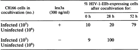
TABLE 1. leu3a retards the spread of HIV-1-IIIb inC8166culturesa
%HIV-1-IIIb-expressing cells C8166cellsin leu3a after cocultivationfor: cocultivation (no.) (300ng/ml)
Oh 28h 52h
Infected(105) + 10 20 79
Uninfected (106)
Infected(105) - 9 100
Uninfected(106)
aExponentiallygrowing C8166cells wereinfectedwith 0.25 to 0.5 infec-tious unit ofHIV-1-IIIbpercellandgrown in the presenceof300 ngof leu3a perml for48 h. At 48h, infected cellswerecocultivated with uninfected C8166 cellsthathad beengrownin thepresence orabsenceof leu3a for the preceding48h. At theindicatedtimes aftercocultivation,HIV-1-expressing cellswerequantitated bythe IFA.
and a receptor-blocking antibody were evaluated for their ability to prevent HIV-1-IIIb infection of C8166 cells. The neutralizing serum was a polyclonal goat antibody raised against the PB1 peptide of the gpl20 envelopeglycoprotein of HIV-1-IIIb (26). The receptor-blocking antibody was
leu3a, a monoclonal antibodyagainst CD4 that blocks infec-tion and syncytium formation (31). The anti-PB1 serumdid notdetectably reduce the titer of the -0.5 x 10⁶ infectious unitspresentin the stockofHIV-1-IIIb. Incontrast, 240 ng of leu3a per ml completely blocked infection (data not
shown).
Since HIV-1canundergocell-to-cell transmission(11) and C8166 cells grow in clumps, the ability of leu3a to block HIV-1 spread within an infected culture was evaluated by cocultivating 106 uninfected C8166 cells with 105 infected cells in the presence and absence of leu3a (Table 1). At various times aftercocultivation, cultureswere assayed for the fraction of cells expressing HIV-1 antigens. Cocultiva-tion in the absence of leu3a resulted in all of the cells expressing HIV-1 antigens within 28 h. In cultures coculti-vated in the presenceof leu3a, asubstantially slower spread of the infection was observed; only 10% of the uninfected cells became positive in the IFA by 28 h after infection. Thus, the presence of 300 ng of leu3a per ml substantially retarded the spread of HIV-1 in C8166 cell cultures.
HIV-1 DNA accumulation in the presence and absence of leu3a. To assessthe roleof infection in the accumulation of viral DNA in C8166 cells, cultures wereinfected with 0.25 to 0.5 infectious units of HIV-1-IIIb per cell. At 3 h after infection, cellswerecollectedand seeded in fresh medium in the presence orabsenceof 300 ng of leu3a per ml. Samples of these cultures were harvested at various times after infection andanalyzedforvirus-expressing cells, dead cells, and the amount and forms of viral DNA in cells (Table 2, Fig. 1and 2).
The presence of leu3a in cultures had little effect on the
amountofviral DNA at 12 h after infection but a substantial and increasing effect on the accumulation of viral DNA at later times after infection (Table 2). By 24 h, cultures grown in the presence of leu3a contained <2 copies of viral DNA per cell, whereas those grown in the absence of leu3a contained -20 copies per cell. By 48 h after infection,cells grownin the presence of leu3a contained -4 copies of viral DNA, whereas cells grown in the absence of leu3a contained -80. Thus, retardation of infection by leu3a resulted in a 20-fold reduction in the amount of viral DNA per cell.
Forms of viral DNA in infected cells. Undigested DNAs
from
various times afterinfection were analyzed onSouth-A
std
5.0 _
2.5 _ 1.25 _ 0.6 0.3 0.15
- leu3a
B
std
St 2.0 _
1:4 1.0 1
1:16 0.5
0.25 _
+ eu3a 0.12 -w
st 0.06
-- leu3a
40 St
- 1:2
+
Ieu3a
_ St-a-. 1 :2
FIG. 1. Exampleof data used for estimationofcopynumbers of
HIV-1 DNAper cell. (A) Copynumber of HIV-1 sequences. The
standard(std) represents known copiesof HIV-1 DNA x 106.(B) Microgramsof cell DNA. The standardrepresentsknownquantities (micrograms) of human DNA. Designations for test samples: st, undiluted sample; 1:4, etc., diluted samples.
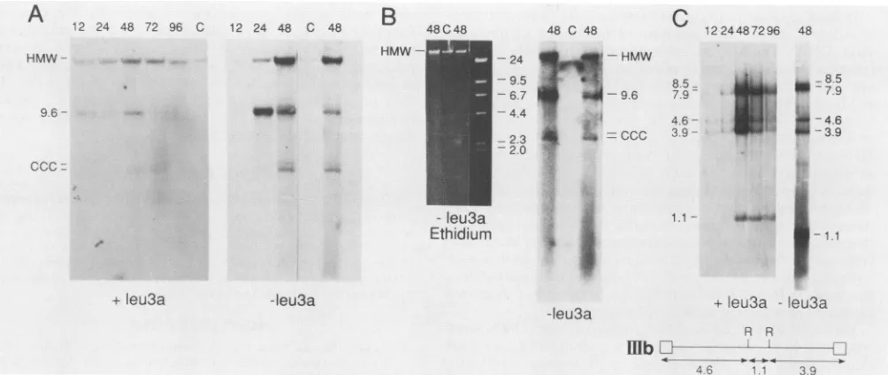
ernblots for the presence ofintegratedviral DNA(presentin high-molecular-weight chromosomal DNA), unintegrated duplex linear DNA(presentas a9.6-kb species),and
cova-lentlyclosedcircularDNA(migratingas adoubletat-5kb) (Fig. 2AandB).Incultures grown inthepresenceofleu3a, muchofthe viral DNA underwentintegration (Fig. 2A). By 24h, most of the 9.6-kb band present at 12 h appearedto haveintegrated. Atthistime20% ofthe cellsexpressedviral antigens. As the first round or progeny virus spread to
uninfectedcells, more9.6-kbDNAappeared;this round of 9.6-kb DNAsynthesisalsoappearedtointegratewithin 24 h ofits appearance.
Incultures growninthe absenceofleu3a(Fig.2AandB), patterns ofrelative band intensities were again consistent with the 9.6-kb linear duplexof viralDNAintegrating within 24 h of itsappearance (see relative intensities ofthe9.6-kb band at24 h after infection and the high-molecular-weight band at48 h after infection in Fig. 2A). By48 h, the most
abundant form ofviral DNA was a broad smear ofviral sequences(mostclearlyseeninFig. 2B). The appearanceof this smear was notaccompanied by obviousdegradationof high-molecular-weight chromosomal DNA(Fig. 2B).
Tofurtheranalyzetheforms ofunintegrated viral DNA, DNAharvestedatvarioustimes afterinfectionwasdigested with EcoRI and analyzed on Southern blots for fragments characteristic oflinear and covalently closed circular viral DNA (Fig. 2C). The EcoRI-digested DNA clearly demon-strated the appearanceoffragments uniqueto unintegrated linearDNA(4.6 and 3.9kb) beforethe appearanceofbands unique to the presumed one-long-terminal-repeat (7.9 kb) and two-long-terminal-repeat forms (8.5 kb) of covalently closedcircular DNA. Fragmentsrepresenting closed circu-lar DNAalwaysmigratedas adoublet,and themorerapidly migratingform of the doublet (presumed one-long-terminal-repeat form) was present at higher levels than the more
slowlymigrating form(mostclearlyexhibited inFig.2Aand C). Analysesof EcoRI fragments of Hirt supernatant DNA from cultures grown in the absenceof leu3a revealed bands representingthelinear and closedcircularforms, the1.1-kb fragment common to all forms, and a smear offragments migrating as approximately 3 kb or shorter linear species (presumably representingthe smearobserved inundigested DNA).
DISCUSSION
Accumulationof HIV-1DNA.Our results demonstrate that theaccumulation of HIV-1DNAin T cellsisduetomultiple
40
4_1
on November 10, 2019 by guest
http://jvi.asm.org/
[image:3.612.61.303.96.184.2]24 48 72 96 C 12 24 48 C 48
_. _
B
HMW
-48C 48 48 C 48
-24
-9.5 - 6.7 -4.4
2.3 2.0
C 1224487296 48
8.5 u:8.5
9 7.9S
4.6
-U
14.6
3.9 --a_ 3.9
1.1 - _$:4
*-1.1
- leu3a
Ethidium
+ Teu3a -Ieu3a
-leu3a + leu3a -leu3a
R R
fb
[image:4.612.65.567.73.285.2]46 11 39
FIG. 2. Forms of HIV-1-IIIb DNA in C8166 cellsatvarious times after infection.(Aand B)Blot analyses of undigested total cell DNA.
Panels A and Bpresentindependent blots ofthesame48-hsamples. (C) Blot analysis of EcoRI-digested total cell DNA (+leu3a samples) and
Hirt supernatant DNA (-leu3a sample). The schematic diagram shows the positions of EcoRI sites (R) in HIV-1-IIIb. DNA blots were
hybridized with nick-translated pS4 sequences. Numbers at the tops of lanes indicate hours after infection. Abbreviations: HMW,
high-molecular-weight chromosomal DNA; CCC, covalently closed circular DNA. The sizes offragments are indicated in kilobases. Designations below data indicate whether DNAs were from cultures grown in the presence orabsence of leu3a. Ethidium indicates an
ethidiumbromide-stained gel. A lane containing HindIII marker fragments is presented for the ethidium bromide-stained gel.
infectionevents(Table 2).Thiswasdemonstrated by
infect-ing cultures and then growinfect-ing the infected cultures in the
presence or absence of an antibody to CD4 that blocks HIV-1 infection (Table 1).During the first 48 h of infection,
culturesgrownin thepresenceof theantibody accumulated
-4 copiesof HIV-1 DNApercell, whereasthose grownin the absence of antibody accumulated -80 copies ofviral DNApercell(Table 2). Inthe lattercultures, mostofthe 80 copies were incomplete viral sequences that migrated as a broad smear(Fig. 2B).
Theaccumulation of highcopynumbers of HIV-1 DNAin C8166 cultures correlated with the appearance of virus-expressing cells. Thus,theproduction ofhighcopynumbers
of viral DNAwasmostlikelyduetoprogeny virusinfecting
cells that had not established superinfection resistance. HIV-1envelope glycoproteinsareabletoestablish superin-fection resistance (4, 36). However, the time between the initiation ofan infection and the establishment of superin-fection resistance may be prolonged by differential gene
expression (16) and the dependence of HIV-1 envelope glycoprotein synthesis on the expression of tat and rev
proteins (17, 18; for a review, see reference 3). Electron
micrographs reveal HIV-1-producing cells releasing high levelsofbuddingvirions(24, 36). Thus, highlevelsofvirion
production coupled with relatively slow establishment of
superinfection resistance could contributetothecopy num-bers of viral DNA present in culturesgrownin theabsence of leu3a.
TABLE 2. HIV-1-IIIbDNAcopiespercella
Treatmentb h after %
IFA`
% Dead DNAcopies/celld % Integrated PV/celfinfection cells cells' xt1Ep DNAe
Withleu3a 12 <1 1 2.6,1 19 0.3
24 20 1 1.6, 1.5 37 0.6
48 70 2 5,3.5 5.6,2 34 1.4
72 90 10 6,4.5 48 2.5
96 95 50 7.3,4 39 2.2
Without leu3a 12 <1 1 2.3,1 <10 <0.2
24 (Syncytia) 24,15 15 3.0
48 (Syncytia) 88,58 75,100 15 12.0
aForexperimentaldetails,seethe text.
bC8166 cellsinfectedwithHIV-1-IIIb were tested with and without leu3a. Dead cells were notquantitatedin culturesundergoing syncytiumformation.
dCopynumbersforculturescontaining syncytiaarenormalized forsingle-cell equivalents ofDNA.
ePercentage ofintegratedviral DNA wasestimatedbydividingcountshybridizedtohigh-molecular-weightDNAbycountshybridizedtoall formsofviral DNA. These represent minimumestimates,because transfer ofhigh-molecular-weightDNAwasincomplete.
fProviruses(PV)(integrated viral DNA)per cell wereestimatedbymultiplyingtheaveragenumberofviral DNAcopiesper cellbythe estimatedpercent integrated.
A
12HMW
-9.6
-CCCI
on November 10, 2019 by guest
http://jvi.asm.org/
[image:4.612.61.562.543.658.2]Defectivereverse-transcript-formingunitscould also have contributed to the accumulation of high copy numbers of viral DNA. The experiments reported in Table 2 were
initiatedat amultiplicity of infectionof 0.25to0.5infectious unit per cell. Infection at this multiplicity resulted in the production ofjust under tworeversetranscriptspercellby 12 h after infection (Table 2). Since there is little virus production during the first 12 h ofinfection, ourstocks of HIV-1-IIIb would appear to contain several
reverse-tran-script-forming units for each infectious unit. Most of the
defective reverse transcripts did not score in the IFA (see Materials andMethods). Thus, most of these may nothave had thepotentialto expresstheviralenvelope
glycoproteins
thatestablish superinfection resistance. There is much pre-cedenceforthepackagingof defective virusbyretroviruses
aswellasforthe presenceofmoredefective than nondefec-tive virus inHIV-1infections(forthefrequencyofdefective
tatgenesin patient samples, see reference20).
Forms of HIV-1 DNA. Four forms of viral DNA were
observed:high-molecular-weight (integrated DNA),a9.6-kb linear duplex, an -5-kb doublet representing covalently closed circular DNA, andabroadsmearofincompleteviral sequences. Incultures grown in the presenceorabsenceof leu3a, the relative hybridization intensities of the 9.6-kb linear duplex and high-molecular-weight sequences were
consistent with the 9.6-kb species being the precursor to
integrated DNA. Irrespective of whether a culture was
grownin the presenceorabsenceofleu3a,the9.6-kbspecies appeared to undergo integration within 24 h of its
appear-ance.Thebroadsmearofviral sequenceswasonlydetected in cultures undergoing active reinfection. This smearcould represent nascent transcripts, since it appeared at at time when the culturewasproducing highcopynumbersofHIV-1 DNA and since smears of viral DNA occur in cultures undergoing active synthesisofhepatitisBvirus DNA(Table 2) (19).Alternatively,thissmearcould representdegradation of DNA as cultures underwent cell death (note that the
smear includes sequences longer than full-length viral DNA), defective-deleted forms of viralDNA,or a combina-tion of the above possibilities. The appearance of thesmear
didnot accompanywidespread degradationof chromosomal DNA, since ethidium bromide stains of gels containing undigested DNA did not reveal smears of chromosomal DNAornucleosomeladders(Fig. 2B).
Generalityof results.Ourexperiments clearlydemonstrate thatTcells accumulateunintegratedHIV-1 DNAas aresult ofmultiple virions entering cells and not by intracellular reverse transcription of newly synthesized HIV-1 RNA. This result is consistent with polymerase chain reaction analyses of the copy number of HIV-1 DNA in peripheral blood lymphocytes from patients with AIDS. These tests
suggest thatthe rareinfectedTcells(-1%of theCD4+ cells) containasingle copyof viral DNA (32). Thissingle copy of DNA presumably arises from reverse transcription of the RNAofaninfecting virus.
Themechanismfor DNA accumulation in HIV-1-infected
Tcells may not necessarily represent the sole mechanism for DNA accumulation in other cell types. In visna virus-infected sheep, -1% of the choroid plexus cells and mono-cytes contain multiple copies of viral DNA (1, 23). The presence of multiple reverse transcripts in the infrequent positive cell suggests that a single infectious event can generate multiple copies of viral DNA. This may correlate with whether virions undergo intracellular assembly.
Elec-tron micrographs of HIV-1-infected monocytes or
macro-phages
reveal intracellular assembly, with the assemblingvirus budding into
cytoplasmic
vacuoles(9).
Thus,
an in-fected monocyte ormacrophage
may have thepotential
to reversetranscribenewly synthesized
HIV-1 RNA. In con-trast, electronmicrographs
ofHIV-1-infectedT cells donotreveal intracellular
assembly
andbudding
of virus(9, 24).
This would be consistent with the data in Table
2,
which indicate thatnewly
synthesized
HIV-1 RNA does notun-dergo
reversetranscription
inTcells.ACKNOWLEDGMENTS
Wethank D. W.Brown,C. A.Holland,and C. Mulder for critical
comments onthemanuscript. WeareindebtedtoR. C. Gallo and co-workers fortheprovisionofHIV-1-IIIb,C8166 and H9cells,and pHXB-2, to the AIDS repository for 8E5 cells, and to Becton Dickinsonforfacilitatingourpurchaseof volumequantitiesof leu3a. This researchwassupportedbyPublicHealth Serviceaward Al 24474fromtheNationalInstitutesofHealth.
LITERATURECITED
1. Brahic, M.,L. Stowring, P. Ventura, and A. T. Haase. 1981. Gene expression visna virus infection in sheep. Nature (Lon-don)292:240-242.
2. Brown,P. O.,B.Bowerman,H. E. Varmus,andJ.M. Bishop. 1989. Retroviral integration: structure of the initial covalent
product and its precursor, and a role for the viral INtegrase protein.Proc. Natl.Acad. Sci. USA 86:2525-2529.
3. Cullen, B.R., and W. C. Greene. 1989. Regulatory pathways governingHIV-1 replication. Cell 58:423-426.
4. Dalgliesh, A., P. Beverley, P. Clapham, D. Crawford, M. Greaves, and R. Weiss. 1984. The CD4 (T4) antigen is an essential component ofthereceptor for the AIDS retrovirus. Nature(London)312:763-766.
5. Fisher, A. G., E. Collatti, L. Ratner, R. C. Galo, and F. Wong-Staal.1985. Amolecularclone ofHTLV-IIIwith
biolog-icalactivity. Nature(London)316:262-265.
6. Folks,T.M.,D.Powell,M.Lightfoote,S.Koenig,A.S.Fauci,S. Benn, A. Rabson, D. Daugherty, H. E. Gendelman, M. D. Hoggan,S.Venkatesan,and M. A. Martin. 1986.Biologicaland biochemicalcharacterizationofaclonedLeu-3-cell surviving
infectionwith theacquiredimmunedeficiency syndrome
retro-virus.J. Exp. Med. 164:280-290.
7. Fujiwara,T., and K.Mizuuchi. 1988. Retroviral DNA
integra-tion: structureofanintegrationintermediate. Cell 54:497-504. 8. Gallo,R.C.,S. Z.Salahuddin,M. Popovic,G.M.Shearer,M. Kaplan,B. F. Haynes,T. J.Palker, R. Redfield,J. Oleske, B. Safai,G.White,P.Foster,and P. D. Markham.1984.Frequent
detection and isolation ofcytopathic retroviruses (HTLV-III)
frompatientswithAIDS andatriskfor AIDS. Science 224:500-503.
9. Gendelman,H.E.,J.M.Orenstein, M. A.Martin,C.Ferrura, R.Mitra,T.Phipps,L. A.Wahl,H.C.Lane,A.S.Fauci,D.S. Burke,D.Skillman,and M. S.Meltzer. 1988.Efficientisolation andpropagationofhumanimmunodeficiencyvirus in
recombi-nant colony stimulating factor 1-treated monocytes. J. Exp.
Med. 167:1428-1441.
10. Giam, C. Z., M. Nerenberg, G. Khoury, and G. Jay. 1986. Expressionof thecompletehuman T-cell leukemiavirustype I pXcodingsequenceas afunctionalproteininEscherichiacoli. Proc.Natl. Acad. Sci. USA 83:7192-7196.
11. Gupta, P.,R.Balachandran,M.Ho,A.Enrico,and C. Rinaldo. 1989.Celltocell transmission of humanimmunodeficiencyvirus type I in the presence of azidothymidine and neutralizing antibody.J. Virol.63:2361-2365.
12. Haase,A.T.,L.Stowring,J.D.Harris,B.Traynor,P.Ventura, R.Peluso,and M. Brahic. 1982. Visna DNAsynthesisandthe tempoof infection in vitro. Virology119:399-410.
13. Hirt, B. 1967. Selective extraction of polyoma DNA from infectedmouse cellcultures. J. Mol. Biol.26:365-369. 14. Johnson, D. A., J. W. Gautsch, J. R. Sportsman, and J. H.
Elder. 1984. Improved technique utilizingnonfat drymilkfor
analysisofproteins andnucleic acidstransferredto
on November 10, 2019 by guest
http://jvi.asm.org/
lose. Gene Anal. Tech. 1:3-8.
15. Keshet, E., and H. M. Temin. 1979. Cell killing by spleen necrosis virus is correlated with a transient accumulation of spleennecrosisvirus DNA. J. Virol.31:376-388.
16. Kim, S., R. Byrn, J. Groopman, and D. Baltimore. 1989. Temporal aspects of DNA and RNA synthesis during human immunodeficiencyvirusinfection: Evidencefordifferentialgene expression. J. Virol. 63:3708-3713.
17. Laspia, M. F., A. P. Rice, and M. B. Mathews. 1989. HIV-1 Tat proteinincreasestranscriptionalinitiation andstabilizes elonga-tion. Cell59:283-292.
18. Malim, H. M., J. Hauber, S.-Y. Le, J. V. Maizel, and B. R. Cullen. 1989. The HIV-1 rev trans-activator acts through a structured target sequence to activate nuclear export of un-splicedviral mRNA. Nature(London)338:254-257.
19. Mason, W. S., C. Aldrich, J. Summers, and J. M. Taylor.1982. Asymmetric replication of duckhepatitis B virus DNAin liver cells: free minus-strand DNA. Proc. Natl. Acad. Sci. USA 79:3997-4001.
20. Meyerhans, A., R. Cheynier, J. Albert, M. Seth, S. Kwik, J. Sninsky, L. Morfeldt-Manson, B. Asjo, and S. Wain-Hobson. 1989.Temporalfluctuations inHIVquasispecies in vivo are not reflected by sequentialHIVisolations. Cell58:901-910. 21. Muesing, M. A., D. H. Smith, C. D. Cabradilla, C. V. Benton,
L. A.Lasky, and D. J. Capon.1985. Nucleic acidstructureand expression of the human AIDS/lympadenopathy retrovirus. Nature(London)313:450-458.
22. Mullins, J. I., C. S. Chen, and E. A. Hoover. 1986. Disease-specific and tissue-specific production of unintegrated feline leukemiavirus variantDNA infelineAIDS. Nature (London) 319:333-336.
23. Peluso, R., A. Haase, L. Stowring, M. Edwards, and P. Ventura. 1985. Atrojan horse mechanism for the spread of visna virus in monocytes. Virology147:231-236.
24. Poli, G., J. M. Orenstein, A. Kinter, T. M. Folks, and A. S. Fauci. 1989. Interferon-abut not AZT supresses HIV expres-sion inchronicallyinfected cell lines. Science244:575-577. 25. Popovic, M., M. G. Sarngadharan, E. Read, and R. C. Gallo.
1984. Detection, isolationand continuous production of cyto-pathic retroviruses (HTLV-III) from patients with AIDS and Pre-AIDS. Science 224:497-500.
26. Putney, S. D., T. J. Matthews, W. G. Robey, D. Lynn, M. Buroff, W. T. Mueller, A. J. Langlois, J. Ghrayeb, S. R. Petteway, Jr., K. J. Weinhold, P. J. Fischinger, F. Wong-Staal, R.C.Gallo,and D. P. Bolognesi. 1986. HTLV-III/LAV-neutral-izing antibodies to an E. coli-produced fragment of the virus envelope. Science234:1392-1395.
27. Rice, N. R., A. S. Lequarre, J. W. Casey, S. Lahn, R. M.
Stephens,andJ. Edwards. 1989. Viral DNA in horses infected withequineinfectiousanemia virus. J. Virol. 63:5194-5200. 28. Robinson, H. L., and B. D. Miles. 1985. Avian leukosis
virus-inducedosteopetrosisis associated withthepersistentsynthesis of viral DNA. Virology 141:130-143.
29. Roth,M.J.,P. L.Schwartzberg, and S. P. Goff. 1989. Structure of the termini of DNA intermediates in the integration of retroviral DNA: dependence on IN function and terminal DNA sequence. Cell 58:47-54.
30. Salahuddin, S. Z., P. D. Markham, F. Wong-Staal, G.Franchini, V. S.Kalyanaraman,andR. C. Gallo. 1983. Restricted expres-sion of human T-cell leukemia-lymphoma virus (HTLV) in transformedhuman umbilical cord blood lymphocytes.Virology 129:51-64.
31. Sattentau, Q. J., A. G. Dalgleish, R. A. Weiss, and P. C. L. Beverley. 1986. Epitopes of the CD4antigen and HIV infection. Science234:1120-1123.
32. Schnittman, S. M., M. C. Psallidopoulos, H. C. Lane, L. Thompson, M. Baseler, F. Massari, C. H. Fox, N. P. Salzman, and A. S. Fauci. 1989. The reservoir for HIV-1 in human peripheral blood is aT cell thatmaintains expression of CD4. Science 245:305-308.
33. Shaw, G. M., B. H. Hahn, S. K. Arya, J. E. Groopman, R.C. Gallo, and F. Wong-Staal. 1984. Molecular characterization of human T-cell leukemia (lymphotropic) virus type III in the acquired immunedeficiency syndrome. Science 226:1165-1171. 34. Somasundaran,M., and H. L. Robinson. 1987. A major mecha-nism of humanimmunodeficiency virus-induced cell killing does notinvolve cell fusion. J.Virol. 61:3114-3119.
35. Somasundaran, M., and H. L. Robinson. 1988. Unexpectedly high levels of HIV-1 RNA and protein synthesis in acytocidal infection. Science 242:1554-1557. (Correction: Science 247: 1531, 1990.)
36. Stevenson, M., C. Meier, A. M. Mann, N. Chapman, and A. Wasiak. 1988. Envelope glycoproteinof HIV induces interfer-ence and cytolysis resistance in CD4+ cells: mechanism for persistance in AIDS. Cell53:483-496.
37. Varmus, H., and R. Swanstrom. 1984. Replication of retrovi-ruses, p. 369-512. In R. Weiss, N. Teich, H. Varmus, andJ. Coffin (ed.), RNA tumorviruses, 2nd ed. ColdSpring Harbor Laboratory, ColdSpringHarbor,N.Y.
38. Vogt, P. K., and R. Ishizaki. 1966. Patternsof viral interference in the avian leukosis and sarcomacomplex. Virology 30:368-374.
39. Weller, S. K., A. E. Joy, and H. M. Temen. 1980. Correlation between cellkillingandmassivesecond roundsuperinfectionby members of some subgroups of avianleukosis virus. J. Virol. 33:494-506.