This is a repository copy of
Plastid redox state and sugars: Interactive regulators of
nuclear-encoded photosynthetic gene expression
.
White Rose Research Online URL for this paper:
http://eprints.whiterose.ac.uk/547/
Article:
Oswald, O, Martin, T, Dominy, P J et al. (1 more author) (2001) Plastid redox state and
sugars: Interactive regulators of nuclear-encoded photosynthetic gene expression.
Proceedings of the National Academy of Sciences of the United States of America. pp.
2047-2052. ISSN 1091-6490
https://doi.org/10.1073/pnas.021449998
[email protected]
https://eprints.whiterose.ac.uk/
Reuse
Items deposited in White Rose Research Online are protected by copyright, with all rights reserved unless
indicated otherwise. They may be downloaded and/or printed for private study, or other acts as permitted by
national copyright laws. The publisher or other rights holders may allow further reproduction and re-use of
the full text version. This is indicated by the licence information on the White Rose Research Online record
for the item.
Takedown
If you consider content in White Rose Research Online to be in breach of UK law, please notify us by
Plastid redox state and sugars: Interactive regulators
of nuclear-encoded photosynthetic gene expression
Oliver Oswald*, Thomas Martin*†, Peter J. Dominy*, and Ian A. Graham‡§
*Plant Molecular Science Group, Division of Biochemistry and Molecular Biology, Institute of Biomedical and Life Sciences, University of Glasgow, Glasgow G12 8QQ, United Kingdom; and‡Centre for Novel Agricultural Products, Department of Biology, University of York,
York YO10 5DD, United Kingdom
Edited by Bob B. Buchanan, University of California, Berkeley, CA, and approved November 27, 2000 (received for review September 19, 2000)
Feedback regulation of photosynthesis by carbon metabolites has long been recognized, but the underlying cellular mechanisms that control this process remain unclear. By using anArabidopsiscell culture, we show that a block in photosynthetic electron flux prevents the increase in transcript levels of chlorophyll ayb-binding protein and the small subunit of Rubisco that typically occurs when intracellular sugar levels are depleted. In contrast, the expression of the nitrate reductase gene, which is induced by sugars, is not affected. These findings were confirmedin plantaby using Arabi-dopsiscarrying the firefly luciferase reporter gene fused to the plastocyanin and chlorophyll ayb-binding protein 2 gene promot-ers. Transcription from both promoters increases on carbohydrate depletion. Blocking photosynthetic electron transport with 3-(3*, 4*-dichlorophenyl)-1,1*-dimethylurea prevents this increase in transcription. We conclude that plastid-derived redox signaling can override the sugar-regulated expression of nuclear-encoded pho-tosynthetic genes. In the sugar-response mutant,sucrose uncou-pled 6(sun6), plastocyanin-firefly luciferase transcription actually increases in response to exogenous sucrose rather than decreasing as in the wild type. Interestingly, plastid-derived redox signals do not influence this defective pattern of sugar-regulated gene ex-pression in thesun6mutant. A model, which invokes a positive inducer originating from the photosynthetic electron transport chain, is proposed to explain the nature of the plastid-derived signal.
P
lant cells are capable of a remarkable diversity of metabolic processes. Which processes operate at any moment in time is largely determined by environmental cues, but how these signals are perceived and transduced into appropriate metabolic re-sponses remains unclear. For example, processes that produce soluble sugars (photosynthesis, starch degradation, gluconeo-genesis, etc.) must balance those utilizing them (respiration, growth processes, etc.). A similar, although somewhat simpler, situation occurs in yeast, and it is now generally accepted that hexokinase senses soluble sugar levels in the cytoplasm and exerts transcriptional control on cell carbon metabolism. How-ever, despite much effort, the details on how hexokinase regu-lates transcription in yeast is unclear. Evidence has also been provided that hexokinase senses hexose sugars in plant cells and generates a signal that results in decreased transcription of nuclear-encoded photosynthetic and glyoxylate cycle genes (1– 5). Other studies have provided evidence for a hexokinase-independent sensing mechanism for hexose sugars as well as a separate mechanism involving sucrose transport across the plasma membrane (6–8).Recent work performed on isolated chloroplasts of higher plants (9) has suggested that plastoquinone (PQ) redox status regulates the transcription of genes localized in the plastid genome coding for the core proteins of photosystems (PS) I and II (psaAyB andpsbA, respectively). Similar experiments with cyanobacteria have also implicated a role for photosynthetic electron transport (PET) in regulating the expression of core PS I and II proteins (10). More surprisingly, the plastid redox state has also been implicated, albeit indirectly, in the regulation of
various nuclear-encoded genes associated with PET (11–13), photomorphogenesis (14), and antioxidant levels (15).
We were interested in further exploring a possible role for plastid redox signaling on nuclear-encoded gene expression, and establish-ing whether this and the sugar-sensestablish-ing mechanism operate on the same genes through a common signaling pathway. By using both cell cultures and transgenic plants carrying promoter-reporter gene fusions, we show that the same nuclear-encoded photosynthetic genes respond to both sugars and plastid-derived redox signals and that the plastid-derived signal can override the sugar response. Furthermore, we show that photosynthetic gene expression in theArabidopsis sucrose uncoupled 6(sun6) sugar response mutant (insensitive to the phytohormone abscisic acid), rather than being repressed, is stimulated by addition of sucrose, and unresponsive to the plastid redox status.
Materials and Methods
Cell Suspension Culture Growth Conditions.Cell suspension cultures were initiated by May and Leaver (16). Cultures were grown as described (17), except that Murashige–Skoog medium with minimal organics was used.
Starvation Time Course.Suspension cultures in the early exponen-tial phase (3 day old) were washed and resuspended to the original cell density in medium without sugar. Aliquots of 8 ml were transferred into 25-cm3tissue culture flasks and put back into the growth room but shaken at 80 rpm. Samples were taken both before and after the wash, and at the times indicated. After harvesting, all samples were washed with distilled water and either used immediately or pelleted and stored at220°C until required.
Sugar Assays.Frozen tissue was extracted with 80% ethanol and sugar levels measured by enzyme assays as described (18).
Chlorophyll Assays.Chlorophyll was extracted with minor adjust-ments for cell culture and assayed as described (19).
RNA Analysis. Total RNA was isolated based on the method described (20). RNA blotting onto nylon membrane was fol-lowed by hybridization as reported (21). Quantification was
This paper was submitted directly (Track II) to the PNAS office.
Abbreviations:CAB,chlorophyll ayb-binding protein;RBCS, small subunit of Rubisco;NR,
nitrate reductase; PET, photosynthetic electron transport; DCMU, 3-(39, 49
-dichlorophenyl)-1,19-dimethylurea; PQ, plastoquinone;PC,plastocyanin;LUC,firefly luciferase; PS,
photo-system; PAR, photosynthetically active radiation;sun,sucrose uncoupled;abi,abscisic acid
insensitive.
†Present address: University of Cambridge, Department of Plant Sciences, Downing Street,
Cambridge CB2 3EA, United Kingdom.
§To whom reprint requests should be addressed. E-mail: [email protected].
The publication costs of this article were defrayed in part by page charge payment. This
article must therefore be hereby marked “advertisement” in accordance with 18 U.S.C.
§1734 solely to indicate this fact.
Article published online before print:Proc. Natl. Acad. Sci. USA, 10.1073ypnas.021449998.
Article and publication date are at www.pnas.orgycgiydoiy10.1073ypnas.021449998
PLANT
performed by using a Fuji phosphoimager. Probes used were the
Arabidopsischlorophyll ayb-binding protein gene 2 (CAB2) (22), an expressed sequence tag for the small subunit of Rubisco (RBCS) (GenBank accession no. T04228), an expressed se-quence tag for nitrate reductase (NR) (GenBank accession no.T88297), a genomic clone ofArabidopsischalcone synthase (23), and as constitutive control, an ADP-ribosylation factor clone (24).
Culture Treatments.Three-day-old cultures were washed as de-scribed above. Controls were resuspended in medium with 3% (wtyvol) sucrose. Where appropriate, 3-(39, 49 -dichlorophenyl)-1,19-dimethylurea (DCMU) in ethanol was added at time 0 to a final concentration of 10mM; controls were treated with ethanol (0.1%). Aliquots (8 ml) were incubated for 24 h under the conditions given earlier.
Respiration and Photosynthesis Measurements.After a 24-h incu-bation, samples were diluted to a cell density of 560.5 mg fresh weightyml with cell culture medium with or without 3% (wtyvol) sucrose. Sodium bicarbonate, freshly buffered to pH 5.8, was added to a concentration of 3 mM to avoid limitation of photosynthesis by CO2availability. Respiration and photosyn-thesis rates were measured by using a Hansatech (King’s Lynn, U.K.) oxygen electrode thermostated at 22 6 0.05°C. Where appropriate, samples were irradiated with a 50-watt quartz-halogen dichroic lamp (Osram, Berlin) attenuated with Balzers (Vaduz, Lichtenstein) neutral density filters to provide an intensity of 20mmol of photonsym2ys photosynthetically active radiation (PAR).
Measurement of PQ Redox State.Culture samples were prepared as described above. Samples were placed in a stirred thermo-stated (22°C) cuvette located in a Perkin–Elmer LS-50 lumi-nometer operating in the prompt f luorescence time base mode. Measurements were made as described (25) with the following modifications (excitation, 440 nm; emission, 685 nm; and 2.5-nm slit width delivering 0.5 mmolym2ys onto the cuvette face). Actinic light intensities were attenuated with Balzers neutral density filters and measured by using a Li-Cor (Lincoln, NE) LI-190SB quantum sensor. Samples were ex-posed to the measuring beam for a minimum of 2 min and the resultingF0f luorescence level (PQ pool fully oxidized) deter-mined. Cells were then exposed to 20mmolym2ys PAR (unless indicated otherwise) for 5 min and the new steady-state level
FSdetermined (PQ redox state partially reduced and poised by conditions). Finally, DCMU was added (10mM final concen-tration) and the resulting steady-state levelFmaxmeasured (PQ pool fully reduced). The redox state of the PQ pool was then determined as (FS2 F0)y(Fmax2F0) (26).
Luminescence Measurements.Plants were grown in soil at 22°C in a greenhouse with a 16-h lighty8-h dark cycle. Rosette stage plants were placed in the dark for 24 h to allow leaf starch breakdown. Leaves were then detached from submerged plants or 4-mm diameter leaf discs removed. Leaf tissue was then floated on half-strength Murashige–Skoog medium with either 3% (wtyvol) sucrose or 1.6% (wtyvol) mannitol as an osmotic control. Where appropriate, 10mM DCMU was added to the incubation media. Samples were prepared by using a modified method of Millar (27). Leaf tissue was prepared with luciferin and images acquired and quantified after 20 min by using a photon-counting camera (Photek, St. Leonards on Sea, U.K.) andIFS32software (4 C, Maidenhead, U.K. and Photek).
Results
Carbohydrate Regulation.Fig. 1 presents data on the effects of carbohydrate starvation (i.e., removal of sucrose) onArabidopsis
[image:3.612.324.550.52.469.2]thalianacell suspension cultures. Intracellular levels of glucose, fructose, and sucrose began to decline after 2 h of starvation, and reached a basal minimal level by 24 h (Fig. 1A). Starch levels remained low at all times (data not shown). Four hours after removal of sucrose, a significant increase (P , 0.05) in total chlorophyll levels was observed, suggesting the induction of genes associated with the photosynthetic apparatus (Fig. 1B). No concomitant increase in the chlorophyll ayb ratio was detected, indicating the synthesis of both the chlorophyll a-containing core complexes and the peripheral light harvesting chlorophyll ayb complexes (Fig. 1B). RNA blot analysis on samples starved for
Fig. 1. Response of suspension culture cells to starvation. Three-day-oldA.
thalianasuspension culture was washed and resuspended in medium with or without 3% (wtyvol) sucrose as described in the text. Prewash and time 0 samples were taken in addition to those over the indicated time course. After starvation, where indicated, sucrose was added to a final concentration of 3% (wtyvol). All values are the average and standard errors of the given number of replicates. P, prewash; 0, postwash; all numbers represent hours of starva-tion; and F6 and F12 cells were starved for 48 h and then refed sucrose for 6 or 12 h, respectively, before harvesting. (A) Intracellular glucose (■); fructose ( ); and sucrose ( ) levels during starvation;n53 independent samples. (B) Total chlorophyll ( ) and chlorophyll aybratios (F); n 53 independent samples. (C) RNA blot analysis on 7mg of total mRNA by using theCAB2gene
as probe. (D) Respiration (black bars) and photosynthesis (white bars) rates after 24 h of sucrose or starvation treatment;n53.
different periods of time suggest thatCABtranscript levels are inversely correlated with intracellular sugar concentrations, be-ing suppressed when sugar levels are high (pre- and postwash), and increasing only when sugar levels began to decline (.24 h). Furthermore, refeeding cells with sucrose after 48 h of starvation resulted in a strong decrease in theCABtranscript (Fig. 1C) and chlorophyll (Fig. 1B) levels, and concomitant increase in intra-cellular sugars (Fig. 1A). Both starved and sucrose-replete cells showed a significant capacity for photosynthesis and respiration, indicating functional electron transport chains (Fig. 1D). These results are consistent with the findings from other plant systems which show that expression of nuclear-encoded photosynthetic genes is inversely correlated with intracellular soluble sugar levels (6, 28, 29).
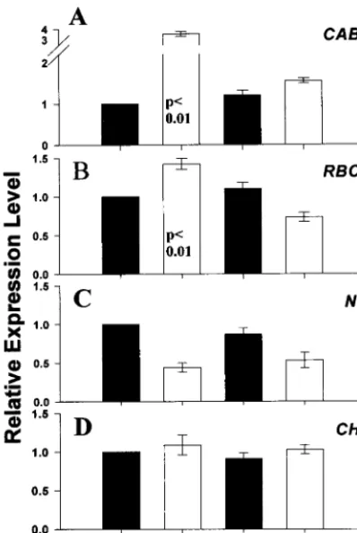
Plastid-Derived Redox Signal.RNA blot analyses on other nuclear-encoded genes suggests that the response reported above is specific for the transcript levels of photosynthetic genes (Fig. 2). After 24 h of starvation,CAB(Fig. 2A) and RBCS(Fig. 2B) transcript levels increased significantly (P,0.01) over those of sucrose-fed cells. In contrast, the corresponding levels forNR
showed a positive correlation with intracellular sugars (Fig. 2C), whereas those of chalcone synthase were insensitive to sugar
status (Fig. 2D). Addition of DCMU, an inhibitor of PET, to samples abolished the starvation-induced increase in bothCAB
andRBCStranscript levels (Fig. 2AandB), but had no effect onNRtranscript abundance (Fig. 2C). DCMU addition had no effect on either the respiration rate, or the rates at which sugars were depleted on starvation (data not shown). These results suggest that the normal starvation-induced increase in the transcript levels of nuclear-encoded photosynthetic genes re-quires PET and is modulated through a plastid-derived signal. The redox status of the PQ pool has been widely implicated as a regulatory signal controlling chloroplast activity (30, 31) and the induction of chloroplast-encoded photosynthetic genes (9). To determine whether the effects reported above are mediated through a similar mechanism, the PQ redox state was monitored by using chlorophyll fluorescence. With both starved and su-crose-fed cells, increasing light intensity resulted in a progressive reduction of the steady-state PQ pool redox potential. However, at the experimental growth conditions of 20mmol of photonsy m2ys PAR, the PQ pool redox state of both starved and sucrose-fed cells were not significantly different, and remained almost fully oxidized (Fig. 3). In the DCMU-treated cells reported above, the PQ pool would also have been fully oxidized because PQ reduction (by PS II) but not its oxidation (by PS I) would have been blocked.
The evidence presented above demonstrates that high intra-cellular sugars repress the abundance of nuclear-encoded pho-tosynthetic gene transcripts through the action of an uncharac-terized cytosolic signaling mechanism that may involve hexokinase. However, the removal of the sugar-repression signal alone does not result in increased transcript levels for these genes; the experiments with DCMU clearly indicate that an additional signal is required that depends on PET.
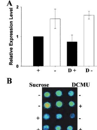
Transcriptional Control.TransgenicArabidopsislines [CAB2-LUC
[image:4.612.338.536.51.179.2](27) and PC-LUC (32)] carrying constructs of the luciferase reporter gene (LUC) fused behind the endogenous CAB2and the plastocyanin gene (PC) promoters were used to test this effect in planta. Fig. 4A presents the results from a typical experiment on detached leaves from thePC-LUCline. Lumi-nescence was low in the presence of sugar (1) and strongly increased in starved samples (2). However, luminescence re-mained low when leaves were starved for 24 h in the presence of DCMU (D2). Similar results were obtained with detached leaves fromCAB2-LUClines (images not presented). To quan-tify the luminescence, experiments were performed on leaf discs
Fig. 2. DCMU prevents the starvation-induced increase in transcript levels of
nuclear-encoded photosynthetic genes. Samples were prepared as described in Fig. 1. DCMU in ethanol was added at time 0 to a final concentration of 10
mM; controls were treated with ethanol (0.1%). Samples were removed after
[image:4.612.77.276.54.351.2]24 h and RNA extracted for RNA blot analysis. Autoradiographs of the result-ing RNA blots were scanned and signal intensity quantified; the values pre-sented are the averages and standard errors from six independent experi-ments; the values have been normalized on the constitutively expressed gene (ARF), and are presented relative to the expression level in sucrose-treated cells. An analysis of variance test was performed on the data and significant probability levels given on the graph. (A)CAB; (B)RBCS; (C)NR; (D) chalcone synthase,CHS; (2) and (1), absence and presence of 3% (wtyvol) sucrose; and D, presence of 10mM DCMU.
Fig. 3. The effect of light on PQ redox state in starved and sucrose-replete
cells. Three-day-old cultures were prepared as described in Fig. 1 andMaterials and Methods. Chlorophyll fluorescence emanating from PS II was measured over the range of indicated light intensities (5–155mmol of photonsym2ys
PAR) and the redox state of PQ was calculated as (FS2F0)y(Fmax2F0) (see
Materials and Methodsfor details). Each value represents the average and SEM ofn53 independent samples. (E) 3% (wtyvol) sucrose; and (F) starved for 24 h.
PLANT
from the CAB2-LUC (diagonal shading) and PC-LUC (solid shading) lines (Fig. 4B). The results from the detached leaves and leaf discs are entirely consistent with those obtained from the cell suspension cultures (Figs. 1 and 2).
Fig. 4Cpresents detached leaf images from a similar experi-ment by using the Arabidopsis mutantsun6, also carrying the
PC-LUC reporter gene construct. Quantified data from leaf discs of the sun6 mutant is presented in Fig. 4B (hatched shading). Sun6was isolated as a line in which sugar failed to repressPCexpression (32), and it has been shown recently that
sun6 is allelic to abscisic acid insensitive (abi4) because of a disruption of anAPETELLA 2-like transcription factor (33). In sharp contrast to the wild-type lines, sugar produced a 3-fold increase in luciferase activity over the levels in starved tissues. Our results suggest that, rather than being repressed by sugars, PC transcription is actually increased in the rosette leaves of the
sun6mutant. Furthermore, blocking PET with DCMU does not affect the sucrose-dependent increase of PC transcription in the
sun6mutant. Therefore, it can be concluded that a lesion in the
sun6yabi4gene, which disrupts the sugar repression response in young seedlings, causes sugar hypersensitivity in fully expanded leaves. This hypersensitive response is not influenced by the plastid-derived signal.
In vivoluciferase activity is dependent on endogenous ATP. To ensure that the effects of sugar and DCMU seen in the
CAB2-LUCandPC-LUClines did not arise as a consequence of altered ATP levels, experiments were carried out on a transgenic line carrying the constitutively expressedCaMV35S-LUC con-struct. Neither sugar nor DCMU affect luciferase activity in this line (Fig. 4D; quantified data not presented).
The requirement for PET for the induction of nuclear-encoded photosynthetic gene transcription was uncovered by DCMU addition at the time of sugar removal from transcrip-tionally repressed samples. To investigate the effects of PET on transcriptionally active samples, DCMU was added to starved cell cultures (Fig. 5A) and starvedPC-LUCleaf discs (Fig. 5B). In both cases, DCMU had no effect on gene expression. Longer treatments of cell cultures and leaf discs with DCMU gave similar results (data not shown).
To summarize, the experiments on the effects of sugar and DCMU on CAB2 and PC expression in planta support the conclusion drawn from RNA analysis on cell suspension cultures that a chloroplast-derived signal modulates the derepression of nuclear-encoded photosynthetic genes. However, this same sig-nal does not have an effect onCABandPCgenes once they are derepressed.
Discussion
[image:5.612.61.292.49.412.2]Plastids are the site of photosynthesis and numerous other essential metabolic processes in higher plants. To perform these tasks, both plastid- and nuclear-encoded proteins are required, but to function optimally, plastid metabolism must be tightly
Fig. 4. DCMU prevents induction of photosynthetic genes in transgenic
plants, but not in the sugar response mutantsun6. Luminescence from de-tachedA.thalianaleaves carrying thePC-LUC(A);PC-LUCinsun6background (C); andCaMV35S-LUC(D) constructs. Detached leaves were harvested and treated as described inMaterials and Methods. Pseudocolors represent lumi-nescence intensity with low (dark blue) and very high (red). (B) Quantified luminescence of 4-mm diameter leaf disks fromCAB2-LUC(diagonal shading);
PC-LUCwild type (solid shading); andsun6(hatched shading) plants. Values represent the averages and SEM of the number of photons emitted per leaf diskys. Treatment codes: (2) and (1), absence and presence of 3% (wtyvol) sucrose for 24 h; and D, presence of 10mM DCMU for 24 h before
[image:5.612.354.523.56.281.2]measurement.
Fig. 5. DCMU does not repress photosynthetic genes of the nucleus. Cell
cultures and leaf disks from thePC-LUClines were prepared as described in
Materials and Methodsand Figs. 2 and 4 with the exception that DCMU was added after samples had been starved for 24 h. (A)CABtranscript levels were determined 3 h after DCMU addition by RNA blot analysis as described in Fig. 2. The averages and SEM are shown (n53); (2) and (1), absence and presence of 3% (wtyvol) sucrose; D, presence of 10mM DCMU. (B) Pseudocolor
lumi-nescence images ofPC-LUCleaf disks 4 h after DCMU addition (blue, low luminescence; red, high luminescence).
integrated with that of the whole cell. Within the plastid, a complex array of mechanisms operating at the level of transcrip-tion and posttranslatranscrip-tion have been identified (34, 35) including redox regulation of both C-3 cycle enzymes (36–38) as well as transcript abundance (9) and translation (39) of the plastid-encoded genes for the photosynthetic reaction centers. Whereas the redox state of the photosynthetic electron transport chain has been shown to regulate the expression of plastid-encoded pho-tosynthetic genes (9), there have been no convincing reports to date of a plastid-derived redox signal operating to regulate nuclear-encoded photosynthetic genes in higher plants. In con-trast, a large volume of literature has accumulated in support of a sugar-sensing mechanism. Here, high levels of intracellular sugars are thought to represses the expression of the nuclear-encoded photosynthetic genesCAB, PC, and RBCS, and high rates of transcription occur only when sugar levels fall below some threshold level. We set out to establish whether any plastid-derived signals influence nuclear-encoded photosyn-thetic gene expression, and if so, whether they operate through a completely separate signaling mechanism, or by interaction with the sugar-sensing signaling mechanism.
Our experiments with cell cultures clearly demonstrate that intracellular sugar levels are inversely correlated withCABand
RBCStranscript levels. However, low endogenous sugar levels alone do not result in increased transcript levels; the experiments with DCMU demonstrate that a plastid-derived signal which is generated by PET is also required. It is well established that DCMU is a specific inhibitor blocking electron flow beyond the
QA-binding site in PS II. No other sites of DCMU action in plant cells are documented, and our results confirm that DCMU does not affect dark respiration rates or the sugar status of cells during starvation. The luminescence experiments with thePC-LUCand
CAB2-LUC Arabidopsislines gave results that are entirely con-sistent with those from the cell suspension cultures, and allow us to conclude that the sugar-sensing and chloroplast-derived sig-nals modulate the rates of gene transcription rather than tran-script stability.
RNA blot analysis on starved and sucrose-fed suspension cell cultures suggest that theNRgene is responsive to sugars (Fig. 2), but unlike CABand RBCS, shows a positive correlation with sugar levels and is not affected by DCMU. These results demonstrate the specificity of the DCMU effect on photosyn-thetic genes that are typically repressed by sugars.
What redox component in the chloroplast evokes nuclear-encoded gene transcription? The observation that in our exper-iments PQ remained almost fully oxidized in both starved (inducing) and sucrose-replete (suppressing) conditions (Fig. 3) suggests that the PQ redox state is not involved. Furthermore, because the oxidationyreduction of PQ is the rate-limiting step
in PET, we conclude all components of the PET chain were oxidized, regardless of sugar status, and therefore none of these give rise to the redox signal affecting nuclear gene transcription. Our experiments using inhibitors of PET support this view. With DCMU, all components downstream of its site of action (i.e., distal of theQB-binding site), including PQ, would have been fully oxidized, whereas those upstream (proximal ofQB), would have been fully reduced. It is difficult to reconcile the observed gene expression patterns in DCMU-treated samples (always suppressed, reduced proximalyoxidized distal components) and untreated samples (induced by starvation, fully oxidized PET) with the concept that the redox status of a PET chain component gives rise to nuclear gene transcription. Extensive experiments to confirm this by using the inhibitor 2,5-dibromo-3-methyl-6-isopropyl-p-benzoquinone, which blocks downstream of PQ, were inconclusive because of the labile nature of 2,5-dibromo-3-methyl-6-isopropyl-p-benzoquinone in our experimental con-ditions. Taken together, we conclude that PET is required to evoke the redox signal mechanism, but that this probably oper-ates through a stromal redox component. Possible candidoper-ates include thioredoxin or glutathione (15). However, it is conceiv-able that subtle changes (within the 0–10% reduced range) in the redox state of a PET chain component are sufficient to trigger nuclear gene transcription. Careful and detailed analysis of electron flux through the components of the photosynthetic electron transport chain, including cyclic electron transport, by using a range of light intensities should resolve this issue.
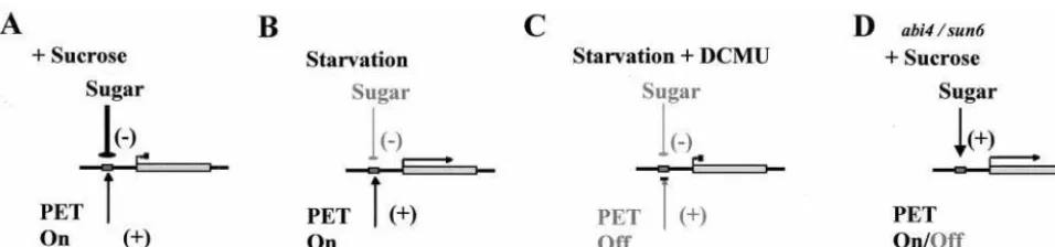
[image:6.612.73.551.52.164.2]A model may be proposed to account for our observations on the dependence of nuclear-encoded photosynthetic gene expres-sion on PET. This model relies on the generation of a positive regulator (inducer) by active PET and is summarized in Fig. 6. The positive regulator model predicts that under the experimen-tal conditions used here (20 mmol photonsym2ys PAR), a moderately weak inductive signal arises from PET, but the strong antagonistic suppression arising from exogenous sugars prevents transcription (Fig. 6A). Starvation in the light results in the removal of the sugar-dependent suppression allowing the chlo-roplast-derived positive regulator to induce transcription (Fig. 6B). However, transcription in starved cells is blocked if PET is inhibited (Fig. 6C). From this model, it follows that altering either the sugar status of the cell or the rate of PET can set the balance between sugar-dependent gene suppression and redox-dependent gene transcription. The experiments described in this study do not completely rule out the possibility that inactive PET could generate a negative signal regulating photosynthetic gene expression in the nucleus, rather than the positive regulator model as proposed. However, evidence fromArabidopsis genome uncoupledmutants that exhibit nuclear-encoded photosynthetic gene expression in the absence of chloroplast development (40),
Fig. 6. Model showing control of nuclear gene transcription by cytosolic sugar status and photosynthetic electron transport. PET gives rise to a positive signal
that enhances nuclear gene transcription. However, the low light levels used in this study (20mmol photonsym2ys PAR) evoke only a moderately weak signal,
and in the presence of the strong antagonistic repression arising from exogenous sugars, transcription is suppressed (A). Starvation in the light results in the removal of the sugar-dependent repression allowing the chloroplast-derived positive regulator to induce transcription (B). However, derepression in starved cells is blocked if PET is inhibited (C). In thesun6mutant, sugars induce photosynthetic gene expression irrespective of the status of PET (D).
PLANT
and from experiments with the inhibitors of carotenoid biosyn-thesis, Norflurazon and Amitrole (41), supports the positive regulator model.
Several recent reports have demonstrated the involvement of the plant hormone abscisic acid in sugar-mediated responses (33, 42, 43). By using theArabidopsis sun6mutant, which is allelic to the abscisic acid-insensitive mutantabi4, we demonstrate thatPC
transcription is induced rather than repressed by sugars and PET has no effect (Figs. 4Cand 6D). It is interesting to note that in
sun6,PCtranscription is behaving in a similar fashion to that of
NRin that it is induced by sugars (Figs. 2Cand 4B). In the case ofNR, PET also had no effect on transcript abundance. It is remarkable that the sugar-repressible, PET-sensitive PC pro-moter is converted to being sugar inducible and PET insensitive by a mutation in theABI4gene.
The model we have proposed provides plants with a flexible mechanism for tailoring cell metabolic processes to the appro-priate tasks determined by the whole plant in its unique envi-ronment. Clearly, the presence of abundant light should trigger the synthesis of new photosynthetic apparatus. However, this is
an appropriate response only when there is a requirement for energy or carbon fixation to meet either the immediate demands of the photosynthetic cell or the demands from sink tissues. When soluble sugar levels rise, for example, because of an impairment of phloem loading by either genetic or environmen-tally determined growth cessation, the synthesis of new photo-synthetic apparatus will be curtailed until the demand for carbon increases. A major challenge for future research will be to elucidate how sugar-, PET-, and abscisic acid-derived regulatory mechanisms interact to optimize photosynthetic gene expression in response to demand.
We thank Andrew Millar for providing the CAB2-luciferase and
CaMV35S-luciferase plants; Sjef Smeekens for providing the PC -luciferase and sun6 plants; Elaine Tobin for theCAB2 clone; Farid Regad and Claude Bardet for theARFclone; G. Trezzini for theCHS
clone; the Arabidopsis Biological Resource Centre for providing the expressed sequence tags for RBCSand NR; and the Central Science Laboratory greenhouse staff and David Neale for maintaining the plants. O.O. was funded through a University of Glasgow, Institute of Biomedical and Life Sciences Ph.D. studentship.
1. Graham, I. A., Baker, C. J. & Leaver, C. J. (1994)Plant J.6,893–902. 2. Graham, I. A., Denby, K. J. & Leaver, C. J. (1994)Plant Cell6,761–772. 3. Jang, J. C., Leon, P., Zhou, L. & Sheen, J. (1997)Plant Cell9,5–19. 4. Sheen, J. (1990)Plant Cell2,1027–1038.
5. Jang, J. C. & Sheen, J. (1997)Trends Plant Sci.2,208–214.
6. Koch, K. E. (1996)Annu. Rev. Plant Physiol. Plant Mol. Biol.47,509–540. 7. Halford, N. G., Purcell, P. C. & Hardie, D. G. (1999)Trends Plant Sci.4,
117–120.
8. Lalonde, S., Boles, E., Hellmann, H., Barker, L., Patrick, J. W., Frommer, W. B. & Ward, J. M. (1999)Plant Cell11,707–726.
9. Pfannschmidt, T., Nilsson, A. & Allen, J. F. (1999)Nature (London) 397,
625–628.
10. Alfonso, M., Perewoska, I. & Kirilovsky, D. (2000)Plant Physiol.122,505–516. 11. Petracek, M. E., Dickey, L. F., Nguyen, T. T., Gatz, C., Sowinski, D. A., Allen, G. C. & Thompson, W. F. (1998)Proc. Natl. Acad. Sci. USA95,9009–9013. 12. Escoubas, J. M., Lomas, M., Laroche, J. & Falkowski, P. G. (1995)Proc. Natl.
Acad. Sci. USA92,10237–10241.
13. Maxwell, D. P., Laudenbach, D. E. & Huner, N. P. A. (1995)Plant Physiol.109,
787–795.
14. Montane, M. H., Tardy, F., Kloppstech, K. & Havaux, M. (1998)Plant Physiol.
118,227–235.
15. Karpinski, S., Escobar, C., Karpinska, B., Creissen, G. & Mullineaux, P. M. (1997)Plant Cell9,627–640.
16. May, M. J. & Leaver, C. J. (1993)Plant Physiol.103,621–627. 17. Christie, J. M. & Jenkins, G. I. (1996)Plant Cell8,1555–1567.
18. Stitt, M., Lilley, R. M., Gerhardt, R. & Heldt, H. W. (1989)Methods Enzymol.
174,518–552.
19. Wintermans, J. F. & Mots, A. D. (1965)Biochim. Biophys. Acta109,448–453. 20. Kay, R., Chan, A., Daly, M. & McPherson, J. (1987)Science236,1299–1302. 21. Church, G. M. & Gilbert, W. (1984)Proc. Natl. Acad. Sci. USA81,1991–1995. 22. Leutwiler, L. S., Meyerowitz, E. M. & Tobin, E. M. (1986)Nucleic Acids Res.
14,4051–4064.
23. Trezzini, G. F., Horrichs, A. & Somssich, I. E. (1993)Plant Mol. Biol.21,
385–389.
24. Regad, F., Bardet, C., Tremousaygue, D., Moisan, A., Lescure, B. & Axelos, M. (1993)FEBS Lett.316,133–136.
25. Rouag, D. & Dominy, P. (1994)Photosynth. Res.40,107–117.
26. Bradbury, M. & Baker, N. R. (1981)Biochim. Biophys. Acta635,542–551. 27. Millar, A. J., Short, S. R., Hiratsuka, K., Chua, N. H. & Kay, S. A. (1992)Plant
Mol. Biol. Rep.10,324–337.
28. Sheen, J. (1994)Photosynth. Res.39,427–438.
29. Stitt, M., Krapp, A., Klein, D., Roeper-Schwarz, U. & Paul, M. (1995) inCarbon Partitioning and Source-Sink Interactions in Plants, eds. Madore, A. M. & Lucas, W. J. (Am. Soc. Plant Physiologists, Rockville, MD), pp. 68–77.
30. Allen, J. F., Alexciev, K. & Hakansson, G. (1995)Curr. Biol.5,869–872. 31. Allen, J. F. & Nilsson, A. (1997)Physiol. Plant.100,863–868.
32. Van Oosten, J. J. M., Gerbaud, A., Huijser, C., Dijkwel, P. P., Chua, N. H. & Smeekens, S. C. M. (1997)Plant J.12,1011–1020.
33. Huijser, C., Kortstee, A., Pego, J. V., Weisbeek, P., Wisman, E. & Smeekens, S. C. M. (2000)Plant J.23,577–585.
34. Link, G. (1996)BioEssays18,465–471.
35. Somanchi, A. & Mayfield, S. P. (1999)Curr. Opin. Plant Biol.2,404–409. 36. Buchanan, B. B. (1984)Bioscience34,378–383.
37. Raines, C. A., Lloyd, J. C. & Dyer, T. A. (1999)J. Exp. Bot.50,1–8. 38. Ruuska, S. A., Andrews, T. J., Badger, M. R., Price, G. D. & von Caemmerer,
S. (2000)Plant Physiol.122,491–504.
39. Danon, A. & Mayfield, S. P. (1994)Science266,1717–1719. 40. Susek, R. E., Ausubel, F. M. & Chory, J. (1993)Cell74,787–799. 41. LaRocca, V., DallaVecchia, F., Barbato, R., Bonora, A., Bergantino, E. &
Rascio, N. (2000)Physiol. Plant.109,51–57.
42. Laby, R. J., Kincaid, S., Kim, D. & Gibson, S. I. (2000)Plant J.223,587–596. 43. Arenas-Huertero, F., Arroyo, A., Zhou, L., Sheen, J. & Leon, P. (2000)Genes
Dev.14,2085–2096.