A Study of the Relationship of Abnormal
Reward Processing and Dopamine Signalling in
Adults with High Functioning Autistic
Spectrum Disorder
Thesis submitted in accordance with the requirements
of the University of Liverpool for the degree of
Master of Philosophy by
Eusra Hassan
2
Acknowledgements
I would first of all like to thank my supervisors Dr. Andrew Curran
and Dr. Kevin Southern for their continual support, advice and guidance
throughout this year, for which I am very grateful. I would like to thank
Unilever and Francis McGlone for providing the funding for the costs of
the study. I would also like to thank Val Adams, Graham Kemp and the
rest of the staff from MARIARC who have provided me with much
assistance during the scanning phase of the study, which I greatly
appreciate. Finally I would like to thank the participants who volunteered
to be a part of the study and without whom the study would not have been
3
Abstract
Introduction: Autistic spectrum disorder (ASD) is a neurodevelopmental condition which is defined by language and communication difficulties,
impairment in social skills and restrictive and repetitive patterns of
behaviour. Functional deficits have often been observed in individuals with
autistic spectrum disorder, which could be partially due to an early failure
of the amygdala. Motor activity, attentional skills, social behaviour and
perception of the outside world are all implicated in autism and are all also
modulated by the neurotransmitter dopamine. Past evidence has suggested
that dopamine neuron activation aids a person on learning to identify the
association of particular stimuli with reward.
Aims: The main objective of this preliminary study is to examine the neural activation during the visualisation of grouped images from the IAPS library
in normal participants and to observe if the activation follows a pattern in
the neural reward circuitary. Methodology: Participants underwent functional magnetic resonance imaging (fMRI), during which they
performed four tasks to test their reward processing; a visual paradigm, in
which the participants saw a display of images with differing emotional
weightings, a gambling paradigm, a soft touch paradigm and a materials
paradigm. The visual paradigm fMRI data was analysed using Brain
Voyager version 1.3. Results: The limbic lobe, anterior cingulate gyrus and the frontal lobe were the regions of the brain most activated for all of the
visual conditions in the control participants. The ASD participant activated
more neural regions, especially the limbic regions, than the control
participant in the Case vs. Control comparison. Discussion: Common areas of activation in the control group were the anterior cingulate gyrus, the
limbic lobe and the medial frontal gyrus. These areas all modulate facial
and image recognition, emotional processes and the evaluation of reward.
The ASD participant demonstrated more activation in limbic regions of the
brain than the control participant. These results support the idea that
individuals with ASD have difficulties in controlling their arousal state and
often have high levels of arousal. This can have implications on the
4
Nomenclature
ASD Autistic Spectrum Disorder
MRI Magnetic Resonance Imaging
fMRI Functional Magnetic Resonance Imaging
IAPS International Affective Picture System
ABA Applied Behavioural Analysis
5
Contents
Acknowledgments 2
Abstract 3
Nomenclature 4
Chapter 1 – Introduction and literature review 7
- 1.1i ASD: definitions, diagnosis and management. 7
∙ History 7
∙ Epidemiology 8
∙ Terminology and diagnosis 9
∙ Differences between Asperger’s syndrome and autism 12
∙ Functional deficits in ASD 14
∙ Management 15
- 1.1ii Brain development in ASD 20
- 1.1iiiNeurochemistry in ASD 22
∙ Serotonin 22 ∙ Dopamine 23
- 1.2i Reward and neurobiology 25
- 1.2ii Addiction and similarities to ASD 27
- 1.2iii Functional Magnetic Resonance Imaging (fMRI) and 28
activation of brain areas involved in reward processing
∙ Introduction to functional MRI 28
∙ fMRI studies investigating autism and reward 31
- 1.3i Reasoning for methodology 33
∙ International Affective Picture System (IAPS) and visual reward 33
- 1.3ii Aims and long term objectives of study 35
Chapter 2 – Methodology 38
∙ Ethical Approval 38
∙ Recruitment 38
6
∙ Evolution of methodology 39
∙ Phase 1 of study 42
∙ Phase 2 of study 44
∙ fMRI aquisition 44
∙ fMRI analysis 50
Chapter 3 – Results 51
∙ Recruitment 51
∙ Phase 1 results – Participant recruitment 52
- Participant characteristics 53
∙ Phase 2 results – Visual paradigm fMRI results for control 53
group batch analysis - Case vs. Control comparison 59
- ASD Circumscribed interests results 69
Chapter 4 – Discussion 71
∙ Control group: Common areas of activation between 71
the conditions ∙ Case vs. Control comparison 75
∙ Limitations 77
∙ Further work and Conclusions 79
References 81
Appendix A - Table of images from IAPS database 99
Appendix B - Sequence of exposure of images, fixation cross & their timings 102 Appendix C – Actual areas of activation in control group vs. predicted areas of activation 107
Appendix D – Actual areas of activation in ASD participant vs. predicted areas of activation 108
7
Chapter 1 – Introduction & literature review
1.1i Autistic spectrum disorder: definitions, diagnosis and management.
A person‟s social, academic and emotional life depends on adequate
communication with the world around them. Each individual must exchange some
sort of contact with people that they meet in order to sufficiently move forward in
their lives. Autistic spectrum disorder (ASD) is a neuro-developmental condition
which is defined by language and communication difficulties, impairment in social
skills and restrictive and repetitive patterns of behaviour. The term „spectrum‟ is used
due to the variation in the severity and pattern of symptoms from person to person. It
was not until the early 1940‟s that a label was introduced for this disorder that has
been found to affect many children and adults.
History:
In 1943 Dr. Leo Kanner assessed a group of 11 children and introduced the
name of early infantile autism; whilst at the same time Dr. Hans Asperger, an
Austrian paediatrician, described a milder form of the disorder that became known as
Asperger‟s syndrome (1). These two disorders are now listed in the Diagnostic and
Statistical Manual of Mental Disorders DSM-IV-TR (2) as two of the five pervasive
developmental disorders (PDD), referred to today as autistic spectrum disorders
(ASD). These disorders are all characterized by delays in the development of
multiple basic functions including socialization and communication, ranging from
the severe form, autistic disorder, to a milder form, Asperger‟s syndrome. If a child
has symptoms of autistic disorder or Asperger‟s syndrome, but does not meet the full
criteria of either of these, their diagnosis is called pervasive development disorder
not otherwise specified (PDD-NOS). Other conditions included in the autistic
spectrum are Rett‟s syndrome and childhood disintegrative disorder. Rett‟s syndrome
is a progressive neurodevelopmental disorder affecting 1 in 10000 females (3).
Usually patients develop normally until approximately six to eighteen months of age,
following which the child regresses, losing their acquired skills such as speech,
motor skills and purposeful hand movements (4). Girls who suffer from Rett‟s
syndrome will then frequently develop microcephaly, seizures, ataxia and autism (5).
8 syndrome live well into their middle ages and beyond (6). Childhood disintegrative
disorder is a condition which occurs in three to four year olds who develop normally
until approximately two years of age, following which they suddenly deteriorate in
their intellectual, social and motor functioning. It has been noted that children who
suffer from this rare syndrome demonstrate autistic like tendencies such as
impairments in non verbal behaviour and a loss of social and language skills (7).
Over the years various terms such as disintegrative disorder, disintegrative psychosis
and Heller‟s syndrome have been employed to describe this condition (8). There is
no known etiology for this disorder and the developmental delay continues
throughout the individual‟s adult life (9).
Epidemiology:
ASD affects approximately 1 in 100 children in the UK, as estimated by the
National Autistic Society (10) , with the risk 3-4 times higher in males than in
females. The prevalence of autism has been calculated in many areas and there have
been differences in the exact prevalence of autism due to the varied methods of
diagnosis between health authorities and individual health professionals (11). In
recent years the estimated prevalence of ASD has ranged between 12.2 to 67.4 cases
per 10,000 population depending on the location and the research undertaken (12).
There has been an increase in the number of reported cases of ASD in the last two
decades (13). It is argued that this increase could be due to a number of factors. The
body of knowledge about the condition is rapidly expanding along with an escalation
in public awareness. The increased knowledge and the development of the concept of
the wide autistic spectrum allows for improved case recognition and the possibility
of a genuine increase in numbers (14).
A number of environmental causes for a rise in incidence have been
discussed by researchers (14), including the question as to whether the triple vaccine
for measles, mumps and rubella could have been a potential cause for a few cases of
childhood autism. However, this concern was not confirmed by scientific backing
and was denied after various investigations (14)(15). There is strong evidence to
suggest that genetic factors have a significant role to play in the aetiology of ASD. It
has been shown that the suggested phenotype for autism is more likely to appear in a
9 dizygotic twin (16)(17). A review by Rutter showed data indicating that siblings of
individuals with autism were more likely to be affected by autism compared to the
general population and subsequent studies confirmed those results(18) (19)(20).
Another argument which favours the genetic basis of autism is the broad phenotype
of the disorder, which includes a range of symptoms from social communication
difficulties shown in Asperger‟s syndrome to mild autistic features such as highly
focused interests and activities (21). First degree relatives of individuals with ASD
tend to have an increased risk of displaying this broader phenotype.
The improved recognition of ASD traits at a younger age may have
contributed to the increased number of diagnosed cases of ASD over recent years
(22). There was little interest in ASD before the 1960‟s. Parent and support groups
began to form in USA and UK, which encouraged medical and educational
professionals to become more informed about the symptoms and methods of
management for the disorder (14). Following the initial surge of groups forming and
their push to have ASD publicised in the media, awareness of the wider view of ASD
began to expand (14). Broadening of the criteria used to assess and diagnose ASD
has contributed to health professionals feeling more prepared to diagnose individuals
with the disorder. A characteristic of ASD is the increased prevalence in males, with
autistic individuals showing a sex ratio of 4:1 (male: female) and individuals with
Asperger‟s syndrome having a sex ratio of 9:1 (male: female) (23)(21). Whether the
apparent increase in prevalence of ASD is due to a true increase in the condition or
an increase in knowledge and recognition of the disorder (24,25), the increase in
prevalence clearly demonstrates the need for a greater understanding of the
condition.
Terminology and diagnosis:
The current diagnostic criteria for autistic disorder, from the Diagnostic and
statistical manual of mental disorders: DSM IV, contain three core domains and for a
diagnosis of autism to be made, an individual must exhibit at least six symptoms
falling within the three core domains (socialisation, communication and restricted
10 A. Qualitative impairment in social interaction, with at least two of the
following:
- impairment in the use of non-verbal behaviours
- inability to develop peer relationships
- lack of ability to seek sharing enjoyment
- interests or achievements and lack of capability of social or emotional
reciprocity.
B. Qualitative impairment in communication, with at least one of the
following:
- delay or lack of spoken language
- inability to begin or sustain a conversation
- repetitive language or lack of make believe play.
C. Restricted repetitive and stereotyped patterns of behaviour, interests and
activities, with at least one of the following:
- a preoccupation with a stereotyped behaviour, interest or activity
- a rigid obedience in following specific routines or rituals
- repetitive motor mannerisms
- preoccupation with parts of an object.
To complete the diagnosis of autism, an individual must demonstrate delays in one or
more of the following areas, with the onset before the age of 3 years:
1. Social interaction.
2. Language as used in social communication.
3. Symbolic or imaginative play.
Asperger‟s syndrome is characterised by impairments in social skills, such as
difficulties in conversation and play and an inability to make appropriate eye contact
or to recognise facial expressions (13), but without difficulties in language fluency or
academic abilities. The diagnostic criteria for Asperger‟s syndrome are similar to that
for autistic syndrome, with two of the core domains (A and C) used to diagnose
autistic syndrome, also seen in Asperger‟s syndrome (26)(27). There also exist
11 autistic syndrome, as stated in the Diagnostic and statistical manual of mental
disorders: DSM IV:
1. The disturbance causes clinically significant impairments in social,
occupational, or other important areas of functioning.
2. There is no clinically significant general delay in language.
3. There is no clinically significant delay in cognitive development or in the
development of age-appropriate self help skills, adaptive behaviour (other
than in social interaction) and curiosity about the environment in
childhood.
4. Criteria are not met for another specific Pervasive Developmental
Disorder or Schizophrenia.
All people with ASD demonstrate difficulties with social interaction and
verbal and non-verbal communication. Additionally, repetitive, bordering on
obsessive, behaviours and interests are common in children and adults with ASD. As
described in Kanner‟s work on autism, the group of children that he studied seemed
to appear aloof and lack concern for other people (1), whereas Asperger‟s published
work depicting children who showed similar symptoms to those from Kanner‟s
work. The only difference between the groups of children was that Asperger‟s group
of children demonstrated higher verbal and cognitive skills.
ASD can very often be recognised in children as young as 1 year by their
parents picking up warning signs that their child is not behaving in the way that they
would expect. Their child may seem to be unresponsive to playful behaviour and
may be quiet and withdrawn in comparison to children of a similar age.
The surfacing of these warning signs should indicate the need for the child to be
evaluated by a professional who specialises in ASD.
The manifestations of autism range from people with severe impairments
who may have severe learning difficulties and undertake repetitive actions, to high
12 and can, to a certain extent, cope in many social circumstances. Autism is often
divided into low, medium or high functioning autism, related to the individual‟s
intellectual abilities (28), although this method of categorising people with autism is
not used in all centres that diagnose the condition.
Differences between Asperger’s syndrome and autism:
The pervasive developmental disorders (Asperger‟s syndrome, disintegrative
disorder and atypical autism) show phenotypic overlaps with core autism, so autistic
spectrum is increasingly being used to demonstrate the continuum between the
conditions (21). However, it is unknown whether autism consists of a variable single
syndrome or a collection of individual syndromes that share similar features.
Many researchers argue that there are differences in the early presentation of children
with high functioning autism and Asperger‟s syndrome, as on the whole Asperger‟s
syndrome presents without learning difficulties, although it can co-exist with
learning problems (26,29,30). In 1994 the Diagnostic and Statistical Manual of
Mental Disorders (DSM-IV), described the diagnostic criteria for Asperger‟s
syndrome. The authors of the manual, revised in 2000 DSM-IV-TR (2), stated their
opinions that Asperger‟s syndrome could be differentiated from autism by
examination of the child‟s early development. Asperger‟s syndrome differs from
autism in that individuals show no general delay in language or cognitive
development (30). Certain characteristics such as early language development and
cognitive skills are delayed in children with autism; however these features are not
significantly slower in children who have Asperger‟s syndrome (31). It is noted that
many people with autism isolate themselves and make no attempts to socialize with
others in their peer groups, whereas individuals with Asperger‟s syndrome by and
large make an effort to interact with other people, even if their manner of socializing
may be repetitive and unusual to others.
The term of Autistic spectrum disorder is used to group together individuals who
diagnostically suffer from different conditions, however clinically experience similar
symptoms and deficits in certain areas of their functioning albeit in various
13 “It must be remembered that autism is diagnosed by the existence of the full triad of
impairments and the particular manifestation of the triad will vary among
individuals. There are no behaviours per se that by their presence or absence indicate
autistic spectrum disorders; it is the overall pattern and underlying difficulties that
14
Functional Deficits:
There are a number of functional brain deficits that have been identified in
people suffering from ASD (33-36). These functional deficits are well defined and
consist of deficits in executive function (33), verbal comprehension, vocabulary and
comprehension (35). Executive dysfunction is another broad category that has been
observed in not only developing children who have ASD, but also adults (37).
Executive function refers to a set of cognitive abilities that control other behaviours
and are essential for goal directed, voluntary decision making. Some clinical features
of high functioning individuals with ASD such as obsessional behaviour and
repetitive actions have been related to executive functional deficits (38).
Tasks measuring drives for central coherence (39) and theory of mind
abilities, which relates to the ability to attribute mental states to others, are related to
abnormalities in the frontal cortex and basal ganglia structures (40). One of the most
powerful theories regarding the aetiology of ASD is the theory of mind deficit
account, which hypothesises that the social difficulties apparent in ASD are due to a
lack of understanding of feelings, thoughts and intentions in themselves and other
people (22,41). This social cognitive approach to looking at autism is employed by
Baron Cohen et al (42), who consider that autistic individuals who lack a theory of
mind do not understand that people have different thoughts in their head regarding
the world around them. The implications for the social interactions of these
individuals include problems in considering other people‟s feelings and an inability
to realise that people have different levels of knowledge compared to them.
The characteristic triad of communication, social and imagination
dysfunctions have been successfully explained by the hypothesis of individuals with
ASD lacking a theory of mind (43,44). However, the non-social impairments shown
by people with ASD cannot be explained by this theory. A further theory exists
which states that the behaviours and actions of people with ASD are due to the lack
of central coherence, which simply means that an individual is unable to incorporate
their senses and their behaviour into reasonable actions (45). This theory was
15 These non-social features consist of restricted interests, obsessive actions to
continually have the same environment, restricted areas of ability, an excellent
memory and perceptual abnormalities (hypo/hypersensitive to sound, touch, vision,
taste and hearing) (44). It is suggested that these features of ASD may be due to a
failure to process incoming stimuli in context (44).
The sensory perceptual problems that are commonly reported in ASD can
severely affect an individual‟s daily quality of life (48). These sensory abnormalities
can manifest as extreme hypersensitivity to everyday sensory stimulation, causing
intolerance to particular situations or sensations, such as loud noises, crowds or tags
on clothing. On the other hand, individuals with ASD may be hyposensitive and have
an underdeveloped response to the same stimulation types. A hyposensitive
individual may use abnormal means to experience sensory input, such as head
banging, or bringing everything into close visual range. Dysfunction in tactile
perception in ASD can also be seen in withdrawal from social touch, refusal to eat
certain textures of food, discomfort in wearing certain materials of clothing, or the
use of fingertips rather than the whole hand when manipulating objects (49).
Management:
The three core difficulties that characterize autistic disorders, social
interaction, communication and restrictive or repetitive behaviours and interests,
manifest in varying degrees of intellectual ability, language skills and behavioural
severity in different individuals. ASD covers similar disorders that share a broad
behavioural phenotype, that seem to follow the same common pathway of atypical
neurodevelopment (31). Health professionals have an important role in recognizing
symptoms of ASD and developing strategic management programmes for each
individual diagnosed with ASD. The main goals in the management of individuals
with ASD is to encourage independent functioning, to promote the development of
socialization, reducing maladaptive behaviours, to improve their quality of life and to
educate and support each person and their family (50). ASD is generally not termed
as a „curable‟ condition; therefore management is aimed at long term therapies. Most
children with ASD are highly likely to remain in the spectrum as adults and may
16 employment and mental health (50)(51,52). Therefore, it is important for the
individual to minimize the core features of the condition and to maximize their
independent function and their quality of life through educational development and
learning (50). The key management strategies include the following:
- Educational interventions:
1. Behavioural strategies andtherapy to develop speech and language are the
cornerstones of management ofASD. Not only do these interventions
tackle communication and social skills, butdaily-living skills, play and
leisure skills and academic achievement are also addressed (53). The
development of all of these skills that an individual with ASD lacks,
allows them to progress in these areas of their lives. In recent years,
research looking into management for ASD has focused on young
children with ASD, due to the evidence that earlier intervention can
significantly improve the future outcome (53). It has been demonstrated
that when interventions of this kind are provided from an early age, the
effectiveness of therapies are noted in the large number of children
benefiting from such therapies (53). Behavioural management therapies
focus on reinforcing wanted behaviours and reducing unwanted
behaviours.
2. Areas of education and support that are thought to be beneficial for
children, who have ASD, are a curriculum that encourages interaction
with people, a predictable routine for the child to feel at ease, a calming
approach to dealing with problem behaviour and definite family
involvement (54). Various specific strategies have been created to be used
in educational programmes for children with ASD. Applied Behaviour
Analysis (ABA) uses principals from experimental psychology to create
interventions that change unwanted behaviours (50). ABA is used to
create and maintain adaptable behaviours, reduce unwanted behaviours,
teach new skills and provide various environments for continuous
improvements in behaviour in various situations (55). Structured teaching
methods such as the TEACCH method, place an emphasis on the
organisation of the physical environment, predictable timetable for
17 3. A very important factor of management for a child with ASD is parental
support and education (57). The provision of support groups can allow
parents and carers of people with ASD to have contact with individuals
who deal with the same trials and tribulations, who may be able to offer
supportive advice (53,54).
- Therapy
The forms of therapy which have a significant impact on an individual‟s
improvement are:
1. Speech and language therapy (as people with ASD usually have deficits
in social communication) (50).
2. Social skills instruction (targeting the initiation of social behaviour,
minimising stereotyped persistent behaviours and teaching social skills)
(58).
3. Occupational therapy (to promote the development of self care and
academic skills) (50).
- Medical management
1. Autism therapies are designed to treat the symptoms of autism rather than
treat the condition itself. The core problems of autism can be complicated
by behavioural problems such as aggression, temper outbursts and
irritability (59). Children with autistic spectrum disorder also often suffer
from co-existing hyperactivity and lability of mood (60). When behavior
therapy and environmental changes do not improve the situation, drug
treatment may be considered. Antipsychotic drugs are the mainstay of
drug therapy for behavioural problems of children and adolescents with
ASD.
Haloperidol is one of the most widely studied antipsychotic drugs used to
reduce symptoms in children and adolescents with ASD (60-62). Safety
concerns exist regarding the risk of drug related dyskinesias with the
usage of haloperidol, meaning that when prescribed haloperidol must be
monitored carefully (62,63).
2. Neurobiological research has associated disruption in the dopamine and
Serotonin2A-18 dopamine D2 antagonist risperidone, an atypical antipsychotic, has been
studied extensively and has been deemed to be safe and efficacious at
reducing the disruptive symptoms observed in autism (67-69).
Risperidone must also be monitored when prescribed for behavioural
symptoms in autistic children and adolescents due to the risk of drug
induced weight gain and metabolic disturbances (62).
3. Selective serotonin reuptake inhibitors (SSRI‟s) have been utilised to treat
the behavioural symptoms and functional impairments of autism in
children and adults (70). Studies have been carried out which suggest
effectiveness of fluvoxamine (in children and adults) and fluoxetine (in
children); however, there is an increased awareness of the agitation risk in
autistic individuals who have been prescribed an SSRI (71,72). There are
questions regarding the tolerability and suitable dosing of SSRIs in
children and therefore these drugs are monitored vigilantly when
prescribed to children (71).
4. General medical management of children and adults with ASD consists of
the same health advice and disease prevention that people without ASD
also require. However in addition, they may require extra health care
needs related to underlying conditions that they may suffer from such as
fragile X syndrome or tuberous sclerosis and other health conditions such
as epilepsy (50,53). The range for the risk of children with ASD
developing epilepsy has varied between 10-30% (73). The prevalence of
epilepsy in children with autistic spectrum disorder has varied between
various studies, due to the differences in the guidelines used for diagnosis.
5. Many members of a multi-disciplinary team have a role in caring for an
individual with ASD. The theory of mind deficits, described in the
functional deficits section (page 14), can be recognised by members of
the professional team, from signs such as difficulties interacting with
others, a lack of understanding of others intentions or feelings and
difficulties in social situations. Social workers, care workers and teachers
can play a vital role in an individual‟s development. By finding methods
of teaching an individual with ASD and helping them progress in their
learning, professionals may be able to find ways of improving the social
19 professionals can also play a part in helping individuals with ASD in
improving their social skills by identifying the signs and by informing the
appropriate specialist, such as a psychologist, who can aid each individual
in their development.
20
1.1ii Brain development and ASD
The functional deficits often observed in individuals with ASD have been
analysed in a recent study and it has been reasoned that these deficits may be
partially due to an early failure of a particular cerebral structure called the amygdala
(74). Functional magnetic resonance imaging studies have established that the
amygdala shows increased activation when social intelligence is being performed
(75). Social intelligence is considered as an individual‟s ability to interpret another
person‟s behaviour in terms of their mental state, an ability to interact in close social
groups and in close relationships, the capability to empathise with another‟s mental
state and to be able to predict how another person feels and how they are going to
behave (76). It has been proposed that the amygdala plays a role in the neural
network that controls social behaviour (77).The overall volume of the amygdala is
significantly reduced and in post mortem studies shows neuropathology in autistic
individual‟s brains in comparison to their age and gender matched controls (78)(79).
The malfunction of the amygdala has a large influence on a particular area of the
brain involved in visual-social perception such as the fusiform (face area) of the
ventral temporal lobe. Neuroimaging work has regularly indicated the large
involvement of the fusiform gyrus and the amygdala in the processing of emotion
and the medial prefrontal and frontal cortex in tasks involving theory of mind (80). A
consistent finding in the neurobiology of autism is the reduced activity of the
fusiform gyrus and other cortical areas of the brain which assist with face recognition
and visual perception in people with autism (81,82).
The amygdala theory of autism was proposed by Baron Cohen et al after
reviewing the evidence of a social function of the amygdala (83). The fMRI study
involved autistic and non-autistic participants judging from a person‟s eyes how they
may be feeling, illustrating that the amygdala showed activation whilst non autistic
participants were making mental inferences about people‟s eyes. However,
participants with autism did not demonstrate as much amygdala activity as the
control participants during the task. The amygdala is therefore proposed to be one of
the neural areas that are abnormal in autism.
Recent imaging studies carried out on autistic populations have demonstrated
21 control group comparison (84,85), which is consistent with the results of previous
studies showing cerebellar hypoplasia in autistic individuals (86,87). Cerebellar
abnormalities, such as hypoplasia and hyperplasia of the posterior vermis and
hemispheres, in autistic individuals have been reported in past studies. However
these are not only seen in autism but also in neurogenetic syndromes and children
with leukaemia who have had radiation to the brain.
Theories have been developed from work showing altered c-fibre
functioning in adults with ASD (88,89). The main class of c-fibres are in the skin and
they react to painful stimuli. C-tactile fibres are responsible for the pleasurable
feeling that touch produces on hairy skin and track directly back to the orbitofrontal
cortex and amygdala. It is a possibility that the altered sensory input from these
fibres is due to an impairment in normal amygdala development from early
childhood. This altered c-tactile fibre function goes hand in hand with the sensitivity
deficits observed in individuals with ASD such as the hyper/hyposensitive response
to sensory stimuli (90). Some people with autism may be unable to tolerate a well
meaning pat on the back, whereas others may engage in self injury without seeming
to feel pain. A class of unmyelinated tactile mechanoreceptors that have recently
been identified in humans, known as CT afferents, are unmyelinated c fibres that
respond to slow, light, stroking touch stimuli (91). These fibres are thought to be
only distributed in hairy skin. With their preferred response as pleasant stroking
touch, this class of fibres compose a social touch system, which could play a role in
the hyper/hyposensitivity associated in autism (91-93).
The failure of development in key brain structures can also be interpreted as a
failure in learning, as the common deficits in ASD are in functional areas where
learning is vital to normal development. Recent studies regarding the neurobiology
of autism support this hypothesis by developing the theories that since the joining of
neurons is the neurobiological trigger behind learning, cortical connectivity must be
22
1.1iii Neurochemistry in ASD
Central to explaining the deficits found in ASD, is the exploration of possible
neurochemical mechanisms that may underlie the abnormal brain functioning. Whilst
the role of serotonin and other neurochemicals has been explored, there is little work
on the role of dopamine in these disorders. This is an important short fall as the
functional deficits seen in ASD are in brain areas where dopamine is the predominant
neurochemical. Researchers who have studied the neurochemistry of ASD have
identified chemical changes that have been found in individuals with ASD (73).
Serotonin
Autistic syndromes have a strong genetic component (98) and a category of
genes that have received much attention is the group that encodes proteins necessary
for brain serotonin metabolism and neurotransmission (99,100). This interest was
due to early findings of increased serotonin in autistic individuals (101)(102) and this
topic has been further researched in current studies (66)(65). A number of studies
have observed hyperserotonaemia in a third of autistic individuals and their first
degree relatives, which suggests that elevated blood serotonin levels could be a
marker for genetic susceptibility of autistic spectrum disorders (99,103). However,
the difficulty with measuring the levels of serotonin or, for that matter dopamine, in
the blood relates to the fact that both of these neurotransmitters cannot cross the
blood-brain barrier. Accurate measurements of the concentrations of serotonin and
dopamine in the brain consequently cannot be quantified by measuring the amount in
the blood.
Further evidence to show the involvement of the serotonin system in autism is the
considerable decrease in obsessive compulsive behaviours, anxiety and anger
observed in autistic individuals after they receive selective serotonin reuptake
inhibitors (104). In the past 10 years many studies have considered the various
options of pharmacological treatment for the behavioural aspects of autistic spectrum
disorder through considerations of the effects of serotonin and another
neurotransmitter, dopamine, on these behaviours (105)(106)(107).
23
Dopamine
The effects of serotonin levels on behaviours in autistic individuals have been
researched in detail; however the effects of dopamine, another neurotransmitter in
the brain, have been studied in less depth. Conflicting evidence has been produced
from studies assessing the role of the dopamine metabolite homovanillic acid (HVA),
in autistic children. Several pieces of evidence showed a difference in levels of HVA
in autistic children compared to non autistic children and some evidence showed no
difference (73,108,109). The pharmacologic management of certain unwanted
behaviours often observed in autistic individuals have shown great results of
reducing these behaviours. Dopamine blockers such as haloperidol (110) and
pimozide (111) have been shown to reduce hyperactivity, stereotypies and negative
behaviours. Neuroleptics which block dopamine and serotonin receptors, such as
risperidone (112) have also shown clinical improvement of behaviours associated
with autism. Such findings argue for a role of dopamine in autism.
Dopamine is a neurotransmitter that produces pleasure and arousal.
Dopamine has many functions in the brain including roles in behaviour, cognition,
motivation, reward, inhibition of prolactin production, sleep, mood, attention and
learning (113,114). Dopaminergic neurons are primarily present in the ventral
tegmental area of the midbrain, the substantia nigra pars compacta and the arcuate
nucleus of the hypothalamus (115). Dopamine neurons are essential for tasks such as
motor functions, motivation and working memory (116). Another central role for
dopamine neurons is the brain reward system, which controls the learning process of
many behavioural actions (117,118).
Motor activity, attentional skills, social behaviour and perception of the
outside world, which are all implicated in autism and other conditions within the
autistic spectrum, are all also modulated by dopamine. The involvement of dopamine
in movement control has long been highlighted due to the discovery of the
association between dopamine depletion and motor deficits in Parkinson‟s disease
(119). This instigated many clinical investigations into different therapies, such
L-Dopa, to improve patient‟s symptoms. Neuroloeptics were characterised soon after
the discovery of dopamine dysfunction in Parkinson‟s, as powerful dopamine
receptor blockers after it was observed that dopamine agonists exacerbate psychosis
24 action. Therefore dopamine is seen to be one of the key transmitters in drug abuse
and dependency. An artificial increase in dopamine transmission is the mechanism
of action for drugs of abuse that lead to addiction.
Dopamine has been identified as an important neurochemical in prefrontal
brain function. PET (Positron Emission Tomography) scans have identified low
medial dopaminergic activity in autistic children in comparison to a control group of
non autistic children and these findings lead researchers to believe that a
dopaminergic deficit may contribute to the cognitive impairment seen in autism, such
as the deficit in anticipatory behaviour and difficulty in shifting attention
(117)(64)(121).
Reward related dopaminergic effects on learning are well established.
Dopamine projections from the midbrain regions, such as the nucleus accumbens, to
the frontal cortex and striatum are a part of the behavioural actions that come from
reward (122,123). This is supported by the dopamine deficits in Parkinsonism,
schizophrenia and drug addiction (124-127). Past evidence has suggested that
dopamine neuron activation aids a person in learning to identify the association of
particular stimuli with reward (128). Di Chiara and North suggested that the
dopaminergic reward system is associated with incentive, preparation of acquiring
reward which can be experienced as urgency, thrill and cravings (129). Recent
research found evidence for the existence of a relationship between dopamine levels
and sensory reward based learning in adults, with results confirming that the effects
of reward on sensory processing could be influenced by dopamine (130).
The recent research into the link of dopamine and reward leads to the
hypothesis that dopamine is central to the processing of reward, which in turn
controls the learning process of many behaviours and actions. The following section
25
1.2i Reward and Neurobiology
The concept of reward is complex. Reward can be perceived as cognitive
representations such as novelty, challenge, acclaim, power, money, territory, and
security (122). Shultz defines rewards as having three basic functions:
1. Rewards serve as goals for voluntary behaviour. Therefore, rewards can cut
short ongoing behaviour and change the priorities of an individual‟s
behavioural actions to more frequently and successfully achieve reward.
2. Rewards have positive reinforcing effects, which mean that the individual
gaining reward wants more. Learning seems to progress when rewards occur
unpredictably and slows as rewards become more predicted.
3. Rewards produce subjective feelings of pleasure and positive emotional
states.
Reward is intimately linked with learning, and through learning, with adjusting
behaviours. Reward must also be associated with attention. An individual must be
attentive to the environment that a reward may occur in order to achieve the reward.
Power and acclaim are forms of complex reward that require cognitive processes to
plan for the reward and anticipate it. Processes are then required to recognise if the
planning was successful to achieving the reward that was envisaged (131).
FMRI studies in non-addicted individuals have shown that reward motivation
and rewards are processed by an interconnecting network of dopamine related brain
areas called the „neural reward system‟ (132-136). This network comprises of a
system made up of the nucleus accumbens, amygdala and the orbitofrontal cortex
(137,138). Each of these structures has a specific role in the response to reward. The
nucleus accumbens has a number of roles in reward such as responding to the
expectation and detection of reward (both the anticipation of reward and the reward
itself) and preparing actions in response to the reward. Neurons in the orbitofrontal
cortex differentiate amongst different types rewards.
Recent research has also shown that activation in the nucleus accumbens
increases with an increased probability of the reward (139). Therefore it is possible
26 rewarding stimulus and to approximate the level of perceived reward by measuring
the degree of activation of the nucleus accumbens and other limbic structures.
It has been found that there is an increase in dopamine release in the ventral
striatum during a reward producing situation (140). The ventral striatum is further
linked with reward related information due to its connections with the orbitofrontal
cortex and limbic regions like the amygdala (136).
Both the amygdala and the hippocampus project to the nucleus accumbens, which is
a component of the ventral striatum. Both the amygdala and the nucleus accumbens
demonstrate increased activity during its recognition of pleasure and reward (141).
Dawson et al. carried out a recent study comparing the ability of autistic
children, children with Down‟s syndrome and children with typical development at
visual comparisons, novelty preference and visual object recognition (142). Although
it was found that autistic children had no problems with visual object recognition,
their ability to form rules regarding the relation between a stimulus and a reward
revealed a difficulty to produce abstract rules.
As described above, dopamine activation aids learning and is involved in the
processing of reward. Therefore, the dopamine deficit seen in people with ASD can
affect the learning and reward system, which could explain the problems in
processing reward in ASD, demonstrated in the above studies. The characteristics
that people with ASD display, such as a lack social interaction and spending time on
their own, could be due to these reward processing problems. If the reward from
positive social interaction is not felt by an individual, the need to be close to other
people will not be felt, which people with ASD, and especially autism, demonstrate.
Consequently, the neurobiology of ASD, which includes a dopamine deficit, enables
a concept to put forward that the abnormal reward brain circuitry in people with ASD
is directly related to the dopamine deficit which is released from those same areas
27
1.2ii Addiction
Individuals addicted to alcohol, drug substances and food, have reward
patterns which show an immediate thrill and a desire to obtain the reward from their
addiction and these reward patterns have been shown to be dopamine dependent
(129). Individuals with ASD display similar reward processing to people with drug
addictions.
Natural rewards stimulate the release of dopamine from the nucleus accumbens
(143). Similarly, the reinforcing effects of drugs of addiction rely on the generation
of dopamine in the nucleus accumbens, which is part of the reward system (144). As
described in the section about dopamine, it is a neurotransmitter with multiple
properties from motor and cognitive functions, modulating of attention and
processing of reward and motivation. The response to drug addiction is not subjected
to the adaptive changes that occur in reward related learning, therefore, the drug
activates dopamine transmission without decreasing with increasing drug use
(143,145,146). This disruption of dopamine activation in the reward areas has been
observed in individuals with ASD. It is well recognised that people with ASD have
restricted and specific interests, usually only one at a time (147). There are obvious
similarities between the single minded tracking down of a single reward and the
behaviours of addicts. These similarities and the knowledge that there is a disruption
in dopamine function in ASD, links the reward processing of people with ASD along
28
1.2iii Functional Magnetic Resonance Imaging (fMRI) and activation of brain areas involved in reward processing
Introduction to functional MRI
Magnetic resonance imaging (MRI) is an imaging technique which is used
clinically and for research to visualise the internal structure and in functional MRI
displays the functions of the body. MRI allows for a much greater contrast between
different soft tissues than computed tomography (CT) which makes it a very useful
apparatus for visualising the brain, musculoskeletal system, cardiovascular system
and for accurately visualising cancers.
In brain imaging, MRI uses magnetic fields and radio waves to generate two
dimensional images of the brain structures without using radioactive tracers, which
can then be used to construct 3D images. The MRI scanner contains a magnet which
produces a magnetic field approximately a thousand times greater than the earth‟s
magnetic field. The magnet in the scanner is rated using a unit of measure known as
„tesla‟ or „gauss‟ (1 tesla = 10,000 gauss) (148). A typical research scanner has field
strength of 3 teslas (149). Hydrogen atoms in the body, which are usually randomly orientated, are forced by the magnetic field to become aligned to the direction of the
field. Radio waves are then sent towards the hydrogen atoms, which then bounce off
and a computer records the signal. Different tissue types such as fat, muscle, grey
matter, white matter and fluid produce different signals and the MRI scanner will
produce a 3 dimensional image of the various tissue types. This is due to the varying
number of hydrogen atoms in different tissues. The tissue that has the least number
of hydrogen atoms, such as bone, is dark in an MRI image, whereas tissue which
contains more hydrogen atoms, such as fatty tissue, appears brighter on the image. In
order to gain information about the different tissue types, the timing of the radio
wave pulses must be changed. Therefore for brain imaging, MRI provides a method
of discriminating between grey and white matter, cerebral spinal fluid and other
structures in the brain.
Functional magnetic resonance imaging, or fMRI, is a method used to
measure brain activity. It works by identifying the changes in blood oxygenation and
flow that take place in response to neural activity. When a particular area of the brain
29 active area. FMRI can be used to produce activation maps showing which parts of
the brain are involved in a particular process (150)(151). Haemoglobin is
diamagnetic (repelled by a magnet) whilst oxygenated but paramagnetic (magnetic)
whilst deoxygenated (152). This difference in magnetic properties leads to
differences in the MR signal of blood depending on the extent of oxygenation. As
mentioned above, since blood oxygenation varies depending on the levels of neural
activity, these differences can be used to measure brain activity. This form of MRI is
known as blood oxygenation level dependant (BOLD) imaging.
It is also important to note the direction of oxygenation change with increased
neuronal activity. There is a short decrease in blood oxygenation instantly after
neural activity increases, which is known as the initial dip in the haemodynamic
response. Following this is a stage where blood flow increases to a level that not only
meets the oxygen demand but overcompensates for the increased demand, meaning
that blood oxygenation increases following neural activation (153). The blood flow
peaks after 6 seconds and then falls back to baseline.
A voxel is a volume of element, representing a value on a regular grid in a
three dimensional space. In MRI the unit of analysis is a single voxel, identified by
its coordinates x, y and z. By identifying which voxels are significant demonstrates
the region of the brain activated by a specific task. The number of voxels activated in
a particular region indicates the area activated by a particular task, therefore a high
number of voxels in a region of interest indicate highly activated region of the brain.
FMRI as a brain imaging technique has several considerable advantages. The
scan is non invasive and does not involve radiation, therefore it is deemed safe for
patients. FMRI is also safe and straightforward for the experimenter to use (154).
These advantages have made fMRI a popular technique for imaging normal brain
function. Over the last decade fMRI has provided new means to investigate a vast
number of research areas including how memories are formed, language, learning,
pain and emotion (155-157).
FMRI is a useful tool in assessing which areas of the brain are activated after a
particular stimulus or activity. For this reason, fMRI has been utilised by many
30 Some general limitations of using MRI scanning are that high quality images can
only be attained if the participant stays very still, however this is not always
managed, whether it be due to the patient or due to the task itself, consequently
movement errors always have to be taken into consideration. Another few problems
that cannot be overcome due to the shape of the scanner are its inability to hold a
large participant and that it may cause some people to feel claustrophobic. An MRI is
not suitable for people who have been acutely injured, due to the fact that life saving
equipment is kept away from the scanner and also because the actual scan takes
longer than other imaging techniques which may cause discomfort for participants. A
limitation of functional MRI scanning, related to the conclusions that are made about
the relation between fMRI and neuronal activity, is that generalisations are often
made about individual‟s brains when the analysis often involves using group
averages (158).
Sensitivity and specificity: Sensitivity in terms of fMRI relates to the
sensitivity of the group analysis in allowing for unusual individual results (159). A
method of ensuring that similarities in a group are apparent and differences are
visible is by using the Talairach brain atlas in the analysis, which encourages each
individual brain to fit into a model brain, without losing each individual‟s activation
results. Specificity in light of fMRI data analysis is demonstrated by interpreting
areas with higher number of voxels in activated areas of interest.
Test repeatability would only be possible if the participants had previously
not seen the images in the visual task as memory may affect the brain activation from
viewing the images. If they had not seen the images before then the test could be
repeated with the same results for each individual participant. However, with a
different number of participants, the group results may differ as the average
31
FMRI studies investigating autism and reward
Structural imaging studies have shown that the various anatomical areas in
the brain are abnormal in individuals with ASD, such as irregularities in the
frontostriatal limbic system, age related changes in grey matter volume and
abnormalities in the cerebellar, caudate nucleus, thalamus, amygdala and
hippocampal regions of the brain (160-162). Impairments in reward association have
also been discovered in children and adults with ASD (163,164). However, only one
study has studied the brain‟s response to reward in people who have ASD using
functional magnetic resonance imaging (38). This study used event related functional
magnetic resonance imaging to examine the neural activation during reward
achievement in individuals with ASD, compared to matched controls. Individuals in
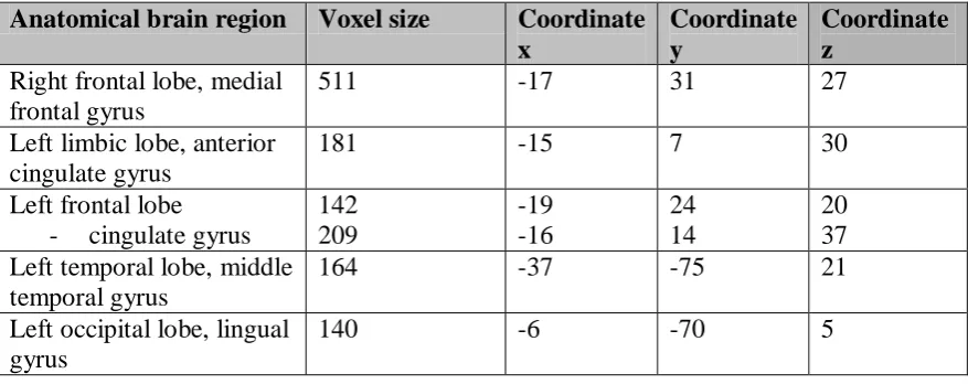
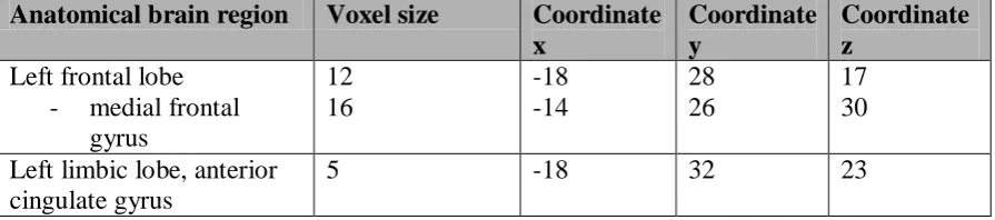
the control group showed significant brain activation which included the bilateral
anterior cingulate and frontal cortices and also the right insula. Individuals in the
autism group showed significant activation in the left anterior cingulate, the middle
and superior frontal gyrus and the right parietal lobe. The autistic individuals in this
study demonstrated significantly greater activation in the left anterior cingulate gyrus
during reward achievement in comparison to the control group. The anterior
cingulate gyrus is one of the areas that mediate reward feedback during cognitive
tasks, playing an important role in cognitive function such as executive attention,
conflict recognition, motivation and arousal (165). In past studies which examined
tasks of theory of mind in autistic individuals, anterior cingulate gyrus activation was
minimal (40,166). A positive correlation was detected between decreased anterior
cingulate gyrus activation and deficits in theory of mind tasks. In the study by
Schmitz et al. 2006 the findings of increased neural activation in the anterior
cingulate cortex during the attainment of reward in people with ASD may reflect an
increased requirement for feedback-related performance monitoring in ASD or an
increase in arousal and attention to rewarded stimuli.
An interesting consideration from the results of that study is that since the
cognitive area of the rostral anterior cingulate cortex demonstrated increased
activation during reward attainment in ASD individuals, it may be due to the
increased effort to achieve a desired result by actively directing their goal to an
32 with autistic spectrum disorder lack the capacity to wait for a reward when they can
attain a reward immediately (129,167).
The study by Schmitz et al. 2006 is the only one of its kind which assesses
reward processes in people with autistic spectrum disorder in comparison to controls
using functional magnetic resonance imaging. However, it only considers monetary
reward in the assessment of neural reward functioning in ASD. There are other
processes such as visual reward and sensory reward that have not yet been studied in
33
1.3i Reasoning for methodology
International Affective Picture System (IAPS) and visual reward
Researchers have used many methods of investigating emotional states, such
as film clips and still pictures (168). A widely employed set images used for visually
testing emotions is the International Affective Picture System (IAPS) (169).
Complex images in the IAPS database demonstrate emotion evoking images of
different weightings. The goal was to demonstrate a large set of standardised,
emotionally evocative colour images that can be easily accessed and used in
research. The standardised set of images have been rated between 1-10 in their
ability to provoke valance (unpleasant/pleasant, 1-10), arousal (calm/excited, 1-10)
and dominance (dominated/in control, 1-10). Neutral images have scores
approximately around 5. The study by Lang consisted of 100 adult volunteers, which
was a limitation to the Lang study, however other studies have carried out the same
experiment to verify the results on more volunteers (170,171). The original IAPS
study by Lang divided the results into adult female, adult male and then adult male
and female together. There was then a separate table with results from the group of
children. This validation process allows researchers to use all groups of images in
their studies, or just particular groups of images, such as those validated by male and
female adults, which is the validated group of images used for this study. The reason
for using this group instead of just the male validated images was due to the
knowledge that the future expanded study may include female participants; therefore
the joint male and female adult validated images were employed.
Work has been done to identify the difference in the emotional processing
and difference in brain activation in fMRI between images of emotional faces and
IAPS images (172). Although this study assessed the sets of brain regions that were
activated in processing facial and the IAPS images, this study utilised IAPS images
depicting emotions such as sadness, anger, happiness, which were selected by the
researchers. However, work has not been carried out to identify which brain areas are
engaged from complex combinations of different emotionally weighted IAPS
images. The images selected for this study from the IAPS database demonstrated
pleasure and displeasure. However the IAPS database does not allow for the images
34 demonstrated pleasurable images in different forms, and one displeasure group of
images was selected. The groups of images were divided into the following:
- Control images (neutral)
- Aroused, in control, pleasure (high valance, high arousal, high dominance)
- Non aroused, in control, pleasure (high valance, high dominance, low
arousal)
- Aroused, not in control, pleasure (high valance, high arousal, low dominance)
- Displeasure (high arousal, low valance, low dominance)
The images were not selected by simply viewing the images, instead the rating of
reach of the emotional categories (valance, arousal, and dominance) was set for each
of the five groups (stated above) and the images were selected via the ratings, which
prevented researcher bias in the choice of visual stimuli.
In addition research has not been undertaken to study the effects of these
emotional stimuli on the reward circuitry in the brain. These complex combinations
of rated emotional images can reveal the impact of various emotionally rewarding
and non-rewarding stimuli on the reward circuitary in the brain. With areas of the
reward circuitry such as the amygdala and the nucleus accumbens seen to have
deficits in people who have ASD, testing a hypothesis that emotion evoking images
from IAPS database activate areas known to process reward in normal participants,
could allow for a hypothesis that people with ASD process reward in a different
manner to the non ASD population. This hypothesis could be extended to investigate
individuals with ASD and observe whether the areas of their brains activated from
these emotionally charged images and other rewarding stimuli, differ to the areas of
activation in controls due to known deficits in brain reward areas in ASD.
35
1.3ii Aims for the study and long term objectives
Specific aims for this study:
- Developing the four paradigms; gambling, visual, touch and materials, and
exploring how best to apply the paradigms in a functional MRI setting.
- Focused development of the visual paradigm using the IAPS image database, with
the aim of creating a task which assesses positive emotional stimuli (rewarding
images) and negative emotional stimuli. The advantages of focusing on this visual
paradigm are that through investigating which groups of images activate which area
in the brain, we can not only explore the differences between the groups of IAPS
images, but we can also identify which areas of the brain are activated during
rewarding stimuli. This information is beneficial when developing the project and
comparing the brain activation in an ASD population and a normal population.
- To assess which anatomical and functional area of the brain is significantly
activated (p < 0.05) from each of the groups of images as stated above in the control
participants and the ASD participants.
- To assess the similarities and differences between the neural areas activated from
the different image groups in the control participants and the ASD participants.
- To assess whether areas in the reward circuitry of the brain, including the amygdala
and the nucleus accumbens are activated from visualising rewarding images in the
MRI scanner.
- To explore for the similarities and differences in which area of the brain is activated
for a particular image category, between the control group and the ASD group.
- Investigate the differences between the ASD participant and a control participant in
brain region activation, allowing for the exploration of the hypothesis of abnormal
reward circuitry in ASD, aiding the development of a larger scale project.
Long term aims
The long term aims of the project are to examine the neural activation during the
visualisation of images from the IAPS library, differing in emotional weightings,
along with the testing of sensory reward through two touch paradigms and risk taking
36 compared to controls. The reasoning for these different reward testing paradigms is
stated below:
- Hyper/hyposensitivity to pleasurable sensory stimuli
It is recognised that a large proportion of individuals with ASD suffer from
hyper/hyposensitivity (88). This includes sensitivity to temperature, noise and touch
(173,174). As mentioned in section 1.1ii, individuals with ASD have been found to
have altered c-fibre functioning (88,89). This information, along with findings of a
class of c fibres in hairy skin that respond to pleasant stroking touch (91), allow the
development of the theory that the hyper/hyposensitivity in autistic individuals could
be due to deficits in c-fibre responses. This leads to a hypothesis that individuals with
ASD may have deficits in the experience of sensory stimulation. In order to test this
hypothesis, a stroking stimulator producing a soft and light stroking action can be
applied to the hairy skin of individuals with ASD whilst they have a functional MRI
scan.
Along with the hyper/hyposensitivity to noise, temperature and soft touch, people
with ASD often have a great like or dislike to an everyday fabric type (175,176). For
this reason, a hypothesis can be built regarding whether everyday materials cause an
abnormal brain activation pattern during fMRI in the brains of people with ASD in
comparison to controls.
- Gambling reward
A study was carried out examining the neural circuitry associated with
immediate versus delayed reward processing in adults with attention deficit
hyperactivity disorder (ADHD) (177). The results of the study demonstrated that in
people with ADHD, a delay of rewards produces hyper activity in the neural circuits
involved in motivation, emotion and reward, which could contribute to the
intolerance of delays in reward associated with ADHD. It is known that individuals
with ASD also demonstrate impairments in the brain regions involved in motivation
and emotion, along with people who have ADHD (167). Therefore, it is
acknowledged that individuals with ASD have difficulties with the ability to wait for
a reward that they can attain. This piece of information is vital in understanding how
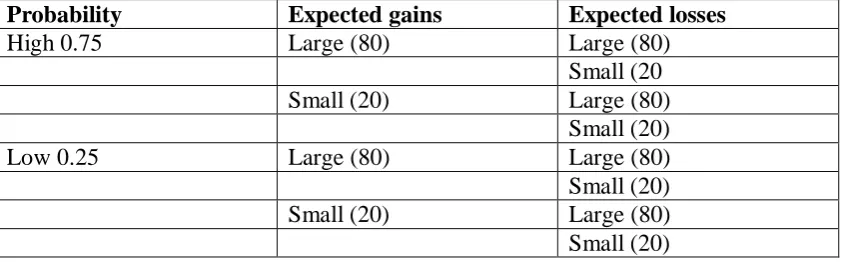
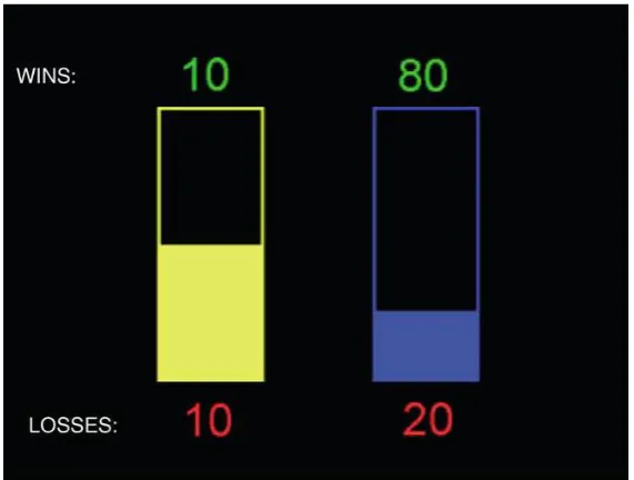
37 The gambling task described in Rogers at al. 2003 (178) is a decision making
task in which participants decide between two risky gambles in order to maximise
monetary reward and then have a short period of time to process the outcomes of
their choice, whether they be positive or negative.
A hypothesis can be formed that since individuals with ASD have difficulties
in waiting for reward, in a gambling situation they must choose an option that allows
them to win their reward straight away, regardless of what it is and whether their
reward can increase if they wait. The gambling task above allows this hypothesis to
be investigated in an fMRI scanner in order to visualise the brain areas of activation