Open Access
Research
Induction of HO-1 in tissue macrophages and monocytes in fatal
falciparum malaria and sepsis
Ian A Clark*
1, Melissa M Awburn
1, Clive G Harper
2, N George Liomba
3and
Malcolm E Molyneux
4,5Address: 1Dept of Biochemistry, Australian National University, Canberra, Australia, 2Dept of Pathology, University of Sydney, Australia, 3Dept of Histopathology, College of Medicine, University of Malawi, Blantyre, Malawi, 4Wellcome Trust Laboratories and Malaria Project, College of Medicine, University of Malawi and 5School of Tropical Medicine, University of Liverpool, UK
Email: Ian A Clark* - [email protected]; Melissa M Awburn - [email protected]; Clive G Harper - [email protected]; N George Liomba - [email protected]; Malcolm E Molyneux - [email protected]
* Corresponding author
Abstract
Background: As well as being inducible by haem, haemoxygenase -1 (HO-1) is also induced by interleukin-10 and an anti-inflammatory prostaglandin, 15d PGJ2, the carbon monoxide thus produced mediating the anti-inflammatory effects of these molecules. The cellular distribution of HO-1, by immunohistochemistry, in brain, lung and liver in fatal falciparum malaria, and in sepsis, is reported.
Methods: Wax sections were stained, at a 1:1000 dilution of primary antibody, for HO-1 in tissues collected during paediatric autopsies in Blantyre, Malawi. These comprised 37 acutely ill comatose patients, 32 of whom were diagnosed clinically as cerebral malaria and the other 5 as bacterial diseases with coma. Another 3 died unexpectedly from an alert state. Other control tissues were from Australian adults.
Results: Apart from its presence in splenic red pulp macrophages and microhaemorrhages, staining for HO-1 was confined to intravascular monocytes and certain tissue macrophages. Of the 32 clinically diagnosed cerebral malaria cases, 11 (category A) cases had negligible histological change in the brain and absence of or scanty intravascular sequestration of parasitized erythrocytes. Of these 11 cases, eight proved at autopsy to have other pathological changes as well, and none of these eight showed HO-1 staining within the brain apart from isolated moderate staining in one case. Two of the three without another pathological diagnosis showed moderate staining of scattered monocytes in brain vessels. Six of these 11 (category A) cases exhibited strong lung staining, and the Kupffer cells of nine of them were intensely stained. Of the seven (category B) cases with no histological changes in the brain, but appreciable sequestered parasitised erythrocytes present, one was without staining, and the other six showed strongly staining, rare or scattered monocytes in cerebral vessels. All six lung sections not obscured by neutrophils showed strong staining of monocytes and alveolar macrophages, and all six available liver sections showed moderate or strong staining of Kupffer cells. Of the 14 (category C) cases, in which brains showed micro-haemorrhages and intravascular mononuclear cell accumulations, plus sequestered parasitised erythrocytes, all exhibited strong monocyte HO-1 staining in cells forming accumulations and scattered singly within cerebral blood vessels. Eleven of the available and readable 13 lung sections showed strongly staining monocytes and alveolar macrophages, and one stained moderately. All of the 14 livers had strongly stained Kupffer cells. Of five cases of comatose culture-defined bacterial infection, three showed a scattering of stained monocytes in vessels within the brain parenchyma, three had stained cells in lung sections, and all five demonstrated moderately or strongly staining Kupffer cells. Brain sections from all three African controls, lung sections from two of them, and liver from one, showed no staining for HO-1, and other control lung and liver sections showed few, palely stained cells only. Australian-origin adult brains exhibited no staining, whether the patients had died from coronary artery disease or from non-infectious, non-cerebral conditions
Conclusions: Clinically diagnosed 'cerebral malaria' in children includes some cases in whom malaria is not the only diagnosis with the hindsight afforded by autopsy. In these patients there is widespread systemic inflammation, judged by HO-1 induction, at the time of death, but minimal intracerebral inflammation. In other cases with no pathological diagnosis except malaria, there is evidence of widespread inflammatory responses both in the brain and in other major organs. The relative contributions of intracerebral and systemic host inflammatory responses in the pathogenesis of coma and death in malaria deserve further investigation.
Published: 19 November 2003
Malaria Journal 2003, 2:41
Received: 20 August 2003 Accepted: 19 November 2003
This article is available from: http://www.malariajournal.com/content/2/1/41
Introduction
Falciparum malaria is a complex multi-organ disease. There is no simple or accepted explanation for how small numbers of parasites can cause such severe illness, or how this infection can cause such wide-spread pathology, since only hepatocytes and erythrocytes are invaded by the pathogen. Undoubtedly parasitized red cells sequester in capillaries and venules, but in recent times the traditional idea that this is the primary cause of organ failure and death through obstructing blood flow has needed modi-fying. In particular, it has had to accommodate the evi-dent involvement of excessive systemic release of pro-inflammatory cytokines, triggered by malarial toxins. For the last decade, many researchers have focussed their efforts on the pathophysiological implications of the abil-ity of these mediators to generate inducible nitric oxide synthase (iNOS), and thus produce a continuous, poten-tially large, supply of nitric oxide in tissues that normally experience only low, tightly controlled, levels of this ubiq-uitous cellular messenger. Despite the harmful effects of iNOS-induced nitric oxide (NO) when produced in unu-sually large amounts [1-3], more commonly it provides negative feedback that suppresses production of the inflammatory cytokines that generate it, and a range of other downstream harmful mediators, through inhibiting NF kappa B, a major activator of protein transcription [4].
Carbon monoxide (CO), another endogenous gas with a similar structure, also inhibits TNF generation [5] again through inhibiting NF kappa B [6]. Both molecules are generated by enzymes that have at least one constitutive form, and another, iNOS and haemoxygenase-1 (HO-1) respectively, induced by inflammatory cytokines. NO and CO act interactively as second messengers in ways that are still being elucidated [7]. For instance, both NO and CO can activate soluble guanylate cyclase to generate cyclic GMP [8], and thus dilate blood vessel walls, as well as per-form their immunosuppressive roles.
This shared activity of NO and CO duplicates that of inter-leukin-10 (IL-10), the prototype anti-inflammatory cytokine, which also suppresses generation of tumour necrosis factor (TNF) and interleukin-1β through inhibit-ing NF kappa B [9]. Thus the high circulatinhibit-ing levels of IL-10 seen in human malaria [IL-10,11] and sepsis [12], have been proposed to suppress disease severity through inhib-iting the systemic inflammatory effects of TNF [13]. These apparently disparate observations are now appreciated to be different parts of the same chain of events, with IL-10 producing its strong anti-TNF effect through inducing HO-1, and thus generating CO [14]. The most plausible explanation for the early observation that TNF induces HO-1 [15] is now therefore its ability to induce IL-10 [16]. Likewise, the anti-inflammatory effect of 15d
prostaglan-din J2 (15d PGJ2), which is present in tissues during
inflammation [17], also operates through HO-1 induc-tion and subsequent generainduc-tion of CO [18]. As in sepsis,
cyclooxygenase-2, which generates 15d PGJ2, is induced
in severe malaria [19]. Thus, rather than HO-1 simply being a marker for haem degradation and a generator of anti-oxidant defences, it is now recognized to be an inte-gral part of the network of inflammatory mediators. It therefore serves as a convenient and sensitive marker for such activity.
Accordingly, brain, lung and liver from 40 African chil-dren who had died of malaria, sepsis or unrelated condi-tions were stained for HO-1 in order to identify cellular sites where the CO-mediated anti-inflammatory activity of IL-10 might be located in these infectious diseases. These tissues had previously been stained for migration inhibitory factor (MIF) and inducible nitric oxide syn-thase (iNOS) [20]. Before the IL-10 or prostaglandin links of HO-1 were appreciated, others [21,22] have immunos-tained brains, but no other tissues, from adult malaria cases, to detect this enzyme. The present study provides further evidence for the presence of multi-organ inflam-matory changes in children fulfilling the clinical criteria of 'cerebral malaria', whether or not malaria was the princi-pal or only pathological diagnosis.
Materials and Methods
Case TissuesAs described earlier [20], all 40 subjects (age range six months to 12 years; 22 females) were children who had been admitted to the Malaria Project wards in the Depart-ment of Paediatrics at the Queen Elizabeth Central Hospi-tal in Blantyre, Malawi (Table). Evaluation, diagnoses, treatment, autopsy permissions were as previously described [20]. Autopsies were performed as quickly after death as possible, with post-mortem intervals ranging from two to 14.5 hrs. Tissue samples were placed into 10% neutral buffered formalin for fixation. The project was approved by the ethics and research boards of the College of Medicine (University of Malawi), the Univer-sity of Liverpool and the Australian National UniverUniver-sity.
Control tissues
Tissues from three Malawian children who were enrolled in this study served as local non-comatose controls. No coma was present at any stage in two of these (patients 41
and 50; see Table). The former grew Salmonella
of five blocks of tissue, trimmed from the periphery of tumour excisions from adult chest wall, and containing skeletal muscle, adipose tissue and small blood vessels. Midbrain sections from three adults who had died of cor-onary artery disease, and from another three who died of non-infectious, non-cerebral conditions (Brain Bank for Sydney Central Area Health Science Approval X980216), were also stained. A section of an inflamed pilonidal sinus was routinely included as a positive control.
Immunohistochemistry
Formalin-fixed tissue samples were embedded in paraffin, sectioned (4 microns) on to polylysine-coated slides, and stained with haematoxylin and eosin (H&E) for routine morphology. A monoclonal anti-HO-1 antibody was pur-chased from StressGen; (Cat. No. OSA-110). Other mon-oclonals were used as irrelevant primary control antibodies, and in other controls the primary antibody was omitted. As previously [20], antigen retrieval was per-formed by immersion in 0.01 M citrate buffer, pH 6.0, in a waterbath at 95°C for 20 min and then cooling to room temperature while still immersed in buffer. After
quench-ing with 3% H2O2 and treating with primary antibody
(dilution of the stock solution 1:500 to 1:2000) at room temperature for 1 hr, biotin-conjugated secondary anti-body and streptavidin-conjugated horseradish peroxidase from an LSAB+ kit (DAKO) were applied to sections for 20 min at room temperature to amplify the antigen signal for subsequent 3,3'-diaminobenzidine (DAB) staining. Known positive controls were stained in each run, and runs were often duplicated on different days to confirm repeatability. Sections were counterstained with haema-toxylin, and outcomes with a dilution of primary anti-body of 1:1000 are shown to illustrate the observed changes. Anti-CD68 antibody (Clone PG-M1) was obtained from DAKO, and used, with antigen retrieval, at a primary antibody dilution of 1:500.
Histological examination
In a recent investigation of the distribution of MIF and iNOS [20], in which 32 cases that had been clinically diag-nosed as cerebral malaria were studied, they were classi-fied into three categories on the basis of the presence or absence of sequestered intracerebral parasites and brain pathology. Category A (n = 11) had no or scanty intracer-ebral parasites and negligible brain pathology detected, category B (n=seven) had sequestered parasites in brain vessels, again with negligible brain pathology, and cate-gory C (n = 14) had both sequestered parasites and inflammatory brain pathology in the form of intravascu-lar monocyte aggregations, fibrin deposition and/or microhaemorrhages. Here this same terminology is retained, and the outcome of immunostaining to detect HO-1 in the brain, lung and liver of these same cases is reported. In most of the category A patients, autopsy
revealed another likely cause of death: this was pneumo-nia in five cases, hepatic necrosis in one, severe anaemia with pulmonary oedema in one, and ruptured cerebral aneurysm in one. No alternative causes were identified in category B and C patients.
One hundred and forty sections from 49 brains were stained for HO-1 and examined. Samples were from fron-tal lobe, pariefron-tal lobe, temporal lobe, occipifron-tal calcarine fissure, hippocampus, caudate nucleus, basal ganglia, tha-lamus, midbrain, pons and medulla, with frontal lobe, occipital region, midbrain and pons most commonly included. Two sections were considered ample to record a result on sections where staining was readily detected, since up to seven were examined in some cases, with no difference in outcome from the opinion formed after the first section. Up to seven sections per brain were examined in those cases with no staining detectable, and these comprised sections from two to four blocks, which pro-vided an element of depth within individual blocks as well as a spread of location across the brain. With few exceptions, staining was either absent, moderate, or strong, and if strong was sometimes remarkably intense and even, despite the high dilution of primary antibody. Mid-brain sections from three adults who had died of cor-onary artery disease, and from another three who died of non-infectious, non-cerebral conditions (Brain Bank for Sydney Central Area Health Science Approval X980216), were included. Single blocks only were available for lung and liver. Two examiners (IC and CH), blinded to the diagnosis, examined the sections independently.
Results
Cerebral malaria
Category A cerebral malaria
Table 1: CM(A), insignificant physically apparent brain pathology
Patient no. Age (mo) Sex Parasitaemia (Pf) &/or Organism cultured
Admitting coma score
Parasites in brain vessels
Brain vessel monocyte HO-1
Lung mono & alveol M HO-1
Liver Kupffer cell HO-1
4 20 M 19% 2 1+ isolated, mod strong, fewer strong
7 29 F 4+ 0 1+ absent strong, fewer strong
22 18 M 2% 0 1+ absent strong, common strong
23 30 F 29% 0 1+ absent strong, common strong
31 39 M 49% 1 1+ absent PMN-obscured strong
33 25 M <1% 0 1+ absent pale strong
37 6 M 34% & P. mal.
trace
2 1+ scattered, mod strong, fewer strong
38 84 F 22% 0 1+ scattered, mod strong, fewer strong
45 34 M 3% 0 1+ absent PMN-obscured moderate
47 22 F 11% 1 1+ absent pale strong
49 17 M 0% 1 1+ absent negative moderate
CM(B), sequestered parasites only
1 27 F 31% 0 2+ rare, strong mod, fewer strong
5 14 M 4% 1 4+ scattered, strong strong, common strong
16 51 F 6% 1 2+ rare, strong strong, fewer strong
21 25 F <1% 2 4+ scattered, strong strong, common strong
25 44 F <1% 2 4+ scattered, strong strong, common strong
39 18 M <1% 2 4+ scattered, strong strong, fewer strong
42 37 F 20% 0 4+ absent PMN-obscured NA
CM(C), sequestered parasites plus local intravascular inflammation
6 17 F 6% 0 3+ accumul, strong strong, common strong
9 16 M <1% 1 3+ accumul, strong strong, fewer strong
11 29 F 29% & H. influ. B
1 2+ accumul, strong PMN-obscured strong
13 22 M 37% 0–1 3+ scattered, strong strong, common strong
15 8 F 1% 2 2+ accumul, strong strong, fewer strong
26 30 M 19% 1 4+ accumul, strong strong, common strong
27 20 M 53% & Sal.
typhi-m
0 3+ accumul, strong strong, fewer strong
28 61 F 23% 0 2+ scattered, strong NA strong
29 43 M 33% 2 3+ accumul, strong strong, fewer strong
32 18 F 47% 0 3+ accumul, strong strong, fewer strong
34 70 M 2% 1 4+ accumul, strong strong, common strong
35 114 M <1% 1 3+ accumul, strong strong, common strong
36 21 F 1% 2 2+ accumul, strong strong, fewer strong
48 48 M <1% 2 3+ accumul, strong mod, fewer strong
COMA (bacteria cultured, and clinically judged not cerebral malaria)
12 6 M Sal. enteritidis 2 negative scattered, strong strong strong, autolys
14 9 F E. coli 0 negative absent strong strong
18 7 F H. influenzae B 1 negative absent absent strong
20 96 F Tuberculosis 0 negative regionally strong regionally strong strong
24 48 F Strep.
pneumoniae
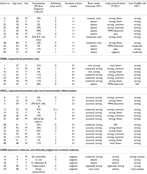
[image:4.612.61.553.113.692.2]In the 11 cases that comprised category A (no discernible histological changes, only the occasional rare malarial pigment or parasites within vessels, and in most cases an additional explanation for death identified at general autopsy) eight had no detectable HO-1 staining. in the brain (Table 1). One showed isolated moderately stained monocytes, and the other two showed a light scattering of strongly stained monocytes across the brain parenchyma. Examples are shown in Fig. 1. The identity of these cells was confirmed with CD68 staining (not shown). Of the 11 lung sections, six exhibited strongly stained monocytes and alveolar macrophages (Fig. 1), two were pale, and one was negative. In the other two the section was largely obscured by a heavy influx of neutrophils, a cell type we have not seen stain for HO-1. Nine of the 11 liver sections showed strongly staining Kupffer cells (Fig. 1), and in the other two these cells were moderately stained only.
Category B cerebral malaria
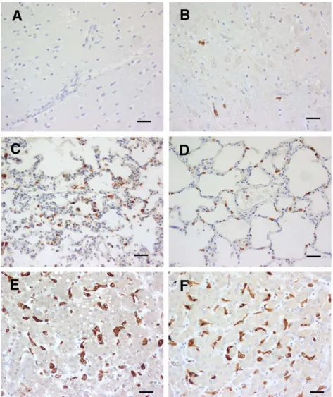
In the seven category B cases (no discernible pathological changes in the examined sections, but sequestered parasi-tised erythrocytes present) 2 had rare strongly staining monocytes in cerebral blood vessels, and another four showed a scattering of strongly stained monocytes (Table 1). The identity of these cells was confirmed with CD68 staining (not shown) An example of each type of density is shown in Fig. 2. Five out of seven lung sections showed strong staining of monocytes and alveolar macrophages, one was moderately stained, and the other section was largely obscured by a heavy influx of neutrophils, with no HO-1 staining discernible. All six of the available seven liver sections showed strong staining of Kupffer cells, again as illustrated in Fig. 2.
Category C cerebral malaria
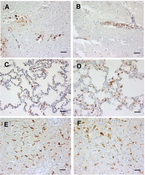
The brains of 14 category C cases (microhaemorrhages, intravascular mononuclear cell accumulations, and also sequestered parasites) all exhibited strong monocyte
HO-1 staining in mononuclear leucocytes forming accumula-tions, and also in those scattered singly, within blood ves-sels (Table 1). The two cases illustrated in Fig. 3 had neither a low haematocrit, nor hypoglycaemia. Monocyte accumulations were not seen in sections of two of the brains, and thus the individual cells are referred to in the Table as "scattered, strong". In addition the macrophages that had aggregated at microhaemorrhages were stained strongly (Fig. 3). The identity of these cells was confirmed with CD68 staining (not shown). Eleven of the 13 availa-ble lung sections showed strongly staining monocytes and alveolar macrophages (Fig. 3), with the monocytes often in accumulations similar to those observed in the brain. One section was moderately stained, and the other section was largely obscured by a heavy influx of neutrophils. As shown in the examples in Fig. 3, all of the 14 livers had strongly stained Kupffer cells.
Coma associated with bacterial infections
Another five patients were comatose on admission and before death, had very low or no peripheral malaria para-sitaemia, and were considered on clinical grounds to be suffering from a disease other than malaria [20]. Two had
septic meningitis (one each culture positive for
Haemo-philus influenzae and S. pneumoniae), two had a positive blood culture (one Salmonella enteritidis, one Escherichia coli) and one, with a lymphocytic infiltrate in the cerebro-spinal fluid, was diagnosed histologically, including the presence of acid-fast organisms, as tuberculous meningi-tis. The E. coli and H. influenzae cases had no detectable HO-1 staining. in the brain, and the other three showed a variable scattering of stained monocytes (Table 1). In the tuberculosis case staining was limited to the areas adja-cent to large blood vessels. Two of the five lung sections (S. enteritidis and E. coli cases) showed strongly staining monocytes and alveolar macrophages, while none were evident in the H. influenzae and S. pneumonia cases, and they were restricted to the region near chronic lesions in
MALAWIAN CONTROLS (without terminal coma)
41 29 F Sal.
typhimurium
5 negative absent absent absent
43 53 M suspect malaria 2, then 5 negative absent absent pale, v. occas
* 50 144 F Strep.
pneumoniae
5 negative absent pale pale, occas
AUSTRALIAN CONTROLS
n= 6 F/
M
NA NA negative negative NA NA
* Serologically positive for HIV (n) Number of brains areas examined Abbreviations: NA = not applicable Sal. typhi-m. = Salmonella typhimurium H. influ. B = H. influenzae type B
CM (A). HO-1 staining of tissues from cerebral malaria cases showing apparently absent physical brain pathology
Figure 1
CM (B). HO-1 staining of tissues from cerebral malaria cases showing apparently absent physical brain pathology, but seques-tered parasites common
Figure 2
CM (C). HO-1 staining of tissues from cerebral malaria cases showing physically apparent brain pathology, plus sequestered parasites present
Figure 3
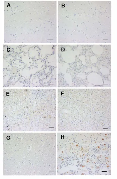
the tuberculosis case. All five livers showed HO-1 staining of the Kupffer cells, although in one case this was moder-ate rather than strong (Table 1). As examples, the changes in brain, lung and liver of the E. coli and H. influenzae cases are shown in Fig. 4.
Control Tissues
Brain sections from all three African controls, and lung sections from two of them, and the liver of one, showed no staining for HO-1 (Table 1, Fig. 5). The lung of the one, and the liver of two, showed few, palely stained cells only. Likewise, the 6 Australian-origin adult brains showed no staining, whether the patients had died from coronary artery disease or non-infectious, non-cerebral conditions (Fig. 5G). The subcutaneous tissues from Australian adult controls were also negative (not shown). In contrast, mac-rophages in the section of inflamed pilonidal sinus included in every staining run invariably were strongly positive (Fig. 5H), as were the macrophages in the splenic red pulp, a location where free haem can be expected to induce HO-1, in all African cases (not shown).
CD 68 staining
CD36 staining confirmed the identity of the mononuclear phagocytic lineage of stained cells, including the cells attracted to ring haemorrhages, that were positive for HO-1. Both single and double staining was undertaken (not shown). This confirmed that negative HO-1 staining of tissues could not be attributed to an absolute absence of monocytes and macrophages in various locations, but to them being present and not staining.
Discussion
Here is described the cellular distribution of HO-1 in sev-eral key organs in African children who died of clinically defined cerebral malaria or coma accompanying a bacte-rial infection. Endothelial cells and vascular smooth mus-cle, and skeletal musmus-cle, so often MIF and iNOS positive [20], were devoid of HO-1. Previous reports of HO-1 staining of human tissues includes the cytotrophoblast cells within the placental bed of the normal human pla-centa [23], the alveolar macrophages of normal and inflamed lung [24], endothelium and macrophages of atherosclerosis lesions [25], Kupffer cells and hepatocytes in portal hypertension [26], tubular epithelial cells in kid-ney diseases [27], and microglia and macrophages in focal cerebral infarcts and brain trauma [28]. Malarial brains, but no other organ, from Asian [22] and European adults [21] have been stained for HO-1, and, consistent with the literature of the time, HO-1 was discussed only in terms of being a stress protein [22] and a generator of CO that could contribute to cerebral malaria by influencing neu-rons directly [21].
What induced HO-1 in these tissues remains unclear. It is well accepted that a high concentration of haem induces HO-1 [29], and the invariable staining observed in spleen red pulp in all cases (not shown), and at cerebral haemor-rhage sites where red cells have been phagocytosed (Fig. 3B) in category C brains is consistent with this. Haem from local haemorrhage cannot, however, account for the presence of HO-1 in mononuclear phagocytes in CM(A) or CM(B) brains, and other organs In addition, cellular deprivation of glucose has been shown to induce the HO-gene 1 in vitro [30], raising the possibility of the hypogly-caemia sometimes seen in severe childhood falciparum malaria being instrumental in generating the observed HO-1. No difference was seen in the intensity of HO-1 staining between cases with normal peripheral blood glu-cose concentration and those with systemic hypoglycae-mia, but this observation does not exclude local tissue hypoglycaemia as a possible stimulus to HO-1 induction. IL-10, a major modulator of inflammation increased in malaria [10], is now known to induce HO-1 [14]. Plasmas for IL-10 assay were not available from these cases, but the precedence exists of high levels being associated with severe malarial illness [10,11].
In view of the evidence that the inflammatory cascade is activated in this Malawian [20,31] and other [32-34] pop-ulations with severe malaria infection, the observed HO-1 is additional evidence that in fatal malaria a widespread host inflammatory response occurs, similar to that seen in other acute infections (reviewed in [35]). This is consist-ent with the HO-1 staining seen in the sepsis cases in this series (Fig. 4), in that systemic bacterial infections are broadly accepted to be examples of systemic inflamma-tion. This is not to suggest that some of the cerebral malaria cases in this series did not have additional or alternative disease mechanisms, arising from local cere-bral lesions, such as vessel obstruction from large parasite loads in the apparent absence of other lesions (Fig. 2B). Nevertheless, strong systemic inflammation was present in all these cases. Local cerebral changes arising from post-schizogony secondary inflammatory events, as demon-strated by the presence of monocytes accumulations, high iNOS, haemorrhages and fibrin in other CM(C) cases (Fig. 3A and 3B) may well also contribute significantly.
non-HO-1 staining of tissues from bacterial sepsis cases
Figure 4
HO-1 staining of tissues from controls
Figure 5
malarial comatose illness, illustrate the fact that coma may accompany infections with extensive systemic inflammation but without evidence of intracerebral inflammatory responses. It is, therefore, possible that, in the CM(B) and CM(C) patients (with cerebral sequestra-tion and cerebral intravascular HO-1 inducsequestra-tion), the observed widespread systemic inflammation may have been similarly important in the pathogenesis of coma. This adds strength to the argument that systemic events, not only those in the brain, should be borne in mind when attempting to understand disease outcome in Afri-can children clinically diagnosed with cerebral malaria. The CM(A) group, one third of the cases, in which malaria was only one of several conditions that could have con-tributed to death, might have provided insight into the state the cerebral vasculature of the CM(B) and CM(C) cases after they had become comatose, but some time before death.
Finally, the inhibitory activity of CO, the product of HO-1, against TNF [4] parallels that of NO, the product of iNOS [5]. Ideas on how iNOS polymorphisms might have been selected for in African populations [36,37] through their interaction with malarial disease might also, from these results, apply to known HO-1 polymorphisms [38,39] in these populations.
Acknowledgments
This work was supported by the Australian National Health and Medical Research Council, Grant 148902, the National Institute of Allergy and Infectious Diseases, USA (Grant 1-RO1-AI 34969) and the Wellcome Trust. We thank Dr Biziwick Mwale, Director of the Queen Elizabeth Cen-tral Hospital, and Professor Robin Broadhead, Department of Paediatrics of the College of Medicine, University of Malawi, for their hospitality and for access to patients in the Malaria Ward, and we thank Professor Terrie Taylor, who played a major part in running the Severe Malaria Ward and in arranging and conducting autopsies. We appreciate the cooperation of par-ents and guardians of the children studied, and the devoted care provided by the nursing and laboratory staff within the Paediatric Department and the research programme. This study received funding from the Australian Health and Medical Research Council, the National Institutes of Health, USA, and The Wellcome Trust, UK.
References
1. Boczkowski J, Lanone S, Ungureanulongrois D, Danialou G, Fournier T, Aubier M: Induction of diaphragmatic nitric oxide synthase after endotoxin administration in rats – role in diaphrag-matic contractile dysfunction.J Clin Invest 1996, 98:1550-1559. 2. Lanone S, Mebazaa A, Heymes C, Henin D, Poderoso JJ, Panis Y,
Zedda C, Billiar T, Payen D, Aubier M et al.: Muscular contractile failure in septic patients – Role of the inducible nitric oxide synthase pathway.Am J Resp Crit Care Med 2000, 162:2308-2315. 3. Mitaka C, Hirata Y, Yokoyama K, Wakimoto H, Hirokawa M, Nosaka
T, Imai T: Relationships of circulating nitrite/nitrate levels to severity and multiple organ dysfunction syndrome in sys-temic inflammatory response syndrome. Shock 2003,
19:305-309.
4. Peng HB, Libby P, Liao JK: Induction and stabilization of I kappa B alpha by nitric oxide mediates inhibition of NF-kappa B.J Biol Chem 1995, 270:14214-14219.
5. Otterbein LE, Bach FH, Alam J, Soares M, Tao Lu H, Wysk M, Davis
tory effects involving the mitogen-activated protein kinase pathway.Nat Med 2000, 6:422-428.
6. Sarady JK, Otterbein SL, Liu F, Otterbein LE, Choi AM: Carbon monoxide modulates endotoxin-induced production of gran-ulocyte macrophage colony-stimulating factor in macrophages.Am J Respir Cell Mol Biol 2002, 27:739-745. 7. Hartsfield CL: Cross talk between carbon monoxide and nitric
oxide.Antioxid Redox Signal 2002, 4:301-307.
8. Schmidt HHHW: NO., CO and .OH – Endogenous soluble gua-nylyl cyclase-activating factors.FEBS Letters 1992, 307:102-107. 9. Wang P, Wu P, Siegel MI, Egan RW, Billah MM: Interleukin (IL)-10
inhibits nuclear factor kappa B (NF kappa B) activation in human monocytes. IL-10 and IL-4 suppress cytokine synthe-sis by different mechanisms.J Biol Chem 1995, 270:9558-9563. 10. Peyron F, Burdin N, Ringwald P, Vuillez JP, Rousset F, Banchereau J:
High levels of circulating IL-10 in human malaria. Clin Exp Immunol 1994, 95:300-303.
11. Wenisch C, Parschalk B, Narzt E, Looareesuwan S, Graninger W:
Elevated serum levels of IL-10 and IFN-gamma in patients with acute Plasmodium falciparum malaria. Clin Immunol Immunopathol 1995, 74:115-117.
12. Lin RY, Astiz ME, Saxon JC, Saha DC, Rackow EC: Relationships between plasma cytokine concentrations and leukocyte functional antigen expression in patients with sepsis.Crit Care Med 1994, 22:1595-1602.
13. Ho M, Sexton MM, Tongtawe P, Looareesuwan S, Suntharasamai P, Webster HK: Interleukin-10 inhibits tumor necrosis factor production but not antigen-specific lymphoproliferation in acute Plasmodium falciparum malaria. J Infect Dis 1995,
172:838-844.
14. Lee TS, Chau LY: Heme oxygenase-1 mediates the anti-inflam-matory effect of IL-10 in mice.Nat Med 2002, 8:240-246. 15. Cantoni L, Rossi C, Rizzardini M, Gadina M, Gheezi P: Interleukin-1
and tumor necrosis factor induce hepatic haem oxygenase. Biochem J 1991:891-894.
16. Platzer C, Meisel C, Vogt K, Platzer M, Volk HD: Up-regulation of monocytic IL-10 by tumor necrosis factor-alpha and cAMP elevating drugs.Int Immunol 1995, 7:517-523.
17. Gilroy DW, Colville Nash PR, Willis D, Chivers J, Paul Clark MJ, Willoughby DA: Inducible cyclooxygenase may have anti-inflammatory properties.Nat Med 1999, 5:698-701.
18. Lee TS, Tsai HL, Chau LY: Induction of heme oxygenase-1 expression in murine macrophages is essential for the anti-inflammatory effect of low dose 15-deoxy-Delta(12,14)-pros-taglandin J(2).J Biol Chem 2003, 278:19325-19330.
19. Deininger MH, Kremsner PG, Meyermann R, Schluesener HJ: Focal accumulation of cyclooxygenase-1 (COX-1) and COX-2 expressing cells in cerebral malaria. J Neuroimmunol 2000,
106:198-205.
20. Clark IA, Awburn MM, Whitten RO, Harper CG, Liomba NG, Molyneux ME, Taylor TE: Tissue distribution of migration inhib-itory factor and inducible nitric oxide synthase in falciparum malaria and sepsis in African children.Malaria J 2003, 2:6. 21. Schluesener HJ, Kremsner PG, Meyermann R: Heme oxygenase-1
in lesions of human cerebral malaria.Acta Neuropathol 2001,
101:65-68.
22. Medana IM, Mai NTH, Day NPJ, Hien TT, Bethell D, Phu NH, Farrar J, White NJ, Turner GDH: Cellular stress and injury responses in the brains of adult Vietnamese patients with fatal Plasmo-dium falciparum malaria. Neuropath Appl Neurobiol 2001,
27:421-433.
23. Lyall F, Barber A, Myatt L, Bulmer JN, Robson SC: Hemeoxygenase expression in human placenta and placental bed implies a role in regulation of trophoblast invasion and placental function.FASEB J 2000, 14:208-219.
24. Lakari E, Pylkas P, Pietarinen Runtti P, Paakko P, Soini Y, Kinnula VL:
Expression and regulation of hemeoxygenase 1 in healthy human lung and interstitial lung disorders.Hum Pathol 2001,
32:1257-1263.
25. Wang LJ, Lee TS, Lee FY, Pai RC, Chau LY: Expression of heme oxygenase-1 in atherosclerotic lesions. Am J Pathol 1998,
152:711-720.
Publish with BioMed Central and every scientist can read your work free of charge
"BioMed Central will be the most significant development for disseminating the results of biomedical researc h in our lifetime."
Sir Paul Nurse, Cancer Research UK
Your research papers will be:
available free of charge to the entire biomedical community
peer reviewed and published immediately upon acceptance
cited in PubMed and archived on PubMed Central
yours — you keep the copyright
Submit your manuscript here:
http://www.biomedcentral.com/info/publishing_adv.asp
BioMedcentral
27. Morimoto K, Ohta K, Yachie A, Yang Y, Shimizu M, Goto C, Toma T, Kasahara Y, Yokoyama H, Miyata T et al.: Cytoprotective role of heme oxygenase (HO)-1 in human kidney with various renal diseases.Kidney Int 2001, 60:1858-1866.
28. Beschorner R, Adjodah D, Schwab JM, Mittelbronn M, Pedal I, Mat-tern R, Schluesener HJ, Meyermann R: Long-term expression of heme oxygenase-1 (HO-1, HSP-32) following focal cerebral infarctions and traumatic brain injury in humans. Acta Neuropathol 2000, 100:377-384.
29. Maines MD, Trakshel GM, Kutty RK: Characterization of two constitutive forms of rat liver microsomal heme oxygenase. Only one molecular species of the enzyme is inducible.J Biol Chem 1986, 261:411-419.
30. Chang SH, Garcia J, Melendez JA, Kilberg MS, Agarwal A: Haem oxy-genase 1 gene induction by glucose deprivation is mediated by reactive oxygen species via the mitochondrial electron-transport chain.Biochem J 2003, 371:877-885.
31. Grau GE, Taylor TE, Molyneux ME, Wirima JJ, Vassalli P, Hommel M, Lambert P-H: Tumor necrosis factor and disease severity in children with falciparum malaria. N Engl J Med 1989,
320:1586-1591.
32. Kern P, Hemmer CJ, Van Damme J, Gruss H-J, Dietrich M: Elevated tumour necrosis factor alpha and interleukin-6 serum levels as markers for complicated Plasmodium falciparum malaria. Am J Med 1989, 87:139-143.
33. Kwiatkowski D, Hill AVS, Sambou I, Twumasi P, Castracane J, Manogue KR, Cerami A, Brewster DR, Greenwood BM: TNF con-centration in fatal cerebral, non-fatal cerebral, and uncom-plicated Plasmodium falciparum malaria. Lancet 1990,
336:1201-1204.
34. Butcher GA, Garland T, Adjukiewicz AB, Clark IA: Serum TNF associated with malaria in patients in the Solomon Islands. Trans R Soc Trop Med Hyg 1990, 84:658-661.
35. Clark IA, Cowden WB: The pathophysiology of falciparum malaria.Pharmacol Ther 2003, 99:221-260.
36. Burgner D, Xu WM, Rockett K, Gravenor M, Charles IG, Hill AV, Kwiatkowski D: Inducible nitric oxide synthase polymorphism and fatal cerebral malaria.Lancet 1998, 352:1193-1194. 37. Hobbs MR, Udhayakumar V, Levesque MC, Booth J, Roberts JM,
Tka-chuk AN, Pole A, Coon H, Kariuki S, Nahlen BL et al.: A new NOS2 promoter polymorphism associated with increased nitric oxide production and protection from severe malaria in Tanzanian and Kenyan children.Lancet 2002, 360:1468-1475. 38. Yamada N, Yamaya M, Okinaga S, Nakayama K, Sekizawa K, Shibahara
S, Sasaki H: Microsatellite polymorphism in the heme oxygen-ase-1 gene promoter is associated with susceptibility to emphysema.Am J Hum Genet 2000, 66:187-195.