Programmed assembly of polymer
–
DNA conjugate
nanoparticles with optical readout and
sequence-speci
fi
c activation of biorecognition
†
Johannes P. Magnusson,aFrancisco Fern´andez-Trillo,bGiovanna Sicilia,a Sebastian G. Spain*aand Cameron Alexander*a
Soft micellar nanoparticles can be prepared from DNA conjugates designed to assembleviabase pairing such that strands containing a polymer corona and a cholesterol tail generate controlled supramolecular architecture. Functionalization of one DNA conjugate strand with a biorecognition ligand results in shielding of the ligand when in the micelle, while encoding of the DNA sequences with overhangs allows supramolecular unpacking by addition of a complementary strand and sequence-specific unshielding of the ligand. The molecular assembly/disassembly and ‘on–off’ switch of the recognition signal is visualized by FRET pair signalling, PAGE and a facile turbidimetric binding assay, allowing direct and amplified readout of nucleic acid sequence recognition.
Introduction
The encoding of information into materials through monomer sequence is a central process in nature, utilising components with˚Angstrom-scale dimensions to build devices that operate over nanometre and micron lengthscales. Natural materials of this type are essentially‘so’ nanomachines, with structures that areexible enough to assemble, dissociate and recombine rapidly, enabling functions such as replication, transcription and translation. Nucleic acid components of this cellular machinery have evolved primarily as biological information storage and transfer materials, but increasingly their potential as synthetic operators and actuators is being realized as chemists exploit new DNA and RNA sequences for functions not
previously seen in nature.1–3 These include molecular
computers,4motors,5nanoreactors,6as well as carriers, sensors
and diagnostics.7–12In addition, the ability to encode‘dormant’ information in DNA sequences,i.e.structures generated from paired nucleic acids that are stable until exposed to a comple-mentary sequence, enables DNA assemblies to be used for logic operations important in a biomedical context.13,14
A common obstacle in translating a drug candidate fromin vitroefficacy to clinical applicability is ensuring that the active compound reaches the disease site while minimising off-target effects. For most anti-cancer drugs this is particularly
problematic as they are oen hydrophobic, leading to indis-criminate diffusion into tissue when administered systemically (intravenously). Combined with the inherent cytotoxicity of these drugs this leads to severe side effects that limit the administrable dose and thus clinical efficacy. Encapsulation of a drug within nanoparticles reduces its ability to permeate into tissues resulting in increased circulation times. Additionally, the enhanced permeation and retention (EPR) effect,15,16where
macromolecules and nanoparticles migrate across the ‘leaky’ vasculature of tumour sites and are retained there, allows for passive targeting of tumours. A more advanced class of delivery vehicles are those which that respond to an additional stimulus so that, once accumulated at the tumour site, they may be activated to either release the drug or promote cellular uptake. For example, Salmaso et al. demonstrated that gold nano-particles coated with a temperature-responsive polymer and non-temperature responsive polymer bearing folate were only taken up by folate-receptor positive cells when the responsive segments were collapsed.17A similar deshielding approach has
recently been demonstrated by Mirkinet al.18These examples
used temperature as a stimulus, however temperature is a
‘crude’ stimulus and difficult to control in vivo, thus replace-ment of the responsive elereplace-ments with nucleic acid assemblies should allow for greater specicity. Many elegant studies have reported the use of DNA-assemblies and logic operators on solid nanoparticle supports, such as gold, silver, metal oxides and quantum dots.19–21 While highly effective in diagnostic appli-cations,‘hard’nanoparticles of this type are less attractive from the viewpoint of combined signalling and therapeutic delivery applications, as the solid core limits their loading with bioactive molecules as well as reducing the inherent exibility of the nanoparticle framework. Furthermore, recent reports have aSchool of Pharmacy, University of Nottingham, University Park, Nottingham, NG7
2RD, UK. E-mail: [email protected]; cameron.alexander@ nottingham.ac.uk; Fax: +44 (0)115 951 5122; Tel: +44 (0)115 846 6272
bSchool of Chemistry, University of Birmingham, Edgbaston, Birmingham, B15 2TT, UK
†Electronic supplementary information (ESI) available: Full experimental procedures and additional supportinggures. See DOI: 10.1039/c3nr04952c Cite this:Nanoscale, 2014,6, 2368
Received 16th September 2013 Accepted 4th November 2013
DOI: 10.1039/c3nr04952c
www.rsc.org/nanoscale
PAPER
Open Access Article. Published on 05 November 2013. Downloaded on 12/04/2016 09:52:25.
This article is licensed under a
Creative Commons Attribution 3.0 Unported Licence.
indicated that some metal-based nanoparticles can induce pro-inammatory and pro-apoptotic effects.22–24Accordingly there are advantages in the biomedicaleld for nanoparticles based solely on organic components.
Here we aim to demonstrate that ‘so’, and serum-stable micellar nanoparticles with a nucleic acid reporter function can be self-assembled from polymer–DNA conjugates. The design motif utilises a strand-matching architecture to project a shield-ing polymer corona that hides a biological recognition signal (biotin). The system was designed to include toehold sequences in the nucleic acid segments such that the micelles may be selectively unshielded by addition of a competing complementary strand resulting in presentation of the signal for binding (Fig. 1).
Experimental section
MaterialsOligonucleotides (HPLC puried) except strand A2 were
purchased from biomers.net GmbH (Ulm, Germany) and were
used without purication. Strand A2 (HPLC puried)
was purchased from Sigma-Aldrich and used without further purication. For sequences and modications see Table 1. Poly(ethylene glycol) monomethyl ether (Mn 1900 Da) was
purchased from Polysciences Europe GmbH (Eppelheim, Germany). N,N0-Disuccinimidyl carbonate (95%), tris-borate-EDTA buffer (TBE, 10concentrate), acrylamide/bis-acrylamide 29/1 (40% solution), ammonium persulfate ($98%),N,N,N0,N0 -tetramethylethylenediamine (TEMED, 99%), orange G, form-amide (>95.5%, BioReagent), ethylenediaminetetraacetic acid disodium salt (EDTA, >99%), urea (electrophoresis grade), tris(hydroxymethyl)aminomethane hydrochloride (tris$HCl, >99%), acetic acid (>99%), triethylamine (TEA, >99%), glycerol
(>98%), 3-hydroxypicolinic acid (3-HPA), diammonium
hydrogen citrate (DAHC), water (BPC grade), Stains-All (95%), methylene blue hydrate (>97%), fetal bovine serum (FBS), avidin (BioUltra, 10–15 units per mg protein) and sodium phospho-tungstate tribasic hydrate (puriss. p.a.) were purchased from Sigma-Aldrich (Gillingham, UK). Dulbecco’s phosphate-buffered saline (DPBS, without Ca2+and Mg2+) was purchased from Lonza. Anhydrous DMSO (>99.5%, <50 ppm water) was
purchased from Acros. All other solvents were Fisher HPLC or analytical grade and used without further purication.
Synthesis ofa-methoxy-u-succinimidyl carbonate poly-[ethylene glycol]1900 (mPEG-NHS)
Poly[ethylene glycol] monomethyl ether (Mn1900 g mol1, 1.9 g,
1 mmol) was dried by azeotropic distillation with toluene and then dissolved in dichloromethane/triethylamine/acetonitrile (4 mL, 2.5/0.5/1). N,N0-Disuccinimidyl carbonate (640 mg, 2.5 mmol) was added and the reaction mixture stirred at room temperature overnight (16 h). The resulting solution was precipitated with diethyl ether/petrol and the resulting solid isolated by centrifugation. The product was puried by disso-lution in DCM and reprecipitation in diethyl ether/petrol for two further times.
1H NMR (400 MHz, chloroform-d)
d¼4.54–4.39 (m, 2H, CH2
adjacent to carbonate), 3.92–3.44 (m, PEG backbone), 3.39 (s, 3H, MeO–), 2.85 (s, 4H, succinimidyl CH2). NMR analysis was
consistent with >95% conversion of the terminal hydroxyl to the succinimidyl carbonate.
Synthesis of PEG-A1
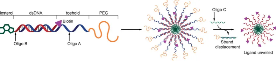
Oligo A1 (10 mg, 1.42mmol) was resuspended in 2 mL of Dul-becco’s PBS (DPBS) at pH 7.5. mPEG-NHS (14.5 mg, 7.1mmol) was dissolved in 200mL of DMSO and added dropwise to the oligonucleotide solution. The conjugation was allowed to proceed for 24 hours. Aer 24 hours the coupling efficiency was examined using high performance liquid chromatography (HPLC, see below for details). HPLC analysis revealed that only around 50% of the starting oligonucleotide had coupled to the PEG in 24 hours, another 2.5 equivalents of mPEG-NHS (7.25 mg, 3.55mmol) in 100mL of DMSO were therefore added to the reaction and it was allowed to proceed for a further 24 hours. The reaction was again monitored by HPLC aer 48 hours, HPLC analysis showed 66% conversion. Another 5 equivalents of mPEG-NHS (14.5 mg, 7.1mmol) in 200mL of DMSO were added to the reaction and leto react for a further 24 hours. Analysis aer 72 hours revealed that conjugation had achieved roughly 80% coupling efficiency. At this time point the reaction was stopped and lyophilized. The crude mixture was resuspended in DNase free water (1 mL) and puried by Fig. 1 Schematic representation of the structure of DNA conjugates and the unshielding of a ligandviastrand displacement. Oligo A holds a PEG chain to provide shielding, oligo B is complementary to part of oligo A and is modified with cholesterol and biotin. When annealed the resulting hybrid forms a micellar structure with the ligand shielded. Oligo C is a DNA sequence complementary to oligo A resulting in strand displacement and unshielding of the ligand (biotin).
Open Access Article. Published on 05 November 2013. Downloaded on 12/04/2016 09:52:25.
This article is licensed under a
[image:2.595.71.536.54.158.2]semi-preparative HPLC (see conditions below). The volume collected was concentrated under reduced pressure (to remove organic solvent) and then subsequently lyophilized to remove the water. PEG-A1 was collected as a white powder, the powder was dissolved in DNase free water and quantied using optical density at 260 nm. In total, 251 OD (7.5 mg, 75% yield) of PEG-A1 were recovered as the pure product. The product was aliquoted into vials, freeze dried and then kept at 20 C prior to being used. The pure product was analysed by MALDI-TOF. Mass expected: 8965 g mol1. Mass found: 9320 g mol1.
Synthesis of PEG-A3
Oligo A3 (0.5 mg, 0.066mmol) was resuspended in 188mL of Dulbecco PBS (DPBS) at pH 7.5. mPEG-NHS (0.67 mg,
0.328 mmol) was dissolved in 60 mL of DMSO and added
dropwise to the oligonucleotide solution. The conjugation was allowed to proceed for 24 hours. Aer 24 hours the coupling efficiency was examined using HPLC (see below for details). HPLC analysis revealed that only around 6% of the starting oligonucleotide had coupled to the PEG in 24 hours, conse-quently another 5 equivalents of mPEG-NHS (1.34 mg, 0.656mmol) in 100mL of DMSO were added to the reaction and it was allowed to proceed for a further 24 hours. The reaction was again monitored by HPLC aer 48 hours, HPLC analysis showed 44% conversion. Another 2.5 equivalents of mPEG-NHS (0.67 mg, 0.328 mmol) in 100 mL of DMSO along with 100mL of THF were added to the reaction and leto react for another 96 hours. Analysis aer 144 hours revealed that conjugation had achieved roughly 80% coupling efficiency. At this time the reaction was stopped, the organic solvent was removed under reduced pressure and the water removed using lyophilization. The crude mixture was aerwards resuspended in DNase free water (1 mL) and puried by semi-preparative HPLC (see conditions below). The solution collected was concentrated under reduced pressure (to remove organic solvent) and then subsequently lyophilized to remove the water. PEG-A3 was collected as a purple powder, which was dissolved in DNase free water and quantied using optical density at 260 nm. PEG-A3 (6.3 OD, 180 mg, 36% yield) was recovered as a pure product. This conjugate was aliquoted into vials, freeze dried and then kept at20C prior to being used. The identity of the product was determined by MALDI-TOF. Mass expected: 9520 g mol1. Mass found: 9840 g mol1.
HPLC analysis and purication of DNA strands
Reverse phase high performance liquid chromatography (RP-HPLC) was performed on a Shimadzu Prominence UPLC systemtted with a DGU-20A5 degasser, LC-20AD low-pressure gradient pump, CBM-20A LITE system controller, SIL-20A autosampler and an SPD-M20A diode array detector. Analytical separations were performed on a Phenomenex Clarity 3 mm Oligo-RP C18 column (4.650 mm) with a gradient of MeOH (10–70% for PEG-A1 and 35–70% for PEG-A3 over 20 min) in 0.1 M triethylammonium acetate (TEAA, pH 7.5)/MeCN (95/5) at aow rate of 1.0 mL min1as a mobile phase. Semi-preparative
separations were performed on a Phenomenex Clarity 3 mm Oligo-RP C18 column (1050 mm) under the same conditions at aow rate of 5.0 mL min1.
MALDI analysis
Matrix-assisted laser desorption/ionization time-of-ight (MALDI-TOF) mass spectrometry was performed on a Bruker MALDI-TOF Ultraex III spectrometer operated in linear, positive ion mode. 3-HPA containing DAHC was used as the matrix for the oligonucleotide analysis. Briey, a saturated solution of 3-HPA (50 mg mL1) was prepared by adding 25 mg of 3-HPA to 500mL of 50% ACN/water. 25mL of DAHC solution (100 mg mL1) was added to 225mL of the 3-HPA solution, to
give a nal DAHC concentration of 10 mg mL1. Equal
volumes of matrix solution and ODN solution (0.2 mM) were mixed and 2mL of the mixture was spotted onto the MALDI plate and allowed to dry.
Polyacrylamide gel electrophoresis (PAGE) Denaturing PAGE
Preparation of samples.Samples were prepared by dilution in denaturing loading buffer and heating to 95C for 2 minutes before rapid cooling ice.
Preparation of gels. Gel casting solution was prepared by mixing the components described in Table S2†and carefully pipetted into preassembled plate. A 10-well comb was inserted and the gel allowed to set for 45 min.
Electrophoresis.Gels were pre-run for 20 min at 200 V in 1 TBE. Wells were then washed with the same buffer and samples loaded at approximately 200 pmoles per well. Gels were run at 200 V until the Orange G loading dye had migrated off the bottom of the gel (approximately 80 min).
Staining.Gels were washed with water then stained with
0.02% methylene blue in 1 TBE for 20 minutes. Excess
stain was removed by destaining with distilled water until the bands were clear against the background. Images
were recorded using a standard at bed scanner (HP
Scanjet 2710).
Native PAGE
Preparation of samples.Samples were prepared by dilution in native loading buffer. Loading dye (Orange G) was added only to the ladder.
Preparation of gels. Gel casting solution was prepared by mixing the components described in Table S2†and carefully pipetted into preassembled plate. A 10-well comb was inserted and the gel allowed to set for 45 min.
Electrophoresis.Gels were pre-run for 20 min at 200 V in 1 TBE. Wells were washed with the same buffer and samples loaded at approximately 200 pmoles per well. Gels were run at 200 V until the Orange G loading dye was approximately 1 cm from the bottom of the gel (approximately 80 min).
Staining. Gels were washed with water then stained with Stains-All staining solution for 20 minutes in the dark. Staining solution was removed and gels destained until bands were clearly visible against background. Gels were imaged as before.
Open Access Article. Published on 05 November 2013. Downloaded on 12/04/2016 09:52:25.
This article is licensed under a
Hybridization of DNA strands
Strands to be hybridized were mixed in equimolar quantities at a concentration of 50–150 mM in hybridization buffer and placed in a 95C water bath. The water bath was allowed to cool to room temperature over a period of 2 hours.
Dynamic light-scattering (DLS)
Dynamic light-scattering was performed using a Viscotek 802 DLS instrument tted with an internal laser (l 830 5 nm, Pmax 60 mW). Hybrid PEG-A1:B1 (50mM in annealing buffer) wasltered through a 0.45mm membrane (Whatman Spartan, regenerated cellulose) prior to analysis. Laser power was adjusted to until detection rate of at least 300 kcps was ach-ieved. A series of 103 seconds experiments was recorded and hydrodynamic radii distributions calculated with Viscotek/ Malvern OmniSize3 soware.
Transmission electron microscopy
Transmission electron microscopy (TEM) was performed on an FEI Tecnai 12 Biotwin microscope. Samples were prepared by rst treating a holey carbon-coated 400 mesh copper TEM grid with graphene oxide solution (0.1 mg mL1) for 30 min. Excess graphene oxide solution was wicked away with the aid oflter paper and the grids dried for 30 min. Oligonucleotide solution (10mL, 5mM) was then placed on the grid; aer 2 minutes the excess liquid was wicked away with the aid oflter paper and the sample allowed to dry for 5 min. Samples were stained by addition of 5mL of a 3% solution of sodium phosphotungstate for 1 min before wicking away as before. Samples were then dried overnight prior to imaging.
Stability assay
Hybrid PEG-A1:B1 (50mM) or A2:C in DPBS were mixed with FBS at a ratio of 9/1 and incubated at 37C. Aliquots were removed at 0, 24, 48 and 72 h. and frozen until later analysis. Samples were analyzed by native PAGE as described previously.
Strand displacement assays
By PAGE. The hybrid to be analyzed was combined with either complementary (strand C) or scrambled (strand D) DNA at equimolar quantities in hybridization buffer at a nal concentration of 10 mM for both hybrid and displacement strand. Samples were incubated at room temperature for 1 h and then analysed by native PAGE as described previously.
By FRET/uorimetry. Fluorescence experiments were per-formed using a Varian Cary Eclipse spectrophotometer (lex494 nm, excitation slit width 2.5 mm;lem519 nm, emission slit width 5 mm). Hybrids PEG-A1:B1 and PEG-A3:B3 were combined at a 4/1 mole ratio in hybridization buffer at a total concentration of 5mM (i.e.1mMuorophore). 500 mL aliquots were added to a uorescence cuvette and theuorescence measured over a period of 10 minutes. Aer this time 25mL of 100mM (1 mole equiv.) of oligonucleotide C or D, or an equivalent volume of buffer, was added to the cuvette and theuorescence measured for a further
30 min. Emission intensities were baseline corrected to the emission intensity att¼0 min.
Percentage emission was calculated using background uo-rescence prior to strand addition as zero and theuorescence of a solution containing 1mM strand B3 as 100%. Full emission spectra were recorded prior to addition of DNA and 30 min post addition.
Avidin–biotin binding assay
Avidin was dissolved in DPBS at a concentration of 0.2 mg mL1 protein (2.5 units per mL) and aliquots (100mL) were transferred to the wells of a 96-well plate. 25mL of PEG-A1:B2 (50mM) was added to each well followed by 75mL of oligo C (100mM, 6 mol equiv.), oligo D (100mM, 6 mol equiv.), or buffer alone. Addi-tional control wells were prepared by combining avidin (100 mL), buffer (25 mL) and oligo C (75 mL). Samples were incubated for 30 min before the absorbance at 550 nm was measured using a plate reader (Tecan Innite M200).
Results and discussion
Design, synthesis and analysis of polymer–DNA conjugates
The use of nanoparticles in diagnosis and therapy requires the solving of numerous synthetic and biological challenges. Systems of this type may need to encapsulate a drug or a signal molecule, traverse complex biological barriers and then report a disease event or release a drug at a determined time point and/or a specic cellular or external cue. A‘so’nanoparticle structure is
advantageous in this environment because biological
membranes are inherentlyexible and a number of studies have shown that micelle- and vesicle-like nanoparticles exhibit better biodistribution and cell uptake properties than more rigid analogues.25–27 In addition, a more uid-like structure at a nanoparticle surface facilitates macromolecular exchange inter-actions, such as ligand–receptor interactions or nucleic acid strand switching. A particularly important example of a switching operation in the drug delivery context is the need to keep a recognition ligand on a polymer or particle hidden/dormant but to expose the specic functionality in response to a biological trigger. Viruses carry out operations of this type to sequentially expose cell entry ligands and endosomal escape functionality, and this‘hide-reveal’concept has been used in synthetic viral-mimetic polymers28,29and thermo-responsive nanoparticles17,30to
enhance drug delivery and selective cell entry.
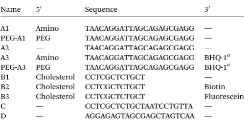
Accordingly, we set out a design concept for ‘hide-reveal’ nanoparticles, utilizing DNA-strand recognition to encode for sequence specic assembly and derivitisation at the respective 30and 50ends of each to direct functionality to the interior and exterior of the particle as shown schematically in Fig. 1. The oligonucleotide sequences and the modications required (a) to assemble the structures, (b) to incorporate an orthogonal bio-logical recognition signal (biotin), (c) to provide a shielding polymer at the exterior (PEG), and (d) to unveil the ligandvia competitive strand displacement are given in Table 1.
The primary chain of Oligo A is 22 bases long and 50-modied with a poly(ethylene glycol) chain to provide
Open Access Article. Published on 05 November 2013. Downloaded on 12/04/2016 09:52:25.
This article is licensed under a
protection and shielding. Oligo B is 50-modied with choles-terol and the 30-terminus of Oligo B is modied with a biotin moiety. Oligo B is complementary to the 12 bases at the 30 terminus of oligo A allowing hybridizationviaWatson–Crick base-pairing to yield a hybrid that shields the biotin moiety within the micelle. The 10 base toehold allows disruption of the hybrid upon addition of Oligo C which is complementary to the full 22 bases of Oligo A. Displacement of Oligo A results in unshielding of the micelle exposing the biotin moieties and making them accessible to bind avidin.
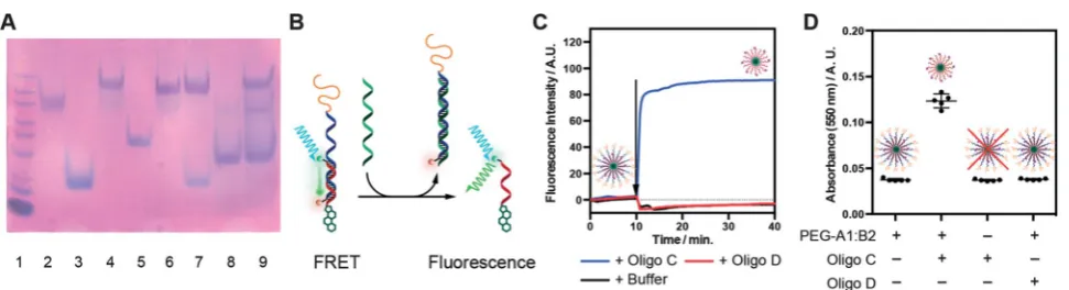
PEGylation of amino-modied oligos A1 and A3 was achieved by reaction with succinimidyl carbonate-activated PEG (Scheme S1†) followed by purication by high pressure liquid chroma-tography (HPLC). Successful conjugation was conrmed by PAGE, HPLC and MALDI-TOF mass spectrometry (Fig. 2A, S2 and S3† respectively). Subsequent assembly of the conjugates into supramolecular structures was achieved by a simple mixing/ annealing process. Hybridization of strand PEG-A1 with strand cholesterol oligo B1 to produce hybrid PEG-A1:B1 led to the formation of discrete objects, as apparent in dynamic light-scattering (DLS, Fig. 2C and S4†) and transmission electron microscopy (TEM). The DLS intensity distribution (Fig. 2C, black line) is dominated by a population centered atRh100 nm with
small populations at 26 and 2 nm. The number distribution (Fig. 2C, red line), calculated from intensity distribution in the range 2 <Rh< 103nm, shows a main population withRh25 nm
and small population at 60 nm. This agrees well with TEM where objects approximately 40 nm diameter are observed although dispersity is higher (Fig. 2D). In addition, labeled hybrids could be formed by hybridization of PEG-A1 with chol-biotin oligo B2 (hybrid PEG-A1:B2), and PEG-A3 (30-quencher) with chol- uo-rescein oligo B3 (hybrid PEG-A3:B3, labeled for FRET).
If DNA conjugates are to be usedin vitro/vivothen they need to be stable under physiological conditions. Consequently, the stability of micellar hybrid PEG-A1:B1 towards degradation by serum enzymes (including nucleases) was determined by incu-bation with 10% fetal calf serum (FCS) at 37C and analysis by native PAGE. Hybrid PEG-A1:B1 was considerably more stable to degradation compared to unmodied (i.e. no 30 or 50 modi -cations) hybrid A2:C (Fig. S6†) with little evident degradation over 72 h; however, the band-broadening imparted by the
PEGylation makes quantitative analysis difficult. The
comparative stability is expected as“spherical nucleic acids”11
have been previously demonstrated to be more stable than the linear form31 and the additional PEG coating should further
reduce the accessibility of the DNA segments to nucleases.
Strand displacement and unshielding of micelles
The“programmable”responsive unshielding was investigated by PAGE and FRET analysis. For PAGE experiments, hybrids were incubated with 1 molar equivalent of either oligonucleo-tide C (complementary) or D (scrambled) at room temperature. Samples were then analyzed by native PAGE (Fig. 3A and S7†). When incubated with complementary oligo C (Lane 7) the band for hybrid PEG-A1:B1 is lost (cf.Lane 4) and bands for chol oligo B1 and hybrid PEG-A1:C found in its place (cf. Lanes 3 and 6 respectively). Conversely, when incubated with scrambled oligo D the PEG-A1:B1 band still remains and no new bands are observed.‡Equivalent bands were observed for the hybrids PEG-A1:B2 and PEG-A3:B3 when analysed in the same manner (Fig. S7A and B†).
For FRET experiments hybrids PEG-A1:B1 and PEG-A3:B3 were mixed at a 4/1 ratio. PEG-A3:B3 has a uorescein and a quencher moiety (Black Hole Quencher 1, BHQ-1, Biosearch Technologies) attached to the 30ends of the strands forming the duplex. While the duplex is intact, FRET from theuorescein to BHQ-1 results in minimaluorescence and an‘off’state. If the double helix is disrupted FRET is lost resulting in switching‘on’ of theuorescence (Fig. 3B). Samples were incubated for 10 min to determine a baseline then 1 equivalent of complementary oligo C or scrambled oligo D, or an equivalent volume of buffer, was added and emission monitored for a further 30 minutes. Addition of either oligo D or buffer alone results in a minimal decrease in emission intensity due to a small dilution of the solution. As expected, addition of oligo C results in a rapid increase in emission before theuorescence reached a plateau value. These data agree with that from the PAGE experiments and conrm that the micelle corona may be removed by addi-tion of a complementary DNA strand.
Comparison of these data to those for an equivalent concentration of the chol-uorescein oligo B3 alone conrms restoration of approximately 45% of the expecteduorescence (Fig. S8B†). Although this is lower than expected it likely results from incomplete unshielding due to steric hindrance resulting in some quencher remaining in close enough proximity to the uorescein to maintain FRET.
[image:5.595.42.296.63.188.2]Finally, to determine if unpacking could be used for the presentation of the targeting ligand for binding, micelles of hybrid PEG-A1:B2 were evaluated in a biotin–avidin binding assay. Briey, PEG-A1:B2 was mixed with avidin in the presence of oligo C, oligo D or buffer alone. If unpacking is successful the biotin will bind to the avidin resulting in crosslinking, and subsequent precipitation, that may be quantied by measuring the solution turbidity (absorbance at 550 nm, Fig. 3D). Turbidity Table 1 Sequences and modifications of oligonucleotides used
Name 50 Sequence 30
A1 Amino TAACAGGATTAGCAGAGCGAGG — PEG-A1 PEG TAACAGGATTAGCAGAGCGAGG — A2 — TAACAGGATTAGCAGAGCGAGG — A3 Amino TAACAGGATTAGCAGAGCGAGG BHQ-1a PEG-A3 PEG TAACAGGATTAGCAGAGCGAGG BHQ-1a B1 Cholesterol CCTCGCTCTGCT — B2 Cholesterol CCTCGCTCTGCT Biotin B3 Cholesterol CCTCGCTCTGCT Fluorescein C — CCTCGCTCTGCTAATCCTGTTA — D — AGGAGAGTAGCGAGCTAGTCAA — aBlack Hole Quencher 1 (Biosearch Technologies).
‡It should be noted that in lanes 8 and 9 in Fig. 3A and S6 there is an extra band observed with a molecular weight between that of oligo D and hybrid PEG-A1:B1. As it also appears in the lane where only oligo D was added we conclude that this is due to self-assembly of oligo D.
Open Access Article. Published on 05 November 2013. Downloaded on 12/04/2016 09:52:25.
This article is licensed under a
measurements demonstrate addition of complementary oligo C results in a signicant increase in absorbance compared to the avidin treated with PEG-A1:B2 alone (p< 0.0001, 1-way ANOVA); addition of scrambled oligo D had no effect on absorbance conrming sequence specic binding. Additionally, to conrm that turbidity was caused by unpacking and not a sequence specic interaction between oligo C and avidin, the same measurement was performed in the absence of PEG-A1:B2 and no effect on turbidity was observed.
Conclusions
In conclusion we have demonstrated that oligonucleotide sequences may be used to direct the supramolecular assembly and unshielding of micellar nanoparticles in a pre-programmable manner. The DNA conjugate micelles formed rapidly and dis-played high stability to serum enzymes as well as securely shielding a recognition ligand in the absence of a specic nucleic acid strand. Unveiling of the ligand by addition of the comple-mentary strand was easily detected by a simple turbidimetry assay
in which the protein, avidin, bound to the exposed biotin signals. Experiments to evaluate the ability of DNA conjugates to switch in response to specic sequencesin vitroandin vivoare continuing in our laboratories.32Programmable control over ligand
presen-tation may have important applications in drug-delivery, e.g. controlled targeting in the presence of a therapeutic nucleic acid sequence. In addition, sensing applications can be envisioned for systems that, like the FRET pair shown herein, can modulate their spectral properties in a programmable way allowing forin vitro andin vivomonitoring of unshielding. Finally, theexibility in the design and synthesis offered by nucleic acid based materials combined with the opportunity to tailor polymers and ligands for specic biomedical tasks suggests materials of this type may prove useful in the personalized diagnostics and patient-group stratied therapeutics.
Acknowledgements
We thank the UK EPSRC (Grants EP/H005625/1, EP/G042462/1) and the University of Nottingham for a Scholarship (GS). Fig. 3 (A) Strand displacement analyzed by PAGE. Lanes: (1) Ladder, (2) PEG-A1, (3) B1, (4) PEG-A1:B1, (5) C, (6) PEG-A1:C, (7) PEG-A1:B1 + C, (8) D, (9) PEG-A1:B1 + D. (B) Schematic of specific oligonucleotide detectionviadisruption of F¨orster Resonance Energy Transfer (FRET). When complementary oligonucleotide is added hybrid PEG-A3:B3 is disrupted resulting in loss of FRET and a consequential increase influorescence. (C) Baseline correctedfluorescence intensity of mixed micelles of PEG-A1:B1 and PEG-A3:B3 (4/1 mole ratio) before and after addition of complementary oligo C (blue), scrambled oligo D (red), or an equivalent volume of buffer (black). (D) Turbidimetry measurements (absorbance at 550 nm) of avidin solutions treated with combinations of PEG-A1:B2, complementary oligo C and scrambled oligo D. Circles represent individual measurements, lines represent mean and bars represent standard deviation. There is only a detectable increase in absorbance when both PEG-A1:B2 and oligo C are present.
Fig. 2 (A) Denaturing PAGE of oligonucleotides before and after PEGylation. Lanes: 1. Ladder, 2. A1, 3. PEG-A1, 4. A3, 5. PEG-A3. (B) Native PAGE of oligonucleotides and hybrids. Lanes: 1. Ladder, 2. PEG-A1, 3. PEG-A3, 4. B1, 5. B2, 6. B3, 7. hybrid PEG-A1:B1, 8. hybrid PEG-A1:B2, 9. hybrid PEG-A3:B3. (C) Dynamic light-scattering of hybrid PEG-A1:B1. Intensity distribution (black line) and number distribution (red line). (D) Trans-mission electron micrograph of hybrid PEG-A1:B1 stained with sodium phosphotungstate.
Open Access Article. Published on 05 November 2013. Downloaded on 12/04/2016 09:52:25.
This article is licensed under a
[image:6.595.61.538.49.164.2] [image:6.595.57.543.235.367.2]Notes and references
1 J. Zheng, J. J. Birkto, Y. Chen, T. Wang, R. Sha,
P. E. Constantinou, S. L. Ginell, C. Mao and N. C. Seeman, Nature, 2009,461, 74–77.
2 D. Y. Zhang, A. J. Turbereld, B. Yurke and E. Winfree, Science, 2007,318, 1121–1125.
3 T. Ciengshin, R. Sha and N. C. Seeman,Angew. Chem., Int. Ed., 2011,50, 4419–4422.
4 B. Chakraborty, N. Jonoska and N. C. Seeman,Chem. Sci., 2012,3, 168–176.
5 J. Bath, S. J. Green and A. J. Turbereld,Angew. Chem., Int. Ed., 2005,44, 4358–4361.
6 M. L. McKee, P. J. Milnes, J. Bath, E. Stulz, A. J. Turbereld and R. K. O’Reilly,Angew. Chem., Int. Ed., 2010,49, 7948– 7951.
7 Y. H. Roh, J. B. Lee, P. Kiatwuthinon, M. R. Hartman, J. J. Cha, S. H. Um, D. A. Muller and D. Luo,Small, 2011,7, 74–78.
8 Y. H. Roh, R. C. H. Ruiz, S. Peng, J. B. Lee and D. Luo,Chem. Soc. Rev., 2011,40, 5730–5744.
9 M.-P. Chien, M. P. Thompson and N. C. Gianneschi,Chem. Commun., 2011,47, 167–169.
10 M.-P. Chien, A. M. Rush, M. P. Thompson and
N. C. Gianneschi, Angew. Chem., Int. Ed., 2010, 49, 5076– 5080.
11 J. I. Cutler, E. Auyeung and C. A. Mirkin,J. Am. Chem. Soc., 2012,134, 1376–1391.
12 J. I. Cutler, K. Zhang, D. Zheng, E. Auyeung, A. E. Prigodich and C. A. Mirkin,J. Am. Chem. Soc., 2011,133, 9254–9257. 13 F. E. Alemdaroglu, N. C. Alemdaroglu, P. Langguth and
A. Herrmann,Adv. Mater., 2008,20, 899–902.
14 H. Lee, A. K. R. Lytton-Jean, Y. Chen, K. T. Love, A. I. Park, E. D. Karagiannis, A. Sehgal, W. Querbes, C. S. Zurenko, M. Jayaraman, C. G. Peng, K. Charisse, A. Borodovsky, M. Manoharan, J. S. Donahoe, J. Truelove, M. Nahrendorf, R. Langer and D. G. Anderson,Nat. Nanotechnol., 2012,7, 389–393.
15 J. Fang, H. Nakamura and H. Maeda,Adv. Drug Delivery Rev., 2011,63, 136–151.
16 V. Torchilin,Adv. Drug Delivery Rev., 2011,63, 131–135.
17 F. Mastrotto, P. Caliceti, V. Amendola, S. Bersani,
J. P. Magnusson, M. Meneghetti, G. Mantovani,
C. Alexander and S. Salmaso, Chem. Commun., 2011, 47, 9846–9848.
18 K. Zhang, X. Zhu, F. Jia, E. Auyeung and C. A. Mirkin,J. Am. Chem. Soc., 2013,135, 14102–14105.
19 S. S. Agasti, S. Rana, M. H. Park, C. K. Kim, C. C. You and V. M. Rotello,Adv. Drug Delivery Rev., 2010,62, 316–328. 20 M. M. C. Cheng, G. Cuda, Y. L. Bunimovich, M. Gaspari,
J. R. Heath, H. D. Hill, C. A. Mirkin, A. J. Nijdam, R. Terracciano, T. Thundat and M. Ferrari, Curr. Opin. Chem. Biol., 2006,10, 11–19.
21 P. Zrazhevskiy, M. Sena and X. Gao,Chem. Soc. Rev., 2010,39, 4326–4354.
22 M. Sharma, R. L. Salisbury, E. I. Maurer, S. M. Hussain and C. E. W. Sulentic,Nanoscale, 2013,5, 3747–3756.
23 D. Choudhury, P. L. Xavier, K. Chaudhari, R. John, A. K. Dasgupta, T. Pradeep and G. Chakrabarti, Nanoscale, 2013,5, 4476–4489.
24 K. Luyts, D. Napierska, B. Nemery and P. H. M. Hoet, Environ. Sci.: Processes Impacts, 2013,15, 23–38.
25 I. Canton and G. Battaglia,Chem. Soc. Rev., 2012,41, 2718– 2739.
26 J. W. Yoo, E. Chambers and S. Mitragotri,Curr. Pharm. Des., 2010,16, 2298–2307.
27 Y. Geng, P. Dalhaimer, S. Cai, R. Tsai, M. Tewari, T. Minko and D. E. Discher,Nat. Nanotechnol., 2007,2, 249–255. 28 M. Soliman, R. Nasanit, S. R. Abulateefeh, S. Allen,
M. C. Davies, S. S. Briggs, L. W. Seymour, J. A. Preece,
A. M. Grabowska, S. A. Watson and C. Alexander, Mol.
Pharmaceutics, 2012,9, 1–13.
29 Y. Nie, D. Schaffert, W. R¨odl, M. Ogris, E. Wagner and M. G¨unther,J. Controlled Release, 2011,152, 127–134. 30 S. R. Abulateefeh, S. G. Spain, K. J. Thurecht, J. W. Aylott,
W. C. Chan, M. C. Garnett and C. Alexander,Biomater. Sci., 2013,1, 434–442.
31 D. S. Seferos, A. E. Prigodich, D. A. Giljohann, P. C. Patel and C. A. Mirkin,Nano Lett., 2009,9, 308–311.
32 G. Yasayan, J. P. Magnusson, G. Sicilia, S. G. Spain, S. Allen, M. C. Davies and C. Alexander, Phys. Chem. Chem. Phys., 2013,15, 16263–16274.
Open Access Article. Published on 05 November 2013. Downloaded on 12/04/2016 09:52:25.
This article is licensed under a