Target for Potent and Broad Neutralization of Simian
Immunodeficiency Viruses
Takeo Kuwata,aKaori Takaki,aKazuhisa Yoshimura,bIkumi Enomoto,aFan Wu,cIlnour Ourmanov,cVanessa M. Hirsch,c Masaru Yokoyama,dHironori Sato,dShuzo Matsushitaa
Center for AIDS Research, Kumamoto University, Kumamoto, Japana; AIDS Research Center, National Institute of Infectious Diseases, Tokyo, Japanb; Laboratory of
Molecular Microbiology, NIAID, NIH, Bethesda, Maryland, USAc; Pathogen Genomics Center, National Institute of Infectious Diseases, Tokyo, Japand
Inducing neutralizing antibodies (NAb) is the key to developing a protective vaccine against human immunodeficiency virus
type 1 (HIV-1). To clarify the neutralization mechanism of simian immunodeficiency virus (SIV), we analyzed NAb B404, which
showed potent and broad neutralizing activity against various SIV strains. In 4 SIVsmH635FC-infected macaques, B404-like
antibodies using the specific VH3 gene with a long complementarity-determining region 3 loop and
light chain were the major
NAbs in terms of the number and neutralizing potency. This biased NAb induction was observed in all 4 SIVsmH635FC-infected
macaques but not in 2 macaques infected with a SIV mix, suggesting that induction of B404-like NAbs depended on the
inocu-lated virus. Analysis using Env mutants revealed that the V3 and V4 loops were critical for B404 binding. The reactivity to the
B404 epitope on trimeric, but not monomeric, Env was enhanced by CD4 ligation. The B404-resistant variant, which was
in-duced by passages with increasing concentrations of B404, accumulated amino acid substitutions in the C2 region of gp120.
Mo-lecular dynamics simulations of the gp120 outer domains indicated that the C2 mutations could effectively alter the structural
dynamics of the V3/V4 loops and their neighboring regions. These results suggest that a conformational epitope consisting of
the V3 and V4 loops is the target for potent and broad neutralization of SIV. Identifying the new neutralizing epitope, as well as
specifying the VH3 gene used for epitope recognition, will help to develop HIV-1 vaccines.
N
eutralizing antibodies (NAb) against human
immunodefi-ciency virus type 1 (HIV-1) protect against viral challenge in
nonhuman primate models (
1
–
5
), suggesting that NAb induction
may be an important key to the development of vaccines against
HIV-1. The role of NAbs in prevention of infection and control of
viral replication has been suggested in several studies using
candi-date vaccines (
6
–
8
). However, the difficulties in inducing NAbs,
especially those that are broadly reactive to various HIV-1 strains,
have hampered the development of such vaccines (
9
–
11
).
Mono-clonal antibodies (MAb) with broad neutralizing activity that
were recently isolated from HIV-1-infected patients have been
characterized to understand the specificities and mechanisms of
broad neutralization (
12
–
16
). The epitopes of these potent and
broad NAbs, such as PG9, PGT128, VRC01, and 10E8, have been
determined precisely (
17
–
19
) and provide an opportunity for
structure-based vaccine design to develop antibody-based
vac-cines for HIV-1 (
11
,
20
–
23
).
Nonhuman primate models of simian immunodeficiency
vi-rus (SIV) infection are commonly used to develop vaccines
against HIV-1 (
6
,
8
,
24
). Various immunogens, vectors, and
reg-imens have been evaluated by challenge infection with SIV.
More-over, immune factors associated with prevention of infection have
been explored in the SIV model. However, epitopes for potent and
broad neutralization of SIV remain unclear because few MAbs
that neutralize a wide range of SIV strains have been available.
Recently, we isolated MAbs from a rhesus macaque infected with
SIVsmH635FC, which was isolated from a rapid progressor
ma-caque (
25
). Infection with SIVsmH635FC, a highly
neutraliza-tion-sensitive molecular clone, resulted in a vigorous and potent
antibody response in all the infected macaques together with viral
mutations to escape antibody recognition (
26
,
27
). MAb B404
bound to a conformational epitope on gp120 of various SIV
strains and did not react to overlapping peptides of SIV Env. The
V3 region was shown to be important by competition
enzyme-linked immunosorbent assay (ELISA) with anti-V3 antibodies
(
25
). The neutralizing activity of B404 against homologous
neutralization-sensitive SIVsmH635FC, genetically divergent
SIVmac316, and neutralization-resistant SIVsmE543-3 was
ob-served.
In this study, we analyzed the epitope of B404 and the
induc-tion of B404-like NAbs in SIV-infected macaques. Analysis of
more than 400 anti-Env MAbs demonstrated that B404-like NAbs
with the same gene usage and specificity were mainly induced in 4
SIVsmH635FC-infected macaques. The B404 epitope was
mapped to a conformational epitope consisting of the V3 and V4
loops exposed on a trimeric Env structure after CD4 binding. The
identification of the new neutralizing epitope and vigorous
anti-body response to this epitope in SIV-infected macaques will help
us to understand broad neutralization in a macaque model of SIV
infection.
MATERIALS AND METHODS
Cells and viruses.PM1 (28) and PM1/CCR5 (29) cells were maintained in
RPMI 1640 medium containing 10% fetal bovine serum (FBS). TZM-bl
Received22 January 2013 Accepted25 February 2013
Published ahead of print6 March 2013
Address correspondence to Shuzo Matsushita, [email protected]. Copyright © 2013, American Society for Microbiology. All Rights Reserved.
doi:10.1128/JVI.00201-13
on November 7, 2019 by guest
http://jvi.asm.org/
(30–33) and 293T (34) cells were maintained in Dulbecco’s modified Ea-gle medium containing 10% FBS. Infectious molecular clones, SIVsmE543-3 (35), SIVsmH635FC (27), SIVmac239 (36), SIVmac316 (37), SIVsmE660FL14, SIVsmH805-24w-3, and SIVsmH807-24w-4 (38) were transfected into 293T cells. After 2 days, the supernatants were fil-tered (0.45m) and stored at⫺80°C as virus stocks.
Construction of Fab libraries from SIV-infected macaques.The Fab
library from SIVsmH635FC-infected rhesus macaque H723 was de-scribed previously (25). The Fab libraries from SIV-infected rhesus ma-caques H704, H709, H714, H711, and H725 (26,27,39) were similarly constructed using the pComb3X system according to the instructions of Barbas et al. (40). Four macaques, H723, H704, H709, and H714, were infected with SIVsmH635FC. H711 was infected with a combination of SIVsmE543-3 and SIVsmH635FC. H725 was infected with plasma sam-ples from 2 SIVsmH445-infected macaques, H631 and H635. Rhesus ma-caques of Indian origin were used in this study. RNA was extracted from lymphocytes from the lymph nodes of these macaques using an RNeasy minikit (Qiagen, Hilden, Germany) and used for subsequent RT-PCR using oligo(dT)20 primer, ReverTra Ace (Toyobo, Osaka, Japan), and Platinum high-fidelityTaqDNA polymerase (Invitrogen, Carlsbad, CA). Two libraries,andlight chains, were constructed for each macaque to examine the frequency of NAbs in each population, although only one library, containing bothandlight chains, was constructed for H723. Immunoglobulin (Ig) genes were inserted into pComb3X, and the liga-tion mix was used for transformaliga-tion of XL1-Blue (Stratagene, La Jolla, CA) by electroporation. Transformed cultures were incubated in super-broth medium with 50g/ml carbenicillin, 10g/ml tetracycline, and 1.4 g/ml kanamycin overnight at 37°C after addition of VCSM13 helper phage (Stratagene). Library phage stock was obtained from the culture medium by polyethylene glycol 8000 –NaCl precipitation. Library size was determined by assessing the number of CFU after infection of XL1-Blue with a diluted phage sample.
Biopanning to obtain anti-Env antibodies.Biopanning was
per-formed using SIV antigen (Ag), which was prepared by infection of PM1 cells with SIVsmE543-3 as previously described (25). To obtain Fab clones against Env, we selected Fab clones from the H723 library using a 96-well plate in which Env was conjugated with anti-Env Fab clones B404, B408, and H301, which recognize gp120 (conformational), gp41 cluster I and gp120 V1, respectively (25). A MaxiSoap 96-well plate (Thermo Fisher Scientific, Waltham, MA) was incubated with 100l of 1.25g/ml B404, 0.625g/ml B408, and 10g/ml H301 for 1 h at 37°C. The wells were washed with phosphate-buffered saline (PBS) containing 0.05% Tween 20 (PBS-T) and blocked with 5% skim milk (Wako Pure Chemical Indus-tries, Osaka, Japan) in PBS (MPBS) for 1 h at 37°C. After the blocking solution was discarded, the wells were incubated with 100l 40-fold-diluted SIV Ag for 1 h at 37°C, washed with PBS-T, and used for panning. After incubation with 50l of phage library for 2 h at 37°C, the wells were washed 5 times with PBS-T, and bound phage was eluted with 50l 100 mM glycine (pH 2.2). Amplified phage was used for the next round of panning, and 3 or 4 rounds of panning were performed. To isolate Fab clones specific to Env, we transformed phagemid DNA into TOP10F=
Escherichia colicells (Invitrogen), and supernatants from isopropyl--D
-thiogalactopyranoside (IPTG; Wako Pure Chemical Industries)-induced cultures were screened for reactivity to SIV Env using ELISA. Fab clones were purified using a His GraviTrap column (GE Healthcare, Bucking-hamshire, United Kingdom), as described previously (25).
Construction of a single-chain variable fragment (scFv) form of
B404.B404 Fab was previously converted into complete rhesus IgG
pro-duced from a stable cell line carrying heavy- and light-chain plasmids pHCG-B404 and pLL-B404 (25). From these plasmids, B404 scFv was constructed using the pComb3X system (40). The heavy-chain variable region (VH) was amplified using pHCG-B404 as a template and primers HSCVH35-FL (5=-GGT GGT TCC TCT AGA TCT TCC TCC TCT GGT GGC GGT GGC TCG GGC GGT GGT GGG GAG GTG CAG CTG GTG SAG TCT GG-3=) and RhSCG404-B (5=-CCT GGC CGG CCT GGC CAC
TAG TGA CCG ATG GGC CCT TGG TGG AGC C-3=). The light-chain variable region (VL) was amplified from pLL-B404 using primers
HSCLam3 (5=-GGG CCC AGG CGG CCG AGC TCG AGC TGA CTC
AGC CAC CCT CAG TGT C-3=) and RhSCJLam404 (5=-GGA AGA TCT AGA GGA ACC ACC GCC TAG GAC GGT CAG CCG GGT CCC-3=). The amplified products were combined by overlapping PCR using prim-ers RSC-F (5=-GAG GAG GAG GAG GAG GAG GCG GGG CCC AGG CGG CCG AGC TC-3=) and RSC-B (5=-GAG GAG GAG GAG GAG GAG CCT GGC CGG CCT GGC CAC TAG TG-3=), digested with SfiI, and inserted into pComb3X in a manner similar to that of Fab construction. The resultant plasmid had the B404 VL and VH regions, which were connected with an 18-amino-acid linker, a histidine tag, and a hemagglu-tinin (HA) tag. This plasmid was transformed into Rosetta 2 (Merck, Darmstadt, Germany), and B404 scFv was purified from the cell pellet using a His GraviTrap column.
ELISA.ELISA was performed to detect antibodies specific to SIV Ag as
previously described (25,41). Briefly, a MaxiSoap 96-well plate was coated with PBS containing 50 ng/ml concanavalin A (Sigma, St. Louis, MO) for 1 h at 37°C, and SIV Env was conjugated by incubation with 50l/well 10-fold diluted SIV Ag for 1 h at 37°C. Samples were added to each well at 50l/well with 50l of MPBS, and the plate was incubated for 1 h at 37°C. When the enhancement effect of soluble CD4 (sCD4) was examined, 25l of sample, 25l of sCD4, and 50l MPBS were added to each well. Fabs specific to SIV Env were detected with anti-HA–peroxidase (1:1,000 dilution; 3F10, Roche Molecular Biochemicals, Mannheim, Germany) and ABTS [2,2=-azinobis(3-ethylbenzthiazolinesulfonic acid)] solution (Roche Molecular Biochemicals).
Competition ELISA was performed similarly using B404 IgG as a com-petitor. Ag-coated wells were incubated with 50l MPBS and 25l serial dilutions of B404 IgG for 1 h at 37°C. After incubation with 25l subsatu-rating concentrations of Fab clones, Fab clone binding was detected by anti-HA–peroxidase (1:1,000) and ABTS solution.
Analysis of neutralizing antibody titers.The neutralizing capability
of Fab samples was measured as the reduction in luciferase activity after infection of TZM-bl cells with various SIV strains (6,25). In addition to Fab samples, plasma samples from SIVsmH635FC-infected macaque H704 (26) and SIVmac239-infected macaque MM324 (42) and MAb M318T (43), which recognizes the V2 region of SIV Env, were used to examine the sensitivity of SIV variants to antibody-mediated neutraliza-tion. Briefly, 100-l portions of serially diluted samples in duplicate were incubated with 50l containing 200 50% tissue culture infectious doses (TCID50) of virus in a 96-well plate. After incubation for 1 h at 37°C, 100 l containing 1⫻105TZM-bl cells/ml with 37.5g/ml DEAE dextran was added. Infected cultures were incubated for 2 days, but cultures in-fected with SIVsmH635FC were incubated for 3 days. After incubation, cells were lysed with 30l cell lysing buffer (Promega, Madison, WI) for 15 min at room temperature (RT), and 10l cell lysate was transferred to a 96-well black solid plate (OptiPlates-96F; Perkin-Elmer, Boston, MA) for measurements of luminescence using a GloMax 96 microplate lumi-nometer (Promega) and a luciferase assay system (Promega). The 50 and 90% inhibitory concentrations (IC50and IC90, respectively) were calcu-lated with nonlinear regression using PRISM5 and defined as the concen-tration that caused 50 and 90% reductions in luciferase activity, respec-tively, compared to that in virus control wells after the subtraction of background.
Construction of Env mutants.Theenvgene was amplified by PCR
using primers SRev-F (5=-GGT TTG GGA ATA TGC TAT GAG-3=) and SEnv-R (5=-CCT ACT AAG TCA TCA TCT T-3=) and SIVsmE543-3 plas-mid as a template. The PCR product was inserted into pcDNA3.1/V5-His-TOPO vector (Invitrogen). After XbaI digestion, the plasmid was ligated with an NheI-XbaI fragment from pLP-IRES2-EGFP (Clontech Labora-tories Inc., Mountain View, CA) to generate a plasmid designated RE543-EGFP that expressed both enhanced green fluorescent protein (RE543-EGFP) and Env. Mutants were constructed from RE543-EGFP using PCR mu-tagenesis. Deletion mutants⌬V1,⌬V2,⌬V3, and⌬V4 were created by Conformational Epitope for Potent SIV Neutralization
on November 7, 2019 by guest
http://jvi.asm.org/
deleting amino acid residues 115 to 149 in the V1 loop, 153 to 209 in the V2 loop, 313 to 342 in the V3 loop, and 404 to 430 in the V4 loop and replacing them with Gly-Ala-Gly, Gly, Gly-Ala, and Gly-Ala-Gly, respec-tively. These mutations were introduced into RE543-EGFP using primers DV1F (5=-ATG TAA TGG AGC CGG CTC TTG CAT AAA AAA-3=) and DV1R (5=-AAG AGC CGG CTC CAT TAC ATC TCA TTG CTA-3=) for ⌬V1, DV2F (5=-ATA GGA GCC GGC CAT TGT AAC ACC AGT-3=) and DV2R (5=-ACA ATG GCC GGC TCC TAT GCA AGA ATC ACC-3=) for ⌬V2, DV3F (5=-TGT AGA GGA GCC GGC TGG TGC CGG TTT GGA-3=) and DV3R (5=-GCA CCA GCC GGC TCC TCT ACA TTT CAT TGT-3=) for⌬V3, and DV4F (5=-AAG AAT TCT TAT ACT GCA AAG GAG CCG GCC CAT GTC ATA TTA GAC AAA-3=) for⌬V4. Mutant⌬Gly was constructed using primers N306AFw2 (5=-TAT TAT GCT CTA ACA ATG AAA TGT AG-3=), N306ARv (5=-CAT TGT TAG AGC ATA ATA CTT-3=), N316AFw (5=-AGA CCA GGA GCT AAG ACA GTT-3=), N316ARv (5=-AAC TGT CTT AGC TCC TGG TCT-3=), N349AFw (5=-GGT TTG GAG GAG CCT GGA GCG-3=), and N349ARv (5=-CGC TCC AGG CTC CTC CAA ACC G-3=) to introduce the mutations N306A, N316A, and N349A at potential N-linked glycosylation sites. Mutant D385R was con-structed using primers S-D368RFw (5=-CCA GCA GGA GGA CGT CCA GAA GTC AC-3=) and S-D368RRv (5=-TTC TGG ACG TCC TCC TGC TGG AGC TGT-3=). The D385R substitution in SIVsmE543-3 corre-sponds to D368R in HIV-1, which interferes with CD4 binding site (CD4bs) antibodies (13,44,45). Mutant I434R was constructed using primers S-I420RFw (5=-GCC ATG TCA TCG TAG ACA AAT AAT CAA C ⫺3=) and S-I420RRv (5=-GAT TAT TTG TCT ACG ATG ACA TGG CAC ⫺3=). The I434R substitution in SIVsmE543-3 corresponds to I420R in HIV-1, which interferes with CD4-induced (CD4i) antibodies (13,45, 46). Amino acid numbering of Env was based on that of SIVmac239, the reference sequence of SIV, and the HIV-2 sequence in the Los Alamos HIV databases (http://www.hiv.lanl.gov/).
Flow-cytometric analysis.Plasmids to express wild-type and mutant
Env were transfected into 293T cells using X-tremeGENE 9 DNA trans-fection reagent (Roche Molecular Biochemicals) according to the manu-facturer’s instructions. After incubation for 2 days, the transfected cells were detached with PBS containing 0.05% trypsin and 0.53 mM EDTA and adjusted to 1⫻107cells/ml in PBS containing 0.2% bovine serum albumin (BSA). To examine the reactivity of Fab, we incubated 50l cells with 10l 50 ng/l Fab for 40 min at RT. After washing with PBS containing 0.2% BSA, the cells were incubated with 50l anti-HA antibody (1:200; 3F10; Roche Molecular Biochemicals) for 20 min at RT, followed by incubation with 50l allophycocyanin (APC)-conju-gated AffiniPure goat anti-rat IgG (H⫹L) F(ab=)2fragment (1:200; Jackson ImmunoResearch Laboratories, Inc., West Grove, PA) for 20 min at RT. When enhancement with sCD4 was examined, cells were resus-pended in PBS containing 0.2% BSA in the presence or absence of 2g/ml sCD4 at 1⫻107cells/ml before staining. After incubation with sCD4 for 15 min at RT, 20l of cells was mixed with 10l 25 ng/l Fab and stained with anti-HA and anti-rat antibodies. Murine MAb KK46 (1:200) was used as a control antibody against the linear V3 epitope (47). KK46-incu-bated cells were stained by APC-conjugated goat anti-mouse Ig (1:200; BD Biosciences, Franklin Lakes, NJ). The stained cells were analyzed using a FACSCalibur flow cytometer (BD Biosciences). The reactivity of Fab to Env was determined by comparison with an unstained control after gating EGFP⫹cells. Data analysis was performed using FlowJo (TreeStar, San Carlos, CA).
Isolation of B404-resistant variants from SIVmac316.The selection
of B404-resistant variants from SIVmac316 was performed as described previously (48,49). Briefly, 5,000 TCID50SIVmac316 was incubated with 5 ng/ml Fab B404 for 30 min at 37°C. Then, 5⫻104PM1/CCR5 cells were added to the virus-Fab mixture. After incubation for 5 h, cells were washed with PBS and resuspended in RPMI 1640 medium supplemented with 10% FBS without Fab B404. The culture supernatant was harvested on day 7 and used to infect fresh PM1/CCR5 cells for the next round of culture in the presence of increasing concentrations of Fab B404. A
B404-resistant virus, P26B404, was recovered from the cell culture supernatant at passage 26 at 400g/ml Fab B404. SIVmac316 was also passaged for the same period in PM1/CCR5 cells in the absence of Fab B404, and the resulting virus was designated P26C. Proviral DNA samples were ex-tracted from PM1/CCR5 cells infected with P26B404 and P26C using a QIAamp DNA blood minikit (Qiagen). The gp120 region was amplified by PCR using primers SEnv-F (5=-ATG GGA TGT CTT GGG AAT CAG C-3=) and SER1 (5=-CCA AGA ACC CTA GCA CAA AGA CCC-3=), cloned using a TA cloning kit (Invitrogen), and subjected to sequencing.
Nucleic acid sequence analysis.The Ig variable regions were
se-quenced using the primers ompseq and pelseq (40), and analyzed with V-QUEST in the International Immunogenetics Database (IMGT;http: //www.imgt.org/) (50). The germ line sequence of the VH gene, from which B404 originated, was determined using the genome database of rhesus macaque (51). Sequences were aligned and phylogenetically ana-lyzed using Molecular Evolutionary Genetics Analysis version 5 (MEGA5) (52).
The gp120 region from P26B404 and P26C was sequenced using prim-ers M13F and M13R in the vector and SE1 (5=-ATA ATA CAG TCA CAG AAC A-3=). Predicted amino acid sequences were aligned using CLC Se-quence Viewer 6 (CLC Bio, Aarhus, Denmark), together with other SIV sequences.
Molecular dynamics (MD) simulation of gp120 from B404-resistant
variants.MD simulations of the gp120 outer domain of SIVmac316 and
the mutants with a F277V or N295S substitution were performed essen-tially as described for MD simulations of the HIV-1 gp120 outer domain (53). SIV gp120 outer domain structures with various V3 regions were constructed using the homology modeling technique with the Molecular Operating Environment (MOE) 2011.10 (Chemical Computing Group Inc., Montreal, Quebec, Canada). The modeling template was the crystal structure of HIV-1 gp120 containing the entire V3 region at a resolution of 3.30 Å (PDB code, 2QAD [54]) and the SIV gp120 core at a resolution of 4.00 Å (PDB code, 3FUS [55]). The 195 amino-terminal and 7 carboxyl-terminal residues were deleted to construct the gp120 outer domain struc-tures. Glycans were added to the gp120 outer domain structures using Online Glycoprotein Builder (56). MD simulations were performed using the SANDER module in the AMBER 10 program package (57,58) and the AMBER force field (59) and GLYCAM06 (60) with the TIP3P water model (61). Bond lengths involving hydrogen were constrained with SHAKE (62), and the time step for all MD simulations was set to 2 fs. A nonbonded cutoff of 12 Å was used. After heating calculations for 20 ps until 310 K using the NVT ensemble, the simulations were executed using the NPT ensemble at 1 atm and at 310 K for 50 ns. To map structurally fluctuating sites in the gp120 outer domain, we calculated the root mean square fluctuation (RMSF) of the main chains of individual amino acid residues as described previously (53). Briefly, the RMSF were calculated using the 90,000 snapshots obtained from MD simulations of 5 to 50 ns. The average structures during these MD simulations were used as refer-ence structures for the calculation of the RMSF using the ptraj module in AMBER 10.
Nucleotide sequence accession numbers.Sequence data for Ig clones
obtained from SIVsmH635FC-infected macaques were submitted to GenBank under accession numbersJF925337toJF925378andJF925380 toJF926116.
RESULTS
Potent and broad neutralizing activity of NAb B404 against
var-ious SIV strains.
SIV-specific Fab clones were previously isolated
from the Fab library from SIVsmH635FC-infected macaque H723
through panning against whole SIV Ag (
25
). Four Fab clones
spe-cific to gp120, represented by B404, showed similar gene usage,
epitope specificity, and neutralizing activity that covered
homol-ogous and heterolhomol-ogous SIV strains. To define the neutralizing
potency of B404 further, IgG, Fab, and scFv with B404 variable
regions were constructed and examined for their neutralizing
on November 7, 2019 by guest
http://jvi.asm.org/
tivity against 7 SIV strains (
Fig. 1
). These SIV strains were
classi-fied into 2 lineages, lineage 1 (SIVsmE660, SIVsmH635FC,
SIVsmE543-3, SIVsmH805, and SIVsmH807) and lineage 8
(SIVmac316 and SIVmac239), according to the phylogenetic
analysis by Apetrei et al. (
63
), which identified nine divergent
lineages in SIVsm/mac corresponding to HIV-1 subtypes.
Sensi-tivity to neutralization was known to be high in SIVmac316,
SIVsmE660, and SIVsmH635FC (
25
,
38
). Infection with these
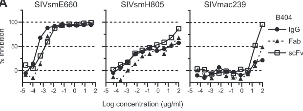
neutralization-sensitive SIV strains was almost completely
blocked by low concentrations of all forms of B404. Although the
potency to inhibit infection was similar among IgG, Fab, and scFv,
B404 IgG was slightly more effective against these SIV strains, as
shown by the neutralizing kinetics of SIVsmE660 (
Fig. 1A
).
Neu-tralization of SIVsmE543-3, SIVsmH805, and SIVsmH807
reached a plateau at 10 to 100 ng/ml IgG B404 and Fab B404, as
represented by neutralization kinetics against SIVsmH805
(
Fig. 1A
). These viruses were moderately sensitive to
B404-medi-ated neutralization, although the IC50s were variable among these
SIV strains (
Fig. 1B
). Interestingly, B404 scFv was more
effec-tive at high concentrations than B404 IgG and B404 Fab in
neu-tralization of these moderately neuneu-tralization-sensitive viruses
and SIVmac239, which is a highly neutralization-resistant strain
(
Fig. 1
). Infection with SIVmac239 was unaffected by the presence
of any form of B404 at a concentration of less than 1
g/ml but was
inhibited more than 90% by 100
g/ml B404 scFv. Neutralization
of 7 of 7 SIV strains, including genetically diverse,
neutralization-resistant SIVmac239, by B404 scFv indicates that B404 is a potent
and broad NAb against SIVsm/mac strains.
Isolation of Env-specific Fab clones from
SIVsmH635FC-in-fected macaques.
To analyze the induction of B404-like
antibod-ies in SIV-infected macaques, Env-specific Fab clones were
iso-lated from 4 SIVsmH635FC-infected macaques: H723, H704,
H709, and H714 (
26
,
39
). Env-specific Fab clones from H723 were
isolated from the previously constructed phage library (
25
)
through panning against Env, which was conjugated by coating
plate wells with the anti-Env Fab clones B404 (anti-gp120
confor-mational), B408 (anti-gp41 cluster I), and H301 (anti-gp120 V1).
Together with anti-Env Fab clones from the previous study, 98
anti-Env Fab clones, including 33 NAbs (33.7%), were obtained
from H723. From 3 other SIVsmH635FC-infected macaques,
and
light-chain phage libraries were separately constructed, and
2 panning series were performed using B404 and H301 to
conju-gate Env. After 4 series of panning in each macaque, we obtained
155, 102, and 53 independent Fab clones from H704, H709, and
FIG 1Potent and broad neutralization by monoclonal antibody B404 from a SIVsmH635FC-infected macaque. (A) The neutralizing potencies of IgG, Fab, and scFv of B404 are shown by inhibition kinetics against SIVsmE660FL14, SIVsmH805-24w-3, and SIVmac239. (B) The neutralizing potencies of IgG, Fab and scFv of B404 are shown by IC50and IC90(g/ml). Seven SIV strains, HIV-2GH123and HIV-1NL432were examined for their neutralizing sensitivities against B404 in
TZM-bl cells. The IC50and IC90values are shown in dark gray (⬍1.0⫻10⫺2g/ml), medium gray (1.0⫻10⫺2to 1.0⫻100g/ml), light gray (1 to 100g/ml)
and white (⬎100g/ml). Percent amino acid differences were calculated by pairwise comparison with SIVsmH635FC.
Conformational Epitope for Potent SIV Neutralization
on November 7, 2019 by guest
http://jvi.asm.org/
[image:4.585.138.448.70.184.2]H714, respectively. Neutralizing activities were observed in 47
clones (30.3%) from H704, 9 clones (8.8%) from H709, and 28
clones (52.8%) from H714 (
Table 1
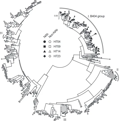
). Phylogenetic analysis of VH
genes revealed that 105 NAbs formed a major NAb cluster with
B404 (
Fig. 2
). The remaining NAbs were separated into 3 minor
clusters containing 3 or 4 NAbs. Fab clones in the major group,
designated the B404 group, were isolated from all 4 macaques
analyzed.
Although Fab clones in the B404 group were genetically similar
to one another, several small clusters were observed in the B404
group, suggesting multiple B cell origins generated by VDJ
recom-bination in these B404-like NAbs. Sequence analysis using the
International Immunogenetics Database (
50
) indicated that the
VH genes of Fab clones in the B404 group were close to human
pseudogene IGHV3-h (approximately 90% identity), but no
functional VH gene showed
⬎
90% identity. Analysis using the
genome database of the rhesus macaque (
51
) revealed a significant
relationship (
⬎
95% identity) between Fab clones in the B404
group and the rhesus macaque VH3 gene in the chromosome 7
scaffold (GenBank accession number NW_001122023). These
re-sults suggest that a major group of NAbs in
SIVsmH635FC-in-fected macaques preferentially use the same rhesus VH3 germ line
that lacks a human counterpart.
Bias in gene usage of NAbs from SIVsmH635FC-infected
macaques.
The genetic features of anti-Env Fab clones are
sum-marized in
Fig. 3
and
Table 1
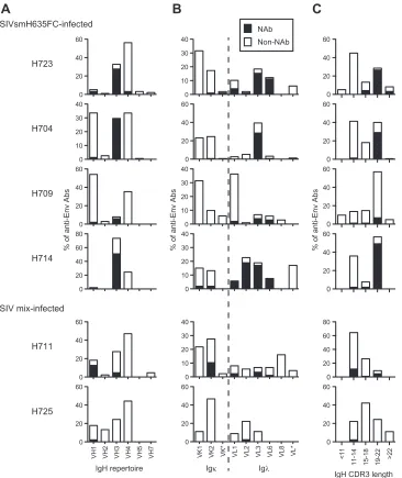
. As mentioned above, a major
pop-ulation of NAbs from SIVsmH635FC-infected macaques used the
same VH3 gene as B404, resulting in a high rate of NAbs using the
VH3 gene (
Fig. 3A
and
Table 1
). A high occupancy of
light
chains was also characteristic of NAbs from
SIVsmH635FC-in-fected macaques (
Fig. 3B
and
Table 1
). Moreover, a long
comple-mentarity-determining region 3 loop of the heavy chain (CDRH3)
was characteristic of the NAbs (
Fig. 3C
and
Table 1
). CDRH3 of
most NAbs had 19 or more amino acids, although the length of
CDRH3 was usually less than 18 amino acids in nonneutralizing
Fab clones. These results clearly showed that B404-like NAbs with
the VH3 gene-encoded heavy chain with a long CDR3 and
light
chain are the main NAb population in SIVsmH635FC-infected
macaques. In contrast, Fab clones from macaques H711 and H725
lacked these remarkable features of B404-like NAbs (
Fig. 3
,
bot-tom;
Table 1
). These 2 macaques were inoculated with a mixture
of SIVs (SIV mix). H711 was infected with a combination of
SIVsmE543-3 and SIVsmH635FC. H725 was infected with plasma
samples from 2 SIVsmH445-infected macaques, H631 and H635,
which SIVsmH635FC was isolated from. Although anti-Env Fab
clones were similarly isolated from these macaques, the frequency
of NAbs from H711 (17.2%) and H725 (2.2%) was lower than that
from SIVsmH635FC-infected macaques (8.8 to 52%). NAbs from
H711 and H725 preferentially used VH1 gene-encoded heavy
chains with a short CDRH3 and
light chains, but NAbs from
these macaques showed a genetic variation, similarly to those in
HIV-1-infected patients (
64
). These results suggested that
B404-like NAbs are induced exclusively in SIVsmH635FC-infected
macaques.
Potent neutralizing activity and the same specificity of NAbs
in the B404 group.
To analyze the epitopes recognized by these
NAbs, we first separated Fabs with neutralizing activity into 2
groups according to the results of competition ELISA with B404
IgG. All the NAbs in the B404 group and group II (
Fig. 2
)
com-TABLE 1Preferential gene usage and competition with B404 of NAbs
Inoculated virus Animal
Frequency (%) of NAbsa
No. of NAbs
% of NAbs with:
Avg CDRH3 lengthb
Competition (%) with B404c
VH3 genes light chains
SIVsmH635FC H723 33.7 33 81.8 93.9 20.0 81.8
H704 30.3 47 95.7 97.9 18.7 100
H709 7.8 8 75.0 100 17.8 100
H714 52.8 28 96.4 92.9 18.7 100
SIV mixd H711 17.2 15 26.7 40.0 15.2 86.7
H725 2.2 1 0.0 100 12 100
a
Frequencies of Fab clones with the VH3 gene andlight chain are shown as percentages of NAbs. bAverage number of amino acids in CDRH3.
c
Competition ELISA with 2g/ml B404 IgG was performed. The frequency of Fabs showing more than 50% inhibition is shown as a percentage of NAbs.
dH711 was inoculated with a mixture of SIVsmE543-3 and SIVsmH635FC. H725 was inoculated with plasma samples from 2 SIVsmH445-infected macaques, H631 and H635.
FIG 2B404-like NAbs formed a major group in anti-Env antibodies from 4 SIVsmH635FC-infected rhesus macaques. NAbs were separated into 4 groups in the phylogenetic tree, which was generated using MEGA5 (70) from heavy-chain genes of 98, 155, 102, and 53 Fab clones from H704, H709, H714, and H723, respectively.
on November 7, 2019 by guest
http://jvi.asm.org/
[image:5.585.39.551.78.179.2] [image:5.585.43.286.430.675.2]peted with B404 IgG (
Fig. 4A
), suggesting that epitopes for these
NAbs overlap or are close to that for B404. Despite the differences
in gene usage, competition with B404 IgG was also observed in
most NAbs from macaques infected with SIV mix (
Table 1
). NAbs
belonging to groups III and IV, with the exception of 1 Fab in
group III, did not compete with B404 IgG (
Fig. 4A
). The binding
ability of these Fabs was even enhanced by the addition of B404.
Competition of the Fabs in groups III and IV with biotinylated K8
in group III suggested that the Fabs in group III and IV share the
same epitope (data not shown). The neutralizing activity of these
Fabs was examined against the genetically divergent SIVmac316
and the neutralization-resistant SIVsmE543-3 (
Fig. 4B
). All of the
Fabs tested showed at least 50% inhibition against both viruses,
and the B404 group included Fabs with potent neutralizing
activ-ity that showed efficient inhibition at low concentrations.
Accord-ingly, the IC50s of the 4 Fabs in the B404 group ranged from 0.8 to
316 ng/ml (average IC
50, 79 ng/ml) against SIVmac316, indicating
the presence of NAbs comparable to B404 (IC50
against
SIVmac316, 0.6 ng/ml) (
Fig. 1
). In contrast, IC
50against
SIVmac316 ranged between 32 and 908 ng/ml (average IC50, 243
ng/ml) in groups III and IV. This result suggests that B404-like
NAbs are the main NAb population in terms of number and
neu-tralizing potency.
Epitope mapping of NAb B404.
To define the region of the
Env targeted by B404, we examined reactivity against mutants of
SIVsmE543-3 Env. Because the V3 loop has been shown to be
important for B404 binding (
25
), mutants with deletions in the
V1, V2, V3, and V4 loops (
⌬
V1,
⌬
V2,
⌬
V3, and
⌬
V4) and a
mu-FIG 3Bias in the Ig gene usage and CDR3 length of neutralizing Fab clones from SIVsmH635FC-infected macaques. The proportions of IgH (A) and Igand Ig(B) repertoires and heavy-chain CDR3 length (C) are shown as percentages of NAbs and non-NAbs. In addition to Fabs isolated from 4 SIVsmH635FC-infected macaques, 87 Fab clones from H711, which was SIVsmH635FC-infected with a combination of SIVsmE543-3 and SIVsmH635FC, and 45 Fab clones from H725, which was infected with plasma samples from 2 SIVsmH445-infected macaques, H631 and H635, were similarly analyzed. Usage of Ig genes was analyzed using V-QUEST in the International Immunogenetics Database (44).
Conformational Epitope for Potent SIV Neutralization
on November 7, 2019 by guest
http://jvi.asm.org/
[image:6.585.112.478.65.500.2]tant lacking 3 glycosylation sites flanking the V3 loop (
⌬
Gly) were
constructed. In addition, mutants carrying single mutations in the
CD4bs (D385R) and CD4i (I434R) sites, corresponding to D368R
and I420R in HIV-1 gp120 (
13
,
44
–
46
), were examined to clarify
the relationship of the B404 epitope to the CD4bs and CD4i sites.
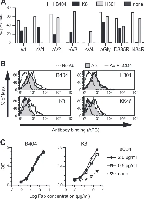
Flow cytometry analysis using cells expressing these Env mutants
revealed that the reactivity of B404 was completely lost in
⌬
V3 and
⌬
V4 mutants, though B404 bound to other mutants even better
than it did to the wild type (
Fig. 5A
). These results suggested that
B404 recognizes a conformational epitope consisting of the V3
and V4 loops.
The reactivity of another Fab, K8, which targets an epitope
other than that of B404 (
Fig. 4A
), was lost in
⌬
V4 and I434R
mutants (
Fig. 5A
). No reactivity to I434R strongly suggested that
K8 is a CD4i antibody. Therefore, the effect of sCD4 ligation on
antibody binding to Env trimers and monomers was examined
using flow cytometry and ELISA, respectively. The reactivity of
B404, K8, and KK46 (murine anti-V3 MAb) to Env on the cell
surface was enhanced by the addition of sCD4, although no effect
was observed in anti-V1 Fab H301 (
Fig. 5B
). This suggested that
epitopes for B404, K8, and KK46 are exposed in the open
confor-mation of the Env trimer triggered by CD4 binding. Consistent
with the analysis of mutant Envs, the reactivity of K8 to Env
monomer was enhanced by the addition of sCD4, but B404
showed no enhancement of reactivity (
Fig. 5C
).
FIG 4The specificity and potency of Fab clones in the B404 group are similar to those of B404. (A) Competition ELISA was performed using serially diluted B404 IgG as a competitor. B404 IgG significantly inhibited the binding of the Fabs in the B404 group (L6, L22, O9, and O19). In contrast, B404 IgG did not compete with the Fabs in groups III (K8 and K10) and IV (K12, K40, and K47) and even enhanced the binding of these Fabs. (B) Neutralization potencies of Fabs in the B404 group (left) and groups III and IV (right) are shown by inhibition of infection to TZM-bl cells with neutralization-resistant SIVsmE543-3 and genetically divergent SIVmac316.
FIG 5B404 recognizes a conformational epitope, including the V3 and V4 loops, and sCD4 enhances the exposure of the epitope in trimeric Env. (A) Reactivity of B404, K8, and H301 (anti-V1 Fab) to Env mutants was examined using 293T cells transfected with plasmids to express SIVsmE543-3 Env (wild-type), mutants with deletions in the V1 (⌬V1), V2 (⌬V2), V3 (⌬V3), and V4 (⌬V4) loops, an N306A/N316A/N349A mutant lacking glycosylation sites near the V3 loop (⌬Gly), a D385R mutant interfering with CD4bs antibodies (D385R), and an I434R mutant interfering with CD4i antibodies (I434R). The transfected cells were stained with Fabs B404, K8, and H301, and the reactivity of Env mutants was analyzed using flow cytometry. The percentage of Fab⫹ cells is shown. (B) Reactivity of Fabs B404, K8, and H301, and murine anti-V3 MAb KK46 to sCD4-treated trimeric Env on the cell surface. Cells transfected with the plasmid to express SIVsmE543-3 Env were incubated with 2g/ml sCD4 for 15 min, and the reactivities of antibodies were similarly examined. The tinted histogram represents cells stained by antibody in the absence of sCD4. The dotted line shows the unstained control. (C) Reactivity of Fab clones B404 and K8 to sCD4-treated monomeric Env. The reactivity of serially diluted Fab to Env was examined by ELISA using SIVsmE543-3 as an antigen in the absence or presence of 0.5 or 2.0g/ml sCD4.
on November 7, 2019 by guest
http://jvi.asm.org/
[image:7.585.42.273.66.458.2] [image:7.585.303.539.70.395.2]The enhancement by sCD4 of reactivity to both monomeric
and trimeric Env and the interference in binding by the I434R
(I420R in HIV-1) mutation in Env, which are features of so-called
CD4i antibodies against HIV-1 (
13
,
45
,
46
), indicate that K8
tar-gets the CD4i epitope. The enhanced reactivity of B404 by sCD4 to
trimeric but not monomeric Env is analogous to the reactivity of
anti-V3 antibodies (
65
). These results suggest that B404
recog-nizes a conformational epitope consisting of the V3 and V4 loops,
which are intensely exposed on the Env trimer after CD4 ligation.
Selection of variants resistant to NAb B404.
To select
B404-resistant variants
in vitro
, we passaged SIVmac316, which is the
most sensitive to B404 of the SIV strains tested (
Fig. 1
), in PM1/
CCR5 cells in the presence of increasing concentrations of B404.
As a control, passage under the same conditions without B404 was
also performed to monitor spontaneous changes during infection
in PM1/CCR5 cells. The concentration of B404 was increased
from 5 ng/ml to 400
g/ml at passage 26. Viruses recovered at
passage 26 in the presence and absence of B404, which were
des-ignated P26B404 and P26C, respectively, were examined for their
sensitivity to antibodies and plasma samples from SIV-infected
macaques (
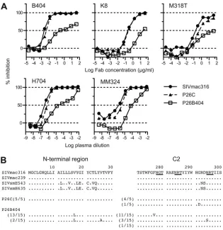
Fig. 6A
). The IC
50for B404 against SIVmac316, P26C,
and P26B404 were 2.8, 4.1, and 240 ng/ml, respectively, showing
an 86-fold resistance of P26B404 to B404 compared with that of
wild-type SIVmac316. P26B404 was also resistant to
neutraliza-tion by MAbs K8 (CD4i) and M318T (V2), which target epitopes
other than that of B404, and plasma samples from SIV-infected
macaques (
Fig. 6A
). These results suggested that P26B404
ac-quired resistance to antibody-mediated neutralization
compara-ble to that observed in neutralization-resistant SIV strains, such as
SIVmac239 and SIVsmE543-3. Sequence analysis of gp120
re-vealed 3 amino acid substitutions specific to P26B404: V17L in the
N-terminal region and F277V and N295S in the C2 region
(
Fig. 6B
). Of these substitutions, the two in the C2 region were
highly conserved among SIVsm/mac and HIV-2 strains. These
substitutions were independently observed, and no variant with
both F277V and N295S was found in the 15 clones sequenced.
MD simulation of gp120 outer domains from B404-sensitive
and B404-resistant variants.
To address structural impacts of
the 2 mutations in the C2 region, we performed MD simulation
of unliganded gp120 outer domains from B404-sensitive
(SIVmac316) and B404-resistant (F277V and N295S) variants. To
map the sites at which structural dynamics were influenced by C2
mutations, we calculated the RMSF of the main chains of
individ-ual amino acid residues using 90,000 snapshots from 5 to 50 ns of
FIG 6Isolation of variants resistant to B404 and amino acid substitutions in gp120. The B404-resistant variant was induced from SIVmac316 by passages of viruses in PM-1/CCR5 cells with increasing concentrations of B404 Fab. A B404-resistant variant, P26B404, was obtained from the supernatant of passage 26. P26C was obtained after 26 passages without B404. (A) The sensitivities of P26B404, P26C, and parental SIVmac316 to neutralization are shown by inhibition of infection of TZM-bl cells. B404 and K8 Fabs, murine MAb M318T, which recognizes the V2 of gp120, and plasma samples from SIVsmH635FC-infected macaque H704 and SIVmac239-infected macaque MM324 were used for the neutralization assay. (B) Amino acid sequences of the N-terminal and C2 regions of gp120 from P26C and P26B404 are aligned with those of parental SIVmac316 and SIV strains, SIVmac239, SIVsmE543-3 and SIVsmH635FC. The number of clones per total number of clones is given in parentheses. Identical amino acids are shown as dots, and potential glycosylation sites in SIVmac316 are indicated with underlining.
Conformational Epitope for Potent SIV Neutralization
on November 7, 2019 by guest
http://jvi.asm.org/
[image:8.585.135.450.64.389.2]each MD simulation (
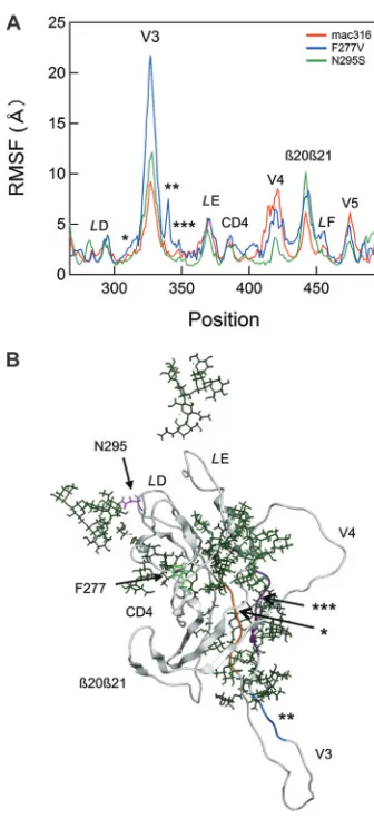
Fig. 7A
). RMSF values provide key
informa-tion about the atomic fluctuainforma-tions of the individual amino acids of
a protein in solution (
57
). These values were maximal at the tip of
the V3 loop and prominent at other loop regions, including
L
D,
L
E, CD4 binding, V4,

20

21/
L
F, and V5 (
Fig. 7A
), suggesting
that these loops fluctuate in solution. Notably, the F277V and
N295S mutations were found to induce changes in RMSF values
mainly at the V3, V4,

20

21/
L
F, and V5 loop regions (
Fig. 7A
,
blue and green lines, respectively). Interestingly, these regions are
located far from the C2 mutation sites compared with the
loca-tions of other loops, such as
L
D,
L
E, and CD4 binding, which had
RMSF values that were similar among wild-type and
B404-resis-tant variants (
Fig. 7B
; F277V in
L
D and N295S in the proximal
region of
L
D). In particular, RMSF changes by the C2 mutations
were the most prominent at the V3/V4 loops and their
neighbor-ing regions (
Fig. 7
). These results suggested that the F277V and
N295S mutations could alter the structural dynamics of V3/V4
loops and their neighboring regions in solution. The structural
alterations would lead to changes in entropy of the regions, which
affect the binding affinity to B404. The finding is consistent with
the results of an epitope mapping study of B404.
DISCUSSION
Potent and broadly neutralizing MAbs have recently been isolated
from HIV-1-infected patients and analyzed to understand the
mechanism of neutralization against a broad spectrum of HIV-1
strains and to design vaccines against their neutralizing epitopes
(
12
–
16
). Although the SIV-macaque model has been used as an
animal model for HIV-1 infection for vaccine development (
6
,
8
,
24
), no potent and broadly neutralizing monoclonal antibody
against SIV was available. Therefore, the epitopes and mechanism
for broad neutralization of SIV remained uncertain. Many
mono-clonal antibodies against SIVsm/mac were isolated from
SIV-in-fected macaques (
66
,
67
) and mice immunized with SIV Env (
43
,
47
,
68
), but few of them showed neutralizing activity against
various SIV strains, including highly neutralization-resistant
SIVmac239 (
68
). In comparison with these monoclonal
antibod-ies against SIV identified so far, B404 apparently has a broadly
neutralizing activity, which enables it to neutralize multiple,
di-verse SIV isolates, and can be defined as the first generation of
broadly NAbs against SIV. The broad and potent neutralizing
ac-tivity of B404 shown in this study indicates that B404 can be used
to analyze broad neutralization against SIV. The B404 epitope, the
newly identified broadly neutralizing epitope against SIV, will
fur-ther understanding of the mechanism of broad neutralization
effective for protection from SIV infection.
The infection of rhesus macaques with SIVsmH635FC, a
highly neutralization-sensitive clone, was chosen for this study
because this SIV strain induced a vigorous and potent antibody
response in all the infected macaques and acquired many viral
mutations to escape antibody recognition (
25
,
26
). The kinetics of
B404 neutralization against various SIV strains were similar to
those observed in the plasma sample of the macaque from which
B404 was isolated, suggesting that B404-like NAbs are
representa-tive of the neutralizing activity in SIVsmH635-infected macaque
H723 (
25
). Consistent with this observation, B404-like NAbs were
shown to be a major group in NAbs genetically and functionally.
Most of the NAbs in the 4 SIVsmH635FC-infected macaques
an-alyzed had the same features, including the use of a specific VH3
germ line and
light chains, a long CDRH3 loop, and competition
with B404. The bias in the specificity and gene usage may be
par-tially enhanced by the screening process, because B404-like NAbs
were predominantly isolated from
light-chain libraries by
pan-ning against H301-conjugated Env. In addition, isolating
antibod-ies against quaternary epitopes constituted by the Env trimer
through panning using monomeric Env was difficult. However,
the presence of many independent B404-like NAbs strongly
sug-gests that B404-like NAbs compose a significant fraction of NAbs
in 4 SIVsmH635FC-infected macaques. Moreover, the vigorous
induction of B404-like NAbs in SIVsmH635FC-infected
ma-caques was also supported by the multiple B cell origins apparent
FIG 7Effects of F277V and N295S mutations on the structural dynamics of the gp120 outer domain. (A) Distribution of RMSF in the gp120 outer domain. MD simulations of gp120 outer domains of SIVmac316, F277V, and N295S were carried out at 1 atm and 310 K for 50 ns as described in Materials and Methods. The RMSF values, which indicate the atomic fluctuations of the main chains of individual amino acids during MD simulations, were calculated using 90,000 snapshots from 9 to 50 ns of each MD simulation. The numbers on the horizontal axes indicate amino acid positions in gp120. The RMSF values of the mutants are significantly different from those of parental SIVmac316 at the V3 loop, the V3 flanking regions (indicated by asterisks), the V4 loop, the2021/LF loop, and the V5 loop regions. (B) A structure at 50 ns of MD simulation of the SIVmac316 gp120 outer domain is shown as a repre-sentative to indicate the steric location of mutation sites and various loops. The regions that are proximal to the V3/V4 loops and displayed fluctuations that differed from those of parental SIVmac316 (Fig. 7A) are highlighted in orange (*), blue (**), and purple (***). Green sticks indicate glycans.
on November 7, 2019 by guest
http://jvi.asm.org/
[image:9.585.77.245.62.430.2]from several subgroups in B404-like NAbs (
Fig. 1
). These
sub-groups originated from distinct B cell precursors generated by
VDJ recombination, because they were often distinguished by the
length and nucleotide sequences of CDRH3.
The observation of few B404-like NAbs from macaques
in-fected with the SIV mix clearly indicates that induction of
B404-like NAbs depends on infection with SIVsmH635FC. The
exclu-sive induction of B404-like NAbs only in SIVsmH635FC-infected
macaques also raises the possibility that Env from SIVsmH635FC
is a highly immunogenic protein that induces antibodies against
the B404 epitope. The use of Env from SIVsmH635FC for
vacci-nation may be advantageous for the induction of broadly
neutral-izing antibodies, because the Env from an HIV-1-infected patient
with broadly neutralizing antibodies induced cross-reactive
anti-HIV-1 NAbs in an animal model (
69
). The Env in this vaccination
study is capable of mediating CD4-independent infection. Since
the Env of SIVsmH635FC has a mutation in the CD4-binding
region (D385N), NAb induction may be affected by CD4
indepen-dency of Env. The development of vaccines aimed at inducing
B404-like NAbs in rhesus macaques will be useful in establishing
models for development of antibody-based vaccines targeting
spe-cific epitopes for broad neutralization.
Biased usage of a specific VH3 germ line gene and
light chain
are remarkable genetic features of B404-like NAbs. The induction
of NAbs with specific germ line genes, such as VH1-69 for CD4i
(
70
) and VH5-51 for V3 (
64
), is frequently observed in
HIV-1-infected patients. A close relationship between VH germ line genes
and target epitopes suggests the importance of Ig gene usage in the
induction of broadly neutralizing antibodies. Therefore, rational
design of vaccines has been undertaken based on reactivity to
an-tibodies with the germ line genes used by known broadly
neutral-izing antibodies (
11
,
71
,
72
). Unfortunately, the VH3 germ line
gene of B404 is divergent from all human VH3 germ line genes,
suggesting the absence of a human counterpart. This may partially
explain why B404-like NAbs have not been identified in
HIV-infected humans, although the structure of HIV-1 Env, which is
different from that of SIV, significantly affects immunogenicity of
the B404 epitope. Rhesus macaque-specific germ line genes were
also used by NAbs against the quaternary epitope of HIV-1 Env
from simian and human immunodeficiency virus
(SHIV)-in-fected macaques, but their germ line genes were different from the
VH3 germ line gene used by B404 (
73
). Even in the presence of a
human VH gene counterpart, the antibody response to a
neutral-izing epitope may differ between rhesus macaques and humans
(
74
). In addition to the genetic diversity of germ line genes, the
rhesus macaque CDRH3 repertoire differs from that of humans,
resulting in species-specific antibody repertoires (
75
). This species
specificity in antibody induction is a problem in the evaluation of
HIV-1 vaccines in animal models, especially those designed for
specific neutralizing epitopes of HIV-1. Conversely, K8, the CD4i
NAb from a SIV-infected macaque, used the rhesus VH1 germ line
gene, an analog of human VH1-69 frequently used by CD4i NAbs
in HIV-1-infected humans (
70
). Thus, the mechanism of
induc-tion of CD4i NAbs with VH1-69 may be common in both humans
and rhesus macaques. To analyze vaccine candidates properly in
nonhuman primates, similarities and differences in antibody
re-sponse between rhesus macaques and humans should be
consid-ered.
B404 recognizes a conformational epitope consisting of the V3
and V4 loops. The enhanced exposure of the epitope in trimeric
Env by sCD4 and efficient neutralization of
neutralization-resis-tant SIV strains by the scFv form of B404 suggests that the B404
epitope is sterically masked by the V1/V2 loops and glycans,
anal-ogous to CD4i and V3 epitopes (
46
,
65
,
76
). Consistent with this
interpretation, B404 reacted more intensely to Env mutants
lack-ing the V1 and V2 loops than to the wild-type Env. MD simulation
supported these observations by indicating changes in the
struc-tural dynamics of V3/V4 loops and their neighboring regions in
gp120 of resistant variants with F277V and N295S mutations in
the C2 region. The acquisition of resistance to the broadly
neu-tralizing antibodies b12, PG9, and PG16 due to C2 mutations far
from the target epitopes was also observed in HIV-1 CRF01_AE
(
77
,
78
). MD simulation is a powerful computational method for
analyzing structural dynamics of proteins in solution on the basis
of theoretical and empirical principles in physical chemistry (
79
)
and has been applied to research on viruses (
80
). The structural
dynamics of protein surfaces in solution plays a key role in protein
interactions, and MD simulation is advantageous in speculation
about interactions between proteins containing flexible regions,
such as V3 and V4 loops. Analysis of fluctuation changes in
mu-tant proteins is useful to identify the regions which affect
protein-protein interaction.
Although the V3 and V4 loops are known to contain linear
epitopes for antibody-mediated neutralization in SIV infection,
no conformational MAb against SIV with characteristics similar
to those of B404 has been reported (
43
,
47
,
66
–
68
). Several MAbs
to conformational epitopes that include the V3 region, such as
PGT antibodies, represented by PGT128 (
15
,
19
), 3BC176, and
3BC315 (
81
), were isolated from HIV-1-infected patients. The
PGT128 epitope consists of the short segment of the V3 loop and
2 neighboring glycans (
19
), but the binding of B404 is
indepen-dent of these glycans near the V3 region. The epitope of 3BC176
and 3BC315 is close to the V3 loop, and their binding is partially
enhanced by CD4 binding, similar to that of B404 (
81
). However,
in contrast to B404, 3BC176 and 3BC315 do not bind to
mono-meric Env and compete with CD4i antibodies, suggesting that
their epitope is different from that of B404. Although we have not
determined the precise B404 epitope, the characteristics of B404
that are similar to those of CD4i and V3 antibodies suggest that
B404 recognizes a conserved region important for binding to the
CCR5 coreceptor (
46
,
65
,
76
,
82
).
Despite recent progress in understanding the broad
neutraliza-tion of HIV-1, epitopes for potent and broad neutralizaneutraliza-tion have
not been analyzed in an SIV model because NAb analysis using
SIV cannot be directly applied to HIV-1. The main disadvantage
of SIV is the antigenicity difference relative to HIV-1, which
makes examination of neutralizing epitopes of HIV-1 impossible.
Although the use of SHIV expressing HIV-1 Env enables the
eval-uation of vaccine candidates designed for target epitopes of HIV-1
in nonhuman primates, an SIV model was more predictive of
vaccine efficacy than a SHIV model in clinical trials of a
T-cell-based vaccine (
83
,
84
). For comprehensive assessment of
immu-nity induced by vaccine candidates, proof-of-concept trials using
the SIV model should be considered before further efficacy trials.
The identification of B404 with its potent and broad neutralizing
activity against SIV will be a useful adjunct for evaluating the
mechanism of neutralization in an SIV-macaque model and will
contribute to the development of HIV-1 vaccines.
Conformational Epitope for Potent SIV Neutralization
on November 7, 2019 by guest
http://jvi.asm.org/
ACKNOWLEDGMENTS
We thank Yuko Katsumata for excellent assistance. The phagemid vector pComb3X was kindly provided by the Scripps Research Institute. The following reagents were obtained through the NIH AIDS Research and Reference Reagent Program, Division of AIDS, NIAID, NIH: TZM-bl cells from John C. Kappes, Xiaoyun Wu, and Tranzyme Inc. and KK46 from Karen Kent and Caroline Powell.
This work was supported in part by the Special Coordination Funds for Promoting Science and Technology, the Program of Founding Re-search Centres for Emerging and Re-emerging Infectious Diseases, the Global COE program Global Education and Research Centre Aiming at the Control of AIDS, a grant-in-aid for scientific research (C-24591484) from the Ministry of Education, Culture, Sport, Science and Technology, Japan, and a grant from the Ministry of Health, Welfare and Labor of Japan (H24-AIDS-007).
REFERENCES
1.Igarashi T, Brown C, Azadegan A, Haigwood N, Dimitrov D, Martin MA, Shibata R.1999. Human immunodeficiency virus type 1 neutraliz-ing antibodies accelerate clearance of cell-free virions from blood plasma. Nat. Med.5:211–216.
2.Shibata R, Igarashi T, Haigwood N, Buckler-White A, Ogert R, Ross W, Willey R, Cho MW, Martin MA.1999. Neutralizing antibody directed against the HIV-1 envelope glycoprotein can completely block HIV-1/SIV chimeric virus infections of macaque monkeys. Nat. Med.5:204 –210. 3.Veazey RS, Shattock RJ, Pope M, Kirijan JC, Jones J, Hu Q, Ketas T,
Marx PA, Klasse PJ, Burton DR, Moore JP.2003. Prevention of virus transmission to macaque monkeys by a vaginally applied monoclonal an-tibody to HIV-1 gp120. Nat. Med.9:343–346.
4.Hessell AJ, Rakasz EG, Poignard P, Hangartner L, Landucci G, Forthal DN, Koff WC, Watkins DI, Burton DR. 2009. Broadly neutralizing human anti-HIV antibody 2G12 is effective in protection against mucosal SHIV challenge even at low serum neutralizing titers. PLoS Pathog.
5:e1000433. doi:10.1371/journal.ppat.1000433.
5.Mascola JR, Stiegler G, VanCott TC, Katinger H, Carpenter CB, Han-son CE, Beary H, Hayes D, Frankel SS, Birx DL, Lewis MG. 2000. Protection of macaques against vaginal transmission of a pathogenic HIV-1/SIV chimeric virus by passive infusion of neutralizing antibodies. Nat. Med.6:207–210.
6.Ourmanov I, Kuwata T, Goeken R, Goldstein S, Iyengar R, Buckler-White A, Lafont B, Hirsch VM. 2009. Improved survival in rhesus macaques immunized with modified vaccinia virus Ankara recombinants expressing simian immunodeficiency virus envelope correlates with re-duction in memory CD4⫹T-cell loss and higher titers of neutralizing antibody. J. Virol.83:5388 –5400.
7.Bogers WM, Davis D, Baak I, Kan E, Hofman S, Sun Y, Mortier D, Lian Y, Oostermeijer H, Fagrouch Z, Dubbes R, van der Maas M, Mooij P, Koopman G, Verschoor E, Langedijk JP, Zhao J, Brocca-Cofano E, Robert-Guroff M, Srivastava I, Barnett S, Heeney JL.2008. Systemic neutralizing antibodies induced by long interval mucosally primed sys-temically boosted immunization correlate with protection from mucosal SHIV challenge. Virology382:217–225.
8.Flatz L, Cheng C, Wang L, Foulds KE, Ko SY, Kong WP, Roychoudhuri R, Shi W, Bao S, Todd JP, Asmal M, Shen L, Donaldson M, Schmidt SD, Gall JG, Pinschewer DD, Letvin NL, Rao S, Mascola JR, Roederer M, Nabel GJ.2012. Gene-based vaccination with a mismatched envelope protects against simian immunodeficiency virus infection in nonhuman primates. J. Virol.86:7760 –7770.
9.Thorner AR, Barouch DH.2007. HIV-1 vaccine development: progress and prospects. Curr. Infect. Dis. Rep.9:71–75.
10. McMichael AJ.2006. HIV vaccines. Annu. Rev. Immunol.24:227–255. 11. Burton DR, Poignard P, Stanfield RL, Wilson IA.2012. Broadly
neu-tralizing antibodies present new prospects to counter highly antigenically diverse viruses. Science337:183–186.
12. D’Souza MP, Livnat D, Bradac JA, Bridges SH. 1997. Evaluation of monoclonal antibodies to human immunodeficiency virus type 1 primary isolates by neutralization assays: performance criteria for selecting candi-date antibodies for clinical trials. AIDS Clinical Trials Group Antibody Selection Working Group. J. Infect. Dis.175:1056 –1062.
13. Scheid JF, Mouquet H, Feldhahn N, Seaman MS, Velinzon K, Pietzsch J, Ott RG, Anthony RM, Zebroski H, Hurley A, Phogat A, Chakrabarti
B, Li Y, Connors M, Pereyra F, Walker BD, Wardemann H, Ho D, Wyatt RT, Mascola JR, Ravetch JV, Nussenzweig MC. 2009. Broad diversity of neutralizing antibodies isolated from memory B cells in HIV-infected individuals. Nature458:636 – 640.
14. Walker LM, Phogat SK, Chan-Hui PY, Wagner D, Phung P, Goss JL, Wrin T, Simek MD, Fling S, Mitcham JL, Lehrman JK, Priddy FH, Olsen OA, Frey SM, Hammond PW, Kaminsky S, Zamb T, Moyle M, Koff WC, Poignard P, Burton DR.2009. Broad and potent neutralizing antibodies from an African donor reveal a new HIV-1 vaccine target. Sci-ence326:285–289.
15. Walker LM, Huber M, Doores KJ, Falkowska E, Pejchal R, Julien JP, Wang SK, Ramos A, Chan-Hui PY, Moyle M, Mitcham JL, Hammond PW, Olsen OA, Phung P, Fling S, Wong CH, Phogat S, Wrin T, Simek MD, Protocol GPI, Koff WC, Wilson IA, Burton DR, Poignard P.2011. Broad neutralization coverage of HIV by multiple highly potent antibod-ies. Nature477:466 – 470.
16. Huang J, Ofek G, Laub L, Louder MK, Doria-Rose NA, Longo NS, Imamichi H, Bailer RT, Chakrabarti B, Sharma SK, Alam SM, Wang T, Yang Y, Zhang B, Migueles SA, Wyatt R, Haynes BF, Kwong PD, Mascola JR, Connors M.2012. Broad and potent neutralization of HIV-1 by a gp41-specific human antibody. Nature491:406 – 412.
17. Zhou T, Georgiev I, Wu X, Yang ZY, Dai K, Finzi A, Kwon YD, Scheid JF, Shi W, Xu L, Yang Y, Zhu J, Nussenzweig MC, Sodroski J, Shapiro L, Nabel GJ, Mascola JR, Kwong PD.2010. Structural basis for broad and potent neutralization of HIV-1 by antibody VRC01. Science329:811– 817. 18. McLellan JS, Pancera M, Carrico C, Gorman J, Julien JP, Khayat R, Louder R, Pejchal R, Sastry M, Dai K, O’Dell S, Patel N, Shahzad-ul Hussan S, Yang Y, Zhang B, Zhou T, Zhu J, Boyington JC, Chuang GY, Diwanji D, Georgiev I, Kwon YD, Lee D, Louder MK, Moquin S, Schmidt SD, Yang ZY, Bonsignori M, Crump JA, Kapiga SH, Sam NE, Haynes BF, Burton DR, Koff WC, Walker LM, Phogat S, Wyatt R, Orwenyo J, Wang LX, Arthos J, Bewley CA, Mascola JR, Nabel GJ, Schief WR, Ward AB, Wilson IA, Kwong PD.2011. Structure of HIV-1 gp120 V1/V2 domain with broadly neutralizing antibody PG9. Nature
480:336 –343.
19. Pejchal R, Doores KJ, Walker LM, Khayat R, Huang PS, Wang SK, Stanfield RL, Julien JP, Ramos A, Crispin M, Depetris R, Katpally U, Marozsan A, Cupo A, Maloveste S, Liu Y, McBride R, Ito Y, Sanders RW, Ogohara C, Paulson JC, Feizi T, Scanlan CN, Wong CH, Moore JP, Olson WC, Ward AB, Poignard P, Schief WR, Burton DR, Wilson IA.2011. A potent and broad neutralizing antibody recognizes and pen-etrates the HIV glycan shield. Science334:1097–1103.
20. Ryu SE, Hendrickson WA.2012. Structure and design of broadly-neutralizing antibodies against HIV. Mol. Cells34:231–237.
21. Stamatatos L, Morris L, Burton DR, Mascola JR. 2009. Neutralizing antibodies generated during natural HIV-1 infection: good news for an HIV-1 vaccine? Nat. Med.15:866 – 870.
22. Saunders KO, Rudicell RS, Nabel GJ.2012. The design and evaluation of HIV-1 vaccines. AIDS26:1293–1302.
23. Haynes BF, Kelsoe G, Harrison SC, Kepler TB. 2012. B-cell-lineage immunogen design in vaccine development with HIV-1 as a case study. Nat. Biotechnol.30:423– 433.
24. Barouch DH, Liu J, Li H, Maxfield LF, Abbink P, Lynch DM, Iampietro MJ, SanMiguel A, Seaman MS, Ferrari G, Forthal DN, Ourmanov I, Hirsch VM, Carville A, Mansfield KG, Stablein D, Pau MG, Schuite-maker H, Sadoff JC, Billings EA, Rao M, Robb ML, Kim JH, Marovich MA, Goudsmit J, Michael NL.2012. Vaccine protection against acquisi-tion of neutralizaacquisi-tion-resistant SIV challenges in rhesus monkeys. Nature
482:89 –93.
25. Kuwata T, Katsumata Y, Takaki K, Miura T, Igarashi T.2011. Isolation of potent neutralizing monoclonal antibodies from an SIV-Infected rhe-sus macaque by phage display. AIDS Res. Hum. Retroviruses27:487–500. 26. Kuwata T, Byrum R, Whitted S, Goeken R, Buckler-White A, Plishka R, Iyengar R, Hirsch VM.2007. A rapid progressor-specific variant clone of simian immunodeficiency virus replicates efficiently in vivo only in the absence of immune responses. J. Virol.81:8891– 8904.
27. Kuwata T, Dehghani H, Brown CR, Plishka R, Buckler-White A, Iga-rashi T, Mattapallil J, Roederer M, Hirsch VM.2006. Infectious molec-ular clones from a simian immunodeficiency virus-infected rapid-progressor (RP) macaque: evidence of differential selection of RP-specific envelope mutations in vitro and in vivo. J. Virol.80:1463–1475. 28. Lusso P, Cocchi F, Balotta C, Markham PD, Louie A, Farci P, Pal R,
Gallo RC, Reitz MS, Jr.1995. Growth of macrophage-tropic and primary
on November 7, 2019 by guest
http://jvi.asm.org/
human immunodeficiency virus type 1 (HIV-1) isolates in a unique CD4⫹T-cell clone (PM1): failure to downregulate CD4 and to interfere with cell-line-tropic HIV-1. J. Virol.69:3712–3720.
29. Yusa K, Maeda Y, Fujioka A, Monde K, Harada S.2005. Isolation of TAK-779-resistant HIV-1 from an R5 HIV-1 GP120 V3 loop library. J. Biol. Chem.280:30083–30090.
30. Takeuchi Y, McClure MO, Pizzato M.2008. Identification of gamma-retroviruses constitutively released from cell lines used for human immu-nodeficiency virus research. J. Virol.82:12585–12588.
31. Wei X, Decker JM, Liu H, Zhang Z, Arani RB, Kilby JM, Saag MS, Wu X, Shaw GM, Kappes JC.2002. Emergence of resistant human immuno-deficiency virus type 1 in patients receiving fusion inhibitor (T-20) mono-therapy. Antimicrob. Agents Chemother.46:1896 –1905.
32. Derdeyn CA, Decker JM, Sfakianos JN, Wu X, O’Brien WA, Ratner L, Kappes JC, Shaw GM, Hunter E.2000. Sensitivity of human immuno-deficiency virus type 1 to the fusion inhibitor T-20 is modulated by core-ceptor specificity defined by the V3 loop of gp120. J. Virol.74:8358 – 8367. 33. Platt EJ, Wehrly K, Kuhmann SE, Chesebro B, Kabat D.1998. Effects of CCR5 and CD4 cell surface concentrations on infections by macrophag-etropic isolates of human immunodeficiency virus type 1. J. Virol.72: 2855–2864.
34. DuBridge RB, Tang P, Hsia HC, Leong PM, Miller JH, Calos MP.1987. Analysis of mutation in human cells by using an Epstein-Barr virus shuttle system. Mol. Cell. Biol.7:379 –387.
35. Hirsch V, Adger-Johnson D, Campbell B, Goldstein S, Brown C, Elkins WR, Montefiori DC.1997. A molecularly cloned, pathogenic, neutraliza-tion-resistant simian immunodeficiency virus, SIVsmE543-3. J. Virol.71: 1608 –1620.
36. Kestler H, Kodama T, Ringler D, Marthas M, Pedersen N, Lackner A, Regier D, Sehgal P, Daniel M, King N, et al.1990. Induction of AIDS in rhesus monkeys by molecularly cloned simian immunodeficiency virus. Science248:1109 –1112.
37. Mori K, Ringler DJ, Kodama T, Desrosiers RC.1992. Complex deter-minants of macrophage tropism in env of simian immunodeficiency virus. J. Virol.66:2067–2075.
38. Wu F, Ourmanov I, Kuwata T, Goeken R, Brown CR, Buckler-White A, Iyengar R, Plishka R, Aoki ST, Hirsch VM.2012. Sequential evolution and escape from neutralization of simian immunodeficiency virus SIVsmE660 clones in rhesus macaques. J. Virol.86:8835– 8847. 39. Kuwata T, Nishimura Y, Whitted S, Ourmanov I, Brown CR, Dang Q,
Buckler-White A, Iyengar R, Brenchley JM, Hirsch VM.2009. Associ-ation of progressive CD4(⫹) T cell decline in SIV infection with the in-duction of autoreactive antibodies. PLoS Pathog.5:e1000372. doi:10.1371 /journal.ppat.1000372.
40. Barbas CF, Scott JM, Silverman G, Burton DR.2001. Phage display: a laboratory manual. Cold Spring Harbor Laboratory Press, Cold Spring Harbor, NY.
41. Robinson JE, Holton D, Liu J, McMurdo H, Murciano A, Gohd R.1990. A novel enzyme-linked immunosorbent assay (ELISA) for the detection of antibodies to HIV-1 envelope glycoproteins based on immobilization of viral glycoproteins in microtiter wells coated with concanavalin A. J. Im-munol. Methods132:63–71.
42. Kuwata T, Kodama M, Sato A, Suzuki H, Miyazaki Y, Miura T, Hayami M.2007. Contribution of monocytes to viral replication in macaques during acute infection with simian immunodeficiency virus. AIDS Res. Hum. Retroviruses23:372–380.
43. Matsumi S, Matsushita S, Yoshimura K, Javaherian K, Takatsuki K.
1995. Neutralizing monoclonal antibody against a external envelope gly-coprotein (gp110) of SIVmac251. AIDS Res. Hum. Retroviruses11:501– 508.
44. Olshevsky U, Helseth E, Furman C, Li J, Haseltine W, Sodroski J.1990. Identification of individual human immunodeficiency virus type 1 gp120 amino acids important for CD4 receptor binding. J. Virol.64:5701–5707. 45. Walker LM, Simek MD, Priddy F, Gach JS, Wagner D, Zwick MB, Phogat SK, Poignard P, Burton DR.2010. A limited number of antibody specificities mediate broad and potent serum neutralization in selected HIV-1 infected individuals. PLoS Pathog. 6:e1001028. doi:10.1371 /journal.ppat.1001028.
46. Thali M, Moore JP, Furman C, Charles M, Ho DD, Robinson J, Sodroski J.1993. Characterization of conserved human immunodefi-ciency virus type 1 gp120 neutralization epitopes exposed upon gp120-CD4 binding. J. Virol.67:3978 –3988.
47. Kent KA, Rud E, Corcoran T, Powell C, Thiriart C, Collignon C, Stott
EJ. 1992. Identification of two neutralizing and 8 nonneutralizing epitopes on simian immunodeficiency virus envelope using monoclonal antibodies. AIDS Res. Hum. Retroviruses8:1147–1151.
48. Hatada M, Yoshimura K, Harada S, Kawanami Y, Shibata J, Matsushita S.2010. Human immunodeficiency virus type 1 evasion of a neutralizing anti-V3 antibody involves acquisition of a potential glycosylation site in V2. J. Gen. Virol.91:1335–1345.
49. Yoshimura K, Shibata J, Kimura T, Honda A, Maeda Y, Koito A, Murakami T, Mitsuya H, Matsushita S.2006. Resistance profile of a neutralizing anti-HIV monoclonal antibody, KD-247, that shows favour-able synergism with anti-CCR5 inhibitors. AIDS.20:2065–2073. 50. Lefranc MP, Giudicelli V, Ginestoux C, Jabado-Michaloud J, Folch G,
Bellahcene F, Wu Y, Gemrot E, Brochet X, Lane J, Regnier L, Ehren-mann F, Lefranc G, Duroux P.2009. IMGT, the international ImMuno-GeneTics information system. Nucleic Acids Res.37:D1006 –D1012. 51. Gibbs RA, Rogers J, Katze MG, Bumgarner R, Weinstock GM, Mardis
ER, Remington KA, Strausberg RL, Venter JC, Wilson RK, Batzer MA, Bustamante CD, Eichler EE, Hahn MW, Hardison RC, Makova KD, Miller W, Milosavljevic A, Palermo RE, Siepel A, Sikela JM, Attaway T, Bell S, Bernard KE, Buhay CJ, Chandrabose MN, Dao M, Davis C, Delehaunty KD, Ding Y, Dinh HH, Dugan-Rocha S, Fulton LA, Gabisi RA, Garner TT, Godfrey J, Hawes AC, Hernandez J, Hines S, Holder M, Hume J, Jhangiani SN, Joshi V, Khan ZM, Kirkness EF, Cree A, Fowler RG, Lee S, Lewis LR, Li Z, Liu YS, et al. 2007. Evolutionary and biomedical insights from the rhesus macaque genome. Science316:222– 234.
52. Tamura K, Peterson D, Peterson N, Stecher G, Nei M, Kumar S.2011. MEGA5: molecular evolutionary genetics analysis using maximum likeli-hood, evolutionary distance, and maximum parsimony methods. Mol. Biol. Evol.28:2731–2739.
53. Yokoyama M, Naganawa S, Yoshimura K, Matsushita S, Sato H.2012. Structural dynamics of HIV-1 envelope Gp1