Efficiency in Complexity: Composition and Dynamic Nature of
Mimivirus Replication Factories
Yael Fridmann-Sirkis,aElad Milrot,aYael Mutsafi,aShifra Ben-Dor,bYishai Levin,cAlon Savidor,cElena Kartvelishvily,d Abraham Minskya
Department of Structural Biology,a
Bioinformatics Unit,b
Proteomics Unit,c
and Department of Chemical Research Support,d
The Weizmann Institute of Science, Rehovot, Israel
ABSTRACT
The recent discovery of multiple giant double-stranded DNA (dsDNA) viruses blurred the consensual distinction between
vi-ruses and cells due to their size, as well as to their structural and genetic complexity. A dramatic feature revealed by these vivi-ruses
as well as by many positive-strand RNA viruses is their ability to rapidly form elaborate intracellular organelles, termed “viral
factories,” where viral progeny are continuously generated. Here we report the first isolation of viral factories at progressive
postinfection time points. The isolated factories were subjected to mass spectrometry-based proteomics, bioinformatics, and
imaging analyses. These analyses revealed that numerous viral proteins are present in the factories but not in mature virions,
thus implying that multiple and diverse proteins are required to promote the efficiency of viral factories as “production lines” of
viral progeny. Moreover, our results highlight the dynamic and highly complex nature of viral factories, provide new and general
insights into viral infection, and substantiate the intriguing notion that viral factories may represent the living state of viruses.
IMPORTANCE
Large dsDNA viruses such as vaccinia virus and the giant mimivirus, as well as many positive-strand RNA viruses, generate
elab-orate cytoplasmic organelles in which the multiple and diverse transactions required for viral replication and assembly occur.
These organelles, which were termed “viral factories,” are attracting much interest due to the increasing realization that the
rapid and continuous production of viral progeny is a direct outcome of the elaborate structure and composition of the factories,
which act as efficient production lines. To get new insights into the nature and function of viral factories, we devised a method
that allows, for the first time, the isolation of these organelles. Analyses of the isolated factories generated at different times
postinfection by mass spectrometry-based proteomics provide new perceptions of their role and reveal the highly dynamic
na-ture of these organelles.
A
n exciting recent development in cellular biology is the
real-ization that intracellular organelles previously considered to
be randomly organized are in fact exquisitely ordered and that this
order crucially affects their function. A prominent example is
pro-vided by replication cycles of positive-strand RNA [(
⫹
)RNA]
vi-ruses, which were shown to involve massive reorganization of the
host cytoskeleton and membrane networks into well-defined
cy-toplasmic platforms, termed “viral factories” (VFs), within which
genome replication and virion assembly are effectively
coordi-nated (1–4). An additional example of highly ordered
virus-gener-ated intracellular organelles is the replication cycle of
nucleocyto-plasmic large DNA viruses (NCLDVs), which include
Poxviridae
,
Phycodnaviridae
,
Iridoviridae
,
Asfarviridae
,
Mimiviridae
, and
Marseilleviridae
(5–7). Specifically, these viruses were shown to
partially or exclusively replicate within large and elaborate
cyto-plasmic VFs. These VFs mediate spatial and temporal
coordina-tion of viral assembly and promote effective recruitment of host
factors, thus enabling rapid, continuous, and efficient generation
of multiple viral progeny (4,
8–12).
Studies of the infection cycles of large DNA viruses such as
orthopoxviruses led to the eukaryogenesis hypothesis, according
to which eukaryotic nuclei have evolved from virus-generated
in-tracellular organelles, specifically, the complex VFs (13–15). The
exquisite spatial order that characterizes mimivirus VFs (10) further
supports the hypothesis that these organelles can be considered
“mini-nuclei” (11) that eventually might have evolved to eukaryotic
nuclei (13,
15–17). This conjecture is intriguing in light of the
realiza-tion that eukaryotic nuclei are highly ordered, as are VFs (18), and
that this order plays a central role in their function (19,
20). It was
further suggested that VFs should be considered the actual living stage
of viruses, whereas mature virions might be viewed as mere seeds that
mediate intercellular transfer of genetic material (21,
22).
The studies reported here were prompted by these findings and
conjectures, as well as by the realization that VFs provide an
ex-citing example of efficient intracellular self-assembly processes, in
addition to the fact that the giant viruses that generate VFs are
abundant and diverse (23,
24). Accordingly, we investigated the
Received6 July 2016 Accepted18 August 2016
Accepted manuscript posted online31 August 2016
CitationFridmann-Sirkis Y, Milrot E, Mutsafi Y, Ben-Dor S, Levin Y, Savidor A, Kartvelishvily E, Minsky A. 2016. Efficiency in complexity: composition and dynamic nature of mimivirus replication factories. J Virol 90:10039 –10047. doi:10.1128/JVI.01319-16.
Editor:G. McFadden, University of Florida
Address correspondence to Yael Fridmann-Sirkis, [email protected], or Abraham Minsky, [email protected].
Supplemental material for this article may be found athttp://dx.doi.org/10.1128 /JVI.01319-16.
Copyright © 2016, American Society for Microbiology. All Rights Reserved.
on November 7, 2019 by guest
http://jvi.asm.org/
structure and dynamic composition of the factories produced by
mimivirus in the cytoplasm of its host,
Acanthamoeba polyphaga
.
With a diameter of 750 nm and harboring about 1,000 genes (25),
the mimivirus, which belongs to the
Mimiviridae
family,
repre-sents one of the largest viruses known to date. Notably, the
mimi-virus infection cycle occurs entirely within the host cytoplasm (12,
26), in contrast to other double-stranded DNA (dsDNA) viruses,
with the exception of vaccinia virus (8,
27) and pithovirus (23).
Thus, host nuclei that generally provide optimal platforms for
replication of dsDNA viruses (18) do not function as such with
these viruses. In and of itself, this observation underscores the
stringent requirements imposed upon cytoplasmic VFs that
en-able them to act as efficient sites for autonomous generation of
viral progeny. The finding that only a relatively small fraction of the
⬃
1,000 proteins encoded by the mimivirus genome are incorporated
into the mature virion (28,
29
[and see below]) further highlights the
central role displayed by VFs by implying that many proteins are
specifically required to produce and propagate these organelles.
MATERIALS AND METHODS
A. polyphagagrowth and mimivirus infection.A. polyphagacells were grown in fresh PYG medium (https://www.dsmz.de/microorganisms /medium/pdf/DSMZ_Medium104.pdf) (including ampicillin [100g/ ml] and kanamycin [50g/ml]) to near-confluence and infected with mimivirus at a multiplicity of infection (MOI) of⬃5 as previously de-scribed (30). The infection was synchronized by washing away virions with PYG 1 h after infection.
Viral factory isolation and purification.At the relevant postinfection (p.i.) time points,A. polyphagacells were shaken vigorously until detach-ment occurred. Cells were centrifuged at 1,000⫻gfor 10 min at 4°C. Pellets were resuspended in ice-cold 20 mM Tris-HCl (pH 7.4)–5 mM MgOAc– 0.5 mM EDTA (pH 8.0)–5% sucrose and centrifuged at 1,000⫻
gat 4°C for 10 min. Pellets were resuspended in 80 mM PIPES [pipera-zine-N,N=-bis(2-ethanesulfonic acid)] (pH 6.8)–1 mM MgCl2–1 mM
EGTA– 0.5% Triton X-100 –10% glycerol and centrifuged at 1,000⫻gfor 10 min at 4°C. The pellet was resuspended in 20 mM Tris-HCl (pH 7.4)–1% Triton X-100 –10 mM CaCl2–150 mM NaCl and centrifuged at
1,000⫻gfor 10 min at 4°C. This step was repeated twice, and the final suspension was subjected to OptiPrep (Sigma) density gradient centrifu-gation (10% to 50%) and mixed with 20 mM Tris-HCl (pH 7.4)–5 mM MgOAc– 0.5 mM EDTA (pH 8.0)–5% sucrose in an ultracentrifuge at 30,000 rpm (Sorvall; SW41 rotor) at 4°C for 2 h. The clearly distinct band containing the viral factories was extracted by pipetting and was dialyzed against 20 mM Tris-HCl (pH 8.0)–1 mM EDTA– 0.5% NP-40 –150 mM NaCl–10% glycerol–1 mM dithiothreitol (DTT). The content of the dial-ysis bag was centrifuged and the pellet resuspended in 50 mM Tris-HCl (pH 8.5)–5 mM DTT– 0.5 M NaCl. The suspension was sonicated using VCX-750 with a tapered tip (Sonics, USA) at 40% amplitude, for 10 cycles (10 s on and 40 s off), on ice.
Mimivirus isolation and purification.A. polyphagacells were in-fected with mimivirus at an MOI of⬃5. Cells were grown in PYG medium (including ampicillin [100g/ml] and kanamycin [50g/ml] at 30°C until they were lysed. Lysed cells were centrifuged at 100⫻gfor 20 min at 4°C. The supernatant was centrifuged at 10,000⫻gfor 30 min at 4°C. The pellet was resuspended in 10 ml phosphate-buffered saline (PBS) and centrifuged at 10,000⫻gfor 30 min at 4°C. The resulting pellet was resuspended in 10 ml PBS (including ampicillin [100g/ml] and kana-mycin [50g/ml]) and filtered through a 1.2-m-pore-size filter. Viruses were further purified by shaking in PBS supplemented with 1% NP-40 for 2 h at room temperature. After 3 washes with PBS, 45 mM CaCl2and 50 g/ml proteinase K were added and the suspension was shaken for 4 h at room temperature and then overnight at 37°C. Viral particles were further purified by the use of an OptiPrep (Sigma) gradient and lysed by sonica-tion under condisonica-tions identical to those used to lyse the viral factories.
Immunofluorescence.Cells were seeded on glass coverslips in PYG medium, infected with mimivirus particles at an MOI of 5, and incubated for the indicated times before being fixed with 4% paraformaldehyde for 15 min. Cells were permeabilized in PBS supplemented with 0.1% Triton X-100 and incubated with anti-mimivirus antibodies raised against virion particles and then counterstained with DAPI (4= ,6-diamidino-2-phenylin-dole). Fluorescence images were obtained with a DeltaVision system and de-convoluted with a conservative SoftWorx package (Applied Precision).
Scanning electron microscopy (SEM).Viral factories isolated and pu-rified as described above were fixed with 2% glutaraldehyde– cacodylate buffer for 1 h. Factories were then deposited on 1 mg/ml poly-L -lysine-coated silicon chips. Dehydration in increasing ethanol concentrations was followed by critical point drying (CPD; Bal-Tec, Lichtenstein). Sam-ples were sputter coated with 2 nm Cr and visualized by SEM using a FEG Ultra55 scanning electron microscope (Zeiss).
Sample preparation for mass spectrometry (MS). Samples from three biological replicates of isolated viral factories from three different time points (4, 5.5, and 7 h postinfection) as well as three biological rep-licates of purified mature virus particles were prepared as described above. Protein concentrations were determined using the bicinchoninic acid (BCA) assay. Proteins were initially reduced through incubation with 5 mM DTT for 30 min at 60°C and alkylated with 10 mM iodoacetamide (Sigma) in the dark for 30 min at 21°C. Proteins were then subjected to trypsin digestion (Promega, Fitchburg, WI) at a 1:50 trypsin/protein ratio for 16 h at 37°C. Digestion was stopped with trifluoroacetic acid (1%). Triton X-100 and NP-40 were removed using detergent removal columns (Pierce, Rockford, IL, USA), and samples were desalted using solid-phase extraction columns (Oasis HLB; Waters, Milford, MA, USA).
Liquid chromatography high-resolution mass spectrometry (LC/ MS).LC/MS-grade solvents were used for all chromatographic steps. Each sample was loaded using splitless nano-ultraperformance liquid chroma-tography (nanoUPLC) (NanoAcquity; Waters, Milford, MA, USA). The mobile phase consisted of solution A (H2O– 0.1% formic acid) and
solu-tion B (acetonitrile– 0.1% formic acid). Sample desalting was performed using a reversed-phase C18trapping column (Waters) (180m internal
diameter, 20 mm length, 5m particle size). Peptides were separated using a HSS T3 nanocolumn (Waters) (75m internal diameter, 250 mm length, 1.8m particle size) at 0.3l/min. Peptides were injected into the mass spectrometer using the following gradient: 4% to 35% solution B for 150 min, 35% to 90% solution B for 5 min, maintained at 95% B for 5 min, and back to the initial conditions. The nanoUPLC was coupled through a nano-electrospray ionization (nanoESI) emitter (New Objec-tive; Woburn, MA, USA) (10m tip) to a quadrupole Orbitrap mass spectrometer (Q Exactive; Thermo Scientific) using a FlexIon nanospray. Data were acquired in DDA mode, using a Top12 method (31). Raw data were imported into Expressionist software (version 9.2.4; Genedata, Switzer-land) and processed as described previously (32). The software was used for retention time alignment and peak detection of precursor peptides. A master peak list was generated from all tandem MS (MS/MS) events and sent for database searching using Mascot v2.5.1 (Matrix Sciences). Data were searched against a database containing mimivirus andAcanthamoeba castel-laniiforward and reversed protein sequences (http://www.uniprot.org/), as well as 125 common laboratory contaminants, for a total of 31,293 searches. Fixed modification was set to carbamidomethylation of cys-teines, and variable modifications were set to oxidation of methionines and deamidation of asparagines or glutamines. Search results were then filtered using the PeptideProphet (33) algorithm to achieve a maximum false-discovery rate of 1% at the protein level. Peptide identifications were imported back to Expressionist to annotate identified peaks. Quantifica-tion of proteins from the peptide data was performed using an in-house script (32). A Student’sttest was used after logarithmic transformation to identify significant differences across the biological replicates. Fold changes were calculated based on the ratio of the geometric means of the different sample groups. Data were normalized on the basis of total ion current. Protein abundance was derived by summing up the three most
Fridmann-Sirkis et al.
on November 7, 2019 by guest
http://jvi.asm.org/
intense peptides per protein, except for the instances in which the protein was detected with two peptides, in which case only the two were used. The mass spectrometry proteomics data have been deposited in the Pro-teomeXchange Consortium via PRIDE (http://www.proteomexchange .org/) (see below).
Statistics.Purified viral factories and mature mimivirus particles as well as noninfectedA. polyphagacells were subjected to mass spectrome-try analyses in a random sequence and with at least three biological repli-cates for each p.i. time point. Only proteins identified by at least two unique peptides whose intensities were at least twice those seen with the uninfected cells were included in our analyses. Data in Table S1 in the supplemental material were determined by averaging the intensities de-termined for the MS results from three biological repeats. Standard devi-ation was calculated according to the following equdevi-ation:
2⫽
兺
共X⫺ 兲2N (1)
whereis the mean andNis the number of samples. The standard error is the standard deviation divided by the root square of the number of indepen-dent biological repeats. Venn diagrams were constructed with Venny 2.0.2 software (http://bioinfogp.cnb.csic.es/tools/venny/index2.0.2).
Accession number(s).The mass spectrometry proteomics data have been deposited in the ProteomeXchange Consortium via PRIDE (http: //www.proteomexchange.org/) (partner repository with data set identi-fier PXD004203).
RESULTS AND DISCUSSION
Previous studies revealed that, shortly after the release of the
mimivirus genome into the host cytoplasm by the opening of a
modified icosahedral capsid vertex, termed the “stargate” (34),
initial replication centers are generated (26). Each replication
center originates from one infecting virus, as was also
demon-strated for the vaccinia virus (27). These centers, already
de-tected at 4 h postinfection (p.i.), progressively increase in size
and eventually coalesce into a single factory that, at 7 h p.i.,
contains viruses at various assembly and DNA-packaging
stages, as well as apparent mature virions (Fig. 1A
to
C).
Isolation of mimivirus viral factories from different
infec-tion stages.
To unravel the composition and dynamic
develop-ment of mimivirus replication centers and VFs, we devised a novel
FIG 1Mimivirus factories within host cells and following isolation. (A to C) Amoeba cells infected with mimivirus at 3 successive postinfection (p.i.) time points (4 h [A], 5.5 h [B], and 7 h [C]) were stained with antibodies against mature virions (red) and counterstained with DAPI (blue). Cell and nucleus contours were derived from differential interference contrast (DIC) micrographs. (D to F) SEM images of isolated viral factories at 4 h (D), 5.5 h (E), and 7 h (F) p.i. Both SEM and fluorescence studies revealed the coalescence of initial viral replication centers into a single viral factory. Insets in panels E and F show stargate structures. (G) Low-magnification SEM micrograph depicting a large field of isolated factories at 7 h p.i. The micrograph reveals that the factories are essentially free from large host components. (H) Purified virions appear pure when surveyed by low-magnification TEM. Scale bars: panels A to C, 5m; panel D, 200 nm; panels E to F, 1m; panel G, 10m; panel H, 500 nm.
on November 7, 2019 by guest
http://jvi.asm.org/
[image:3.585.113.473.65.426.2]methodology of isolating and purifying the organelles generated at
4, 5.5, and 7 h p.i. Isolated VFs were subjected to scanning electron
microscopy (SEM) and immunofluorescence microscopy, as well
as mass spectrometry-based proteomics and bioinformatics
anal-yses, in order to identify the factors responsible for the efficiency
of these platforms in rapidly producing multiple complex viruses.
SEM analyses revealed that the virus-generated organelles were
essentially pure, containing no visible host-derived contaminants
(Fig. 1D
to
G). Replication centers isolated at 4 h p.i. revealed
multiple compact particles that did not yet exhibit obvious viral
characteristics (Fig. 1D). Replication centers isolated at 5.5 h p.i.
appeared as larger, partially fused particles in which stargate
struc-tures were already evident (Fig. 1E). Mimivirus factories isolated
at 7 h p.i. were larger and studded with fibril-coated mature and
fibril-free immature viral particles and exhibited conspicuous
stargates (Fig. 1F). Significantly, the morphologies of the purified
viral replication centers and factories were consistent with their
in
situ
structures within infected amoeba cells (Fig. 1A
to
C),
imply-ing that the isolated virus-generated organelles retained their
in-tegrity.
Protein content in mimivirus factories is highly dynamic.
Purified viral replication centers and factories at each p.i. time
point and mature mimivirus particles, as well as mock-infected
A.
polyphaga
amoeba cells, were subjected to mass spectrometry
(MS) analyses in a random sequence with at least three repeats.
Only proteins that were identified by at least two unique peptides
and whose intensities were at least twice those seen in
mock-in-fected
A. polyphaga
cells were included in our analyses. Since the
A. polyphaga
genome has been sequenced only partially, the data
were searched against
Acanthamoeba castellanii
strain Neff as well
as mimivirus sequences, and we focused only on proteins encoded
by mimivirus in the present study.
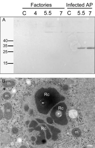
Our proteomic assays were supported by examination of two
viral proteins with known localizations. Specifically, our MS
anal-yses (see Table S1 in the supplemental material) revealed that the
L410 core protein was included in the VFs. This observation was
confirmed by Western blot analyses (Fig. 2A
and
B) as well as by
immuno-transmission electron microscopy (immuno-TEM)
(Fig. 2C). In contrast, eukaryotic translation initiation factor
4E-like protein L496 is present only in the cytoplasm of infected cells
and not at any stage in the VFs (Fig. 3A). This finding is
corrobo-rated by
in situ
TEM studies that showed ribosomes surrounding
the VFs but not incorporated into these organelles (Fig. 3B). These
observations, along with the absence of conserved ribosomal
pro-teins in the VFs (http://www.proteomexchange.org/
[identifier
PXD004203]), imply that translation of viral proteins is carried
out in the host cytoplasm and not in the VFs. Notably, this finding
significantly reinforces the notion of a parallel between viral
fac-tories and eukaryotic nuclei.
Venn diagrams were used to visualize the dynamic nature of
the viral factories and to provide insights into the mimivirus
rep-lication cycle (Fig. 4; see also Tables S1 and S2 in the supplemental
material). A total of 303 mimivirus proteins were detected in viral
factories. Specifically, 201, 255, and 287 proteins were present in
viral factories purified at 4, 5.5, and 7 h p.i., respectively (Fig. 4A).
Only one protein was exclusive to the 4-h-p.i. replication centers
(L538; RNA helicase). Three unique proteins (R721 [chemotaxis
protein CheD], R548 [thioredoxin-like protein], and R841 [an
ankyrin repeat-containing protein]) were found in 5.5-h-p.i. viral
factories, whereas 36 unique proteins were found in 7-h-p.i. viral
factories. These included L540 and R563 (RNA helicases), L425
(capsid), R362 and R443 (thioredoxin domain-containing
pro-teins), L288 (lectin), L484 (ankyrin repeat-containing protein),
L293 (hydrolase), L593 (prolyl 4-hydroxylase), and 20
uncharac-terized proteins. The presence of five annotated structural
pro-teins (L410 [core], L71 and L668 [collagen propro-teins], R440
[cap-sid], and L725 [fibrils]) (35) at both 5.5 and 7 h p.i. is consistent
with previous observations according to which extensive assembly
processes of viral components such as membranes and capsid
gen-eration take place at these p.i. stages (1,
18). It was previously
demonstrated by diverse imaging techniques that membranes are
present in mature mimivirus virions (36) as well as in VFs (10,
37).
Here, 30 membrane proteins were detected in the purified viral
factories at the three p.i. time points (see Table S1). This
observa-FIG 2Isolated viral factories contain the core protein L410. (A) Western blots of total cell lysates at different postinfection time points were incubated with anti-core protein (L410) antibodies. L410 was detected only at later stages of infection, matching our mass spectroscopy findings. (B) Western blots of purified viral factories were incubated with anti-L410 antibodies. L410 was detected in viral factories, as well as in the virion, only at 7 h p.i., thus further substantiating our MS results. (C) Immunolabeling of cryopreserved cells at 8 h p.i. with rabbit anti-L410 antibodies, followed by exposure to gold-conjugated secondary antibody, revealed that the core protein is present in the viral factory as well as in the assembling virions. Scale bar: 1m.
Fridmann-Sirkis et al.
on November 7, 2019 by guest
http://jvi.asm.org/
[image:4.585.113.474.69.243.2]tion suggests that the membrane network remains associated with
the VFs following their isolation and purification.
We found that 177 proteins were common to viral factories at
all three time points (Fig. 4A). This result implies that multiple
infection processes occur continuously during the infection cycle.
Proteins detected throughout the infection cycle include enzymes
involved in DNA replication and transcription, DNA repair, and
protein degradation (Fig. 5), as well as in redox processes and
ankyrin repeat-containing proteins. The presence of multiple viral
DNA replication and transcription enzymes in the cytoplasmic
factories is consistent with continuous generation of new viral
genomes and proteins and corroborates the finding that
mimivi-rus infection occurs exclusively in the host cytoplasm (5,
21,
26,
34). This notion is further supported by the observation that as
many as 14 different putative helicases (of 15 encoded helicases)
are found in the viral factories. Notably, whereas some helicases
were present in factories generated at all of the time points probed,
others were detected only in earlier or later infection stages (Fig.
5A; see also Tables S1 and S2 in the supplemental material),
im-plying that the large number of helicases does not indicate
redun-dancy but instead indicates specific roles required at different
in-fection stages.
Although the mimivirus virion proteome has already been
re-ported (28,
29), we were interested to reexamine the composition
of mature virions by applying mass spectrometry procedures
identical to those used for examination of the factories. A total of
236 mimivirus proteins were detected in mature viral particles
(see Tables S1 and S2 in the supplemental material) in contrast to
the 114 reported in reference
29, a discrepancy likely due to the
higher sensitivity of the instrumentation used in this study.
Com-parison of the protein content of mature virions to the protein
content of 4-h VFs (Fig. 4B) revealed that 139 proteins are shared,
including six helicases and seven transcription factors (see Tables
S1 and S2), supporting the notion that massive replication and
transcription processes occur already at this stage (26). In
com-parisons of the protein content of 7-h viral factories to that of
mature virions, 213 proteins were found to be common (Fig. 4C).
FIG 3Eukaryotic translation initiation factor 4E-like protein L496 and ribo-somes are not incorporated into the viral factories. (A) Western blots of total cell lysates at different postinfection time points were incubated with anti-4E-like protein (L496) antibodies. L496 was detected only in the total cell lysates and not in the factories, as indeed indicated by our MS findings. (B) TEM of intracellular replication centers at 4 h p.i. in the process of coalescing into a single viral factory. The micrograph reveals ribosomes (dark dots with diam-eters of⬃25 to⬃30 nm) that surround the replication centers but are not incorporated into these organelles. Rc and m, replication centers and mito-chondria, respectively. Asterisks represent the cores of the original infecting virus (26). Scale bar: 500 nm.
FIG 4Venn diagrams depicting protein content in mimivirus factories and in mature virions. (A) Proteins detected in isolated viral factories at 4, 5.5, and 7 h p.i. (blue, yellow, and green circles, respectively). The diagram demonstrates that the compositions of viral factories along the infection cycle differ substan-tially, thus underscoring the dynamic nature of these virus-generated organ-elles. (B) Relations between proteins detected in mature virions and proteins present in viral factories at 4 h (magenta and blue circles, respectively). (C) Relations between proteins detected in viral factories at 7 h and proteins found in mature virions (green and magenta circles, respectively). This diagram re-veals that many proteins were present in the factories but not included in mature virions, implying that these proteins act as part of a production line, on par with the notion that viral factories represent intracellular organelles where vigorous viral assembly occurs.
on November 7, 2019 by guest
http://jvi.asm.org/
[image:5.585.81.247.63.321.2] [image:5.585.336.505.65.440.2]There were 13 proteins in the virion that were not detected in the
factories at all stages investigated here. Three were
oxidation-re-lated proteins (R135, R419, and L894), one was an ankyrin
repeat-containing protein (L371), and eight were uncharacterized (see
Table S2). The comparison also demonstrated that the number of
proteins present in the factory is larger than the number detected
in mature virions. This finding supports the conjecture that VFs
should be considered “production lines” of viruses (10,
18) in
which numerous proteins are required for virus production but
not for the onset of the infection process.
Notably, 23 proteins, including R135, a putative
glucose-meth-anol-choline (GMC)-type oxidoreductase shown to be a
compo-nent of mimivirus fibrils (35), were detected in virion particles but
not in our MS studies of the relatively late 7-h VFs (Fig. 4C; see also
Tables S1 and S2 in the supplemental material). In contrast,
anti-bodies raised against fibril-containing virions did interact with
7-h factories (Fig. 1C), implying the presence of R135 in these
factories. A possible interpretation is that R135, which was shown
to be glycosylated (35), may undergo glycosylation on host
mem-branes outside the VFs and may only then be supplemented to the
assembling virions. Another interpretation could be that some
proteins are present in the VFs in small amounts and are therefore
undetectable.
In addition to depicting the highly dynamic distribution of
DNA processing enzymes such as representative helicases, DNA
repair proteins, and DNA-replication-related proteins in factories
from various p.i. times (Fig. 5A,
B, and
C, respectively), the results
shown in
Fig. 5D
imply that significant protein regulation occurs
at the protein level. Specifically, 11 proteins predicted to be
in-volved in protein degradation were detected in the viral factories
as well as in mature virions, indicating that protein degradation
processes are taking place throughout mimivirus infection cycle.
The levels of these proteins from 4-h to 7-h factories differ,
how-ever. Thus, nine degradation-related proteins were found in 4-h
FIG 5Dynamic composition of the mimivirus cytoplasmic factories. (A1 to A3) Distribution of three helicases in viral factories and mature viral particles. (B1 to B3) Distribution of DNA repair proteins in viral factories and in mature viral particles. (C1 to C3) Distribution of DNA-replication-related proteins. (D1 to D3) Distribution of protein-degradation-related proteins. Intensities represent relative protein amounts. The figure highlights the fact that the relative amounts of proteins belonging to similar functional groups in viral factories substantially differ at progressive postinfection time points, as well as in mature virions.
Fridmann-Sirkis et al.
on November 7, 2019 by guest
http://jvi.asm.org/
[image:6.585.42.542.62.470.2]factories, eight were detected in 5.5-h factories, and all 11 enzymes
were present at 7 h. Six degradation-related proteins were present
in mature virions, hinting that degradation of host and viral
pro-teins is already required at the onset of the infection, as well as
throughout the process. The possibility that protein degradation
also occurs in mature virions is intriguing and will be examined.
Genes encoding ankyrin repeat-containing proteins were
pre-viously shown to be the largest group in the mimivirus genome,
with 66 members (38). The ankyrin repeat is 33 amino acids long,
appearing in proteins involved in multiple cellular tasks such as
protein-protein interactions, cytoskeleton generation, and cell
signaling. We manually reannotated the list of ankyrin
repeat-containing proteins in the genome of mimivirus (NC_014649.1)
and found 98 proteins, of which only 18 were found in the
mimi-virus factories; 13 were present from 4 h to 7 h in factories but only
1 in mature virions (see Tables S1 and S2 in the supplemental
material). This observation implies functions carried out mainly
outside the factories. The latter conjecture is supported by the
previous findings indicating that ankyrin repeat-containing
pro-teins in vaccinia virus (39) and in Paramecium bursaria chlorella
virus 1 (PBCV-1) virus (40) are involved in ubiquitination of host
proteins in the host cytoplasm. This finding substantiates the
no-tion suggested above that protein regulano-tion through protein
deg-radation is a crucial process throughout viral infection both inside
and outside the viral factories.
The majority of proteins in the viral factories are encoded by
the central part of the mimivirus genome.
We have mapped the
distribution of proteins detected in mimivirus factories at three
successive time points, as well as in mature virions, on the
mimi-virus genome (Fig. 6). Notably, we found that the majority of these
proteins were encoded by the central part of the genome, whereas
the extremities of the mimivirus genome genes were detected to a
markedly lesser degree. This observation corroborates previous
studies where mimivirus virions propagated under axenic
condi-FIG 6Genomic organization of proteins detected in viral factories and mature virions. Our proteomic analyses indicate that the majority of proteins found in the viral factories of mimivirus are encoded by genes located in the center of the mimivirus chromosome. This observation is consistent with previous findings according to which mimivirus virions propagated under axenic conditions undergo substantial reduction of their genome that specifically occurs at the extremities of the genome and yet is not detrimental to mimivirus infectivity (35). CDSs (coding DNA sequences) are represented as blue arrowheads. Proteins detected in VFs at 4, 5.5, and 7 h p.i. are labeled in blue, purple, and maroon rectangles, respectively. Proteins found in mature mimivirus particles are labeled in green. The linear mimivirus genome is depicted as a circle starting from the site indicated by an arrow. The figure was constructed with the CGView Comparison Tool (CCT) (46).
on November 7, 2019 by guest
http://jvi.asm.org/
[image:7.585.113.475.66.442.2]tions revealed that a substantial reduction of their genome
specif-ically occurred at the edges of the genome (35). It was proposed
that these regions encode proteins involved in control of
compet-itor and hence are redundant under axenic conditions (35). In
addition, it has been demonstrated that the central region of
pox-virus genomes is highly conserved whereas genes at the terminal
regions are more divergent (41,
42). The termini of the genome of
African swine fever virus (ASFV), which also belongs to the
NCLDV family, are similarly variable in different isolates (43).
Proteome studies of large viruses, including Cafeteria
roenber-gensis virus (CroV) (44) and PBCV-1 virions (45), were
con-ducted. The large number of proteins in PBCV-1 raised the issue
of why giant viruses contain so many genes (45). We propose that
a partial answer is provided by the current report, which
high-lights the elaborate and highly dynamic protein composition of
VFs. In addition, the issue of duplicated genes and genomic
re-gions in the mimivirus was raised (38). Our results imply that
proteins that seem to have similar functions actually have different
expression levels at various stages of infection, implying specificity
rather than redundancy. Still, only about 300 proteins are found in
the factories, and 200 of those proteins are uncharacterized; the
roles of these proteins as well as of the remaining
⬃
700 mimivirus
proteins need to be elucidated. Finally, we claim that the
proce-dure reported here for the isolation of mimivirus factories and
their proteomic analyses, along with the fact that this
methodol-ogy can be extended to other large dsDNA and RNA viruses, may
provide new insights into the pathway of viral infection as well as
into intracellular self-assembly processes in general.
ACKNOWLEDGMENTS
We thank Eyal Shimoni and Tamar Unger for helpful discussions. Y.F.-S., E.M., and A.M. designed the research; Y.F.-S., E.M., Y.M., S.B.-D., Y.L., A.S., and E.K. and conducted the experiments; Y.F.-S., E.M., Y.M., S.B.-D., Y.L., A.S., and A.M. analyzed the data; Y.F.-S. and A.M. wrote the paper.
FUNDING INFORMATION
This work, including the efforts of Abraham Minsky, was funded by Israel Science Foundation (ISF) (813/14). This work, including the efforts of Abraham Minsky, was funded by Minerva Foundation (Minerva Stiftung) (910217).
REFERENCES
1.de Castro IF, Volonte L, Risco C.2013. Virus factories: biogenesis and structural design. Cell Microbiol15:24 –34.
2.den Boon JA, Ahlquist P.2010. Organelle-like membrane compartmen-talization of positive-strand RNA virus replication factories. Annu Rev Microbiol64:241–256.http://dx.doi.org/10.1146/annurev.micro.112408 .134012.
3.Netherton C, Wileman T.2011. Virus factories, double membrane vesi-cles and viroplasm generated in animal cells. Curr Opin Virol1:381–387. http://dx.doi.org/10.1016/j.coviro.2011.09.008.
4.Novoa RR, Calderita G, Arranz R, Fontana J, Granzow H, Risco C.
2005. Virus factories: associations of cell organelles for viral replication and morphogenesis. Biol Cell97:147–172.http://dx.doi.org/10.1042 /BC20040058.
5.Abergel C, Legendre M, Claverie JM.2015. The rapidly expanding uni-verse of giant viruses: Mimivirus, Pandoravirus, Pithovirus and Mollivi-rus. FEMS Microbiol Rev39:779 –796.http://dx.doi.org/10.1093/femsre /fuv037.
6.Iyer LA, Balaji S, Koonin EV, Aravind L.2006. Evolutionary genomics of nucleo-cytoplasmic large DNA viruses. Virus Res117:156 –184.http://dx .doi.org/10.1016/j.virusres.2006.01.009.
7.Iyer LM, Aravind L, Koonin EV.2001. Common origin of four diverse
families of large eukaryotic DNA viruses. J Virol75:11720 –11734.http: //dx.doi.org/10.1128/JVI.75.23.11720-11734.2001.
8.Katsafanas GC, Moss B.2007. Colocalization of transcription and trans-lation within cytoplasmic poxvirus factories coordinates viral expression and subjugates host functions. Cell Host Microbe2:221–228.http://dx.doi .org/10.1016/j.chom.2007.08.005.
9.Milrot E, Mutsafi Y, Fridmann-Sirkis Y, Shimoni E, Rechav K, Gurnon J, Van Etten JL, Minsky A.2016. Virus-host interactions: insights from the replication cycle of the largeParamecium bursaria chlorellavirus. Cell Microbiol18:3–16.http://dx.doi.org/10.1111/cmi.12486.
10. Mutsafi Y, Shimoni E, Shimon A, Minsky A.2013. Membrane assembly during the infection cycle of the giant mimivirus. PLoS Pathog
9:e1003367.http://dx.doi.org/10.1371/journal.ppat.1003367.
11. Tolonen N, Doglio L, Schleich S, Krijnse Locker J.2001. Vaccinia virus DNA replication occurs in endoplasmic reticulum-enclosed cytoplasmic mini-nuclei. Mol Biol Cell12:2031–2046.http://dx.doi.org/10.1091/mbc .12.7.2031.
12. Van Etten JL, Lane LC, Dunigan DD.2010. DNA viruses: The really big ones (giruses). Annu Rev Microbiol64:83–99.http://dx.doi.org/10.1146 /annurev.micro.112408.134338.
13. Bell PJL.2001. Viral eukaryogenesis: was the ancestor of the nucleus a complex DNA virus? J Mol Evol53:251–256.http://dx.doi.org/10.1007 /s002390010215.
14. Bell PJL.2006. Sex and the eukaryotic cell cycle is consistent with a viral ancestry for the eukaryotic nucleus. J Theor Biol243:54 – 63.http://dx.doi .org/10.1016/j.jtbi.2006.05.015.
15. Takemura M.2001. Poxviruses and the origin of the eukaryotic nucleus. J Mol Evol52:419 – 425.
16. Forterre P.2006. The origin of viruses and their possible roles in major evolutionary transitions. Virus Res117:5–16.http://dx.doi.org/10.1016/j .virusres.2006.01.010.
17. Forterre P, Prangishvili D.2013. The major role of viruses in cellular evolution: facts and hypotheses. Curr Opin Virol3:558 –565.http://dx.doi .org/10.1016/j.coviro.2013.06.013.
18. Mutsafi Y, Fridmann-Sirkis Y, Milrot E, Hevroni L, Minsky A.2014. Infection cycles of large DNA viruses: emerging themes and underlying questions. Virology466:3–14.
19. Meaburn KJ, Misteli T.2007. Chromosome territories. Nature445:379 – 381.http://dx.doi.org/10.1038/445379a.
20. Misteli T.2004. Spatial positioning: a new dimension in genome function. Cell119:153–156.http://dx.doi.org/10.1016/j.cell.2004.09.035. 21. Claverie JM, Abergel C.2009. Mimivirus and its virophage. Annu Rev Genet
43:49 – 66.http://dx.doi.org/10.1146/annurev-genet-102108-134255. 22. Claverie JM, Abergel C.2010. Mimivirus: the emerging paradox of
quasi-autonomous viruses. Trends Genet 26:431– 437. http://dx.doi.org/10 .1016/j.tig.2010.07.003.
23. Legendre M, Bartoli J, Shmakova L, Jeudy S, Labadie K, Adrait A, Lescot M, Poirot O, Bertaux L, Bruley C, Coute Y, Rivkina E, Abergel C, Claverie JM.2014. Thirty-thousand-year-old distant relative of giant icosahedral DNA viruses with a pandoravirus morphology. Proc Natl Acad Sci U S A 111:4274 – 4279. http://dx.doi.org/10.1073/pnas .1320670111.
24. Philippe N, Legendre M, Doutre G, Coute Y, Poirot O, Lescot M, Arslan D, Seltzer V, Bertaux L, Bruley C, Garin J, Claverie JM, Abergel C.2013. Pandoraviruses: amoeba viruses with genomes up to 2.5 Mb reaching that of parasitic eukaryotes. Science341:281–286.http://dx.doi .org/10.1126/science.1239181.
25. Raoult D, Audic S, Robert C, Abergel C, Renesto P, Ogata H, La Scola B, Suzan M, Claverie JM.2004. The 1.2-megabase genome sequence of mimivirus. Science 306:1344 –1350. http://dx.doi.org/10.1126/science .1101485.
26. Mutsafi Y, Zauberman N, Sabanay I, Minsky A. 2010. Vaccinia-like cytoplasmic replication of the giant mimivirus. Proc Natl Acad Sci U S A
107:5978 –5982.http://dx.doi.org/10.1073/pnas.0912737107.
27. Mallardo M, Leithe E, Schleich S, Roos N, Doglio L, Krijnse Locker J.2002. Relationship between vaccinia virus intracellular cores, early mRNAs, and DNA replication sites. J Virol76:5167–5183.http://dx.doi.org/10.1128/JVI.76 .10.5167-5183.2002.
28. Claverie JM, Abergel C, Ogata H.2009. Mimivirus, p 89 –121.InVan Etten JL (ed), Lesser known large dsDNA viruses, vol 328. Springer-Verlag, Berlin, Germany.
29. Renesto P, Abergel C, Decloquement P, Moinier D, Azza S, Ogata H, Fourquet P, Gorvel JP, Claverie JM.2006. Mimivirus giant particles
Fridmann-Sirkis et al.
on November 7, 2019 by guest
http://jvi.asm.org/
incorporate a large fraction of anonymous and unique gene products. J Virol80:11678 –11685.http://dx.doi.org/10.1128/JVI.00940-06. 30. La Scola B, Audic S, Robert C, Jungang L, de Lamballerie X, Drancourt
M, Birtles R, Claverie JM, Raoult D.2003. A giant virus in amoebae. Science299:2033.http://dx.doi.org/10.1126/science.1081867.
31. Kelstrup CD, Young C, Lavallee R, Nielsen ML, Olsen JV. 2012. Optimized fast and sensitive acquisition methods for shotgun proteomics on a quadrupole orbitrap mass spectrometer. J Proteome Res11:3487– 3497.http://dx.doi.org/10.1021/pr3000249.
32. Shalit T, Elinger D, Savidor A, Gabashvili A, Levin Y.2015. MS1-Based label-free proteomics using a quadrupole orbitrap mass spectrometer. J Proteome Res14:1979 –1986.http://dx.doi.org/10.1021/pr501045t. 33. Keller A, Nesvizhskii AI, Kolker E, Aebersold R.2002. Empirical
statis-tical model to estimate the accuracy of peptide identifications made by MS/MS and database search. Anal Chem74:5383–5392.http://dx.doi.org /10.1021/ac025747h.
34. Zauberman N, Mutsafi Y, Ben Halevy D, Shimoni E, Klein E, Xiao C, Sun S, Minsky A.2008. Distinct DNA exit and packaging portals in the virus Acanthamoeba polyphaga mimivirus. PLoS Biol6:e114.http://dx .doi.org/10.1371/journal.pbio.0060114.
35. Boyer M, Azza S, Barrassi L, Klose T, Campocasso A, Pagnier I, Fournous G, Borg A, Robert C, Zhang XZ, Desnues C, Henrissat B, Rossmann MG, La Scola B, Raoult D.2011. Mimivirus shows dramatic genome reduction after intraamoebal culture. Proc Natl Acad Sci U S A
108:10296 –10301.http://dx.doi.org/10.1073/pnas.1101118108. 36. Xiao CA, Chipman PR, Battisti AJ, Bowman VD, Renesto P, Raoult D,
Rossmann MG.2005. Cryo-electron microscopy of the giant mimivirus. J Mol Biol353:493– 496.http://dx.doi.org/10.1016/j.jmb.2005.08.060. 37. Suárez C, Welsch S, Chlanda P, Hagen W, Hoppe S, Kolovou A,
Pagnier I, Raoult D, Krijnse Locker J.2013. Open membranes are the precursors for assembly of large DNA viruses. Cell Microbiol15:1883– 1895.
38. Suhre K. 2005. Gene and genome duplication in Acanthamoeba
polyphaga mimivirus. J Virol79:14095–14101.http://dx.doi.org/10.1128 /JVI.79.22.14095-14101.2005.
39. Sonnberg S, Seet BT, Pawson T, Fleming SB, Mercer AA.2008. Poxvirus ankyrin repeat proteins are a unique class of F-box proteins that associate with cellular SCF1 ubiquitin ligase complexes. Proc Natl Acad Sci U S A
105:10955–10960.http://dx.doi.org/10.1073/pnas.0802042105. 40. Noel EA, Kang M, Adamec J, Van Etten JL, Oyler GA.2014. Chlorovirus
Skp1-binding ankyrin repeat protein interplay and mimicry of cellular ubiquitin ligase machinery. J Virol88:13798 –13810.http://dx.doi.org/10 .1128/JVI.02109-14.
41. Gubser C, Hué S, Kellam P, Smith GL. 2004. Poxvirus genome: a phylogenetic analysis. J Gen Virol85:105–117.http://dx.doi.org/10.1099 /vir.0.19565-0.
42. Moss B.2006.Poxviridae: the viruses and their replication, p 2905–2945.
InKnipe DM, Howley PM, Griffin DE, Lamb RA, Martin MA, Roizman B, Straus SE (ed), Fields virology, 5th ed. Lippincott Williams & Wilkins, Philadelphia, PA.
43. Dixon LK, Chapman DAG, Netherton CL, Upton C.2013. African swine fever virus replication and genomics. Virus Res173:3–14.http://dx.doi .org/10.1016/j.virusres.2012.10.020.
44. Fischer MG, Kelly I, Foster LJ, Suttle CA.2014. The virion of Cafeteria roenbergensis virus (CroV) contains a complex suite of proteins for tran-scription and DNA repair. Virology466:82–94.
45. Dunigan DD, Cerny RL, Bauman AT, Roach JC, Lane LC, Agarkova IV, Wulser K, Yanai-Balser GM, Gurnon JR, Vitek JC, Kronschnabel BJ, Jeanniard A, Blanc G, Upton C, Duncan GA, McClung OW, Ma F, Van Etten JL.2012. Paramecium bursaria chlorella virus 1 proteome reveals novel architectural and regulatory features of a giant virus. J Virol86:
8821– 8834.http://dx.doi.org/10.1128/JVI.00907-12.
46. Grant JR, Arantes AS, Stothard P.2012. Comparing thousands of circu-lar genomes using the CGView Comparison Tool. BMC Genomics13:202. http://dx.doi.org/10.1186/1471-2164-13-202.
on November 7, 2019 by guest
http://jvi.asm.org/
![FIG 1 Mimivirus factories within host cells and following isolation. (A to C) Amoeba cells infected with mimivirus at 3 successive postinfection (p.i.) time points(4 h [A], 5.5 h [B], and 7 h [C]) were stained with antibodies against mature virions (red) a](https://thumb-us.123doks.com/thumbv2/123dok_us/141659.18803/3.585.113.473.65.426/mimivirus-factories-following-isolation-mimivirus-successive-postinfection-antibodies.webp)