Nonstructural Proteins NS1 and NS2 during Natural Cell Infection
Identifies PACT as an NS1 Target Protein and Antiviral Host Factor
Kazuki Tawaratsumida,aVan Phan,a*Eike R. Hrincius,aAnthony A. High,bRichard Webby,aVanessa Redecke,aHans Häckera
Department of Infectious Diseases, St. Jude Children’s Research Hospital, Memphis, Tennessee, USAa
; Proteomics Core Facility, St. Jude Children’s Research Hospital, Memphis, Tennessee, USAb
ABSTRACT
Influenza A virus (IAV) replication depends on the interaction of virus proteins with host factors. The viral nonstructural
pro-tein 1 (NS1) is essential in this process by targeting diverse cellular functions, including mRNA splicing and translation, cell
sur-vival, and immune defense, in particular the type I interferon (IFN-I) response. In order to identify host proteins targeted by
NS1, we established a replication-competent recombinant IAV that expresses epitope-tagged forms of NS1 and NS2, which are
encoded by the same gene segment, allowing purification of NS proteins during natural cell infection and analysis of interacting
proteins by quantitative mass spectrometry. We identified known NS1- and NS2-interacting proteins but also uncharacterized
proteins, including PACT, an important cofactor for the IFN-I response triggered by the viral RNA-sensor RIG-I. We show here
that NS1 binds PACT during virus replication and blocks PACT/RIG-I-mediated activation of IFN-I, which represents a critical
event for the host defense. Protein interaction and interference with IFN-I activation depended on the functional integrity of the
highly conserved RNA binding domain of NS1. A mutant virus with deletion of NS1 induced high levels of IFN-I in control cells,
as expected; in contrast, shRNA-mediated knockdown of PACT compromised IFN-I activation by the mutant virus, but not
wild-type virus, a finding consistent with the interpretation that PACT (i) is essential for IAV recognition and (ii) is functionally
com-promised by NS1. Together, our data describe a novel approach to identify virus-host protein interactions and demonstrate that
NS1 interferes with PACT, whose function is critical for robust IFN-I production.
IMPORTANCE
Influenza A virus (IAV) is an important human pathogen that is responsible for annual epidemics and occasional devastating
pandemics. Viral replication and pathogenicity depends on the interference of viral factors with components of the host defense
system, particularly the type I interferon (IFN-I) response. The viral NS1 protein is known to counteract virus recognition and
IFN-I production, but the molecular mechanism is only partially defined. We used a novel proteomic approach to identify host
proteins that are bound by NS1 during virus replication and identified the protein PACT, which had previously been shown to
be involved in virus-mediated IFN-I activation. We find that NS1 prevents PACT from interacting with an essential component
of the virus recognition pathway, RIG-I, thereby disabling efficient IFN-I production. These observations provide an important
piece of information on how IAV efficiently counteracts the host immune defense.
I
nfluenza virus replication depends on interactions between
vi-rus-encoded proteins and host factors, controlling key aspects of
host and virus biology, including cell survival, virus production,
and the inhibition of virus recognition and host defense (
1
). The
latter aspect of virus host interaction is highlighted by partially
characterized functions of the IAV nonstructural protein 1 (NS1),
which is essential for virus pathogenicity (
2–4
). During the last
several decades, various cellular NS1 binding partners have been
identified, illustrating protein-protein and protein-RNA
interac-tions as modes of NS1 function (
1
). While an important inhibitory
role of NS1 in IFN-I activation has been established for more than
10 years (
4
), more recent work has started to unravel the
molecu-lar mechanisms involved. Initially, the RNA binding property of
NS1 has been described as a mechanism to prevent viral RNA
from recognition by components of the cellular host defense
sys-tem, including the interferon-induced, double-stranded RNA
(dsRNA)-activated protein kinase (PKR) and the 2
=
-5
=
oligo(A)
synthetase (OAS)/RNase L pathway (
5
,
6
). NS1 has also been
shown to interfere with virus recognition via the pattern
recogni-tion receptor RIG-I, which activates several immune signaling
pathways upon viral RNA recognition, including the type I
inter-feron (IFN-I) pathway that mediates antiviral resistance (
7
).
Spe-cifically, NS1 has been described to interact and interfere directly
with the ubiquitin ligases TRIM25 and RIPLET, which catalyze
the formation of nondegrading polyubiquitin chains that are
re-quired for RIG-I activation and recruitment of the downstream
effector protein MAVS (
8–10
). Still, it is not entirely clear whether
Received20 March 2014Accepted24 May 2014
Published ahead of print4 June 2014
Editor:S. Perlman
Address correspondence to Hans Häcker, [email protected].
* Present address: Van Phan, 583 Ridge Springs Rd., Collierville, Tennessee, USA.
K.T. and V.P. contributed equally to this article.
Supplemental material for this article may be found athttp://dx.doi.org/10.1128 /JVI.00830-14.
Copyright © 2014, American Society for Microbiology. All Rights Reserved.
doi:10.1128/JVI.00830-14
on November 7, 2019 by guest
http://jvi.asm.org/
these functions of NS1 are solely responsible for its inhibitory role
in the IFN-I pathway. One protein that has recently been shown to
increase RIG-I activity independent of viral RNA is the protein
activator of the interferon-induced protein kinase (PACT), which
was originally cloned as a binding partner and activator of PKR, an
IFN-inducible kinase with established antiviral function (
11–13
).
PACT has also been shown to be required for optimal activity of
Dicer, a nuclease required for RNA interference (
14
,
15
).
Al-though the precise mechanism of PACT–RIG-I interaction and
function requires further investigation, gain- and loss-of-function
experiments established a critical function for PACT in RIG-I
ac-tivation (
15
). Very recently, two viral proteins, the herpes simplex
virus (HSV) protein Us11 and the Ebola virus protein VP35 were
shown to interact with PACT and counteract PACT-mediated
IFN activation (
16
,
17
), supporting the idea that PACT-mediated
IFN-I activation is a critical part of the antiviral host defense.
In order to identify novel factors involved in IAV biology, we
devised a proteomic strategy to analyze protein interactions of
NS1 and NS2 during physiological virus replication. While such a
strategy has been used for other viruses, IAV have been difficult to
approach due to their specific genome packaging mechanism and
space constraints. Individual RNA segments of IAV are packed
into RNA/protein complexes, where the first approximately 100
bp serve at least two purposes, i.e., as packaging signal and as
coding sequence (
18
). Thus, insertion of epitope tags for protein
purification at the 5
=
or 3
=
end interferes with genome packaging.
In order to maintain both functions and allow fusion of an epitope
tag for efficient protein purification, we took advantage of the 2A
peptide of the porcine teschovirus-1 (P2A), which mediates
ribo-somal skipping (
19
), allowing duplication of the first 90 bp of the
NS gene segment, thereby functionally separating genome
pack-aging and protein translation (for details, see
Fig. 1A
) (
18
). Using
a recombinant virus based on this technology, followed by cell
infection and quantitative proteomic analysis of
NS1-/NS2-inter-acting proteins, we identified many known NS1-/NS2-interNS1-/NS2-inter-acting
proteins, as expected, but also novel factors, including PACT.
Here, we describe in detail our strategy to investigate virus-host
protein interactions during physiological virus replication and
also characterize PACT function during IAV infection,
demon-strating that PACT is a critical factor for IAV recognition that is
specifically targeted by NS1 to counteract RIG-I-mediated IFN-I
activation.
MATERIALS AND METHODS
Reagents.Antibodies were used against FLAG (M2 [soluble and bead immobilized]; Sigma-Aldrich), PACT (Cell Signaling), NS1 (mouse monoclonal), clone 18/1 (20), and rabbit polyclonal (GeneTex; used in Fig. 3D only), hemagglutinin (HA; bead immobilized, clone 3F10; Roche), NS2 (GeneTex), matrix protein (mouse monoclonal, clone M2-IC6 [21]), nucleoprotein (GeneTex), and p38 (Santa Cruz).
Plasmids and generation of recombinant virus.For immunoprecipi-tation (IP) experiments, FS-tagged NS2 was generated by cloning the full-length coding region of NS2 3-prime of a FS epitope tag as described inFig. 1, contained in a mammalian expression vector with EF1␣ -pro-moter. The same strategy was used for generation of FS-NS1; however, four translationally silent mutations preventing aberrant splicing that tar-get splice donor (G30A, A33T), branch point (A483T), and splice accep-tor (A501T) sites were introduced by PCR. A bacterial expression vecaccep-tor for His6-tagged PR8-NS1 was generated by cloning the coding sequence of
PR8-NS1 containing described splicing-disabling point mutations C-ter-minal of a His6epitope tag into pQE-31 (Qiagen). Sequences of all
plas-mids were verified by DNA sequencing. A His6-tagged form of
TAT-HA-green fluorescent protein (GFP) was kindly provided by Stephen Dowdy (University of California, San Diego) (22). The cDNA of PACT was ob-tained from Addgene (catalog no. 15667) (23) and cloned 3 prime of a triple HA-epitope tag contained in a pcDNA3-based vector (Invitrogen). The pEF-BOS-based expression vector for F-RIG-I was kindly provided by T. Fujita. The recombinant PR8 viruses used (PR8-wt, PR8-NS-FS, and PR8⌬NS1) were rescued by reverse genetics as described previously based on plasmids encoding the gene segments of influenza virus A/Puerto Rico/ 8/34 (PR8) (24). Modifications in the sequence of NS1 (for PR8-NS-FS detailed inFig. 1A) were introduced by standard molecular biology tech-niques and verified by DNA sequencing. For PR8⌬NS1, the sequence encoding AS11-230 was deleted, resulting in one remaining open reading frame encoding NS2, as described previously (4). A high virus titer was produced on Madin-Darby canine kidney (MDCK) cells for proteomic experiments. In experiments where PR8⌬NS1 was used, viruses were grown on African green monkey kidney (Vero) cells.
Virus titration.Virus titration of PR8-wt and PR8-NS-FS was done by limiting dilution on MDCK cells, followed by an HA inhibition assay. Where PR8⌬NS1 was used, the virus titers were determined by intracel-lular staining in HEK293T cells. To this end, HEK293T cells were seeded on poly-L-lysine-coated 96-well plates (4⫻104cells/50l of infection medium) and infected with 20l of virus-containing supernatants (by limiting dilution) for 2 h at 37°C, followed by the addition of 70l of medium (DMEM) containing 20% fetal bovine serum (FBS) and incuba-tion at 37°C for another 22 h. Intracellular virus staining was performed on plate by removing the supernatant, washing with phosphate-buffered saline (PBS), fixing the cells with 2% formaldehyde in PBS for precisely 20 min at 25°C, repeated washing with PBS–1% FBS, and staining with bio-tin-conjugated anti-H1N1 antibody (I7650-05G, US Biological) in PBS containing 1% FBS and 0.5% saponin (PBS-Sap; Sigma-Aldrich) for 20 min at 25°C. After the antiviral antibody was removed, the cells were incubated with streptavidin-phycoerythrin (BD Biosciences) in PBS-Sap for 20 min at 25°C. The cells were washed twice with PBS-Sap and once with PBS, followed by treatment with 30l of trypsin-EDTA (Invitrogen) for 3 min at 37°C, resuspension in 170l of PBS containing 30% FBS, and flow cytometry.
Cell culture and generation of shRNA knockdown cell lines.
HEK293T, A549, MDCK, and Vero cells were cultured in DMEM high glucose (Invitrogen) supplemented with 10% FBS, 50 mM 2-mercapto-ethanol (2-ME), antibiotics (penicillin G at 100 IU/ml and streptomycin sulfate at 100 IU/ml [P/S]), and pyruvate (1 mM). Cell infections were performed in infection medium {DMEM high glucose (Invitrogen) sup-plemented with 2-ME, P/S, 0.1% bovine serum albumin, and TPCK [L -(tosylamido-2-phenyl) ethyl chloromethyl ketone] trypsin at 1g/ml} after the cells were washed twice with PBS.
Retroviral shRNA plasmids for control and humanPACTwere pur-chased from Transomix (TRH1000 (RLGH-GN50894 [PACT sh1], RLGH-GN50896 [PACT sh2]), and TRH1103 [sh ctrl]), and nonreplicat-ing retrovirus was produced in HEK293T cells by cotransfection of helper plasmids and VSV-G expression plasmid. Supernatant was directly used for infection of HEK293T cells, followed by puromycin selection of poly-clonal populations.
Tandem affinity purification.A549 cells (five 15-cm cell culture dishes) were infected with PR8-wt or PR8-NS-FS at a multiplicity of in-fection (MOI) of 0.5 in inin-fection medium. After 1 h of incubation at 37°C, medium was replaced by regular cell growth medium, followed by incu-bation for 24 h at 37°C. Medium was replaced by ice-cold PBS and col-lected by cell scraping and centrifugation. Cell pellets were incubated with lysis buffer (LB; 20 mM HEPES/KOH [pH 7.5], 150 mM KCl, 1.5 mM MgCl, 1 mM EDTA, 10% glycerol, 1 mM orthovanadate, 10 mM -glyc-erophosphate, 5 mM 4-nitrophenyl-phosphate, 10 mM sodium fluoride, Complete protease inhibitors [Roche]) supplemented with 1% NP-40 for 20 min. Samples were sonicated with ten 3-s pulses (level 2) on a Virsonic 60 cell disruptor (Virtis) to disrupt nuclei and then cleared by
on November 7, 2019 by guest
http://jvi.asm.org/
tion. Cleared lysates were loaded five times over anti-FLAG-bead-con-taining columns. Unbound proteins were removed by washing column with LB plus 0.1% NP-40, and proteins were eluted in 1 ml of LB plus 0.1% NP-40 supplemented with 3⫻FLAG peptide (0.2 mg/ml; Sigma-Aldrich). Eluted proteins were loaded five times over a StrepTactin col-umn (IBA), unbound proteins were removed by washing the colcol-umn with LB plus 0.1% NP-40, and proteins were eluted in 0.9 ml of LB plus des-thiobiotin (5 mM; Sigma-Aldrich), followed by concentration using 5K NMWL spin columns (Millipore), separation on a 4 to 12% Bis-Tris gra-dient gel (Bio-Rad), and staining with SYPRO Ruby protein stain (Invit-rogen). Individual bands were cut, in-gel digested using trypsin, and an-alyzed by MALDI-TOF (matrix-assisted laser desorption ionization–time of flight) mass spectrometry (MS) or liquid chromatography-coupled mass spectrometry (LC-MS) using an electrospray ionization (ESI) cou-pled to a linear ion trap mass spectrometer (LTQ-XL; Thermo Fisher), as indicated in the figure legends.
One-step protein purification using SILAC-labeled cells.For stable isotope labeling by amino acids in cell culture (SILAC), HEK293T cells were cultured in arginine- and lysine-free DMEM (Invitrogen) supple-mented with 10% dialyzed FBS (Invitrogen), penicillin-streptomycin, and eitherL-arginine andL-lysine (light) orL-arginine-HCl (13C6; CLM-2265 [R6]) andL-lysine-2HCl (4,4,5,5 D4; DLM-2640 [K4]) (medium) (Cam-bridge Isotope Labs). For complete incorporation of labeled amino acids, cells were passaged three times in SILAC medium over a period of 5 days. The labeled HEK293T cells were infected with wt (light) or PR8-NS-FS (medium) at an MOI of 0.6 in infection medium. After 1 h of incubation at 37°C, the medium was replaced by SILAC medium, fol-lowed by incubation at 37°C for 24 h. The medium was replaced by ice-cold PBS and collected by cell scraping and centrifugation. Cell pellets were incubated with LB plus 1% NP-40 for 20 min. Samples were soni-cated with ten 3-s pulses (level 2) on a Virsonic 60 cell disruptor (Virtis) to disrupt nuclei and cleared by centrifugation. Cleared lysates were loaded five times over anti-FLAG-bead-containing columns. Unbound proteins were removed by washing column with LB plus 0.1% NP-40, and proteins were eluted at pH 3.5 in water supplemented with 100 mM glycine, 50 mM KCl, 0.1% NP-40, and Roche complete protease inhibitors. The proteins were concentrated by trichloroacetic acid precipitation, followed by sep-aration on a 4 to 12% Bis-Tris gradient gel (Bio-Rad) and staining with SYPRO Ruby protein stain (Invitrogen). The entire lane was cut into individual bands (seeFig. 2B) and analyzed by LC-tandem MS (LC-MS/ MS) using ESI coupled to an LTQ Orbitrap XL mass spectrometer (Thermo Fisher).
MALDI-TOF MS analysis.Proteins were reduced and alkylated with iodoacetamide, and a tryptic digest was prepared. MS analysis was performed using a model 4700 proteomics analyzer from Applied Biosys-tems. The digest was introduced into the instrument in a crystalline ma-trix of␣-cyano-4-hydroxycinnamic acid also containing 2 mM ammo-nium citrate to suppress ionization of matrix clusters. Database searches were performed using GPS explorer software (Applied Biosystems), which uses the Mascot search engine. Protein assignments were made on the basis of both MS and MS/MS spectra. The Swiss-Pro database (num-ber 090207) was used for protein identification.
Sample preparation and ESI LC.The protein gel bands were reduced and alkylated with iodoacetamide and digested with trypsin enzyme. The digest was introduced into the instrument via on-line chromatography using reversed-phase (C18) ultra-high-pressure LC on a nanoACQUITY
apparatus (Waters). The column used was a Waters BEHC18 with an inner diameter of 75m and a bed length of 10 cm. The particle size was 1.7m. Tryptic peptides were gradient eluted over a gradient (0 to 70% B for 60 min and 70 to 100% B for 10 min, where B⫽70% [vol/vol] acetonitrile, 0.2% formic acid) at a flow rate of 250 nl/min into the linear ion trap (LTQ-XL or LTQ Orbitrap XL) through a noncoated spray needle with voltage applied to the liquid junction.
MS/MS analysis with LTQ XL (Thermo Fisher) and database analy-sis.Data-dependent scanning was incorporated to select the 10 most abundant ions (one microscan per spectrum; precursor isolation width, 2.0 Da; 35% collision energy; 30-ms ion activation; 30-s exclusion dura-tion; 15-s repeat duradura-tion; and a repeat count of 2) from a full-scan mass spectrum for fragmentation by collision-activated dissociation. Database searches were performed using RAW files in combination with the Mascot search engine. Protein/peptide assignments were made on the basis MS/MS spectra. The NCBInr 20070908 database was used for protein identification. Product ions (b/y-type ions) were queried in an automated database search against a protein database (NCBInr 20070908 [5,454,477 sequences; 1,888,849,002 residues, all species]) by the Mascot search al-gorithm. The following residue modifications were allowed in the search: carbamidomethylation on cysteine (fixed modification) and oxidation on methionine (variable modification). Mascot was searched with a precur-sor ion tolerance of⫾1 Da and a fragment ion tolerance of⫾0.7 Da and using the automatic decoy database-searching tool in Mascot. The iden-tifications from the automated search were verified by manual inspection of the raw data.
MS/MS analysis with LTQ XL Orbitrap (Thermo Fisher) (for SILAC) and MaxQuant analysis.Data-dependent scanning was incorpo-rated to select the 20 most abundant ions (one microscan per spectrum; precursor isolation width, 2.0 Da; 35% collision energy; 30-ms ion acti-vation; 30-s exclusion duration; 15-s repeat duration; and a repeat count of 2) from a full-scan mass spectrum collected in the Orbitrap at a 60,000 resolution for fragmentation by collision-activated dissociation in the ion trap. Database searches are performed using the Andromeda search en-gine that is part of the MaxQuant software (version 1.2.0.34) developed at the Max Planck Institute (25). MaxQuant was also used to quantitate peptides and proteins and to provide ratios and generate a report in Excel format. Protein assignments were made on the basis of both MS and MS/MS spectra, whereas peptide quantitation was based solely on MS data.
Production of recombinant proteins.His6-tagged recombinant pro-teins were expressed inEscherichia colistrain BL21(DE3). Protein expres-sion was induced by 1 mM IPTG (isopropyl--D-thiogalactopyranoside) in Turbo Prime media (Athena Enzyme Systems) at 16°C for 16 h. Cells were lysed in MCAC-0 buffer (50 mM Tris-Cl [pH 7.2], 500 mM NaCl, 10% [wt/vol] glycerol) and lysed by passage through a microfluidizer. The lysate was cleared by centrifugation, and proteins were captured using nickel beads (Gold Biotechnology), followed by elution with imidazole and dialysis.
In vitrotranslation and pulldown experiments.In vitrotranslation and radioactive [35S]methionine labeling of PACT was performed using
the TNT SP6 high-yield wheat germ protein expression system (Promega) according to the manufacturer’s instructions. Pulldown assays were per-formed by incubating 5g of His-GFP or His-NS1 fusion proteins im-mobilized on Ni-NTA beads (Qiagen) within vitro-translated protein for 4 h at 4°C. Bead-immobilized proteins were washed five times with lysis buffer supplemented with 0.1% NP-40, and proteins were eluted with sample buffer (Bio-Rad). Proteins were separated by SDS-PAGE and in-cubated with Amplify Reagent (GE Healthcare) for signal amplification. Gels were dried, scanned by using a phosphorimager (Typhoon FLA 9500; GE Healthcare), and analyzed by using ImageQuant TL software (GE Healthcare).
Immunoblotting (IB) and IP.Cells were lysed in lysis buffer supple-mented with 0.5% NP-40 for 20 min. Lysates were precleared with Sep-harose beads (Sigma-Aldrich) for 1 h, followed by IP overnight using anti-Flag M2 resin (Sigma-Aldrich) or anti-HA affinity matrix (3F10; Roche). The immunoprecipitated samples or cell lysates were resolved by SDS-PAGE (Bio-Rad) and transferred onto nitrocellulose membranes. Membranes were probed with the indicated antibodies and visualized using enhanced chemiluminescence (Pierce) for detection.
Dual luciferase reporter assays.HEK293T cells were transfected with an IFN-promoter (⫺125) reporter (firefly luciferase) vector,Renilla
on November 7, 2019 by guest
http://jvi.asm.org/
luciferase vector (pRL-TK; Promega), and the indicated expression plas-mids using Lipofectamine 2000 (Invitrogen). At 24 h after transfection, the cells were lysed in passive lysis buffer (Promega). The luciferase activ-ity was determined using a dual-luciferase kit (Promega), and the firefly luciferase activity values were normalized to theRenillaluciferase activity.
IFN bioassay.To inactivate virus, 120l of virus-containing cell cul-ture supernatant was treated in 96-well plates by UV light (15 W, 302 nm, XX-15M UV bench lamp; Upland) for 10 min at a 6-cm distance, resulting in the reduction of IAV infectivity below the detection limit. UV-treated cell culture supernatant and recombinant human IFN-(hIFN-; PBL) in limiting dilution as a standard was added to Caki cells (kindly provided by L. Pfeffer) that were stably cotransduced with lentiviral vectors con-taining ISRE-firefly luciferase and EF1␣-Renillaluciferase cassettes. The luciferase activity was determined after 24 h using a dual-luciferase kit (Promega), and the firefly luciferase activity values were normalized to the
Renilla luciferase activity. IFN values were determined based on the hIFN-standard. The detection limit of this bioassay was 1.5 U/ml.
Statistical analysis.Data presented in reporter assays and IFN bioas-says are expressed as means⫾the standard deviations and were compared using the Studentttest. APvalue of⬍0.05 was considered significant.
RESULTS
Generation of recombinant IAV with epitope-tagged NS1 and
NS2.
In order to establish epitope-tagged forms of NS1 and NS2
that could be purified by different affinity purification strategies,
including tandem affinity purification (TAP), we established a
fusion TAP tag, consisting of a triple FLAG tag and a StrepOne tag,
here referred to as the FS tag. The coding sequence of this tag was
introduced into the influenza virus A/PR/8/34 (PR8) NS gene as
illustrated in
Fig. 1A
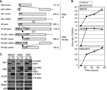
. The upper part of
Fig. 1A
depicts the NS gene
and the two mRNA splice variants encoding NS1 and NS2. To
introduce the FS tag at the N terminus of NS1/2, we duplicated 90
bp of the 5
=
coding region of the NS1 gene in order to avoid
interference with efficient vRNA packaging (
18
). To trigger
re-moval of these additional 30 amino acids from the mature NS1/2
proteins, we introduced downstream thereof the 2A peptide of the
porcine teschovirus-1 (P2A), which confers near-complete
ribo-somal “skipping” (
19
) that is functionally equivalent to cleavage
immediately upstream of the FS tag (
Fig. 1A
, lower part). This NS
gene (and in parallel the PR8 wild-type [PR8-wt] gene as control)
was used, along with the other PR8 gene segments to establish
recombinant virus by reverse genetics (
26
). The recombinant
vi-rus, referred to as PR8-NS-FS, displayed growth characteristics on
MDCK, A549, and HEK293T cells similar to those of the PR8-wt
virus (
Fig. 1B
), suggesting the functional activity of the
recombi-nant FS proteins. We infected the different cell lines with these
viruses and determined the expression of virus proteins by IB. NS1
and NS2 expression of both viruses was detected, with the
FS-FIG 1Generation of a recombinant PR8 virus with epitope-tagged NS1- and NS2 (PR8-NS-FS). (A) IAV NS gene arrangement and structure of recombinant PR8-NS-FS virus through the duplication of 90 bp of the 5=gene segment (hatched), followed by the 2A peptide of the porcine teschovirus-1 (P2A) and a tandem triple-FLAG-StrepOne epitope tag (FS). Different expected mRNA and protein products for PR8-wt (upper part) and PR8-NS-FS (lower part) are shown. Triangle, 2A-encoded “cleavage” site. (B) Replication of PR8-wt and PR8 NS-FS on MDCK, A549, and HEK293T cells was determined by infection at an MOI of 0.001 (MDCK and A549 cells) or at an MOI of 0.01 (HEK293T cells) and determination of the virus titer from the supernatants at the indicated time points. (C) Immunoblot analysis of MDCK, A549, and HEK293T cells, infected with PR8-wt (wt) and PR8-NS-FS (FS) at an MOI of 1 for 24 h. Blots were analyzed with antibodies against proteins indicated (left), and the protein identity is indicated (right).on November 7, 2019 by guest
http://jvi.asm.org/
[image:4.585.112.476.67.375.2]tagged forms displaying the expected increase in molecular size (6
kDa) due to the FS tag (
Fig. 1C
). Although the expression of
ma-trix protein and nucleoprotein was comparable for both viruses
on all cell types investigated, the expression of the FS-tagged form
of NS1 was reduced, whereas expression of FS-NS2 was increased
compared to their wild-type forms (
Fig. 1C
). FS-NS1 and FS-NS2
expression from PR8 NS-FS was also detected when using
FLAG-specific antibodies (
Fig. 1C
, right side), and the identity of both
proteins was confirmed by MS analysis (see below). As such, the
strategy of duplicating part of the genomic sequence, followed by
a P2A peptide, can be used to introduce an epitope tag into
full-length viral proteins maintaining virus replication, at least under
in vitro
conditions, albeit the relative expression levels of wild-type
and epitope-tagged forms of proteins may be affected.
Proteomic analysis of NS1 and NS2 during virus replication.
In order to analyze NS-interacting proteins during virus
replica-tion, we infected A549 lung epithelial cells with the recombinant
PR8-NS-FS and PR8-wt in parallel, followed by TAP, SDS-PAGE,
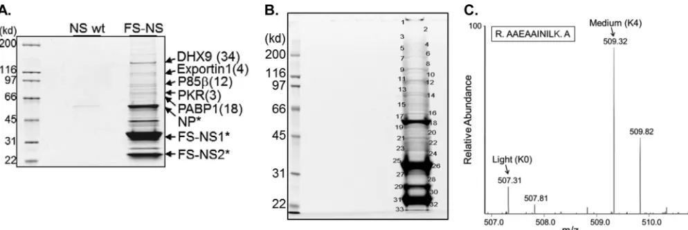
and SYPRO Ruby staining. Several distinct bands were visible
fol-lowing TAP of PR8 NS-FS-infected cells, which were largely
miss-ing in the TAP sample of PR8-wt-infected cells, demonstratmiss-ing the
purification power of the FS-tag (
Fig. 2A
). Several bands were
analyzed by MS, identifying unequivocally NS1 and NS2, but also
several other proteins with a significant number of unique
pep-tides (
Fig. 2A
). These included known NS1- and NS2-interacting
proteins, such as the regulatory beta-subunit of
phosphatidylino-sitol 3-kinase (p85

) and Exportin-1 (XPO1), respectively (
1
,
27
,
28
), indicating that the recombinant PR8-FS-NS virus can be used
to identify NS1/2-interacting proteins during the natural course
of infection. In this TAP-based experiment, the assessment of true
NS1/2-interacting proteins relied largely on quantification by
pro-tein staining. Unequivocal identification of less-abundant
NS1/2-interacting proteins will require more precise methods of
quanti-fication. In the last decade, substantial progress in quantitative
forms of proteomics has been made, allowing the separation of
true interacting proteins from contaminating proteins. One of
these techniques is based on SILAC (
29
). In this procedure, cells
are labeled with different, nonradioactive heavy isotopes of certain
amino acids. For example, control cells are “labeled” with regular,
“light” amino acids, and cells to be infected are “labeled” with
“medium” or “heavy” arginine and lysine. After infection, the
ly-sates of both cell populations are combined, and all following
steps, including protein purification and MS analysis, are
per-formed with one, mixed sample. The source of proteins will then
unequivocally be identified by the specific mass differences of
in-dividual peptides (
29
,
30
). One prerequisite for this approach is
cell culture in the presence of dialyzed FBS, which allows complete
replacement of unlabeled amino acids with exogenous, labeled
amino acids. When culturing A549 cells in the presence of
differ-ent isotopic amino acids followed by IAV infection, we noted a
profound reduction in infection efficiency by more than 10-fold,
preventing complete cell infection (data not shown). We therefore
tested HEK293T cells and found these cells much less affected by
SILAC conditions. We therefore used this cell type for the
follow-ing experiment. HEK293T cells were labeled with different
iso-topes, followed by infection with PR8-wt and PR8-FS-NS. At 18 h
postinfection, lysates were prepared, combined, and subjected to
one-step purification based on anti-FLAG antibodies, which was
followed by acidic elution and LC-MS. The purified samples were
separated by SDS-PAGE, and 33 bands were analyzed individually
by LC-MS, followed by MaxQuant-based analysis (
Fig. 2B
). Some
of the proteins identified are depicted in
Table 1
(for complete
analysis see Table S1 in the supplemental material). As expected,
several known NS1- and NS2-interacting proteins were identified
specifically as NS1/NS2-interacting proteins, as expressed by the
ratio of medium- and light-labeled proteins, representing
pro-teins derived from PR8-FS-NS- and PR8-wt-infected cells,
respec-tively. For example, p85

was identified at 5.9-fold-higher levels
from PR8-FS-NS- than PR8-wt-infected cells. As expected, the
PIK3R2-associated alpha-subunit, PIK3R1, was also enriched,
possibly copurifying with the regulatory

-subunit. Other known
NS1/NS2-interacting proteins, such as PABP-1 and the very
re-cently identified NS1-interacting protein dsRNA-specific
adeno-sine deaminase (ADAR), as well as the NS2-interacting Exportin-1
FIG 2Purification of NS1/2 from IAV-infected cells and identification of associated proteins by MS. (A) SYPRO Ruby-stained SDS-PAGE of samples derived from A549 cells that were infected with PR8-wt and PR8-FS-NS for 24 h at an MOI of 0.5, followed by TAP. (B) SYPRO Ruby-stained SDS-PAGE of purified protein samples from SILAC-labeled HEK293T cells. HEK293T cells were labeled with Lys0/Arg0 (light) and Lys4/Arg6 (medium) and infected with PR8-wt (light) or PR8-NS-FS (medium), followed by protein purification and SDS-PAGE. (C) Bands subjected to LC-MS/MS analysis are indicated. Mass spectrum of a peptide representing PACT, whose differentially labeled forms (Light from PR8-wt-infected cells and medium from PR8-NS-FS-infected cells) can be identified by their specific mass difference of 4 Da (⫽2m/zof double-charged peptide, for lysine). Arrows indicate the C12 monoisotopic peaks of each peptide.on November 7, 2019 by guest
http://jvi.asm.org/
[image:5.585.43.540.65.231.2](XPO1), were identified as specifically associated with the
NS-FS-expressing virus (
Table 1
) (
28
,
31
). Notably, a number of proteins
not characterized as NS1- or NS2-interacting proteins were
iden-tified, including the protein PACT, which was identified
unequiv-ocally with four unique peptides at a ratio of 2.98 over the control
(
Table 1
and see Table S1 in the supplemental material). The MS
spectrum of one PACT-derived peptide is shown in
Fig. 2C
,
illus-trating the relative abundance of PACT from PR8-NS-FS
(me-dium) and PR8-wt (light). Taken together, the data demonstrate
that the recombinant virus can be used to specifically identify
virus-interacting host proteins in a quantitative proteomic
ap-proach. Certainly, in the case of various splice products, as
observed for NS proteins, differentiation between NS1- or
NS2-interacting proteins needs to be established in
indepen-dent experiments (see below).
NS1 interacts with PACT.
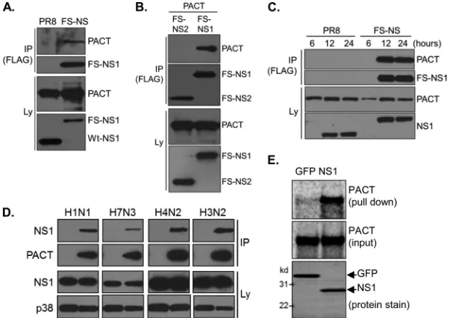
We confirmed interaction of NS1/
NS2 with endogenous PACT during virus infection in small scale
[image:6.585.42.548.77.175.2]experiments using PACT-specific antibodies. After 24 h of
infec-tion, PACT copurified specifically with FS-tagged proteins but
was not identified in samples from PR8-wt-infected cells (
Fig. 3A
).
To determine whether this interaction could be recapitulated in
the absence of virus infection and also to determine whether NS1
or NS2 interacted with PACT, we performed
transient-transfec-tion experiments with epitope-tagged proteins. As shown in
Fig.
3B
, NS1, but not NS2, interacted robustly with PACT. Analyzing
the kinetics of PACT-NS1 interaction by infecting cells for
differ-ent periods of time, we found protein interaction at the earliest
time point of NS1 expression, indicating immediate and efficient
interaction of the two proteins (
Fig. 3C
). To determine whether
binding of PACT is a more general property of IAV NS1 proteins,
we infected cells with different IAV strains and analyzed
PACT-NS1 interaction by IP/IB analysis. Similar to PR8 PACT-NS1, PACT-NS1 of the
three other investigated IAV strains interacted robustly with
PACT, suggesting that PACT binding is a conserved function of
TABLE 1MS-/MaxQuant-based identification of NS1- and NS2-interacting proteins using SILACProtein Gene
No. of unique peptides
M/L ratio (normalized)a
Sequence coverage (%)
IFN-inducible dsRNA-dependent protein kinase activator A (PACT) PRKRA 4 2.98 14.1
Phosphatidylinositol 3-kinase regulatory subunit beta PIK3R2 23 5.90 40.7
Phosphatidylinositol 3-kinase regulatory subunit alpha PIK3R1 4 2.87 7
Double-stranded RNA-specific adenosine deaminase ADAR 34 3.08 30.6
Polyadenylate-binding protein 1 (PABP-1) PABPC1 7 1.90 39.8
Exportin-1 XPO1 7 2.05 7.3
ATP-dependent RNA helicase A DHX9 41 0.99 37.1
aM, medium (proteins derived from PR8-NS-FS-infected cells); L, light (proteins derived from PR8-wt-infected cells).
FIG 3NS1 interacts with PACT during virus replication andin vitro. (A) HEK293T cells were infected with PR8-wt or PR8 NS-FS for 24 h, followed by immunoprecipitation (IP) and immunoblotting (IB) with antibodies against PACT and NS1 (Ly, total lysate). (B) HEK293T cells were transfected with PACT along with FS-tagged NS1 or NS2 for 48 h, followed by IP/IB analysis using antibodies against FLAG (for IP) and antibodies against FLAG (detecting FS-NS1 and FS-NS2) and PACT (for IB). (C) HEK293T cells were infected with PR8-wt or PR8 NS-FS for 6, 12, or 24 h, followed by IP and IB with antibodies against FLAG (for IP) and PACT and NS1 (for IB). (D) HEK293T cells were transfected with control plasmid or HA-tagged PACT, followed by infection with indicated IAV strains for 24 h and IP/IB analysis using antibodies against HA (for IP) and PACT, NS1, and p38 (for IB). H1N1, PR8; H7N3, A/laughing gull/Delaware bay/42/2006; H4N2, A/quail/California/D113023808/2012; H3N2 (A/Perth/16/2009). (E) Pulldown experiment ofin vitro-translated PACT using recombinant, bead-immobilized GFP or NS1. A pull-down sample and 10% of input sample were resolved by SDS-PAGE and analyzed by using a phosphorimager or SYPRO Ruby protein staining, respectively.
on November 7, 2019 by guest
http://jvi.asm.org/
[image:6.585.134.450.415.638.2]NS1 (
Fig. 3D
). To determine whether NS1 could interact directly
with PACT, we established pulldown assays based on bacterially
expressed recombinant NS1 and
in vitro
-translated PACT. NS1
precipitated PACT robustly, whereas GFP, used as a negative
con-trol, did not (
Fig. 3E
). As such, NS1 interacts with PACT during
IAV infection.
NS1 inhibits PACT binding to RIG-I and blocks RIG-I/
PACT-mediated IFN-

promoter activity.
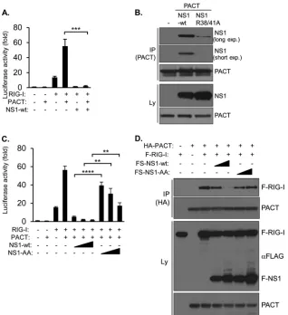
To study the impact
of NS1 on PACT biology, we coexpressed NS1 together with
PACT and RIG-I and measured the activity of a cotransfected
IFN-

-promoter luciferase reporter. RIG-I alone activated the
IFN-

-promoter, whose activity was further increased by
coex-pression of PACT, as expected (
Fig. 4A
) (
15
). However, IFN-

promoter activity induced by either RIG-I alone or PACT/RIG-I
was strongly reduced by NS1, demonstrating that NS1 interfered
with the PACT/RIG-I pathway (
Fig. 4A
).
Previously, the RNA binding domain of NS1 has been
demon-strated to be involved in NS1-mediated inhibition of
virus-medi-ated IFN-I activation, where two highly conserved amino acids,
which confer binding of dsRNA, i.e., R38 and K41 of PR8-NS1,
were found to be essential for inhibition of IFN-I activation (
32
,
33
). To determine whether these amino acids might be involved in
PACT binding, we coexpressed NS1-wt and a R38A/K41A mutant
protein (NS1-AA), along with PACT in HEK293T cells, followed
by IP. Although the expression levels of both NS1 proteins were
comparable, the interaction between PACT and the mutant
pro-tein was strongly reduced, suggesting that the integrity of the RNA
binding domain is important for protein interaction (
Fig. 4B
). To
substantiate these findings in functional assays, we coexpressed
RIG-I, PACT, and NS1 proteins in IFN-

reporter assays.
Al-though NS1 blocked RIG-I/PACT-induced IFN-

-promoter
ac-tivity at all of the concentrations used, NS1-AA exhibited strongly
reduced, albeit not completely abolished, inhibitory activity (
Fig.
4C
). Given that direct interaction of PACT with RIG-I is required
FIG 4NS1 prevents interaction between PACT and RIG-I and blocks PACT/RIG-I-mediated IFN-activation. (A) IFN-reporter assay. HEK293T cells were transfected with an IFN-promoter luciferase vector, either alone (control) or along with expression vectors for RIG-I, PACT, and NS1, as indicated, followed by analysis of luciferase activity after 48 h. Values are presented as the fold changes over the control. Error bars represent the standard deviations of three transfection replicates. ***,P⬍0.001. (B) HEK293T cells transfected with PACT, along with FS-NS1 or FS-NS1 (R38A/K41A) (NS1-AA), for 48 h, followed by IP/IB analysis with antibodies against PACT (for IP) and NS1 and PACT (for IB). Long and short exposures of films are shown, as indicated. (C) IFN-reporter assay. HEK293T cells were transfected with an IFN-promoter luciferase vector, either alone (control) or along with expression vectors for RIG-I, PACT, NS1, and NS1-AA as indicated, followed by analysis of luciferase activity after 48 h. Values are presented as the fold changes over the control. Error bars represent the standard deviations of three transfection replicates. **,P⬍0.01; ****,P⬍0.0001. (D) HEK293T cells were transfected with expression vectors for the indicated proteins for 48 h and analyzed by IP/IB with antibodies against HA (for IP) and antibodies against RIG-I, PACT, and FLAG (recognizing FLAG-RIG-I and FS-NS1) (for IB).on November 7, 2019 by guest
http://jvi.asm.org/
[image:7.585.134.452.65.413.2]for RIG-I activation (
15
), we sought to determine whether NS1
could directly interfere with this protein interaction. Indeed,
whereas PACT precipitated RIG-I efficiently in
transient-transfec-tion experiments, coexpression of NS1-wt, but not NS1-AA,
re-duced RIG-I–PACT interaction significantly (
Fig. 4D
). We
con-clude that the functional integrity of the RNA binding domain of
NS1 is important for PACT binding and interference with RIG-I–
PACT interaction and, as a consequence, the inhibition of PACT/
RIG-I-mediated IFN activation.
PACT is required for virus recognition and IFN activation in
the absence of NS1.
PACT has previously been demonstrated to
contribute to Sendai virus-mediated IFN-I activation (
15
). To
study the impact of PACT for IAV-induced IFN activation, we
established HEK293T cell lines wherein PACT expression was
sta-bly suppressed based on retrovirally delivered shRNA against
en-dogenous
PACT
mRNA. Two independent, polyclonal PACT
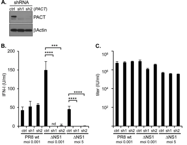
shRNA cell lines (and shRNA control cells) were generated, both
of which exhibited strongly reduced PACT protein levels
com-pared to control cells (
Fig. 5A
). These cells were used for infection
with PR8-wt and a recombinant PR8 mutant, where the
NS1-specific coding region of the NS gene segment was deleted (PR8
⌬
NS1), followed by analysis of IFN-I secretion in a quantitative
bioassay. Both viruses induced IFN-I in control cells, although the
PR8
⌬
NS1 induced more IFN-I, as expected (
4
) (
Fig. 5B
).
How-ever, PR8
⌬
NS1-induced IFN-I secretion in cell lines with
com-promised PACT expression was strongly reduced (
Fig. 5B
). In
contrast, IFN-I secretion upon PR8-wt infection was minimally
affected by the PACT expression levels, which is consistent with
the idea that NS1 efficiently counteracted PACT-mediated IFN
activation, which could not further be reduced by additional
sup-pression of PACT (
Fig. 5B
). It is currently unclear why PR8-wt
induced more IFN-I than PR8
⌬
NS1 on PACT shRNA cells
(
Fig. 5B
). Apparently, this phenomenon was not related to virus
replication since both viruses multiplied to a similar extent during
the time period analyzed, and a slightly reduced proliferative
ca-pacity of PR8
⌬
NS1 on PACT shRNA cells at a lower MOI was not
apparent at a high MOI, where IFN-I release was still fully ablated
(
Fig. 5B
and
C
). Taken together, the data show that PACT is
crit-ical for IAV recognition in the absence of NS1. The data are
con-sistent with the interpretation that PR8-wt-mediated NS1
expres-sion interferes efficiently with PACT biology.
DISCUSSION
Using a novel strategy to identify IAV NS1/2-interacting host
pro-teins, we identified a number of uncharacterized
NS1/2-interact-ing proteins, includNS1/2-interact-ing PACT. The specific IAV construct
devel-oped retains the full-length structure of the tagged proteins and
allows efficient one- and two-step purifications. It is likely that a
similar approach is feasible for other IAV gene segments. While
two-step purifications yield highly purified samples, the
evalua-tion of identified proteins as true interactors depends on
MS-based spectral counting or quantitative assessment of the protein
staining, both of which are difficult for low-abundance proteins.
In addition, the time and sample dilution required for TAP is
likely to result in the loss of low-affinity interactors. In contrast, a
one-step approach reduces the purification time significantly but
depends for thorough interpretation, particularly of
low-abun-dance proteins, on quantitative MS approaches, e.g., SILAC.
Al-though our data demonstrate the feasibility of the approach, we
believe that at least two improvements can be made. First, we
noted that some proteins, e.g., DHX9, which was previously
iden-tified as NS1-interacting protein in a nonquantitative proteomic
FIG 5PACT is required for efficient IFN-I production upon IVA infection in the absence of NS1. (A) Stable knockdown of PACT in HEK293T cells by two different, retrovirally delivered shRNAs against PACT mRNA (sh1, sh2) and control shRNA (ctrl), analyzed by IB with antibodies against PACT and-actin. (B and C) The HEK293T cells described in panel A were infected with PR8-wt and PR8⌬NS1 at an MOI of 0.001 for 72 h or with PR8⌬NS1 at an MOI of 5 for 24 h as indicated. The IFN-I activity (B) and virus titer (C) were determined by IFN bioassay and limiting dilution, after 72 h (MOI of 0.001) and 24 h (MOI of 5), respectively. nd, not detectable; ***,P⬍0.001; ****,P⬍0.0001.on November 7, 2019 by guest
http://jvi.asm.org/
[image:8.585.134.456.68.317.2]approach, a finding confirmed in our TAP approach (
Fig. 2A
), was
identified in the SILAC experiment at similar ratios from the
dif-ferentially labeled cells and thus would be ranked nonspecific
(
Table 1
) (
34
). This phenomenon is most likely due to interaction
of DHX9 with FS-NS1 during the purification process in the
mixed samples, a known problem for dynamic protein
interac-tions (
35
). As such, our approach has most likely sacrificed the
identification of such protein interactions in favor of the
reduc-tion of false-positive identificareduc-tions. A relatively simple remedy to
this problem will be the separate purification of individual
sam-ples, followed by mixing of purified samples for SDS-PAGE and
MS, also referred to as “MAP (mixing after purification)-SILAC”
(
35
). We also noted an unexpected reduction in the infection
ef-ficiency of SILAC-labeled A549 cells, which led us to use
HEK293T cells. For A549 cells (and possibly other cell types),
other forms of quantitative MS seem therefore more suitable,
par-ticularly post-tryptic labeling procedures, such as the use of
tan-dem mass tags (TMT) and isobaric tags for relative and absolute
quantitation (iTRAQ), which have made major progress during
the last few years, overcoming initial quantification problems
(
36
).
One protein unequivocally identified by the proteomic
ap-proach and further confirmed as NS1-interacting partner is
PACT, a protein whose binding to PKR and RIG-I has been
char-acterized in some detail before (
11
,
15
). We found in luciferase
reporter assays based on coexpression of PACT and RIG-I that
NS1 interfered efficiently with IFN-

promoter activation.
Al-though PACT upregulated RIG-I-induced IFN-

promoter
activ-ity significantly, which was almost completely blocked by NS1,
this experimental setting does not differentiate between
NS1-me-diated interference with PACT-induced RIG-I activation and
TRIM25-mediated RIG-I ubiquitination, which was previously
shown to increase RIG-I activity (
8
,
10
). Two sets of experiments,
however, indicate that NS1-PACT interaction is important. First,
knockdown of PACT strongly reduced RIG-I-mediated IFN
acti-vation in the absence of NS1, demonstrating that PACT
contrib-utes significantly to RIG-I activation in IAV-infected cells and,
second, NS1 interfered directly with complex formation between
PACT and RIG-I, which mediates RIG-I activation (
Fig. 4A
and
C
) (
15
). As such, it appears that NS1 has acquired the capacity to
interfere with distinct mechanisms that control RIG-I activation
at different levels of its signaling cascade.
As mentioned, NS1 has also been shown to interact directly
with PKR (
37
) and was found to block PKR activation by dsRNA
and by PACT through interaction with the N-terminal region of
PKR which contains two dsRNA binding domains (
38
). In the
same publication, the authors noted that NS1 pulled down PACT
when overexpressed in HEK293T cells. The interpretation of these
findings was complicated by the fact that other proteins, including
PKR, in these lysates might have contributed to this interaction.
Our results based on recombinant NS1 and
in vitro
-translated
PACT supports the interpretation that the NS1/PACT interaction
is direct. An interesting observation is that the two exposed,
charged amino acids R38/K41 contained in the RNA binding
do-main of NS1 support PACT interaction and contribute to
NS1-mediated interference with PACT-RIG-I activity (
Fig. 4B
and
C
).
This observation contrasts to the mentioned NS1/PKR
interac-tion, where a NS1 dsRNA binding mutant with a single amino acid
substitution (R38A) retained its inhibitory activity against
dsRNA-and PACT-induced PKR activation (
38
). Although it needs to be
formally ruled out that the additional point mutation of K41 used
in our report may account for the PKR-inhibitory function by
NS1, it appears that the dsRNA binding domain is more
specifi-cally involved in interference with the IFN-I pathway than PKR
activation and that both functions of NS1 can thus be separated on
a molecular level. Despite these molecular differences, it is
impor-tant to note that PACT represents a clear common denominator
of two well-characterized antiviral defense systems, i.e., the RIG-I
and PKR pathways, both of which are disabled upon NS1
expres-sion. Interestingly, another mechanism for how these two
path-ways may act interdependently has been proposed based on the
observation that critical components thereof, including PKR and
RIG-I, colocalize upon virus infection in a compartment referred
to as antiviral stress granules (avSGs) (
39
). IAV-mediated avSG
formation and IFN-I production, most likely RIG-I dependent,
was found to depend on (i) the presence of PKR and (ii) the
ab-sence of NS1 (
39
). One possible interpretation of these findings is
that NS1-mediated interference with PKR prevents the formation
of a structural compartment required for orchestration of the
an-tiviral defense, including RIG-I activation. Although the assembly
of the different protein components in avSGs could not be
as-sessed due to solubility issues, it appears that in this model PKR
acts upstream of RIG-I (
39
). As such, it will be interesting to see
how PACT-mediated activation of PKR and RIG-I fits into this
model, which we would assume act in a parallel rather than serial
fashion. Certainly, the two scenarios are not mutually exclusive
and may represent independent mechanisms, possibly acting in a
cell-type-specific or time-dependent way. In fact, it seems possible
that initial virus recognition and IFN-I activation depends on PKR
(and RIG-I) in avSGs, while at later time points during infection
and upon priming with small amounts of IFN-I, a
PACT-RIG-I-dependent (avSG-inPACT-RIG-I-dependent) mechanism prevails. This
hy-pothesis is supported (i) by data from PKR-deficient cells, where a
deficiency of IFN-I production upon poly(I·C) treatment could be
overcome by pretreatment with IFN-I, suggesting a restriction of
PKR function to the initial step of IFN-I activation (
40
), and (ii) by
data from HEK293T cells, where PACT overexpression strongly
supported IFN-

-induced IFN-

promoter activity, suggesting
that PACT represents a critical part of a secondary,
IFN-I-medi-ated RIG-I activation mechanism, akin to the previously
charac-terized positive feedback regulation of IFN production (
15
,
41
).
Certainly, more work will be required to assess the
time-depen-dent, relative contributions of PACT to RIG-I versus PKR
activa-tion.
Although our data indicate a critical role of R38/K41 in protein
interaction, these amino acids are certainly also critical for
NS1-mediated dsRNA binding, a function that has been proposed as
mechanism to prevent recognition of viral dsRNA emerging
dur-ing replication (
6
,
42
). The existence of such immunostimulatory
dsRNA, particularly in the cytoplasm, where RIG-I-mediated
vi-rus sensing most likely occurs, however, has been questioned for
negative-strand RNA viruses such as IAV, at least in the presence
of functional IAV nucleoprotein (
43
,
44
). As such, the binding of
viral dsRNA by NS1 and its relevance for virus recognition
re-quires further investigation. Although our
in vitro
binding assay of
NS1 and PACT does not completely rule out the possibility that
trace amounts of RNA contained in the
in vitro
translation sample
contribute to protein interaction, another emerging possibility is
that the RNA binding domain represents a protein interaction
motif. In this context it is important to note that the two viral
on November 7, 2019 by guest
http://jvi.asm.org/
proteins, i.e., HSV Us11 and Ebola VP35, which have very recently
been shown to counteract PACT-mediated IFN activation (
16
,
17
), both contain dsRNA binding domains that are critical for
PACT interaction. dsRNA seemed dispensable for Us11-PACT
and VP35-PACT interaction, supporting the idea that the RNA
binding domains may act as protein interaction motifs. For VP35,
critical residues involved in RNA binding have indeed been found
to mediate intermolecular VP35 interactions, providing evidence
for the principle possibility of protein interaction of involved RNA
binding domains. More detailed information based on structural
experiments will be required to confirm this hypothesis.
Taken together, the data shown in the present study contribute
to our understanding of how NS1 interferes with pathogen
recog-nition, highlighting yet another host target, PACT, whose
inacti-vation along with other proteins acting at various stages of virus
recognition, including TRIM25, Riplet, and PKR, compromises
and delays host immune defense against IAV infection (
8
,
9
).
ACKNOWLEDGMENTS
We thank Youming Shao and Richard Heath (St. Jude Protein Production Facility) for the production of the recombinant proteins. We thank Ruiq-iong Wu and Liying Chi for excellent technical support.
E.R.H. was supported by the Deutsche Forschungsgemeinschaft. This study was supported by the American Lebanese Syrian Associated Charities.
REFERENCES
1.Hale BG, Randall RE, Ortin J, Jackson D.2008. The multifunctional NS1 protein of influenza A viruses. The J. Gen. Virol.89:2359 –2376.http://dx .doi.org/10.1099/vir.0.2008/004606-0.
2.Egorov A, Brandt S, Sereinig S, Romanova J, Ferko B, Katinger D, Grassauer A, Alexandrova G, Katinger H, Muster T.1998. Transfectant influenza A viruses with long deletions in the NS1 protein grow efficiently in Vero cells. J. Virol.72:6437– 6441.
3.Kochs G, Koerner I, Thiel L, Kothlow S, Kaspers B, Ruggli N, Sum-merfield A, Pavlovic J, Stech J, Staeheli P.2007. Properties of H7N7 influenza A virus strain SC35M lacking interferon antagonist NS1 in mice and chickens. J. Gen. Virol.88:1403–1409.http://dx.doi.org/10.1099/vir .0.82764-0.
4.Garcia-Sastre A, Egorov A, Matassov D, Brandt S, Levy DE, Durbin JE, Palese P, Muster T.1998. Influenza A virus lacking the NS1 gene repli-cates in interferon-deficient systems. Virology252:324 –330.http://dx.doi .org/10.1006/viro.1998.9508.
5.Lu Y, Wambach M, Katze MG, Krug RM.1995. Binding of the influenza virus NS1 protein to double-stranded RNA inhibits the activation of the protein kinase that phosphorylates the elF-2 translation initiation factor. Virology214:222–228.http://dx.doi.org/10.1006/viro.1995.9937. 6.Min JY, Krug RM.2006. The primary function of RNA binding by the
influenza A virus NS1 protein in infected cells: inhibiting the 2=-5= oli-go(A) synthetase/RNase L pathway. Proc. Natl. Acad. Sci. U. S. A.103: 7100 –7105.http://dx.doi.org/10.1073/pnas.0602184103.
7.Kato H, Takeuchi O, Sato S, Yoneyama M, Yamamoto M, Matsui K, Uematsu S, Jung A, Kawai T, Ishii KJ, Yamaguchi O, Otsu K, Tsujimura T, Koh CS, Reis e Sousa C, Matsuura Y, Fujita T, Akira S. 2006. Differential roles of MDA5 and RIG-I helicases in the recognition of RNA viruses. Nature441:101–105.http://dx.doi.org/10.1038/nature04734. 8.Gack MU, Albrecht RA, Urano T, Inn KS, Huang IC, Carnero E, Farzan
M, Inoue S, Jung JU, Garcia-Sastre A.2009. Influenza A virus NS1 targets the ubiquitin ligase TRIM25 to evade recognition by the host viral RNA sensor RIG-I. Cell Host Microbe5:439 – 449.http://dx.doi.org/10.1016/j .chom.2009.04.006.
9.Rajsbaum R, Albrecht RA, Wang MK, Maharaj NP, Versteeg GA, Nistal-Villan E, Garcia-Sastre A, Gack MU.2012. Species-specific inhi-bition of RIG-I ubiquitination and IFN induction by the influenza A virus NS1 protein. PLoS pathogens 8:e1003059. http://dx.doi.org/10.1371 /journal.ppat.1003059.
10. Gack MU, Shin YC, Joo CH, Urano T, Liang C, Sun L, Takeuchi O, Akira S, Chen Z, Inoue S, Jung JU. 2007. TRIM25 RING-finger E3
ubiquitin ligase is essential for RIG-I-mediated antiviral activity. Nature 446:916 –920.http://dx.doi.org/10.1038/nature05732.
11. Patel RC, Sen GC.1998. PACT, a protein activator of the interferon-induced protein kinase, PKR. EMBO J.17:4379 – 4390.http://dx.doi.org /10.1093/emboj/17.15.4379.
12. Meurs E, Chong K, Galabru J, Thomas NS, Kerr IM, Williams BR, Hovanessian AG.1990. Molecular cloning and characterization of the hu-man double-stranded RNA-activated protein kinase induced by interferon. Cell62:379 –390.http://dx.doi.org/10.1016/0092-8674(90)90374-N. 13. Gale M, Jr, Katze MG.1998. Molecular mechanisms of interferon
resis-tance mediated by viral-directed inhibition of PKR, the interferon-induced protein kinase. Pharmacol. Ther.78:29 – 46.
14. Lee Y, Hur I, Park SY, Kim YK, Suh MR, Kim VN.2006. The role of PACT in the RNA silencing pathway. EMBO J.25:522–532.http://dx.doi .org/10.1038/sj.emboj.7600942.
15. Kok KH, Lui PY, Ng MH, Siu KL, Au SW, Jin DY.2011. The double-stranded RNA-binding protein PACT functions as a cellular activator of RIG-I to facilitate innate antiviral response. Cell Host Microbe9:299 –309. http://dx.doi.org/10.1016/j.chom.2011.03.007.
16. Kew C, Lui PY, Chan CP, Liu X, Au SW, Mohr I, Jin DY, Kok KH.2013. Suppression of PACT-induced type I interferon production by herpes simplex virus 1 Us11 protein. J. Virol.87:13141–13149.http://dx.doi.org /10.1128/JVI.02564-13.
17. Luthra P, Ramanan P, Mire CE, Weisend C, Tsuda Y, Yen B, Liu G, Leung DW, Geisbert TW, Ebihara H, Amarasinghe GK, Basler CF. 2013. Mutual antagonism between the Ebola virus VP35 protein and the RIG-I activator PACT determines infection outcome. Cell Host Microbe 14:74 – 84.http://dx.doi.org/10.1016/j.chom.2013.06.010.
18. Dos Santos Afonso E, Escriou N, Leclercq I, van der Werf S, Naffakh N. 2005. The generation of recombinant influenza A viruses expressing a PB2 fusion protein requires the conservation of a packaging signal overlapping the coding and noncoding regions at the 5=end of the PB2 segment. Vi-rology341:34 – 46.http://dx.doi.org/10.1016/j.virol.2005.06.040. 19. Donnelly ML, Luke G, Mehrotra A, Li X, Hughes LE, Gani D, Ryan
MD. 2001. Analysis of the aphthovirus 2A/2B polyprotein “cleavage” mechanism indicates not a proteolytic reaction, but a novel translational effect: a putative ribosomal “skip.” J. Gen. Virol.82:1013–1025. 20. Brown LE, Hinshaw VS, Webster RG.1983. Antigenic variation in the
influenza A virus nonstructural protein, NS1. Virology130:134 –143.http: //dx.doi.org/10.1016/0042-6822(83)90123-X.
21. van Wyke KL, Yewdell JW, Reck LJ, Murphy BR. 1984. Antigenic characterization of influenza A virus matrix protein with monoclonal an-tibodies. J. Virol.49:248 –252.
22. Becker-Hapak M, Dowdy SF.2003. Protein transduction: generation of full-length transducible proteins using the TAT system. Curr. Protoc. Cell Biol. Chapter 20:Unit 20 22. http://dx.doi.org/10.1002/0471143030 .cb2002s18.
23. Kok KH, Ng MH, Ching YP, Jin DY.2007. Human TRBP and PACT directly interact with each other and associate with dicer to facilitate the production of small interfering RNA. J. Biol. Chem.282:17649 –17657. http://dx.doi.org/10.1074/jbc.M611768200.
24. Hoffmann E, Neumann G, Kawaoka Y, Hobom G, Webster RG.2000. A DNA transfection system for generation of influenza A virus from eight plasmids. Proc. Natl. Acad. Sci. U. S. A.97:6108 – 6113.http://dx.doi.org /10.1073/pnas.100133697.
25. Cox J, Mann M.2008. MaxQuant enables high peptide identification rates, individualized p.p.b.-range mass accuracies and proteome-wide protein quantification. Nat. Biotechnol.26:1367–1372.http://dx.doi.org /10.1038/nbt.1511.
26. Hoffmann E, Stech J, Guan Y, Webster RG, Perez DR.2001. Universal primer set for the full-length amplification of all influenza A viruses. Arch. Virol.146:2275–2289.http://dx.doi.org/10.1007/s007050170002. 27. Ehrhardt C, Wolff T, Pleschka S, Planz O, Beermann W, Bode JG,
Schmolke M, Ludwig S.2007. Influenza A virus NS1 protein activates the PI3K/Akt pathway to mediate antiapoptotic signaling responses. J. Virol. 81:3058 –3067.http://dx.doi.org/10.1128/JVI.02082-06.
28. Neumann G, Hughes MT, Kawaoka Y. 2000. Influenza A virus NS2 protein mediates vRNP nuclear export through NES-independent inter-action with hCRM1. EMBO J.19:6751– 6758.http://dx.doi.org/10.1093 /emboj/19.24.6751.
29. Ong SE, Mann M. 2005. Mass spectrometry-based proteomics turns quantitative. Nat. Chem. Biol. 1:252–262. http://dx.doi.org/10.1038 /nchembio736.
on November 7, 2019 by guest
http://jvi.asm.org/
30. Harsha HC, Molina H, Pandey A.2008. Quantitative proteomics using stable isotope labeling with amino acids in cell culture. Nature protocols 3:505–516.http://dx.doi.org/10.1038/nprot.2008.2.
31. de Chassey B, Aublin-Gex A, Ruggieri A, Meyniel-Schicklin L, Pra-dezynski F, Davoust N, Chantier T, Tafforeau L, Mangeot PE, Ciancia C, Perrin-Cocon L, Bartenschlager R, Andre P, Lotteau V.2013. The interactomes of influenza virus NS1 and NS2 proteins identify new host factors and provide insights for ADAR1 playing a supportive role in virus replication. PLoS Pathog.9:e1003440.http://dx.doi.org/10.1371/journal .ppat.1003440.
32. El Hefnawi M, Alaidi O, Mohamed N, Kamar M, El-Azab I, Zada S, Siam R.2011. Identification of novel conserved functional motifs across most Influenza A viral strains. Virol. J.8:44.http://dx.doi.org/10.1186 /1743-422X-8-44.
33. Donelan NR, Basler CF, Garcia-Sastre A.2003. A recombinant influenza A virus expressing an RNA-binding-defective NS1 protein induces high levels of beta interferon and is attenuated in mice. J. Virol.77:13257– 13266.http://dx.doi.org/10.1128/JVI.77.24.13257-13266.2003. 34. Lin L, Li Y, Pyo HM, Lu X, Raman SN, Liu Q, Brown EG, Zhou Y.2012.
Identification of RNA helicase A as a cellular factor that interacts with influenza A virus NS1 protein and its role in the virus life cycle. J. Virol. 86:1942–1954.http://dx.doi.org/10.1128/JVI.06362-11.
35. Wang X, Huang L.2008. Identifying dynamic interactors of protein complexes by quantitative mass spectrometry. Mol. Cell. Proteomics 7:46 –57.http://dx.doi.org/10.1074/mcp.M700261-MCP200.
36. Wuhr M, Haas W, McAlister GC, Peshkin L, Rad R, Kirschner MW, Gygi SP.2012. Accurate multiplexed proteomics at the MS2 level using the complement reporter ion cluster. Anal. Chem.84:9214 –9221.http: //dx.doi.org/10.1021/ac301962s.
37. Tan SL, Katze MG.1998. Biochemical and genetic evidence for complex formation between the influenza A virus NS1 protein and the
interferon-induced PKR protein kinase. J. Interferon Cytokine Res.18:757–766.http: //dx.doi.org/10.1089/jir.1998.18.757.
38. Li S, Min JY, Krug RM, Sen GC.2006. Binding of the influenza A virus NS1 protein to PKR mediates the inhibition of its activation by either PACT or double-stranded RNA. Virology349:13–21.http://dx.doi.org/10 .1016/j.virol.2006.01.005.
39. Onomoto K, Jogi M, Yoo JS, Narita R, Morimoto S, Takemura A, Sambhara S, Kawaguchi A, Osari S, Nagata K, Matsumiya T, Namiki H, Yoneyama M, Fujita T.2012. Critical role of an antiviral stress granule containing RIG-I and PKR in viral detection and innate immunity. PLoS One7:e43031.http://dx.doi.org/10.1371/journal.pone.0043031. 40. Yang YL, Reis LF, Pavlovic J, Aguzzi A, Schafer R, Kumar A, Williams
BR, Aguet M, Weissmann C.1995. Deficient signaling in mice devoid of double-stranded RNA-dependent protein kinase. EMBO J.14:6095– 6106.
41. Marie I, Durbin JE, Levy DE.1998. Differential viral induction of distinct interferon-alpha genes by positive feedback through interferon regulatory factor-7. EMBO J.17:6660 – 6669.http://dx.doi.org/10.1093/emboj/17.22 .6660.
42. Talon J, Horvath CM, Polley R, Basler CF, Muster T, Palese P, Garcia-Sastre A.2000. Activation of interferon regulatory factor 3 is inhibited by the influenza A virus NS1 protein. J. Virol.74:7989 –7996.http://dx.doi .org/10.1128/JVI.74.17.7989-7996.2000.
43. Weber F, Wagner V, Rasmussen SB, Hartmann R, Paludan SR.2006. Double-stranded RNA is produced by positive-strand RNA viruses and DNA viruses but not in detectable amounts by negative-strand RNA vi-ruses. J. Virol.80:5059 –5064. http://dx.doi.org/10.1128/JVI.80.10.5059 -5064.2006.
44. Wisskirchen C, Ludersdorfer TH, Muller DA, Moritz E, Pavlovic J. 2011. The cellular RNA helicase UAP56 is required for prevention of dou-ble-stranded RNA formation during influenza A virus infection. J. Virol. 85:8646 – 8655.http://dx.doi.org/10.1128/JVI.02559-10.
on November 7, 2019 by guest
http://jvi.asm.org/