R E S E A R C H
Open Access
The distribution of
Blastocystis
subtypes in
isolates from Qatar
Marawan Abu-Madi
1*, Mahmoud Aly
2,3, Jerzy M. Behnke
4, C. Graham Clark
5and Hanan Balkhy
2,3Abstract
Background:Blastocystisis a common single-celled intestinal parasite of humans and other animals comprising at
least 17 genetically distinct small subunit ribosomal RNA lineages (subtypes (STs)), nine of which have been found in humans. The geographic distribution ofBlastocystissubtypes is variable, but the subtypes present in Qatar are at present unknown.
Methods:Stool samples were collected from randomly selected, apparently healthy subjects arriving in Qatar for
the first time.Blastocystissubtypes were determined by sequencing of the small subunit rRNA gene (SSU rDNA) PCR products. Phylogenetic analyses were done using Maximum Composite Likelihood method.
Results:71.1 % of samples were positive forBlastocystisinfection based on PCR-detection methodology compared
to only 6.9 % by microscopy. Prevalence ofBlastocystisdid not differ between the sexes nor between age classes. However, there was a regional difference in prevalence with subjects arriving from Africa showing the highest (87.6 %), those from Western Asia intermediate (68.6 %) and from Eastern Asia the lowest prevalence (67.6 %). Genetic analysis detected only three STs. ST3 was the most common (69.3 %) and ST2 was the rarest (3.5 %), while ST1 had a prevalence of 27.2 %. ST2 showed a regional variation, being absent from the 64 Western Asian
Blastocystis-positive subjects. Both ST1 and ST3 showed significant differences in prevalence between the sexes. Conclusions:This is the first report exploring the distribution ofBlastocystissubtypes in our region. We recommend that stool screening via microscopy for the presence ofBlastocystisshould be abandoned since it is extremely insensitive. In future, the prevalence ofBlastocystisinfections should be based on PCR methodology and we predict that in the years ahead diagnostic PCR will become the tool of choice. More work is needed to identify the full range ofBlastocystissubtypes that circulate in our region.
Keywords:Blastocystis, Real time-PCR, Subtype, Small subunit ribosomal DNA, Prevalence, Genotyping, Phylogenetic
analysis, Qatar
Background
Blastocystisis a single-celled intestinal protist, taxonom-ically placed within the Stramenopiles, which colonizes an estimated 1,000,000,000 people globally and a variety
of animal species [1]. Although Blastocystis is also
de-tected in asymptomatic humans [2], some studies link this organism with intestinal and extra-intestinal disease
[3, 4]. To date, despite Blastocystis being the most
fre-quently isolated protist from diarrheal patients in the de-veloped world, a causal link has not yet been established
conclusively [5]. Blastocystis is genetically diverse and
based on small subunit rRNA gene analysis (SSU rDNA), at least 17 subtypes (STs) have been identified in humans, other mammals, and birds. Among these sub-types, ST1-ST4 collectively account for 90 % of human carriage [6], while the ST5-ST9 account for the remaining 10 %. To date ST9 has only been isolated from humans [7]. To better understand the genetic
di-versity and determine the prevalence ofBlastocystis
sub-types around the world, further research is required [8, 9]. The introduction of molecular screening assays such as real-time polymerase chain reaction (RT-PCR) has
demonstrated that the prevalence ofBlastocystisis much
higher than previously reported on the basis of detection by conventional microscopy, in both developed [2, 9, 10] * Correspondence:[email protected]
1
Department of Health Sciences, College of Arts and Science, Biomedical Research Center, Qatar University, P.O. Box 2713, Doha, Qatar
Full list of author information is available at the end of the article
and developing [6] countries. Studying the genetic diver-sity ofBlastocystisin different hosts, age groups and gen-ders, and in different regions of the world, is essential to further our knowledge of the epidemiology and clinical relevance of this organism.
In the past decade, Qatar has seen a fast-paced trans-formation in the standards of living of its citizens. This tre-mendous growth has spawned building and modernization programs. There has been a large influx of migrant workers into Doha to complete very ambitious and large-scale con-struction projects. These workers mostly originate from states in the Middle-East, Asia and Africa, the latter two being regions where intestinal parasites are particularly common [11, 12]. Economic growth has been accompan-ied by rapidly expanding domestic services in the city, which are also dependent on an immigrant labour force. Improvements in the infrastructure have not kept pace with this transformation, particularly the housing and sanitation available for workers. The immigration policy in Qatar applies a quota system to control the number of workers entering Qatar each year according to their coun-try of origin. This creates a dynamic population of workers that changes from year to year and therefore requires con-tinuous monitoring. On arrival in Qatar and before they can obtain work permits, all new immigrants are obliged to report to the Medical Commission for thorough med-ical inspections but fecal examinations of all immigrant workers seeking employment in the food industries and/ or as housemaids were abandoned following the introduc-tion of compulsory treatment with albendazole, which is effective mainly against helminths and not most protozoa
(although it does have activity against Giardia [13]). No
specific treatment is given for protozoan infections, so in contrast to helminth infections these are not eradicated from infected immigrants on arrival in Qatar. Once a worker has been issued a work permit they are then not obliged to undergo further health inspections unless they work in the food industry, in which case annual re-examination is mandatory. As foreign workers form an in-tegral part of the food industry in Qatar, it is imperative to determine their status as a potential reservoir and source of infection for enteric pathogens. The same is true for housemaids, who not only handle the food served to fam-ilies, but also infants and children, who are generally more susceptible to infection than adults.
As prevention is far more effective in the long-term than cure, the identification of carriers and facilitation of the medical management of individuals who shed these patho-gens can be improved through the application of sensitive screening methods, thereby minimizing the risk of infec-tion spreading to other sectors of the community. In this paper we report on the implementation of a molecular screening assay (MSA) based on real-time PCR for the de-tection and subtyping ofBlastocystisin samples collected
from migrant workers newly arriving in Qatar, and we compare the detection rate with that achieved by conven-tional microscopy. We examine prevalence data for evi-dence of regional variation, while controlling for age and sex effects, and in a subset of the data we test whether the prevalence of the different STs of Blastocystis differs be-tween regions of origin of the carriers or is influenced by host intrinsic factors, sex and age.
Methods
Sample collection and DNA extraction
We obtained stool samples from 608 randomly selected immigrant workers (including food handlers, construc-tion workers, and housemaids), arriving for the first time in Qatar, and undergoing mandatory health checks at the Medical Commission during their application for work permits. Samples were collected in individually la-belled, standard sterile disposable containers and were immediately stored on ice until processed for DNA ex-traction and microscopy.
Samples were aliquoted for DNA extraction alongside conventional stool examination by microscopy. Micro-scopic examination was carried out as described by Abu-Madi and others [14]. DNA was extracted using the QIAamp DNA stool minikit (Qiagen, Hilden, Germany)
according to the manufacturer’s instructions. Briefly,
samples were weighed, homogenized in lysis buffer, and incubated at 95 °C for 5 min to ensure lysis of the tar-geted protozoa. After centrifugation, the DNA in the supernatant was purified using a silica column supplied with the kit. The quantity and quality of the DNA was determined using spectrophotometry (Nanodrop, Ther-moScientific) and gel electrophoresis.
Ethical approval
Ethical approval for this project was obtained from the Medical Research Centre and the Research Committee at Hamad Medical Corporation, Qatar (Research proto-col # 11110/11).
RT-PCR amplification and sequencing
To detect the presence of Blastocystis in the DNA
ex-tracted from the samples described above, well-studied primers targeting the SSU rDNA region were used [10, 15, 16]. This region covers approximately 190 bp that are
highly specific for the SSU rDNAs ofBlastocystis STs1-9,
but can also distinguish each ST. This approach has been chosen over other possible approaches as the primer set used is of diagnostic quality yet at the same time it ampli-fies a product that will allow for subtyping once it is se-quenced [15, 16]. DNA extracted from samples that were
positive for Blastocystis based on light microscopy was
used as a positive control. The reaction mixture contained
0.125 μM probe. Samples were processed using an ABI 7500 instrument. The reaction conditions were set as fol-lowing: 95 °C for 5 min, and 50 cycles of denaturation at 95 °C for 15 s followed by annealing and extension at 60 °C for 1 min.
Purified PCR products were sequenced using a Big-Dye® Terminator v3.1 Cycle Sequencing Kit (Applied
Biosystems™Austin, TX, USA). Thermal cycling was 96 °C
for 1 min, 40 cycles of (96 °C for 10 min, 50 °C for 5 s,
60 °C for 4 min) using either Blastocystis Fwd S1,
GGTCCGGTGAACACTTTGGATTT or Blastocystis
RvsS2, CCTACGGAAACCTTGTTACGACTTCA.
Prod-ucts were purified using XTerminator™ and SAM™
solu-tions (Applied Biosystems™ Foster City, CA, USA) and
sequenced on a 3730xl DNA Analyzer (Applied Biosys-tems™, Hitachi, Tokyo, Japan).
Genotyping analysis
To identify Blastocystis subtypes, both strands of the
amplicons were sequenced and were compared with all the SSU rRNA gene sequences available from the Na-tional Centre for Biotechnology Information (NCBI) database using the BLAST program. SSU rDNA subtypes were identified by determining the closest similarity match
against all known Blastocystis STs. All of our sequences
have been deposited in GenBank under Accession num-bers KM438204-30.
Statistical analysis
Prevalence data are presented as percentages with 95 % confidence limits (95 % CL), calculated with be-spoke software based on the tables of Rohlf and Sokal [17]. Analysis comprised three stages. In the first we
compared detection ofBlastocystisusing the PCR method
with conventional detection by routine microscopy. In the second step, we analyzed the factors affecting
prevalence of Blastocystis, based on detection of
in-fected subjects by PCR. For this analysis we used max-imum likelihood techniques based on log linear analysis of contingency tables in the software package IBM SPSS Statistics Version 21 (IBM Corporation). Initially, full factorial models were fitted, incorporating as factors region of origin of subjects (REGION - 3 levels), sex of subjects (SEX, 2 levels, male and female), and age of
sub-jects (AGE, 3 levels). The subsub-jects (n= 608) originated
from 18 countries, but for statistical analysis were assigned to three regions. Region 1, Africa comprised subjects from
Cameroon (n= 1), Egypt (2), Eritrea (3), Ethiopia (34),
Ghana (3), Kenya (27), Nigeria (15), Sudan (2), Chad (1) and Uganda (1). Region 2, Eastern Asia comprised sub-jects from Indonesia (119), Philippines (65), Thailand (2) and Vietnam (2). Region 3, Western Asia, comprised subjects from Bangladesh (71), India (117), Nepal (62) and Sri Lanka (81). Subjects were allocated to three age classes
(age class 1 = 18–29 years old, age class 2 = 30–39 years old and age class 3 = 40–58 years old). The presence /
ab-sence ofBlastocystis(INFECTION) was fitted as a binary
factor. These explanatory factors were fitted initially to all models that were evaluated. For each level of analysis in turn, beginning with the most complex model, involving all possible main effects and interactions, those combina-tions that did not contribute significantly to explaining variation in the data were eliminated in a stepwise fashion beginning with the highest level interaction (backward se-lection procedure). A minimum sufficient model was then obtained, for which the likelihood ratio ofχ2 was not sig-nificant, indicating that the model was sufficient to explain the data. The importance of each term (i.e. interactions in-volving infection) in the final model was assessed by the probability that its exclusion would alter the model signifi-cantly and these values relating to interactions that in-cluded INFECTION are given in the text.
In stage three, analysis of the factors affecting the
prevalence of genotyped Blastocystis isolates was
con-ducted on the smaller data-set of sequenced isolates. The subtype identification of a subset of 132 randomly selected isolates was achieved by sequencing the SSU
rDNA. Some isolates (n= 18) were problematic and not
available for analysis because they could not be subtyped successfully or were associated with missing data. How-ever, 114 isolates were completely subtyped and analysed for the current study. The successfully subtyped isolates originated from donors ranging in age from 20 years to a maximum of 56 years, with an average of 31.9 ± 0.75 (median of 30.0) years. Fifty three subjects were males (mean age = 31.2 ± 1.13 and 61 females (mean age = 32.5 ± 0.99), and there was no significant difference
be-tween the ages of the two sexes (z= 1.055, P= 0.29).
The approach adopted for analysis was similar to that in stage two of the analysis. Again subjects were allo-cated to the same three regions and age classes. Three
models were fitted, one each for Blastocystis ST1, ST2
and ST3, and in each case with AGE, SEX and REGION as explanatory factors and presence/absence of the sub-type (INFECTION) as a binary factor. Reduction to mini-mum sufficient models followed the same procedure as that explained above in stage two of the analysis.
Throughout our cut-off for statistical significance was P= 0.05.
Results
Prevalence ofBlastocystisbased on PCR method of detection
negative by both methods. However, five samples were recorded as positive by microscopy but not detected as such by PCR.
Factors affecting the prevalence ofBlastocystisbased on detection by PCR
Analysis of the prevalence of Blastocystisbased on PCR
revealed that neither host sex nor age affected preva-lence significantly (Table 1). However, there was a highly significant effect of the region from which subjects origi-nated (REGION x INFECTION,χ22= 16.1, p<0.001).
In-fections with Blastocystis were most common among
subjects from Africa and least common among those from Eastern Asia, and the difference in prevalence be-tween these regions was 20.0 %.
Subtype analysis and prevalence
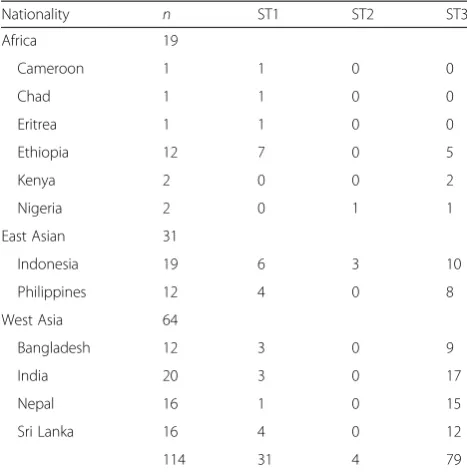
Subtype analysis revealed that only three subtypes were present in the samples analyzed. ST1 was present in 31 samples (27.2 %), ST2 was found in four hosts (3.5 %), one from Nigeria and three from Indonesia, while 79 (69.3 %) samples had ST3. Interestingly, ST3 was found in samples isolated from subjects of all geographical re-gions included in the current study i.e. Africa, East and West Asia. Table 2 shows the occurrence of subtypes and strains among the 114 subjects by country of origin. When the data were subdivided by the 12 countries of origin of the subjects, some subsets became very small, and indeed there was only one subject each from Cameroon, Chad and Eritrea.
Factors affecting the prevalence of subtypes of
Blastocystisin subsets of the subjects in the study
In order to enable statistical analysis, the subjects were allocated to three geographical regions (Tables 2 and 3). There was no regional effect on ST1 and ST3, but with age and host sex taken into account there was a
significant effect of region of origin on ST2 (REGION x INFECTION, χ22= 7.1,P= 0.029), despite the low preva-lence of this ST among the 114 samples. This subtype was not detected among the 64 W. Asian subjects (Table 2). Table 3 shows that ST3 appeared to be more common among W. Asian and ST1 among African sub-jects, but with host sex and age taken into account these differences in prevalence did not reach statistical signifi-cance (effect of REGION on ST1,χ22= 5.1,P= 0.078 and on ST3,χ22= 5.9,P= 0.052).
With region of origin and age taken into account, there were significant effects of host sex on the
preva-lence of ST1 (Table 4; SEX x INFECTION, χ21= 13.4, p
<0.001) and ST3 (Table 4; SEX x INFECTION,χ21= 15.1,
p <0.001). In the case of ST1 the bias was strongly in
favour of female subjects with prevalence of this subtype being 3.6-fold more common among females. ST3, showed a bias in the opposite direction with prevalence among male subject being 1.6-fold higher compared with females.
[image:4.595.304.538.111.348.2]With region of origin and host sex taken into account, there were no significant effects of host age, although as Table 1Factors affecting prevalence ofBlastocystis, based on
detection by PCR
Factor Number Prevalence (%) 95 % CL
Host sex
Males 280 70.7 65.85–75.17
Females 328 71.3 66.10–76.10
Host age
Age class 1 (18–29) 286 73.1 68.25–77.42 Age class 2 (30–39) 233 70.0 65.49–74.10 Age class 3 (40–58) 89 67.4 54.32–78.65 Region of origin
Africa 89 87.6 76.54–94.08
Eastern Asia 188 67.6 57.95–75.86
[image:4.595.56.291.565.732.2]Western Asia 331 68.6 63.23–73.49
Table 2The distribution of subtypes ofBlastocystisamong the 114 subjects by their countries of origin
Nationality n ST1 ST2 ST3
Africa 19
Cameroon 1 1 0 0
Chad 1 1 0 0
Eritrea 1 1 0 0
Ethiopia 12 7 0 5
Kenya 2 0 0 2
Nigeria 2 0 1 1
East Asian 31
Indonesia 19 6 3 10
Philippines 12 4 0 8
West Asia 64
Bangladesh 12 3 0 9
India 20 3 0 17
Nepal 16 1 0 15
Sri Lanka 16 4 0 12
114 31 4 79
Table 3Prevalence of subtypes ofBlastocystisamong the three regions of origin of the subjects in the study
Region n Subtype prevalence as a percentage (95 % CL)
ST1 ST2 ST3
[image:4.595.303.540.664.732.2]can be seen from Table 4, there appeared to be a steady decline in the prevalence of ST1 with increasing age and an increase with age in the case of ST3.
Discussion
The results of our study show clearly that there is an
enormous difference in the detection of Blastocystis in
stool samples by microscopy vs PCR methodology. Based on PCR methodology we concluded that 71.1 % of
the samples contained BlastocystisDNA, whilst
conven-tional microscopy detected only 42 positive samples, giv-ing a prevalence of just 6.9 %. Higher detection rates of
intestinal protozoa, including Blastocystis, by PCR
com-pared with conventional microscopy have also been re-cently reported in asymptomatic individuals from Brazil [18], where microscopy detected 10 infected subjects out of 126 (prevalence = 7.9 %) whereas PCR detected 43 in-fections in the same subjects (prevalence = 34.1 %). This difference in sensitivity of the two assays raises questions about the utility of continuing to employ conventional microscopy, when this method has such a high failure rate in detecting the presence of this parasite. In this study, as in our earlier work from Qatar, the stool sam-ples were all screened by highly trained medical labora-tory technicians and it is unlikely that any further improvement is possible in the accuracy of detection by microscopy. On this basis, we can confidently conclude that earlier estimates of the prevalence ofBlastocystisin Qatar, which were all entirely based on microscopy [14], were very heavily underestimated. Clearly, diagnostic mi-croscopy ofBlastocystisinfections is generally less sensi-tive than PCR-based methodology, resulting in marked underestimation of the true prevalence ofBlastocystis.
Our data showed that there was no difference in prevalence between males and females both having similarly high values for prevalence, although others have found significant differences in prevalence
be-tween the sexes. Higher prevalence of Blastocystis in
male compared with female subjects has been observed, for example, in Libya [19] and in China [20]. Participa-tion in outdoor activities by the adult males, with the associated higher risk of contamination by the faecal-orally transmitted cysts of the parasite, has been
proposed as the underlying explanation. However, there are examples also of bias in the opposite direction with higher prevalence among female subjects, as for ex-ample in Spain [21].
There was no difference in prevalence between the age classes in our data, although there was a consistent, if marginal, drift downwards with increasing age, which was not statistically significant. Some earlier studies con-cur with our results in failing to find any age effects on
the prevalence of Blastocystis. For example, no
signifi-cant associations were found between infections with
any of the observed Blastocystis STs and age classes in
Libya [22]. However, other studies have found signifi-cantly higher infection rates in adults compared with children, with the highest prevalence rate among asymp-tomatic young adults aged between 18 and 30 years [23]. Trends in the opposite direction, with a higher preva-lence rate in children compared to adults, have also been reported, as for example in the Philippines [24]. In sup-port of the latter, a recent study in Thailand has also
re-ported a significant reduction in the Blastocystis
infection with increasing age, the prevalence rate peak-ing in the younger children in the study [25]. These
con-trasting findings suggest that the distribution of
Blastocystis infection among host populations shows spatial heterogeneity with respect to host age and sex, and these inconsistencies are most likely attributable to local factors such as the environmental conditions that influence locally the extent of, and the efficiency of, the faecal-oral route of transmission among host sectors of varying age, and between the two sexes [22].
The most prominent source of variation in prevalence ofBlastocystisthat we detected was in the region of ori-gin of the subjects, with immigrants from Africa show-ing the highest prevalence and those from Eastern Asia, comprising Indonesia and the Philippines, the lowest.
The prevalence ofBlastocystisinfection is known to vary
from country to country, as well as within countries [6]. However, it has been reported that a higher prevalence occurs in developing countries than developed countries [26–28]. Blastocystis is transmitted through the faecal-oral route and among the most obvious risk factors are poor personal and community hygiene, culture, and life-style of a population. All of these vary from country to country, partially depending on the economic status and standard of living, but also on the geographical location and climatic factors [29].
[image:5.595.57.291.112.208.2]Because we used a PCR-based methodology for detec-tion and successfully sequenced the PCR products, we were also able to distinguish between different STs of Blastocystis. Our results revealed some interesting trends with respect to the influence of the region of origin on the prevalence of STs but the only ST that was shown statisti-cally to vary in relation to where the carrier originated Table 4The influence of host sex and age on the prevalence
of ST1 and ST3
Subset ST1 ST3
% 95 % CL % 95 % CL
from was ST2, which was completely absent in the 64 sub-jects from W. Asia. It should be noted that only four samples in total were classified as this ST, and there-fore we have to reserve judgement as to whether the conclusion of a regional influence on the prevalence of this ST is robust. Clearly further work must in-clude a considerably larger sample size, to consolidate or refute this finding.
The high prevalence of ST3 in our study population, es-pecially among immigrants from Western Asia, is consist-ent with other reports in the literature, as for example in Thailand [30], Egypt [31], Singapore [26], Turkey [32, 33], Germany [34], France [35], Malaysia [36], and Lebanon [37]. In a recent study of the distribution of Blastocystis subtypes in three African countries (Libya, Nigeria and Liberia) by Alfellaniet al. [6], four subtypes were detected in the Libyan population with ST1 (50.0 %, 19/38) showing the highest prevalence, followed by ST3 (39.5 %, 15/38), ST2 (7.9 %, 3/38) and ST7 (2.6 %, 1/38). Other studies have also identified ST1 as dominant among the examined out-patients [19].
Perhaps the most interesting finding from our study, was the marked sex-difference in the prevalence of ST1 and ST3, with ST1 being far more common among fe-male subjects and subtype 3 more common among in-fected males. Our finding of female bias with ST1 agrees
with a study in Libya where Blastocystis ST1 infection
was also significantly associated with females but was linked also to a low educational level [19]. Nevertheless, female sex-bias has not been detected in all studies. No significant associations between sex and subtypes of Blas-tocystiswere found in Turkey [33, 34, 38]. The reasons for these marked differences in the relative proportions of subtypes between the sexes in affected populations are not currently understood, but they most likely relate to differ-ences between the sexes in cultural/traditional patterns of behaviour and hence to different exposure rates to the sources of transmission [39].
Conclusions
On the basis of our results, we recommend that stool
screening via microscopy for the presence ofBlastocystis
should be largely abandoned since it is extremely in-sensitive even in the hands of the most experienced technicians. In future, the detection ofBlastocystis infec-tions should be based on PCR methodology and we pre-dict that in the years ahead diagnostic PCR will become the tool of choice [40]. Our results have raised a number of interesting issues that will be addressed in future work, notably the underlying reasons for the regional
trends in the prevalence of the three STs ofBlastocystis
that we detected in our study population, and for the sex bias in the prevalence of ST1 and ST3.
Competing interests
The authors declare that they have no competing interests.
Authors’contributions
MAAM and JMB conceived the study, collected and analyzed the data, and wrote the manuscript. CGC analyzed and interpreted the results and wrote the manuscript. MA and HB performed the genotyping and DNA sequencing. All authors read and approved the final manuscript.
Acknowledgements
We would like to thank the Biomedical Research Centre at Qatar University for providing facilities for this work. This publication was made possible by a NPRP grant number NPRP4-1283-3-327 from the Qatar National Research Fund, Qatar Foundation and HMC grant # 11110/11. The statements made herein are solely the responsibility of the authors. We would like to thank Haneen Al-Bardaweel and Aarti Sharma for their help in processing the samples. All authors read and approved the final version of the manuscript.
Author details
1
Department of Health Sciences, College of Arts and Science, Biomedical Research Center, Qatar University, P.O. Box 2713, Doha, Qatar.2King Abdullah International Medical Research Center, National Guard Health Affairs, Mail Code: 2216, P.O. Box 22490, Riyadh 11426, KSA.3King Saud bin Abdulaziz University for Health Sciences, Riyadh, Saudi Arabia.4School of Life Sciences, University of Nottingham, University Park, Nottingham, United KingdomNG7 2RD.5Faculty of Infectious and Tropical Diseases, London School of Hygiene and Tropical Medicine, Keppel Street, WC1E 7HT London, United Kingdom.
Received: 5 July 2015 Accepted: 3 September 2015
References
1. Scanlan PD, Stensvold CR.Blastocystis: getting to grips with our guileful guest. Trends Parasitol. 2013;29:523–9.
2. Scanlan PD, Marchesi JR. Micro-eukaryotic diversity of the human distal gut microbiota: qualitative assessment using culture-dependent and -independent analysis of faeces. ISME J. 2008;2:1183–93.
3. Jimenez-Gonzalez DE, Martinez-Flores WA, Reyes-Gordillo J, Ramirez-Miranda ME, Arroyo-Escalante S, Romero-Valdovinos M, et al.Blastocystisinfection is associated with irritable bowel syndrome in a Mexican patient population. Parasitol Res. 2012;110:1269–75.
4. Zuel-Fakkar NM, Abdel Hameed DM, Hassanin OM. Study ofBlastocystis hominis
isolates in urticaria: a case–control study. Clin Exp Dermatol. 2011;36:908–10. 5. Fletcher SM, Stark D, Harkness J, Ellis J. Enteric protozoa in the developed
world: a public health perspective. Clin Microbiol Rev. 2012;25:420–49. 6. Alfellani MA, Stensvold CR, Vidal-Lapiedra A, Onuoha ES, Fagbenro-Beyioku
AF, Clark CG. Variable geographic distribution ofBlastocystissubtypes and its potential implications. Acta Trop. 2013;126:11–8. doi:10.1016/ j.actatropica.2012.12.011. PubMed: 23290980.
7. Ramírez JD, Sánchez LV, Bautista DC, Corredor AF, Flórez AC, Stensvold CR.
Blastocystissubtypes detected in humans and animals from Colombia. Infect Genet Evol. 2014;22:223–8.
8. Stensvold CR, Nielsen HV, Mølbak K, Smith HV. Pursuing the clinical significance ofBlastocystis-diagnostic limitations. Trends Parasitol. 2009;25:23–9.
9. Stensvold CR, Christiansen DB, Olsen KE, Nielsen HV.Blastocystissp subtype 4 is common in DanishBlastocystis-positive patients presenting with acute diarrhea. Am J Trop Med Hyg. 2011;84:883–5.
10. Forsell J, Granlund M, Stensvold CR, Clark CG, Evengård B. Subtype analysis ofBlastocystisisolates in Swedish patients. Eur J Clin Microbiol Infect Dis. 2012;31:1689–96.
11. Dib HH, Lu SQ, Wen SF. Prevalence ofGiardia lambliawith or without diarrhea in South East, South East Asia and the Far East. Parasitol Res. 2008;103:239–51.
12. Nkrumah B, Nguah SB.Giardia lamblia: a major parasitic cause of childhood diarrhoea in patients attending a district hospital in Ghana. Parasit Vectors. 2011;4:163. doi:10.1186/1756-3305-4-163.
14. Abu-Madi MA, Behnke JM, Doiphode SH. Intestinal parasitic infections among long-term residents and settled immigrants in Qatar in the period 2005 to 2011. Am J Trop Med Hyg. 2013;88:1185–95. doi:10.4269/ajtmh.13-0006. 15. Stensvold CR, Ahmed UN, Andersen LO, Nielsen HV. Development and
evaluation of a genus-specific, probe-based, internal-process-controlled real-time PCR assay for sensitive and specific detection ofBlastocystis
spp. J Clin Microbiol. 2012;50:1847–51.
16. Stensvold CR, Alfellani MA, Nørskov-Lauritsen S, Prip K, Victory EL, Maddox C, et al. Subtype distribution ofBlastocystisisolates from synanthropic and zoo animals and identification of a new subtype. Int J Parasitol. 2009;39:473–9.
17. Rohlf FJ, Sokal RR.Statistical Tables. San Francisco: Freeman W.H. & Company; 1995.
18. David EB, Guimarães S, de Oliveira AP, de Oliveira-Sequeira TCG, Bittencourt GN, Nardi ARM, et al. Molecular characterization of intestinal protozoa in two poor communities in the State of São Paulo Brazil. Parasit Vectors. 2015;8:103.
19. Abdulsalam AM, Ithoi I, Al-Mekhlafi HM, Al-Mekhlafi AM, Ahmed A, Surin J. Subtype distribution ofBlastocystisisolates in Sebha, Libya. PLoS One. 2013;8:e84372. doi:10.1371/journal.pone.0084372.
20. Wang KX, Li CP, Wang J, Cui YB. Epidemiological survey ofBlastocystis hominisin Huainan City, Anhui Province, China. World J Gastroenterol. 2002;8:928–32.
21. Martin-Sanchez A, Canut-Blasco A, Rodriguez-Hernandez J, Montes-Martinez I, Garcia-Rodriguez J. Epidemiology and clinical significance ofBlastocystis hominisin different population groups in Salamanca (Spain). Eur J Epidemiol. 1992;8:553–9.
22. Abdulsalam AM, Ithoi I, Al-Mekhlafi HM, Khan A, Ahmed A, Surin J, et al. Prevalence, predictors and clinical significance ofBlastocystissp. in Sebha, Libya. Parasit Vectors. 2013;6:86. http://www.biomedcentral.com/content/ pdf/1756-3305-6-86.pdf.
23. Yaicharoen R, Sripochang S, Sermsart B, Pidetcha P. Prevalence of
Blastocystis hominisinfection in asymptomatic individuals from Bangkok, Thailand. Southeast Asian J Trop Med Public Health. 2005;36:17–20. 24. Baldo ET, Belizario VY, De Leon WU, Kong H-H, Chung D-I. Infection status
of intestinal parasites in children living in residential institutions in Metro Manila, the Philippines. Korean J Parasitol. 2004;42:67–70.
25. Pipatsatitpong D, Rangsin R, Leelayoova S, Naaglor T, Mungthin M. Incidence and risk factors ofBlastocystisinfection in an orphanage in Bangkok, Thailand. arasit Vectors. 2012;14:37.
26. Wong KH, Ng GC, Lin RT, Yoshikawa H, Taylor MB, Tan KS. Predominance of subtype 3 amongBlastocystisisolates from a major hospital in Singapore. Parasitol Res. 2008;102:663–70.
27. Hirata T, Nakamura H, Kinjo N, Hokama A, Kinjo F, Yamane N, et al. Prevalence ofBlastocystis hominisandStrongyloides stercoralisinfection in Okinawa, Japan. Parasitol Res. 2007;101:1717–9.
28. Horiki N, Maruyama M, Fujita Y, Yonekura T, Minato S, Keneda Y. Epidemiologic survey ofBlastocystis hominisinfection in Japan. Am J Trop Med Hyg. 1997;56:370–4.
29. Popruk S, Udonsom R, Koompapong K, Mahittikorn A, Kusolsuk T, Ruangsittichai J, et al. Subtype distribution ofBlastocystisin Thai-Myanmar border, Thailand. Korean J Parasitol. 2015;53:13–9. doi:10.3347/
kjp.2015.53.1.13.
30. Yoshikawa H, Wu Z, Kimata I, Iseki M, Ali IK, Hossain MB, et al. Polymerase chain reaction-based genotype classification among human
Blastocystis hominispopulations isolated from different countries. Parasitol Res. 2004;93:22–9.
31. Hussein EM, Hussein AM, Eida MM, Atwa MM. Pathophysiological variability of different genotypes of humanBlastocystis hominisEgyptian isolates in experimentally infected rats. Parasitol Res. 2008;102:853–60.
32. Böhm-Gloning B, Knobloch J, Walderich B. Five subgroups ofBlastocystis hominisisolates from symptomatic and asymptomatic patients revealed by restriction site analysis of PCR amplified 16S-like rDNA. Trop Med Int Health. 1997;2:771–8.
33. Özyurt M, Kurt Ö, Mølbak K, Nielsen HV, Haznedaroglu T, Stensvold CR. Molecular epidemiology ofBlastocystisinfections in Turkey. Parasitol Int. 2008;57:300–6.
34. Dogruman-Al F, Yoshikawa H, Kustimur S, Balaban N. PCR-based subtyping ofBlastocystisisolates from symptomatic and asymptomatic individuals in a major hospital in Ankara, Turkey. Parasitol Res. 2009;106:263–8.
35. Souppart L, Sanciu G, Cian A, Wawrzyniak I, Delbac F, Capron M. Molecular epidemiology of humanBlastocystisisolates in France. Parasitol Res. 2009;105:413–21.
36. Ragavan ND, Govind SK, Chye TT, Mahadeva S. Phenotypic variation in
Blastocystissp. ST3. Parasit Vectors. 2014;7:404.
37. El Safadi D, Meloni D, Poirier P, Osman M, Cian A, Gaayeb L, et al. Molecular epidemiology ofBlastocystisin Lebanon and correlation between Subtype 1 and gastrointestinal symptoms. Am J Trop Med Hyg. 2013;88:1203–6. doi:10.4269/ajtmh.12-0777.
38. Dogruman-Al F, Dagci H, Yoshikawa H, Kurt K, Demirel M. A possible link between subtype 2 and asymptomatic infections ofBlastocystis hominis. Parasitol Res. 2008;103:685–9. doi:10.1007/s00436-008-1031-3. PubMed: 18523804.
39. Li LH, Zhou XN, Du ZW, Wang XZ, Wang LB, Jiang JY, et al. Molecular epidemiology of humanBlastocystisin a village in Yunnan province, China. Parasitol Int. 2007;56:281–6. doi:10.1016/j.parint.2007.06.001. PubMed: 17627869.
40. Clark CG, van der Giezen M, Alfellani MA, Stensvold CR. Recent developments inBlastocystisresearch. Adv Parasitol. 2013;82:1–32. doi:10.1016/B978-0-12-407706-5.00001-0.
Submit your next manuscript to BioMed Central and take full advantage of:
• Convenient online submission
• Thorough peer review
• No space constraints or color figure charges
• Immediate publication on acceptance
• Inclusion in PubMed, CAS, Scopus and Google Scholar
• Research which is freely available for redistribution