JOURNALOFVIROLOGY, May 1991, p. 2415-2421 0022-538X/91/052415-07$02.00/0
CopyrightC) 1991, American Society for Microbiology
A
Single-Stranded Gap in
Human
Immunodeficiency Virus
Unintegrated Linear DNA Defined by
a
Central
Copy
of
the
Polypurine Tract
PIERRE CHARNEAU AND
FRANQOIS
CLAVEL*United'Oncologie Virale, Departement SIDAetRetrovirus, Institut Pasteur, 25 ruedu Dr Roux,
75724ParisCedex 15,France
Received9November1990/Accepted 7 February 1991
Thestructure ofunintegrated human immunodeficiency virus type 1 (HIV-1) DNAfrom acutely infected humanlymphoid cellswasanalyzed by nucleaseS1cleavage. Weobserved aunique, discrete single-stranded
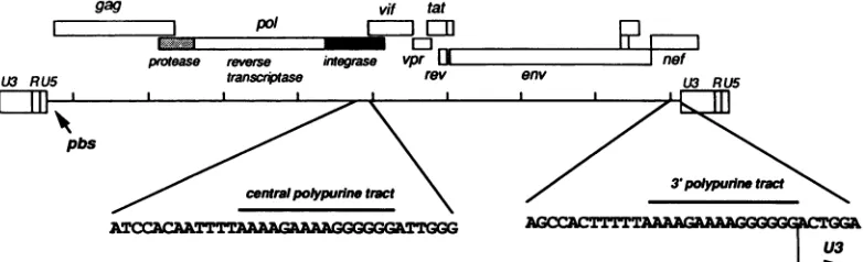
gap in unintegrated linear DNA molecules, located nearthecenter ofthe genome. Oligonucleotide primer extensionexperiments determinedthat thedownstreamlimitofthisgapcoincideswiththe last nucleotide ofa centralcopyof the polypurinetractfoundin all sequencedlentivirusgenomes.Other retroviruses have onlyone copy of the polypurine tract at the 5' boundary ofthe 3' long terminal repeat, which has been shown to determine initiation ofretroviral DNA plus-strand synthesis. We conclude from our observations that the central repeat of the polypurine tract can create an additional site for plus-strand synthesis initiation in
lentiviruses. The central single-stranded gap was notfound in circular DNA molecules, the vastmajority of them carrying only one long terminal repeat. This finding suggests that the generation of such circular molecules is associatedwithearly DNA ligation events.
Retroviruses replicate through reverse transcription of their RNAgenomesinto adouble-stranded DNA molecule.
Retroviral genes are expressed from an integrated copy of this double-stranded DNA genome, the provirus. Both strandsof the retroviral DNAgenome aresynthesizedby the virus-encoded reverse transcriptase, which has both RNA-andDNA-dependentDNApolymerase activities (10, 38, 41).
The template for minus-strand synthesis is viral genomic RNA,and thetemplate for plus-strand synthesisis thenewly reverse-transcribedminusstrand, following removal ofRNA
from theRNA-DNA hybrid bythe RNase H activity asso-ciated with reverse transcriptase (8, 11). Synthesis of the minus strand andsubsequently of theplusstrand is initiated nearthe 5'endof the respective template. The
correspond-ing short segments (minus- and plus-strand strong-stop
DNAs) are further transferred to the other end of the template, resulting in the formation of the long terminal repeats (LTRs), present ateachendoftheprovirus (10, 30, 37).Theprimer forminus-strandsynthesisisthe 3' end ofa tRNA moleculepackaged in the viral particle togetherwith
genomicviral RNA andhybridizedtotheprimer bindingsite
locatedatthe3' boundary of the U5 region (35, 36, 39). The initiation site of the plus strand has been deduced from analysisofreversetranscriptionreactions productsobtained either invitroorfromdetergent-disrupted virions andfrom the sequences of proviral molecular clones. This site is locatedimmediately3' ofapolypurinetract(PPT) represent-ingthe 5' boundaryof the U3region (21,22) (Fig. 1). Ithas beenproposedthat the PPT is usedtodefineanRNAprimer by specific cleavage of the RNAtemplateatthis sitebythe
reverse transcriptase-associated RNase H (20, 23, 27, 32).
Indeed,inmurineretroviruses, invitroreversetranscription reactions reveal RNA primers that remain associated with the elongating plus strand and are heterogeneous in length (7, 26). Unlike mostretroviruses, human immunodeficiency
*Correspondingauthor.
viruses (HIV) and other lentiviruses have twocopies of the PPT, oneat the border of the 3' LTR and the other located
nearthe middleof thegenome, withinthepol coding region (5, 12, 33, 43). Previous experiments on visna virus, the
prototypic lentivirus, have shown that unintegrated DNA
molecules display a single-stranded gap located approxi-mately in the same area(3, 14). Other viral DNA genomes carry single-stranded gaps or nicks. This is the case for
hepatitis B virus and cauliflower mosaic virus, in which synthesisof viral DNAgenomeinvolvesareverse transcrip-tion step.
We have tested here the hypothesis that the HIV central PPTrepresents anadditional initiation site for thesynthesis of theplus strand of HIV DNA. We show that HIV linear
unintegrated DNA molecules carry a discrete
single-stranded gapwhose downstream limit is the lastnucleotide of thecentralPPT, indicatingthatthisstructureislikelyused toinitiateplus-strand synthesisatthecenterof thegenome.
We also show that the single-stranded gap is absent from circular molecules. Since it is known that such circular molecules are formed only aftertransport ofreverse
tran-scription products into thenucleus,weproposethat nuclear
ligationand DNArepaireventsresult inboth theclosing of circular DNA molecules andfillingof thegap.
MATERIALS AND METHODS
Cells and viruses. MT4 cells were a gift from M. David
Hogan, Laboratory of Molecular Microbiology, National InstituteofAllergyand Infectious Diseases, Bethesda,Md. These cells, which are transformed by human T-cell leuke-mia virus type I, were shown to allow acute cytopathic
HIV-1 infection(13). CEM clone 13 cells (28)were derived from the human lymphoid cell line CEM (ATCC CCL119)
and express high levels of CD4 antigen. Cells were main-tained in RPMI 1640medium(GIBCOLaboratories)
supple-mented with 10% fetal calfserum.
The viral isolate used inourexperimentswasHIV-lb,,, (2),
2415
Vol. 65, No. 5
on November 10, 2019 by guest
http://jvi.asm.org/
gag
I I
U13
LUI
RU.vif tat
pol r
-m
rmn
- [Ir
protease reverse integrase vpr IUI
i5 transcrjptase rev
I X I I
pbs
centralpolypurinetract
m
,
J nefenv U3 RU5
I
3Ip3'polypvrdnetrc
ATC_CACATTTTAAAAGAAAAGGGGGGATTGGG
FIG. 1. Positions ofthe two PPTs onthe HIV-1 genome. Shadedareas in thepolopen reading frame indicate the regions codingfor different functions. pbs, Primerbindingsite,representingtheinitiation site forminus-strand synthesis.
recovered followingtransfection of COS cellswithinfectious
proviral molecular clone pBRU-2 (24a). Cellswere infected
atamultiplicity of 1:10 (1 50% tissueculture infective dose
per 10 cells) with virus from a frozen (-80°C) stock
pro-duced on MT4 cells and titrating 8 x 10⁵ tissue culture
infective doses per ml on MT4 cells. Following infection,
cultures weremonitoredfor cytopathic effect, which in the
described conditions of infection appeared approximately 3 days after infection. At that time, cells were harvested for DNA isolation.
Analysis of viral DNA. Low-molecular-weight DNA was extracted frominfected cells byHirtextraction(15).
Nucle-aseSi (Appligene, Strasbourg, France)wasusedat1.5U/,Ig
ofDNA afteradditionof1:10 volume of lOx S1buffer(300
mMsodiumacetate [pH 4.6], 500mMNaCl, 10mMZnCl2)
andincubatedat37°C. Fordouble digestions witha restric-tionenzymeandnuclease S1,DNA(10,ug)wasfirstdigested
with therestrictionenzyme;then lOx nucleaseS1 bufferand
nuclease S1 (15 U)wereaddedtothereaction mixture, and
the sample was further incubated at 37°C. DNA was then
subjected toelectrophoresison 1%agarosegelsthat didnot containethidiumbromideandanalyzed bySouthernblotting (34).
The nucleotide position numbers used here to describe DNA fragments startatthe firstnucleotide ofalinearHIV
DNA genome(5' end oftheU3 region inthe 5' LTR).
Twoprobes wereusedinhybridization experiments. The 5'probe was aPstIfragment spanningthegagregion from
positions1415to2839,andthe3' probewas aKpnIfragment
spanning the env region from positions 6343 to 9005. Both
probeswere obtainedfrom HIV-1 molecular clone pNL4-3
(1)andlabeled bythe randomhexamer method (6).
Primer extension. Amodification of theprimer extension techniquewasused, basedon TaqDNApolymerase, which
allowsmultiple cycles of denaturation, annealing, and
poly-merization. Thereaction mix contained 10 ,ug ofHirtDNA,
0.2 mMeachdeoxynucleotide, 1.25 U ofTaqDNA
polymer-ase (Perkin-Elmer Cetus N801-0045), 10 pmol of 5'-end-labeledprimer, and 0.5 ,ul of PMPEreagent (Stratagene) in 10 mM Tris-HCl (pH 8.3)-50mM KCl-2 mM MgCl2-0.01% gelatin, fora total reaction volume of50 ,ul. The first cycle included30sfor denaturationat92°C, 1minfor annealingat
50°C,and 1minforpolymerizationat72°C. Thefollowing59 cycles included 10 s at 92°C, 1 min at 50°C, and 1 min at 72°C. Asonlyoneprimerwasincludedinthereaction, this
multicycle primer extension method is distinct froma
poly-merase chain reaction and resulted in linear, not
exponen-tial,amplification ofthe reactionproduct.
Oligonucleotide primers were 5' end labeled with T4
polynucleotide kinase in the presence of [-y-32P]ATP to a
specific activityof 5 x 10⁵to106
cpm/10
pmol.Primerpol-1 (5'-ACA ATC ATC ACC TGC CAT CTG) anneals to theplus strand at position 5085. Primer pol-2 (5'-TCC AAA
GTG GAT CTC TGC TGT) anneals to the plus strand at
position4951.
The sameoligonucleotide primerswereusedtogeneratea
sequenceladderfromasingle-stranded M13template carry-ing the
HIV-lbFU
plus strand from positions 4688 to 5129.Sequencing was done by the method ofSangeret al. (29).
Primer extension and sequencereactions were analyzed in parallelon a6%polyacrylamide-8 Murea sequencinggel.
RESULTS
A single-stranded gap in HIV-1 unintegrated DNA. We
hypothesizedthat the existence ofanadditional plus-strand initiationsite in the HIV-1 genome could berevealedbythe presence ofa single-stranded region in HIV-1 unintegrated
DNAmolecules.Toexaminethispossibility,
low-molecular-weight DNA was selectively extracted (15) from acutelyHIV-1-infected MT4 cells(13), treated withnucleaseS1, and analyzedbySouthernblottingwithaprobe
spanning
mostofthe3'half of the HIV-1 genome. Figure2shows thekinetics of nuclease S1 digestion of HIV-1
unintegrated
DNA. Inundigested DNA,three bandswere observed. The
position
of the middle band, around 9.5 kb, isconsistentwith linear full-length molecules, and the position of the lowerband,
around 6kb, isconsistent with closed
(supercoiled)
circles. The upper band, which has an apparent size of approxi-mately 15 kb, is likely to be open (relaxed) circular mole-cules. After S min ofdigestionwith nuclease S1(15 U/10,ugofDNA),major changes occurred.The band
corresponding
tolinear molecules turned intoadoublet; the upper band had the size ofintact linear molecules (9.5 kb), and the lower one, at
approximately
9kb,
was consistentwith the sizeoflinearized circles withoneLTR. Simultaneously,the
closed-circles 6-kb band completely disappeared, while the
inten-sity oftheopen-circles bandincreased slightly.More
strik-ingly,anew5-kbbandappeared,correspondingtothe 3'half of linear molecules cut intotwo fragments by nuclease S1.
The otherfragment, of similar size, could be detected in a
distinct hybridization experiment with a 5' probe (data not
shown). This finding indicates the presence of a unique
nuclease S1-sensitive site in HIV unintegrated DNA mole-cules locatedapproximately atthe centerof the genome.
The complete disappearance of the closed circles, even
I I L-A
.W,CCACTTTTTAAAAC4AAAC,GGGGC-#k..
1
'0111..
on November 10, 2019 by guest
http://jvi.asm.org/
[image:2.612.114.505.74.193.2]STRUCTURE OF UNINTEGRATED HIV-1 DNA 2417
NT 5 15 30 60
kb -12
A 5 probe 1 2 3 4 5 6
3probe
1 2 3 4 5 6
kb
-12
9
." m s
Om 4m Mi -s
- 9
-3 - 5
-3
2
FIG. 2. Kinetics of nuclease S1 action on HIV-1
unintegr-.--DNA.Shown isaSouthern blot oflow-molecular-weight DNA from HIV-1-infected MT4 cells, hybridizedtoaproberepresentingmost
of the 3' half ofthe HIV-1 genome(see Fig. 3B). Lanes: NT, no
nuclease Si treatment;5, 15, 30, and 60,treatmentwith S1 nuclease (1.5 U/,ug of DNA) for 5, 15, 30, and 60 min, respectively. 0,Open
circles, L, linearmolecules; C, closed circles.
after5 min of nuclease Si treatment, can bebestexplained
by a previously described high sensitivity of supercoiled circular DNAmoleculesto nuclease Si (19). Therefore, we assumedthat at this stage ofincomplete nuclease Si diges-tion, the closed circles were cleaved at random sites, into
open circles ifonly one strand was cut (this explains the increase in open circles) and into linear molecules when Si nuclease further cleaved atthe resulting nicks.
With further digestion, the open-circles band gradually disappeared, togetherwith thefull-length linear molecules, while the band correspondingtolinearized one-LTRcircles remained unaffected. The gradual disappearance of open circleswasduetotheircleavageatrandom nicksthat relate to their relaxed physical state: Si cleavage at those sites linearized them. Meanwhile, linear molecules were further cleaved atthe central Si-sensitive site, generatingmore of theprobe-reactive 5-kb fragment: after 1 h oftreatment,very littleof the full-length linearmolecules remained.
The centralsingle-strandedgapisdiscreteand unique. We furtherattemptedto localize and define thestructureofthe HIV-1centralnucleaseSi-sensitive site. Figure 3A showsa Southern blot analysis ofnuclease Si cleavage products of
HIV-1 unintegrated DNA, obtained from acutely
HIV-1-infected MT4cells,combined withdigestionbytwodifferent restrictionenzymes. Intheseexperiments, complete
restric-tionenzymedigestionswereperformed, followed by partial
nucleaseSi digestion (20minwith15U/10 ,LgofDNA). The
digestion products were examined by hybridization to two different HIV-1 probes: a 5' probe, in the gag region,
spanning from nucleotide positions 1415 (from the start of
U3)to2839,anda3'probe,spanningmostof theenvandnef
regions, fromnucleotide positions 6343to 9005.
Inthe
HIV-ibr,
genome,PstI hasaunique site inthegagcoding region atposition 1415. Digestion ofHIV-1
uninte-grated DNA with PstI (Fig. 3A, lanes 3) cut the linear
molecules intwofragmentsof 8.3 and1.4 kb,the latterbeing
undetected by eitherof thetwo probes used in this
experi-B 5probe
Pst
ceitraI ppt 3probe
_
FIG. 3. Position of the single-stranded gap on the HIV-1 ge-nome. (A) Two autoradiograms from the same Southern blot,
hybridized to two different probes as indicated at the top. Low-molecular-weight DNA from HIV-1-infected MT4 cells was
ana-lyzedby nuclease S1 treatmenteither withnoprevious restriction enzymedigestion (lanes 1 and 2), after digestion with PstI (lanes 3 and4),orafterdigestion with BamHI (lanes 5 and 6). Lanes 1, 3, and
5, No nuclease S1 treatment; lanes 2, 4, and 6, treatment with nuclease S1 (1.5U/Iugof DNA).(B) Positions of the corresponding probesonthe HIV-1genome,relativetothe central PPT andtothe unique PstI andBamHI restrictionsites.
ment.PstI alsolinearizes circlesintoa9-kbproduct.
Nucle-aseSi treatmentofPstIdigestion products (lanes 4) yielded
a 5-kb fragment reactive with the 3' probe and a 3.3-kb
fragment reactive with the 5' probe. This result establishes
thatboth boundaries of the central single-stranded gap are discrete.
Afterdigestionwith BamHI (Fig. 3A, lanes5), whichhas a unique site in the env region at position 8520, the linear DNAmoleculeswerecleavedintotwofragmentsof
approx-imately 8.5 and 1.2 kb, both reactive with the 3' probe.
Treatment with nuclease Si (lanes 6) released a 3.7-kb
fragment detected by the 3' probe and a 4.9-kb fragment reactive with the 5' probe. The 1.2-kb fragment, which includes the 3' PPT, remained unaffected by nuclease Si treatment. When no restriction enzyme digestion was
per-formed before nuclease Si treatment, Si cleavage (lanes 2)
released a 5' 4.9-kb fragment and a 3' fragment of similar size. Overall, the results from these single- and
double-digestion experiments were consistent and could locate the
single-stranded gap aroundposition 4900, within 0.2 kb of the central PPTrepeatfound in the HIV-lbru sequence.
These experiments show that the central single-stranded
gapisuniquein the HIV-1genome. Indeed,the sizes of the
fragmentsreleasedby both the singleand double digestions
excluded thepresence of anothergap.
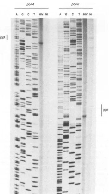
The3' boundaryof the gapis the central copyof the PPT. To locate the 3' boundary of the single-stranded gapmore
precisely relative to the central PPT found in all lentiviral
genomes, primer extension experiments were conducted,
using low-molecular-weight DNA from acutely HIV-1-in-fectedlymphoidcellsas atemplate.We usedoligonucleotide primers complementary to the viral plus strand, located
0-C
-VOL.65, 1991
on November 10, 2019 by guest
http://jvi.asm.org/
[image:3.612.316.555.74.279.2] [image:3.612.97.257.75.264.2]2418 CHARNEAU AND CLAVEL
pol-I pol-2
A G C T HIVNI A G C T HIVNl
pptl _
~~~ ~ ~ ~ ~ ~~ ~~~~~n
ppt
4-w
* _
_-FIG~~s.4. Loaizto
_o~~
of the..dontra limiothsige
stranded gap by oligonucleotide primer extension. Two primer extension reactions with two different oligonucleotide primers are
presented.Primerpol-1annealstothe HIV-1plusstrandatpositions 5085 to 5105, and primerpol-2 anneals at positions 4951 to 4971. Botholigonucleotides were12P5' end labeled and allowedtoprime
anextensionreaction,using low-molecular-weightDNAfrom HIV-1-infected cells (lanes HIV) or noninfected cells (lanes NI) as
templates as described in Materials and Methods. LanesA, G, C, and Tare sequencereactions fromasingle-stranded M13 template
carryingthe HIV-lbru,plusstrandfrompositions4688to5129,using
the indicated oligonucleotide asa primer. ppt,Centralcopy ofthe
PPT.
downstream to the central copy of the PPT, expecting
elongationof theseprimerstostoppreciselyatany
interrup-tion of the plus strand. To increase the sensitivity, the enzymeusedwasTaqlpolymerase,whichallowedustoheat denature extension reaction mixes and to carry several
cycles of extension for each reaction. Two different
an-tisense 5'-end-labeled oligonucleotide primers were used. Primer pol-2 anneals to the plus-strand 135 nucleotides
downstream of the PPT, and primer pol-i anneals 134
nucleotides further downstream. In parallel with extension
reactions, DNA sequence reactions were conducted, using
the same oligonucleotide primers on single-stranded
M13-HIVdlbrutemplates, enabling us toprecisely locate the stop in primer extension on the HIV-1bru genome (Fig. 4). With
both primers we observed a single, discrete stop in primer
extension that coincided with the last nucleotide of the
centralPPT. No equivalent stopwasfound with
low-molec-ular-weight DNA from uninfected cells. In addition, no
HIV-specific signal could be observed with a primer
com-plementary to the minus strand, located upstream of the
central PPT(datanot shown). This result demonstrates that the 3' boundary of the HIV-1 central single-stranded gap is defined by the central copy of the PPT. We can infer from this observation that the central PPT is used as an additional initiation site for the synthesis of the plus strand of HIV-1 DNA.
Thesingle-strandedgap isfoundexclusivelyonlinear DNA
molecules. Electrophoretic analysis of undigested and of PstI- orBamHI-digested unintegrated HIV-1 DNAshowed
an approximately equal proportion of linear and circular molecules inacutely HIV-infected MT4 cells. Indeed, after
PstI or BamHI digestion, the intensity of the shortened product resulting from digestion of linear molecules was
approximately equal to that of the 9-kb linearized circles,
found in both BamHI and PstI digestion reactions. Of interest is that no band with a size similar to that of the full-length linear molecules (corresponding to two-LTR lin-earized circles) could be observed in these reactions. This
means that the vast majority of circular molecules in the acutely HIV-1-infected MT4 system have onlyone LTR.
Several lines of evidence indicate that circular molecules do not carrya single-stranded gap. First, the closed circles by definition cannot be gapped, although they are highly sensitive to nuclease Si digestion (19). Second, the SI
digestion kinetics experiment (Fig. 2) shows that the 9-kb productcorrespondingtothelinearized one-LTR circleswas
still present, even after 1 h of treatment with nuclease Si, when native linear moleculeshad almost completely disap-peared. Third, afterdigestion with PstI andBamHI, which
cutonlyoncein the
HIV-1bru
genome,nucleaseSitreatment did not generate products of a size compatible withfrag-ments releasedfrom gapped circular molecules. For
exam-ple, the BamHI-nuclease Si double digestion failed to
re-leasea5.5-kbfragment, which would span from the BamHI sitetothe gap in acircularone-LTRmolecule andwouldbe reactive with the 5' probe. Similarly, the PstI-nuclease S1 doubledigestiondid notrelease any3'-probe-reactive
prod-uctof5.7kb, which would be the distance between the gap site and the PstI site in a circular one-LTR molecule.
Overall, theproductsobserved in the doubledigestswereof
a sizecompatible only with DNA fragments released from
native, linear two-LTR molecules.
Finally, we haveobserved that theproportion of circular
versus linear molecules could vary with the source of the
analyzedDNA. Figure 5 showsacomparisonofnucleaseSi
treatment of low-molecular-weight DNA from MT4 cells harvested 2daysafter HIV-1 infection(lanes1 to4) and from CEM cells harvested 9days after infection (lanes 5to8). In the CEM cells, theproportion oflinear molecules waslow,
as shown by the lower proportion of 9.5-kb band than of open andclosedcirclesonundigestedlane 5. Anothersign of the lowproportion of linear moleculesis visible on lane 7,
where the intensity of the 1.2-kb fragment released by BamHI is weaker than that of the 9-kb linearized circles.
Coincidentally, theamountofsubgenomicproduct released
bynucleaseS1digestionof thesameDNAsampleswaslow.
In the MT4 cells, in which more linear molecules were
found, thequantityofsubgenomic S1 digestion productwas
proportionally muchhigher than intheCEM cells. DISCUSSION
We have observed that HIV-1 unintegrated linear DNA
molecules, which accumulate in acutely infected CD4+
lymphoidcells, carryashort, unique, single-stranded region
at the center of the genome. Both ends of this
single-J. VIROL.
on November 10, 2019 by guest
http://jvi.asm.org/
[image:4.612.97.273.71.383.2]STRUCTURE OF UNINTEGRATED HIV-1 DNA 2419
1 2 3 4 5 6 7 8 q U R
U3 R
kb
kb 1U3RU5
3'ppt
- 12
nL
-9
- 5
- 3
a p
Asy~~~C"- PptU
41
-2
CWOO~L~
[image:5.612.58.300.75.285.2]_ 1 a
FIG. 5. Nuclease S1 sensitivity of HIV-1 unintegrated DNA fromtwodifferent cell cultures. Lanes: 1to4,low-molecular-weight DNA from MT4 cells harvested 2 days after infection; 5 to 8, low-molecular-weightDNA fromCEM cells harvested 9 days after infection. Unintegrated HIV-1 DNAwasanalysed by nuclease S1
treatment either with no previous restriction enzyme digestion
(lanes 1, 2, 5, and 6)orafter digestion with BamHI (lanes 3,4, 7,and 8). Lanes 1, 3, 5, and 7, No nuclease S1treatment;lanes 2, 4, 6, and 8, treatment with nuclease S1 (1.5 U/Ipgof DNA). Both Southern blots were hybridized to the 3' probe shown in Fig. 3B. Both autoradiogramsareovernightexposures.
stranded gap are discrete. We have established that the 3'
boundary of this single-stranded element coincides with a PPT found at position 4800 in the HIV genome, an exact repeat of the structure normally found in all retroviruses next to the U3 region ofthe LTR (at position 9070 in the HIV-1provirus).We havenotdeterminedthe 5'boundaryof thegapandthereforecannotbecertainofitssize.However, analysis of the sizes of fragments released from nuclease
Sl-treated linear molecules following restriction enzyme
digestions and correspondingtoeither the3' orthe5' side of thegapsuggeststhat thisgapisshort, probably lessthan 100
nucleotides inlength.
In other retroviral models, the PPT has been shown to determine in vitro the initiation site of retroviral DNA
plus-strand synthesis (20, 23, 27, 32). The DNAgenomes of
hepatitis B and cauliflower mosaic viruses, which are syn-thesizedthroughreverse transcription ofanRNAtemplate,
carry severalsingle-stranded structures. Inparticular, there aretwoplus-strand discontinuitiesin the cauliflower mosaic
virus genome that are defined by short PPTs which likely
correspondto plus-strand initiation sites (25, 42). Our
find-ings indicate that in HIV-1, and most likely also in other
lentiviruses,thecentralPPTis used in vivoas anadditional
primingsite forplus-strand synthesis. Indeed, primer
exten-sion experiments shown here reveal that the plus-strand
DNA3'tothegapstartsexactlyatthelast nucleotide of this PPT. This findingestablishes that this structure determines
precise andspecificprimingofplus-strandDNAatacentral
position in the genome. Because we used Taq polymerase
andnotreversetranscriptase in theprimerextension
exper-iments, we could not conclude whether the RNA primer
FIG. 6. Model for HIV reverse transcription. 1, Minus strong-stop synthesis; 2, first (minus-strand) template transfer and plus-strandstrong-stopsynthesis, initiatedatthe 3'PPT; 3, progression ofminus-strandsynthesis andinitiation ofplus-strand synthesisat
the centralPPT; 4, second (plus-strand) template transfer; 5,
forma-tion of the LTRsby stranddisplacement and synthesis and
progres-sion ofsynthesisof the 5' half of the plus strand; 6, lineargapped DNAmolecule; 7, followingstep4, ligation atboth boundaries of the LTR, before strand displacement and synthesis canstart, and
progression of plus-strand synthesis; 8, ligation at the gap and
formationofaone-LTRclosed circle.
correspondingto the central PPT remained attachedtothe nascentDNAstrand, asdescribed for other retroviruses at the 3' PPT (7, 26).
It has been shown in avian retroviruses that plus-strand synthesiscanstart atsitesdistinct from the 3'PPT, resulting
inplus-stranddiscontinuities (16, 18). However,these initi-ation sites donotseemtobeuniqueorwelldefined,andthey
donot result in adiscrete single-stranded structure
compa-rabletowhatwedescribe here. Itislikelythattheresulting plus-strand segmentsareeliminatedby stranddisplacement
events, as was shown in mellitin-permeabilized virions (4).
InHIV, sucha stranddisplacement islikely tooccuratthe 3' PPT, to generate the 5' end of linear molecules (Fig. 6, steps4and5),butseemsnottooccuratthe central PPT.We
cannot explain why the upstream limit of the gap remains discrete. The gapped linear molecules represent a defined species: they are full-length, double-stranded molecules on
which synthesis of the 5' half of the viral plus strand is
O- ^ -12
L- 9-9
C- I
- 5
f-13
0-
4l.IJ
L
-c-
4M
-1 VOL. 65, 1991
I
9
(ij19
on November 10, 2019 by guest
http://jvi.asm.org/
[image:5.612.317.551.79.418.2]stopped near the initiation site ofthe 3' half. It is possible, however, that similar to what is found incauliflower mosaic virus (25), elongation of the upstream HIV plus strand engages in a brief stranddisplacement event. Further studies
are needed to clarify this point.
As a whole, our findings support a model of retroviral DNA synthesis (Fig. 6) in which linear molecules require a
stranddisplacement step for LTR synthesis (4, 26), whereas one-LTR circles are the results of ligation events. Indeed, closed circular DNA molecules, whicharefound exclusively in the nucleus (17, 31), could be generated by ligation at both boundaries of the LTR and at the gap (Fig. 6, steps 7 and 8), following the proposed "intrastrand" plus-strand template transfer (24). This, in turn, could result from early nuclear transport ofuncompleted DNA molecules.
Finally, it remains to be understood why lentiviruses, unlike other retroviruses, have developed and conserved a repeat of the PPT at the center of the genome. The most likely explanation is that it allows progression of plus-strand
synthesis before elongation of the minus strand is complete (Fig. 6, step 3), probably resulting in a gain of time in DNA synthesis. The precise location of the PPT repeat at the
centerof the genome supports this hypothesis: an additional
plus-strandinitiation at this site could allow its elongation to reach the 3' PPT approximately at the same time minus-strand synthesis is completed, the latest being required for
plus-strand template transfer (Fig. 6, step 4). The LTRs can then be synthesized as plus-strand synthesis is being
com-pleted in the 5' half of the genome (Fig. 6, step 5). Since lentiviruses, which are not transforming viruses, rely
essen-tially on reverse transcription for their propagation, this feature could constitute an evolutionary advantage. The initiation of HIV plus-strand synthesis at the center of the genomeis also interesting in view of recent observations that in unstimulated normal human lymphocytes, synthesis of the HIV minus strand appears to be arrested approximately halfway along the genomic RNA template (44). Dependence offull-length DNA synthesis on cell growth had also been described for avian viruses (9, 40). If the described stop in HIV minus-strand synthesis is located beyond the central PPT, it may allow early synthesis of a plus strand covering the whole 3' half of the genome and may direct faster
completion of double-stranded full-length DNA molecules uponfurther lymphocyte activation.
ACKNOWLEDGMENTS
We thank Luc Montagnier, in whose laboratory these studies wereconducted, Marc Alizon, Olivier Danos, and Dominic Dwyer for helpful comments during preparation of the manuscript, and Agnes Cordonnier,who gave us some of the DNAs analyzed here. P.C. is a recipient of a grant from the Agence Nationale de Recherches surle SIDA.
REFERENCES
1. Adachi, A., H. E. Gendelmann, S. Koenig, T. Folks, R. Willey, A. Rabson, and M. A. Martin. 1986. Production of acquired immunodeficiency syndrome-associated retrovirus in human and nonhuman cells transfected with an infectious molecular clone. J. Virol. 59:284-291.
2. Barre-Sinoussi, F., J. C.Chermann, F. Rey, M. T. Nugeyre, S. Chamaret, J. Gruest, C. Dauguet, C. Axler-Blin, F. Brun-Vezinet, C. Rouzioux, W. Rozenbaum, and L. Montagnier. 1983. Isolation of a T-lymphotropic retrovirus from a patient at risk for acquired immune deficiency syndrome (AIDS). Science 220:868-871.
3. Blum,H.E., J. D. Harris, P.Ventura, D. Walker, K. Staskus, E. Retzel, and A. T. Haase. 1985. Synthesis in cell culture of the
gapped linear duplex DNA of the slow virus visna. Virology 142:270-277.
4. Boone, L. R., and A. M. Skalka.1981.Viral DNAsynthesizedin vitro by avianretrovirus particles permeabilized with mellitin. II. Evidence for a strand displacement mechanism in plus-strandsynthesis. J. Virol. 37:117-126.
5. Chakrabarti, L., M. Guyader, M. Alizon, M. D. Daniel, R. C. Desrosiers, P. Tiollais, and P. Sonigo. 1987. Molecularcloning andnucleotide sequence of simian immunodeficiency virus from macaques. Nature (London) 328:543-547.
6. Feinberg, A. P., and B. Vogelstein. 1983. A technique for radiolabeling DNArestriction endonuclease fragments to high specific activity. Anal. Biochem. 132:6-13.
7. Finston, W. I., and J. J.Champoux. 1984. RNA-primed initia-tion of Moloney murine leukemia virus plus strands by reverse transcriptase in vitro. J. Virol. 51:26-33.
8. Friedrich, R., and K. Moelling. 1979. Effect of viral RNAse H on the avian sarcomaviral genome during early transcription invitro. J. Virol. 31:630-638.
9. Fritsch, E. M., and H. M.Temin. 1977. Inhibition of viral DNA synthesis in stationary chicken embryo fibroblasts infected with avian retroviruses. J. Virol. 24:461-469.
10. Gilboa, E., S. Mitra, S. Goff, and D. Baltimore. 1979. Adetailed model of reverse transcription and tests of crucial aspects. Cell 18:93-100.
11. Grandgenett, D. P., G. F. Gerard, and M. Green. 1972. Ribo-nuclease H: a ubiquitous activity in virions of ribonucleic acid tumorviruses. J. Virol. 10:1136-1142.
12. Guyader, M., M. Emerman, P. Sonigo, F. Clavel, L. Montagnier, and M. Alizon. 1987.Geneticorganization and transactivation of human immunodeficiency virus type 2. Nature (London) 326: 662-669.
13. Harada, S., Y. Koyanagi, and N. Yamamoto. 1985.Infection of HTLV-III/LAV in HTLV-I-carryingcells MT-2 and MT-4and application in a plaque assay. Science 229:568-566.
14. Harris, J. D., J. V. Scott, B. Traynor, M. Brahic, L.Stowring, P. Ventura, A. T. Haase, and R. Peluso. 1981. Visna virus DNA: discovery of a novelgapped structure. Virology 113:573-583. 15. Hirt, B. 1967. Selective extraction of polyoma DNA from
infected mouse cell cultures. J. Mol. Biol. 26:365-369. 16. Hsu, T. W., and J. M.Taylor. 1982. Single-stranded regions on
unintegratedavianretrovirus DNA. J. Virol. 44:47-53. 17. Kim, S., R. Byrn, J. Groopman, and D. Baltimore. 1989.
Temporal aspects of DNA and RNA synthesis during human immunodeficiency virus infection: evidence for differential gene expression.J. Virol. 63:3708-3713.
18. Kung, H. J., Y. K. Fung, J. E. Majors, J. M. Bishop, and H. E. Varmus. 1981. Synthesis of plus strands of retroviral DNA in cells infected with avian sarcoma virus and mouse mammary tumorvirus. J. Virol. 37:127-138.
19. Mechali,M., A. M. deRecondo, and M. Girard. 1973. Actionof theS1endonuclease from Aspergillus oryzae on simian virus 40 supercoiled component I DNA. Biochem. Biophys. Res. Com-mun.54:1306-1320.
20. Mitra, S., M. Chow, J. J. Champoux, and D. Baltimore. 1982. Synthesisof murine leukemia virus plus strong stop initiates at aunique site.J.Biol. Chem. 257:5983-5986.
21. Mitra, S., S.Goff,E.Gilboa, and D. Baltimore. 1979. Synthesis of a 600 nucleotide-long plus-strand DNA by virions of Moloney murine leukemia virus. Proc. Natl. Acad. Sci. USA 76:4355-4359.
22. Olsen, J., and K. Watson. 1982.Reverse transcription of avian myeloblastosis 35S RNA. Early synthesis of plus-strand DNA of discrete size in reconstructed reactions. Nucleic Acids Res. 10:1009-1027.
23. Omer, C. A., R. Resnick, and A. J. Faras. 1984. Evidence for involvement of an RNA primer in initiation of strong-stop plus-strand DNA synthesis during reverse transcription. J. Virol. 50:465-470.
24. Panganiban, A. T., and D. Fiore. 1988. Ordered interstrand and intrastrand DNA transfer during reverse transcription. Science 241:1064-1069.
24a.Peden, K.Unpublished data.
on November 10, 2019 by guest
http://jvi.asm.org/
STRUCTURE OF UNINTEGRATED HIV-1 DNA 2421 25. Pfeiffer, P., and T. Hohn. 1983. Involvement of reverse
tran-scription in the replication of cauliflower mosaic virus: a de-tailed model and test of some aspects. Cell 31:781-789. 26. Rattray,A. J., and J. J. Champoux. 1987. The role of Moloney
murine leukemia virus RNase H activity in the formation of plus-strand primers. J. Virol. 61:2843-2851.
27. Resnick,R., C. A. Omer, and A. J. Faras. 1984. Involvement of retrovirus reverse transcriptase-associated RNase H in the initiation of strong-stop (+) DNA synthesis and the generation ofthelongterminalrepeat. J. Virol. 51:813-821.
28. Rey, M.A., B. Krust, A. G. Laurent, L.Montagnier, and A. G. Hovanessian. 1989. Characterization of human immunodefi-ciency virustype 2envelope glycoproteins: dimerization ofthe glycoproteinprecursorduring processing. J. Virol. 63:647-658. 29. Sanger,F., S. Nicklen, and A. R. Coulson. 1977. DNA sequenc-ing with chain-terminating inhibitors. Proc. Natl. Acad. Sci. USA74:5463-5468.
30. Shank, P. R., S. H.Hughes, H. J. Kung, M. J. E., L. Quintrell, R. V.Guntaka, J. M. Bishop, and H. E. Varmus. 1978. Mapping unintegratedavian sarcoma virus DNA: termini of linear DNA bear300 nucleotides present once ortwice in two species of circular DNA.Cell 15:1383-1395.
31. Shank, P. R., and H. E. Varmus. 1978. Virus-specific DNA in the cytoplasmof avian sarcoma virus-infected cells is a precur-sor to covalently closed circularviral DNA in the nucleus. J. Virol. 25:104-114.
32. Smith, J. K., A.Cywinski, and J. M. Taylor. 1984. Initiationof plus-strand DNA synthesis during reversetranscription ofan avianretrovirus genome. J.Virol.49:200-204.
33. Sonigo, P., M. Alizon, K. Staskus, D. Klatzmann, S. Cole,0. Danos, R. E., P.Tiollais, A. Haase, and S. Wain-Hobson. 1985. Nucleotide sequenceofthevisnalentivirus: relationshiptothe AIDS virus. Cell43:369-382.
34. Southern,E.1975. Detection ofspecificsequencesamong DNA fragments separated by gel electrophoresis. J. Mol. Biol. 98: 503-517.
35. Staskus,K.A., M. S.Collett, and A. J. Faras. 1976.Initiation of
DNAsynthesisby the avian oncormavirus RNA directed DNA polymerase: structural andfunctional localizationof the major speciesofprimerRNAontheoncornavirus genome. Virology
71:162-168.
36. Taylor, J. M., and R. Ilmensee. 1975. Site onthe RNA ofan
avian sarcoma virus at which primer is bound. J. Virol.
16:553-558.
37. Temin, H. M. 1981.Structure,variation andsynthesisof
retro-viruslong terminal repeats. Cell 27:1-3.
38. Varmus, H., and R. Swanstrom. 1982. Replication of retrovi-ruses, p. 369-512. In R. Weiss, N. Teich, H. Varmus, and J. Coffin (ed.), RNA tumorviruses, vol. 1. Cold Spring Harbor Laboratory, Cold Spring Harbor, N.Y.
39. Varmus, H. E., S. Heasley, H. J. Kung, H. Oppermann, V. Smith,J. M.Bishop, and P. Shank. 1978. Kinetics ofsynthesis, structureandpurification of avian sarcoma virus-specific DNA made in the cytoplasm of acutely infected cells. J. Mol. Biol. 120:55-82.
40. Varmus, H. E., T. Padgett, S. Heasley, G. Simon, and J. M. Bishop. 1977. Cellular functions are required for the synthesis and integration of avian sarcoma virus-specific DNA. Cell 11:307-319.
41. Verma, I. M. 1975. Studies on reverse transcriptase of RNA tumorviruses. III. properties of purified Moloney murine leu-kemia virus DNApolymerase and associated RNase H. J. Virol. 15:843-854.
42. Volovitch, M., G. Grugeon, and P. Yot. 1978. Studies on the single strand discontinuities in the cauliflower mosaic virus genome.NucleicAcids Res. 5:2913-2925.
43. Wain-Hobson, S., P. Sonigo, 0. Danos,S.Cole, and M. Alizon. 1985. Nucleotide sequence of the AIDS virus, LAV. Cell 40:9-17.
44. Zack, J. A., S. J. Arrigo, S. R. Weitzman, A. S. Go, A. Haislip, and I. S. Y. Chen. 1990. HIV-1 entry into quiescent primary lymphocytes: molecular analysis reveals a labile, latent viral structure. Cell 61:213-222.
VOL.65, 1991