Dissertation submitted for
BRANCH V DEGREE EXAMINATION
THE TAMILNADU
Dr.M.G.R. MEDICAL UNIVERSITY
CHENNAI, TAMILNADU
CERTIFICATE
This is to certify that the Dissertation on “MORPHOLOGY AND MORPHOMETRIC ANALYSIS OF THE HUMAN MITRAL VALVE COMPLEX”, is a bonafide work, carried out in the Upgraded Institute of Anatomy, Madras Medical College, Chennai - 600 003, during 2003 - 2006 by
Dr.M.Kavimani, under my Supervision and Guidance in partial fulfillment of
the regulation laid down by the Tamilnadu Dr.M.G.R. Medical University, M.S.Anatomy Branch-V Degree Examination to be held in September 2006.
DEAN
Madras Medical College
Chennai - 600 003. Prof.Dr.CHRISTILDA FELICIA JEBAKANI Director and Professor, M.S., Upgraded Institute of Anatomy ,
ACKNOWLEDGEMENT
I wish to express my sincere gratitude to my most beloved and respected teacher and guide, Prof. Dr. CHRISTILDA FELICIA JEBAKANI, M.S., Director and Professor, Upgraded Institute of Anatomy, Madras Medical College, Chennai, who has motivated me to conduct this study and provided all the guidance and facilities available in the Institution. But for her untiring and enthusiastic support and valuable suggestions this study would not have been possible.
My sincere thanks to Dr.KALAVATHI PONNIRAIVAN, B.Sc., M.D., Dean, Madras Medical College, Chennai for permitting me to avail the facilities in the Institution during the course of my study.
I express my sincere thanks to Director and Professor, Institute of forensic medicine, Madras Medical College, Chennai for permitting me to collect the specimens from the Institute of forensic medicine for my study.
I wish to thank Dr.RAJA SANTHOSAM, Director and Professor, Institute of Cardiothoracic Surgery, Madras Medical College, Chennai for his
excellent guidance and unflinching support showed me the right path to complete this work.
I am extremely thankful to cardiothoracic surgery postgraduate students
DR.RAMAKRISHNAN, Dr.PONNUSWAMY, Dr.GOPAL, Dr.GIRISH,
for their help to conduct the clinical studies.
I am grateful to Prof.Dr.T.R.KALAVATHY, M.S., (Rtd.), Director and Professor, Upgraded Institute Anatomy, Madras Medical College, Chennai for her constant encouragement and support during the study.
My sincere thanks to Dr.I.JEYARAJ, M.S., Tmt.M.S. THENMOZHI,
Tmt.M.C.INDIRAKUMARI, Dr.CHEZIAN for their appreciation and
encouragement throughout my dissections.
I am thankful to Dr.IWAN JAMES, Dr.A.SHARMILA, Dr.V.SATHIALAKSHMI, Dr.M.VIJAYALAKSHMI, Dr.A.SENTHAMIL
SELVI, Dr.S.SUMATHILATHA for their constant support and
encouragement.
I wish to thank Dr.P.MURUGESAN, Dr.SATHISH, Dr.KARTHIK
for their constant support.
I am thankful to Dr.PREETHI, Dr.JAYANTHI NANADEEPAM,
Dr.JAYANTHI, Dr.ANITA for their supportive and encouraging throughout
my study.
I express my sincere thanks to Mr.JABBAR, P.DEVARAJAN,
G.ANKIAH, Mr.SUGENDRAN, who helped with making arrangements for
I am thankful to the workers in the Forensic Medicine Department who helped me in collecting specimens
I am very grateful to my wife Dr. M. SARASWATHI M.V.Sc.,
(Surgery) and my parents Mr.G.MOGANARANGAN and M.VASANTHA,
Dr.M.LATHASARATHY, Dr.MURUGASARATHY and Dr.M.ANBUMALAR,
CONTENTS
CHAPTER
NO. TITLE
PAGE NO.
1 INTRODUCTION 1
2. AIM OF THE STUDY 4
3. REVIEW OF LITERATURE 5
4. MATERIALS AND METHODS 30
5. DEVELOPMENTAL ANATOMY 35
6. OBSERVATION 36
7. DISCUSSION 53
8. CONCLUSION 71
1
INTRODUCTION
The Search for TRUTH is in one way hard And in another easy
For it is evident that no one can master it fully Nor miss it entirely
But each adds a little to our knowledge of Nature And from the facts assembled
There arises grandeur
- Aristotle (384 – 322Bc)
The opening of a new field of surgical endeavor often arouses interest in the detailed study of the anatomy of the involved part of the body.
As a result of such studies, current notions may be so changed and extended so as to understand better the morphologic structures of the organ and to provide a scientific basis for its function.
The impetus given to mitral valve surgery in the course of the last few years has prompted a thorough revision of our knowledge concerning the anatomy of the normal mitral valve.
2
Normal anatomy of mitral value complex
The mitral value complex includes mitral annulus, leaflets of the mitral valve, chordae tendineae and papillary muscles. Mitral annulus – the mitral annulus consists of a collagenous ring where the lamina fibrosa of the valve leaflets are attached.
Leaflets of the mitral valve – The leaflets of the mitral valve are two, anterior and posterior hence called bicuspid valve. These leaflets are separated by two deep indentations are known as commissures, namely anterolateral and posteromedial commissures.
The base of anterior leaflet is attached to the anteriomedial margin of the annulus and extends between right and left fibrous trigones. The anterior leaflet consist of a thick rough zone close to the free margin and a thin clear zone extending upto the annulus (Basal zone is absent in this leaflet).
The free margin and the ventricular surface of the rough zone provide attachments to free chordae and rough zone chordae. The anterior leaflet presents two peculiar and strongest strands of rough zone chordae derived from anterolateral and posteromedial papillary muscles. These are called strut chordae.
3
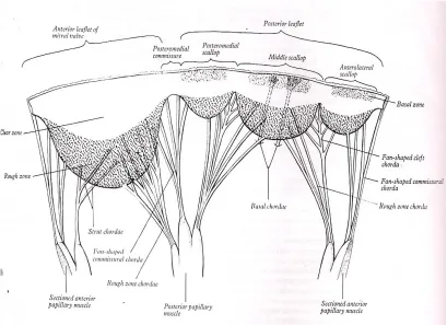
The clefts and commissures give attachment to fan shaped chordae. Each scallop of the posterior leaflet consists of three zones – rough, clear and basal. Ventricular surfaces of the rough and basal zones are connected to the chordae tendineae.
Chordae tendineae – There are endothelial covered collagenous threads. The chordae are classified according to their attachments to the leaflets as follows – commissural, cleft, rough zone, strut and basal chordae.
The commissural and cleft chordae are fan shaped, and are attached to the indentations and margins of adjacent leaflets or scallops. The rough zone chordae attached to close to the free margin. Strut chordae is two peculiar and strongest strands of rough zone chordae. Basal chordae extend from the ventricular wall to the basal zone of the leaflets.
Papillary muscles
These are conical muscular projections, usually two in number – anterior (anterolateral) and posterior (Posteromedial). The base of the anterior papillary muscle is attached to the sternocostal surface. The posterior papillary muscle is attached to the inferior wall of the left ventricle.
AIM OF THE STUDY
Normal mitral valve function depends upon the anatomic and
mechanical integrity of the atrioventricular ring, the valve leaflets, chordae
tendineae and the papillary muscles.
While there is no dispute in accepting that the mitral valve forms a
continuous skirt hanging down from the mitral annulus, there is no consensus
as to how many leaflets are in the valve or as to what constitutes the point of
separation of one leaflet from the other.
Advances in echocardiography, invasive cardiology (including balloon
mitral valvuloplasty) and surgical reconstruction of mitral valves necessitate an
appreciation of the many variations in the anatomy of the mitral valve.
The classical description of the mitral valve found in the textbooks of
anatomy is inadequate for the need of the cardiac surgeon. Similarly the
importance of the valvular structures and the myocardium in the mechanism of
valve closure requires a new appraisal in view of recent observations.
The aim of the present study is to analyse the morphologic and morpho
metric details of the human mitral valve complex.
This study will also be of much help for mitral valve procedures such as
mitral commissurotomy, commissuroplasty, valvuloplasty and artificial
REVIEW OF LITERATURE
MITRAL ANNULUS
Henry Gray (1858) said that the mitral annulus is not a simple fibrous
ring, but comprises elements varying greatly in consistency, with which the
valvular laminae fibrosae become continuous. These variations are of the
greatest functional significance, allowing major changes in the annular shape
and dimensions at different stages of the cardiac cycle, ensuring optimal
efficiency in valvular actions of the mitral complex and its equally important
role in controlling inflow / outflow patterns through the left ventricle.
MC Alpine WA (1975) disfavoured the term annulus, preferring to
describe the sheetlike fibrous area as the aortoventricular membrane that
extended around the subvalvar region.
A.K.Datta (1986) described that the mitral annulus consists of a
collagenous ring where the lamina fibrosa of the valve leaflets are attached.
Hutchins GM et al., (1986) reported that the floppy mitral valve is
associated with disjunction at the mitral annulus. A separation between the
atrial wall - mitral valve junction and the left ventricular attachment is known
as disjunction.
Angelini A, et al., (1988) studied about mitral annular circumference
and demonstrated pronounced variations not only from heart to heart but with
They traced prongs of fibrous tissue from each of the fibrous trigones
which were not continuous around the orifice. They also say that the mitral
annulus opposite the area of valvar fibrous continuity tends to be "weaker" in
terms of lacking a well formed fibours cord. This is the area affected in
"annular dilatation" and also most often involved in calcification of the mitral
annulus. With severe dilatation, the minor axis of the valvar orifice becomes so
distended that the leaflets which are of fixed lengths, become unable to
approximate each other.
Kitzman DW, Edwards WD (1990) found that with advancing age
there are significant increases in heart weight, ventricular septal and probably
left ventricular free wall thickness and in valve circumferences. In the
myocardium there are increases in fat, collagen, elastin and lipofuscin. The
geometry of the heart changes as well, due to decreasing base to apex
dimension, right ward shift and dilatation of the aortic root, and left atrial
dilatation. The aortic and mitral valves thicken and become fibrotic along their
oppositional surfaces, and their mitral annuli are the sites of collagen
degeneration, lipid accumulation and calcification.
Tirone E, David M.D., (1994) described that the interactions between
the mitral valve and left ventricle are complex and not yet completely
understood. However, continuity between the papillary muscles and the mitral
annulus is probably the most important factor in this relationship because
severance of the chordae tendineae in experimental animals causes a significant
drop in left ventricular systolic function as assessed by load independent
parameters. This deterioration in ventricular function is even more marked in
evidence that maintenance of papillary muscle - mitral annular continuity
during mitral valve surgery is beneficial to left ventricular function and clinical
outcome. That is one of the reasons why the mitral valve should be repaired
rather than replaced in patients who need mitral valve surgery. If replacement
is necessary, the chordae tendineae should be preserved. If preservation of
chordae tendineae is difficult because of calcification, fibrosis or for other
reasons it is possible to resuspend the papillary muscle with expanded tetra
fluoroethylene sutures. Preservation of chordae tendineae during mitral valve
replacement is also important because it prevents spontaneous rupture of the
posterior wall of the left ventricle, a rare but dreaded complication of this type
of surgery.
Gerda L. Van Rijk-Zwikker, M.D. et al., (1994) viewed mitral valve
from left atrium and described the mitral annulus consisting arbitrarily of two
areas - Anterior and posterior to the commissures. The anterior commissure is
also called the anterolateral or superior commissure (SC) Starting at the
superior commissure and rotating clockwise, the annulus (and left atrium) is
adjacent to a small part of the left ventricular free wall (LVFW), the left fibrous
trigone (LFT) and the left coronary cusp of the aortic valve (LCC). The
non-coronary cusp (NCC) is the next structure that borders the annulus, then the
right fibrous trigone (RFT), the interventricular septum and the posteromedial
or inferior commissure (IC) (Fig.1).
Tetsuro Sakai et al., (1999) measured the distance from the tip of the
papillary muscle to its corresponding mitral annulus in 57 normal cadaveric
hearts for a guide to the resuspension procedure in mitral valve replacement.
muscle distances of the mitral apparatus are similar in 2, 4, 8 'O' clock positions
and correlate with the mitral annular diameter (Fig.2).
Syho (2002) reported that the mitral annulus marking the hinge line of
the valvar leaflets is more D shaped than the circular shape portrayed by
prosthetic valves. The straight border accommodates the aortic valve allowing
the latter to be wedged between the ventricular septum and the mitral valve. In
this region, the aortic valve is in fibrous continuity with one of the two leaflets
of the mitral valve. Expansions of fibrous tissues at either extreme of the area
of continuity form the right and left fibrous trigones. The atrioventricular
conduction bundle passes through the right fibrous trigone. Although the term
mitral annulus implies a solid ring-like fibrous cord to which the leaflets are
attached, this is far from the case. In the area of aortic - mitral fibrous
continuity, the distal margin of atrial myocardium over the leaflet defines the
hinge line. When viewed from the ventricular aspect however the hinge line is
indistinct since the fibrous continuity is an extensive sheet (Fig.3).
Skandalaki's Surgical Anatomy (2004) said that the mitral valve
leaflets insert on the left atrioventricular fibrous mitral annulus.
COMMISSURES
Henry Gray (1858) said that the two deep indentations which are
regularly positioned and receiving unique fan shaped commissural chordae
tendineae are anterolateral and posteromedial commissures. Mitral
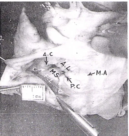
Rusted.I.E., et al., (1952) emphasized the localizing importance of the
anterior muscle in "Commissurotomy" (Valvulotomy) for mitral stenosis,
stating that in only 1.5 percent of their specimens was the muscle atypical and a
poor guide to the commissure between the CUSPS, as determined both by
inspection and palpation. A single muscle points directly toward the
commissure, either by a well defined apex, by a groove upon it or by an
imaginary line projected along it (Fig.4). When the muscle is double, a line
along the posterior aspect of the more anterior muscle points toward it and
when triple the middle of the three muscles points toward it. The chordae from
this muscle may also serve as a guide to the commissure (since they go to both
anterior and posterior cusps), for in more than half the cases a distinct,
hammock like groove leading to the commissure could be palpated (the
chordae of the posteromedial muscle were rarely (10%) a good guide to the
commissure). They suggested that the chordae from the anterior muscle might
be especially useful as a guide in those cases of mitral stenosis. He presented
further anatomical data including measurements of the normal bicuspid valve.
They emphasized also the difference between the normal commissures
or areas of fusion at the bases of valves and the pathological commissures
produced by further fusion of the edges of the valves. In their material the
normal commissures averaged about 0.7 to 0.8 cm in length, so that a
commissure which upon palpation is much greater than 1 cm long can be
supposed to be formed in part by abnormal fusion. They also pointed out that
thickening of the chordae tendineae may interfere with the function of the
valvulotomy cannot be expected to relieve valvular stenosis.
Michele A. Chiechi et al., (1956) studied 105 normal human hearts and
stated that the posterior portion of the mitral valve is the least gifted with
valvular tissue and therefore the weakest part of the valve. the preponderance
of valvular incompetence at the region of the posterior commissure is probably
due to this shortage of valvular tissue in this region. The posterior junctional
tissue could be destroyed by a rheumatic process more easily than the anterior
which is richer in valvular tissue. They came to a conclusion that no
commissurotomy should ever reach the mitral ring, because of the importance
of maintaining a cuff of valvular tissue to insure closure of the orifice. By
splitting the valvular tissue upto the annulus a fatal regurgitation might be
produced. It is well known that a posterior commissurotomy more often is
followed by a surgically produced valvular incompetence there is an anterior
separation. If the operation had been adequate that is if the division has been in
the line of the commissure, the production of incompetence could be explained
only by the fact that the junctional tissue has been divided along with the
commissure. This is much more easily done posteriorly because the posterior
commissure is usually less deep than the anterior and the atrioventricular
annulus is reached promptly by a stroke of the finger or of the knife.
Hollinshed (1957) reported that both mitral valve leaflets are
continuous with each other at their bases, and accessory leaflets may
sometimes be found in the angles at which they meet. These angles are again
known as commissures.
junctional zones of valvular tissue.
DU Plessis LA. Marchand P (1964) reported that commissures are
points of attachment of the mitral annulus to the fibrous trigones.
Ranganathan N, et al., (1970) described the commissures as
indentations at either end of the anterior leaflet or aortic leaflet.
Lam JHC. et al., (1970) stated that the areas of leaflets covered by
typical commissure chordae have been considered as the commissural area
which would include adjacent leaflet tissue.
Yacoub M. (1976) considered the commissural scallops as separate
leaflets and states that mitral valve has four leaflets and is a quadricuspid valve.
Anderson R.H., Becker A.E. (1980) described about clefts separating
anterior and posterior leaflets.
A.K.Datta (1986) reported that the two leaflets of mitral valve are
separated by two deep indentations, anterolateral and posteromedial
commissures.
Wilcox B.R, Anderson RH, (1992) defines commissure as the space
between identifiable components of the skirt of leaflet tissue.
Solomon Victor, Vijaya M.Nayak (1994) reported that the junctions
between the two leaflets commonly called commissures.
Syho (2002) stated that the anterior and posterior leaflets meet to form
commissure. They are designated as the anterolateral and posteromedial
commissures. It is worth noting however, that the indentations between leaflets
do not reach the annulus.
MITRAL VALVE LEAFLETS
Henry Gray (1858) described the anterior leaflet of mitral valve as
large, triangular with no marginal indentations. Its lamina fibrosa is
peripherally continuous, beyond the margins of the fibrous sub-aortic curtain
with the mitral aspects of the right and left fibrous trigones, between there with
fibrous curtain itself and beyond the trigones with the trigonal roots of the fila
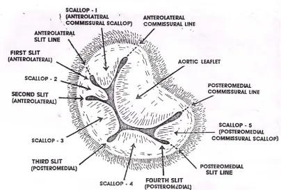
coronaria. The posterior leaflet has usually two minor indentations or clefts.
Clefts divide the posterior leaflet into a relatively large middle scallop and
smaller anterolateral commissural and posteromedial commissural scallops.
T.Walmsley (1929) states that Andreas Vesalius was the person who
first suggested the picturesque term "mitral" to describe the left atrioventricular
valve owing to its resemblance to a plan view of the bishop's mitre.
Harken (1948) described anteromedial leaflet as the aortic baffle
because it is the only dividing structure between the mitral and the aortic
orifice. Directly inferior to the aortic canal, it constitutes an integral part of the
outflow tract of the left ventricle. Due to this anatomic location a considerable
portion of its function is believed to be the direction of the flow of blood
toward the aorta.
In other words the leaflet apparently acts as a watershed which deflects
He reported that 26 of 35 hearts had two additional or commissural
leaflets which are the scallops just behind commissural line.
Hollinshed (1957) stated that bicuspid valve consists only of an anterior
and posterior leaflets, the anterior being the larger of the two; as in the case of
the tricuspid valve.
Zimmerman J (1966) studied about prongs of fibrous tissues from each
of the fibrous trigones which are expansions of fibrous tissue at either end of
the area of aortic and mitral valvar continuity.
Ariela Pomerance (1967) studied ageing in 805 human hearts and
observed the following changes in the mitral valve.
Anterior leaflet or anterior cusp - nodular thickening at the free edge,
which when marked is generally referred to as senile sclerosis, and lipoid
deposition nearer the attached part.
Atheromatosis of the anterior cusp of the mitral valves is also
considered to be related to age and results of his study confirm this view. He
found 4 changes in posterior cusp or leaflet that is -
(1) The most striking change with age was the rise in incidence of
mitral ring calcification.
(2) Small puckered scars were seen in about 5 percent of men over
65 and 3 percent of women.
(3) A more constant finding particularly in the female, was diffuse
(4) The cusps became thickened opaque and voluminous resembling
a parachute.
N. Ranganathan, et al. (1970) studied fifty normal mitral valves from
adults and observed the following:
Commissures which are identified by the commissural partition the
mitral valvular tissue into anterior and posterior leaflets.
The posterior leaflet is further divided into scallops by cleft in its tissue
and the posterior leaflet which is partitioned this way there was triscalloped in
46 hearts.
In 42 hearts a large middle scallop was present with two smaller scallops
on either side. They also state that rough and clear zones can be defined on the
anterior leaflet and rough, clear and basal zones on the posterior leaflet.
A.K. Datta et al. (1984) studied morphology and morphometry of the
mitral valve in 30 normal human hearts collected from postmortem room
(Fig.5a,b,c).
Precise demarcation of mitral leaflets subdivision of posterior leaflet
into three scallops, rough and clear zones of both leaflets and pattern of chordal
attachments are observed and their age changes are noted by them and they
quote that the morphometry reveals that the basal width of the posterior leaflet
is distinctly more extensive than that of the anterior leaflet. The height of the
anterior leaflet, however is comparatively more than that of the posterior
central height than its clear zone, whereas in the middle scallop of posterior
leaflet the height of the rough zone is slightly longer than the clear zone. Their
findings provide morphological evidence of the mechanism of closure of the
normal mitral valve.
Carpentier A. et al., (1971) who did a new reconstructive operation for
correction of mitral and tricuspid insufficiency state that free edge of the mural
leaflet is often divided into three or more scallops or segments described as
lateral, middle and medial or assigned terms like, P1, P2 and P3 (Fig.6).
Perloff JK, and Roberts WC (1972) emphasized the important role of
the atrial wall though it was not a part of mitral apparatus since left atrial
enlargement can contribute to mitral regurgitation.
The continuity of the atrial myocardium over the atrial surface of the
mural (posterior) leaflet makes this leaflet vulnerable to being displaced when
the atrial chamber enlarges.
Bulkley B.H. and Robert W.C. (1975) stated that in deformation of the
mural leaflet, the middle scallop is most often affected. This is thought to be a
consequence of mitral annular dilatation in this region.
Solomon Victor, Vijaya M. Nayak (1994) studied one hundred human
hearts from autopsies to clarify controversies in the literature about
commissures, slits, chordae and leaflets of the mitral valve. They have
designated perpendiculars drawn from the annulus to the free edge at the
shortest height of the mitral veil on either side of the aortic leaflet as the
mitral veil precisely into aortic and mural leaflets. Slits in the leaflets were
easily identified by the dipping in of the free edge into the cusp tissue. There
was no slits in the aortic leaflet. Ninety eight hearts had slits in mural leaflet, in
the anterolateral halves in 81, posteromedial halves in 76, and in the centre in
one. The number of slits in the mural leaflet varied from one to five but was
commonly two. Unlike the relatively static and straight annulus of the aortic
leaflet, the curved annulus of the mitral leaflet contracts and changes in contour
during systole. Hence slits are necessary in the mural leaflet to help it fold and
adapt to the reduced orifice during systole and unfold during the diastole. In
addition to this coarse adjustment of the mural leaflet, both leaflets are pleated
due to "Hooding up" of the leaflet tissue between chordal attachments during
systole, providing the fine tuning to enable the leaflets to adapt themselves to
the reduced systolic orifice (Fig.7a,b,c).
The nodular appearance of the line of apposition of the leaflet is
evidence of this pleating mechanism during valve closure. Slit lines which they
have designated as perpendiculars drawn from the summit of the slits to the
annulus, were used arbitrarily to divide the mural leaflet into 2 to 6 scallops.
When slits are absent there are no scallops. The slits and scallops are best
serially numbered counter clockwise from the surgeon's view through the
atrium. The scallops immediately behind the commissural lines, customarily
labelled as the commissural scallops, are best defined as mural leaflet tissue
between the anterolateral or posteromedial commissural line and the closest
anterolateral or posteromedial slit line. Anterolateral commissural scallops
were seen in 81 hearts and posteromedial commissural scallops in 76 hearts.
the slits. However the chordae arising from each papillary muscle group in toto
form a fan, reaching out to the corresponding adjacent halves of the two
leaflets, restraining their splaying out in diastole and upward bulge during
systole. This findings are relevant to the pathogenesis of mitral valvular disease
and reparative procedures.
Gerde L van Rijk - Z Wikker, MD et al., (1994) reported that the edge
of the anterior leaflet is smooth and the edge of the posterior leaflet often has
multiple indentation or clefts bridged by fan like chords. Just as in the actual
commissures the indentations never reach the mitral annulus itself, otherwise
the valve would be incompetent. The same applies to mitral valve
commissurotomy where the incision intended to open the commissures should
not reach the mitral annulus itself but leave at least 2 mm of valve tissue. In
diastole the combined surface area of the two leaflets is 1½ to 2 times the
surface area of the functional mitral orifice, while in systole than anterior
leaflet alone may cover the effective mitral orifice.
Therefore in mitral valve incompetence in conjunction with mitral
annular dilatation, the extent of reduction of the annulus to obtain a competent
valve can be estimated from the size of the anterior leaflet, which has to cover
the mitral orifice at the annulus level. The redundancy of valvular tissue
indicates that the mitral valve will not rapidly become incompetent with mitral
annular dilatation alone.
He also state that the atrial side of the valve leaflets has a smooth
surface. One half of two thirds of the ventricular surface is irregular, in
distance. The smooth part of the ventricular surface of the anterior leaflet is the
continuation of the aorto mitral membrane and is roughly one third of the total
surface. The site of insertion of the chords on the ventricular surface called the
rough zone of the leaflets, serves as a fibrous re-enforcement of the leaflets.
Although there is considerable variation, the rough zone is larger in the anterior
leaflet, because the chords insert over a large area in the central part of the
leaflet. The rough zone is smaller in the posterior leaflets, tapers off toward the
clefts in the leaflet and toward the commissures, and is absent in the
commissural areas. The absence of chordal support and the relative immobility
of the mitral annulus in the commissural areas makes reconstruction of the
valve in these areas more difficult.
Cheng To et al., (1997) studied the mitral valve by echo cardiography
and stated when the closed mitral valve is seen in profile, the major part of the
closure line lies below the plane of the atrioventricular junction rising toward
the commissures at the peripheral ends so that the atrial surface of the leaflet
has a saddle like configuration. Being tethered by the tensor apparatus, the line
of coaptation in a normal valve does not extend above the level of the junction
during ventricular systole.
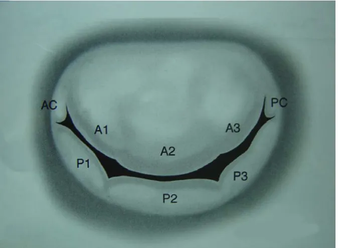
Syho (2002) described that the aortic leaflet which is in fibrous
continuity with the aortic valve has a rounded free edge and occupies a third of
the mitral annular circumference, whereas the other leaflet is long and narrow,
lining the remainder of the circumference. The aortic leaflet hangs like a
curtain between the left ventricular inflow and outflow tracts. When the valve
is closed, this leaflet appears to form the greater part of the atrial floor but is
form an arc shaped closure line, or zone of apposition, that is obliquely situated
relative to the orthogonal planes of the body. With the leaflets meeting, the
view of the valve from the atrium resembles a smile.
Skandalaki (2004) in his book of Surgical Anatomy reported that two
leaflets guard the opening forming the mitral valve. The anterior leaflet is large
and triangular in shape. The smaller posterior leaflet is known as the Merklin
leaflet. The leaflets insert on the left atrioventricular fibrous annulus. Their free
margins are attached by chordae tendineae to the papillary muscles. The
scalloped margin of the posterior leaflet gives the impression of indistinct
minor leaflets, often at opposite sides of the valve.
CHORDAE TENDINEAE
Henry Gray (1858) reported that the false chordae interconnect
papillary muscles, extend from the latter to a point on the ventricular wall or
interconnect two points on the ventricular walls. They are ignored in many
accounts and when mentioned considered of little or no functional significance.
True chordae of the mitral valve complex may first be divided into inter
leaflet or commissural chordae and varieties of leaflet chordae. Of the latter
those of the anterior leaflet are rough zone chordae, including special strut
chordae. Those of the posterior leaflet include rough zone chordae, cleft
chordae and basal chordae (Fig.8a).
Most true chordae divide into branches from a single stem soon after its
origin from the apical third of a papillary muscle, or proceed as single cords
Anterolateral and posteromedial commissural chordae arise near the tips
of papillary muscles by a single stem fanning out at once into radiating strands
attached to the smooth free margin of the commissure.
Tandler J. (1913) suggested most commonly used classifications of
chordae tendineae into three orders.
Ist order - chordae tendineae inserted into the leaflet's edge.
IInd order - chordae tendineae inserted 6 to 8 mm beyond the free
margins.
IIIrd order - chordae tendineae inserted into the basal portion of the
ventricular aspect of the posterior leaflet.
Quain's elements of Anatomy (1929) distinguishes three orders of
chordae tendineae according to the site of attachment to the leaflets.
Ist order - chordaes are those inserted on the free edge. They are
numerous, delicate and often form networks near the edge.
IInd order chordaes insert on the ventricular surface of the leaflets
beyond the free edge forming the rough zone. These are thicker than first order
chordaes.
IIIrd order - chordaes attach only to the mural leaflet since they arise
directly from the ventricular wall or from small trabeculations. They insert to
the basal portion of the leaflet and run only a short distance toward the free
Lam JHC et al., (1970) did a study on chordae tendineae from 50
normal mitral valves and they distinguished "four main types by their mode of
insertion" (Fig.9).
1. Commissural chordae inserted into the commissures between
anterior and posterior leaflets.
2. Rough zone chordae inserted into the ventricular aspect of the
distal rough portion of the anterior and posterior leaflets.
In this rough zone chordae two of the anterior leaflet rough zone
chordae are thicker than the others and are called strut chordae.
3. Cleft chordae inserted into the clefts between the scallops of the
posterior, leaflet.
4. Basal chordae are single strands that arise from the posterior
ventricular wall and inserted into the basal zone of the posterior
leaflet.
They say that this classification gives clear definition of mitral valve
anatomy and forms a sound basis for functional studies of chordae tendineae.
Becker A.E. and DW Wit APM (1980) studied 100 hearts and showed
that uniformity of the chordal attachment was uncommon. Normal valves have
a spectrum of chordal support and this lack of uniformity could lead to leaflet
A.K. Datta et al. (1984) studied thirty human hearts and observed the
following (Fig.10a,b,c):
The rough zone chordae are attached to the free margin and the
adjoining ventricular aspect of both leaflets of the mitral valve. Each rough
zone chordae on approaching the leaflet splits into three strands : one is
attached to the free margin, the other to the junction of rough and clear zones,
and the third to a point between them.
Two rough zone chordae known as strut chordae are thicker than the rest
and are attached to the anterior leaflet on either side of the tip, one arising from
the apex of anterior papillary muscle and other from the posterior papillary
muscle.
The basal chordae are observed only in the posterior leaflet of all age
groups but are more prominent in the third age group. These are attached along
a narrow basal strip close to the mitral annulus.
Vander Bel. Kahn J, Becker A.E. (1986) did a study on a series of
floppy valves revealed pronounced derangements in chordal branching and in
their attachments to the leaflets, leaving part of the affected leaflet less well
supported.
Solomon Victor, Vijaya M. Nayak (1995) reported that there is no
consistent fan-shaped chordal pattern at the commissural lines or at the slits.
However the chordae arising from each papillary muscle group, in toto, form a
fan, reaching out to the corresponding adjacent halves of the 2 leaflets,
(Fig.11).
Syho (2002) described that the tendinous cords are string like structures
that attach the ventricular surface or the free edge of the leaflets to the papillary
muscles. The tendinous cords of the mitral valve are attached to two groups of
papillary muscles or directly to the posterio inferior ventricular wall to form the
tensor apparatus of the valve. Cords that arise from the apices of the papillary
muscles attach to both aortic and mural leaflets of the valve. Since cords
usually branch distal to their muscular origins, there are five times as many
cords attached to the leaflets as to the papillary muscles. Normally there are
only two commissural cords, one supporting each free margin of the
commissural region. These cords arise as a single stem but branch like the
struts of a fan that, on closing and opening, allow the adjacent leaflets to coapt
and to move apart:
According to him leaflet cords are of several forms. The most numerous
are rough zone cords. Rough zone cords in the mural leaflet are generally
shorter and thinner than those found in the aortic leaflet. Among the rough
zone cords of the aortic leaflet two are the largest and thickest, termed strut
cords. They arise from the tip of each papillary muscle and are thought to be
the strongest. Basal cords are unique to the mural leaflet.
Another type of cord, the cleft cord, is found only in the mural leaflet.
Each is a miniature version of a commissural cord whose branches insert into
the free margin between adjacent segments while the main stem runs into the
He says, according to Toronto classification, the strut cords and parts of
the rough zone and cleft cords correspond to second order cords.
Parts of the rough zone cords, cleft cords, and the commissural cords
that insert to the free edge of the leaflets correspond to first order cords.
Yoav Turgeman MD (2003) says that Tandler's classification of
chordae tendineae into Ist order, second order, and third order based on the site
of insertion of chordae tendineae into the valvular leaflet is simple to use and it
neither emphasizes the morphologic differences between chordae tendineae nor
relates their sites of insertion to their function.
Papillary muscles
HenryGray (1858) stated that the two left ventricular papillary muscles
vary in length and breadth and may be bifid. The anterior papillary muscle
arises from the sternocostal mural myocardium, the posterior from the
diaphragmatic region.
Chordae tendineae arise mostly from the tip and apical third of each
muscle, but sometimes near its base. They diverge and are attached to
corresponding points on both mitral leaflets.
Rusted I.E. (1951) did an elaborate study of papillary muscles in 200
hearts found that the anterior muscle (anterolateral) was single, though most
frequently grooved lengthwise in 76.5 percent, double in 12 percent, triple in
9.5 percent, and more than triple in 4 percent.
30 percent, double in 26.5 percent, triple in 37 percent, and there were more
than three in 6 percent. They emphasized the localizing importance of the
anterior (anterolateral) papillary muscle in commissurotomy for mitral stenosis
since incidence of atypical anterior papillary muscle is less.
Brock R.C. (1952) says that the papillary muscles normally arise from
the apical and middle thirds of the left ventricular wall.
N.Ranganathan M.D. and G.E. Burch M.D. (1969) studied the gross
morphologic characteristics of the anterolateral and posteromedial papillary
muscles of the left ventricles in the ten hearts and classified the papillary
muscles into 3 broad categories.
1. Completely tethered papillary muscle i.e. a papillary muscle fully
adherent to the subjacent ventricular myocardium and protruding very
little into the ventricular cavity with a few trabecular attachments.
2. Finger like papillary muscle. (ie) papillary muscle with one third or
more of the body protruding freely into the ventricular cavity with a
very few or no trabecular attachments.
3. Mixed type papillary muscle (ie) a papillary muscle with part of the
body protruding freely into the ventricular cavity, but also with
considerable trabecular attachments and tethering.
Tirone E.David M.D. (1994) stated that the interactions between the
mitral valve and left ventricle are complex and not yet completely understood.
probably the most important factor in this relationship because severance of the
chordae tendineae in experimental animals causes a significant drop in left
ventricular systolic function as assessed by load independent parameters. This
deterioration in ventricular function is even more marked in dilated hearts due
to chronic mitral regurgitation. There is strong clinical evidence that
maintenance of papillary muscle - annular continuity during mitral valve
surgery is beneficial to left ventricular function and clinical outcome. That is
one of the reasons why the mitral valve should be repaired rather than replaced
in patients who need mitral valve surgery. If replacement is necessary, the
chordae tendineae should be preserved. If preservation of chordae tendineae is
difficult because of calcification, fibrosis or for other reasons it is possible to
resuspend the papillary muscle with expanded tetrafluoroethylene sutures.
Preservation of chordae tendineae during mitral valve replacement is also
important because it prevents spontaneous rupture of the posterior wall of the
left ventricle, a rare but dreaded complication of this type of surgery.
Gerda L. Van Rijk - Zwikker, MD (1994) described that the two
papillary muscles supporting the chords of the mitral valve have a variable
configuration and size. Both muscles are located at the posterior free wall of
the left ventricle.
The anterolateral muscle is usually single, can be distinguished from the
posterior muscle, because it is larger and is located at the posterior and left side
of the left ventricle, underneath the obtuse margin.
The function of the papillary muscles is closely related to the function of
amount of tension exerted by the papillary muscles to remain within strict
limits.
Transient mitral prolapse and incompetence caused by myocardial
ischemia result from the inability of the papillary muscle to contract and
maintain adequate coaptation of the valve leaflets. Infarction of the ventricular
wall in the vicinity of the papillary muscles will also cause mitral incompetence
due to either elongation of the infarcted papillary muscle or outward motion of
the ventricular wall. The first will lead to prolapse of the mitral valve. The
second causes incomplete coaptation of the leaflets because of retraction.
Surgical correction of the mitral incompetence based on papillary muscle
dysfunction is often unsuccessful due to the dynamic character of this part of
the subvalvular apparatus.
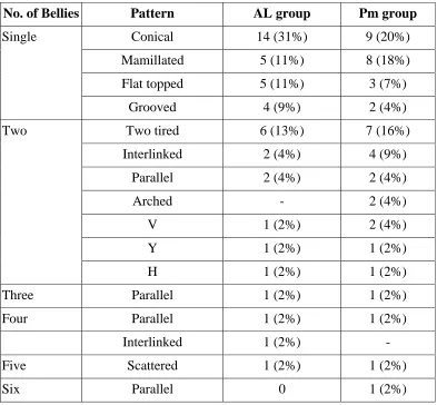
Solomn Victor, Vijaya M. Nayak (1995) studied the papillary muscles
of the mitral valve in 100 human autopsy hearts and state that a mid-mitral
plane passing through the middle of the aortic and mural leaflets divides the
chorda papillary support of the mitral valve into anterolateral and
posteromedial halves (Fig.12a,b,c,d). The anterolateral papillary support had 1
belly in 67 hearts, 2 in 27, 3 in 4, 4 in 1, and 5 in 1 heart. Likewise, the
posteromedial papillary support had 1 muscle belly in 50 hearts, 2 in 36, 3 in
11, and 4 in 3. The single papillary muscles were conical, mammillated, flat
topped, grooved, stepped, wavy, arched, sloped or saucerized. When there were
two bellies they presented a two tiered, interlinked parallel or arched, V, Y or
H configuration. Three papillary muscles formed a parallel, interlinked or
arrangement with the third belly separate. When four or five bellies existed,
they were parallel or interlinked. In the anterolateral and posteromedial group,
the papillary muscle bellies were mostly intraluminal in 14% and 11%, mostly
intraluminal with the tip anchored in 19% and 28%, equally sessile and
intraluminal in 54.5% and 41.5% mostly sessile in 12.5% and 19.5%,
respectively. In the anterolateral group 19% of papillary muscle bellies arose
from the upper third of the ventricle, 79.5% from middle third and 1.5% from
lower third. The corresponding figures for posteromedial group are 6%, 92.5%,
and 1.5%, respectively. Four to 22 chordae originated from the anterolateral
papillary group, ending in 14 to 72 chordal insertions into the corresponding
half of the valve. Likewise, 2 to 18 chordae arose from the posteromedial
papillary group ended in 12 to 80 leaflet insertions. The chordae in each group
are best considered in toto as a fan. The configuration of the fan is unique in
each heart. Imaging techniques need to be refined to outline those variations
more precisely.
Petra W. Oosthoek et al. (1998) did a study in normal hearts at
between 5 and 19 weeks of development of papillary muscles with
immunohistochemistry, three dimensional reconstructions, and gross
inspection. Scanning electron microscopy was used to study human hearts. In
embryonic hearts a prominent horse shoe shaped myocardial ridge runs from
the anterior wall through the apex to the posterior wall of the left ventricle.
In the atrioventricular region this ridge is continuous with atrial
myocardium and covered with cushion tissue. The anterior and posterior parts
myocardium. Their lateral sides gradually delaminate from the left ventricular
wall, and the continuity between the two parts is incorporated in the apical
trabecular network. In this way the anterior and posterior parts of the ridge
transform into the anterolateral and posteromedial papillary muscles,
respectively. Simultaneously, the cushions remodel its valve leaflet and
chordae. Only the chordal part of the cushions remains attached to the
developing papillary muscles. The conclusion is that the disturbed
delamination of the anterior or posterior part of the trabecular ridge from the
ventricular wall, combined with underdevelopment of chordae, seems to be the
cause of asymmetric mitral valves. Parachute valves however develop when the
connection between the posterior and anterior part of the ridge condenses to
from one single papillary muscle (Fig.13).
Syho (2002) stated that the cords arise from the tips of the papillary
muscles. Alterations in the size and shape of the left ventricle can distort the
locations of the papillary muscles, resulting in valvar function being disturbed.
The papillary muscles normally arise from the apical and middle thirds
of the left ventricular wall. Described in most text books as two in number,
however there are usually groups of papillary muscles arranged fairly close
together. At their bases, the muscles sometimes false or have bridges of
muscular or fibrous continuity before attaching to the ventricular wall. Extreme
MATERIAL AND METHODS
Study material
The study material consists of
(a) 45 normal human hearts (40 Adult [25 male 15 female] and 5
Full term Foetuses)
(b) 5 clinical cases
Methods of study
I. Conventional dissection method
(a) In adult hearts
(b) Foetal hearts
II. Clinical study of 5 cases
I. CONVENTIONAL DISSECTION METHOD
Collection of specimens
45 fresh adult human postmortem heart specimens were from taken at
autopsy from the Institute of forensic Medicine, Madras Medical College,
Chennai, within 24 hours from the time of death.
The autopsies had been carried out by the conventional 'I' shaped
incision from the hyoid bone to the pubic symphysis. The thoracic cage was
opened by cutting through the costochondral junctions and the heart had been
removed.
The heart thus removed was emptied of blood inside, washed thoroughly
in running tap water. It was then transported in closed plastic containers to the
Institute of Anatomy, Madras Medical College, Chennai for further dissection.
Dissection of specimens
Position and orientation of the heart and its chambers was confirmed.
Left atrium wall was then opened by an incision through the right and
left inferior pulmonary veins and the upper part of left atrial auricle was
dissected and the mitral valve was inspected from above.
Out flow tract of the left ventricle was opened by an incision on the
sternocostal surface of the heart extending from the apex, parallel and close to
the interventricular septum upto the aortic orifice (Fig.14, a,b).
In each heart a detailed examination was made of the mitral valve
annulus, commissures, valve leaflets, chordae tendineae and papillary muscles.
The collected specimens were immersed in the following specific
preservative solution - 10 litres of normal saline, 1 litre of 10% formalin, 50 ml
of glycerine, 5 gms of powdered thymol.
The following morphological features of mitral valve complex were
1. Mitral valve annulus - (a) Shape
(b) Circumference
(c) Age and sex differences
2. Commissures - (a) Number of Commissures
(b) Position of Commissures
3. Mitral valve leaflets - (a) Number of Leaflets and position
of leaflets
In posterior leaflet - (a) Number of slits or clefts
(b) Number of scallops -
4. Chordae tendineae - (a) Types of chordae
i) Rough Zone chordae
ii) Strut chordae
iii) Commissural chordae
iv) Cleft Chordae
v) Basal Zone Chordae
(b) Number of chordae at origin
(c) Number of chordae at insertion
into leaflets
5. Papillary muscles - (a) Number of papillary muscles
(b) Spatial orientation
(c) Pattern (shape) of papillary
muscles
(d) Site of origin
(e) Extent of protrusion of papillary
muscles into the left ventricular
MORPHOMETRIC ANALYSIS OF MITRAL VALUE COMPLEX
Measurements were taken with the help of a divider, thread and a
millimeter scale under the following parameters:
1. Circumference of Mitral Annulus was taken by keeping a thread
in the sulcal margin and the point is marked where it meets the beginning of the
thread and this distance in the thread is measured with millimeter scale
(Fig.15).
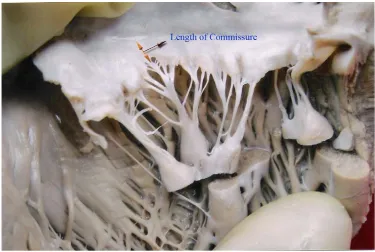
2. Commissures
(a) Length of anterior commissure
(b) Length of posterior commissure
are measured from the annulus to the commissure with a divider and
millimeter scale as shown in Fig. 16.
Using divider and millimeter scale
3. Mitral value leaflets : (a) The height of each single valve leaflet
measured by perpendicular line from
the annulus to the centre of the free
edge of the leaflet.
(b) The breadth of the single valve leaflet
at the line of insertion at the valve ring
(Fig.17).
(c) Vertical height of Rough zone is
measured which is the distal rough
portion on the ventricular aspect of the
(d) Vertical height of clear zone is
measured which is identified towards
the annular side as a smooth and
membranous area without any chordal
attachment on the ventricular aspect of
the anterior and posterior leaflets.
4. Length of chordae tendineae is measured from tip of papillary
muscle to insertion in the leaflet using divider and millimeter scale (Fig.18).
II. CLINICAL STUDY
Clinical study was done on 5 patients having mitral valve disease. These
patients were diagnosed and taking treatment at the cardiology department,
Government General hospital, Chennai – 3. They included 4 male and 1
Female patient of age ranging from 45 to 60 years with the following
diagnosis.
S. No. Name Age Diagnosis
1. Ramanathan 48 years Mitral stenosis
2. Govindan 55 years Mitral regurgitation
3. Parvathy 45 years Mitral valve prolapse
4. Ganeshan 58 years Mitral stenosis
5. Kumar 60 years Mitral regurgitation
DEVELOPMENTAL ANATOMY
MITRAL
VALVE COMPLEXMitral valve is formed by proliferation of connective tissue under the
endocardium of the left atrioventricular canal.
The atrioventricular valve develops as shelf like projections from the
margins of the atrioventricular orifice, directed as almost complete conical
sheets towards the ventricles, their advancing edges continuing, initially as
trabecular ridges, deep into the ventricular cavity. With continued differential
growth and excavation on their ventricular aspects, each sheet develops two
marginal indentations, defining the principal valve leaflets and minor marginal
indentations (clefts) subidividing some leaflets into scallops. Each leaflet
develops functionally significant regional variations in surface texture. Its core
condenses as a collagenous lamina fibrosa. The latter blends at its
atrioventricular base with the inappropriately named fibro areolar valve
"annulus" - each a part of the complex, functionally crucial, fibrous skeleton of
the heart. The papillary muscles are the ventricular ends of the original
trabeculae and, whilst free throughout their length, their mural ends are
confluent with mural ventricular musculature and receive a dense population of
its nerves and specialized conducting tissues.
Degenerating ventricular muscle forms chordae tendineae, thick stringy
attachments which connect the valve leaflets to the papillary muscles of the
OBSERVATION
The findings observed in 45 hearts (40 adult (25 male & 15 female) + 5
foetus) and clinical case study are analysed.
45 human hearts collected are arranged in four age groups 0-1, 1-20,
21-40, above 40 years and studied.
In each heart a detailed examination was made of the mitral annulus,
commissures, valve leaflets, chordae tendineae and papillary muscles.
Measurements were taken with the help of a divider, thread and a
millimeter scale for morphometric analysis of mitral valve complex and the
following details were observed.
MITRAL ANNULUS
The peripheral margin of the mitral annulus is represented by a sulcal
margin. The shape of the mitral annulus is D shaped in all 45 specimens
(Fig.19a).
The superomedial part of the mitral annulus gave attachment to the
anterior leaflet in 45 specimens (Fig.19b,c).
Rest of the mitral annulus provides attachment to the posterior leaflet in
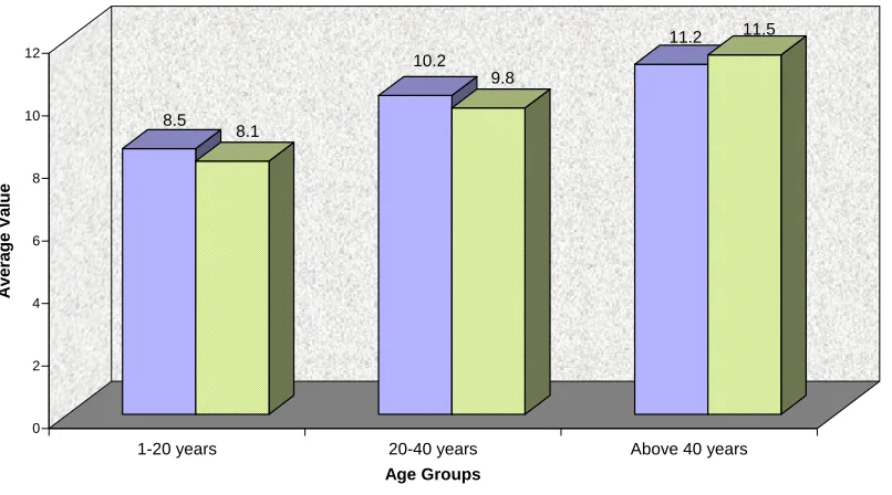
Circumference of the mitral annulus was studied with the help of a
thread and millimeter scale. An increase in the mean value of the circumference
with advancing age was observed. The circumference of the mitral annulus in
the age group of 0-1 year ranged from 0.3-0.5cm with a mean value of 0.32 ±
0.07, in the age group of 1-20 years ranged from 6.5 - 9.6cm with a mean
value of
8.11 ± 1.25, in 21-40 years ranged from 9.1-10.8cm with a man value of
9.79 ± 0.43, and above 40 years it ranged from 10.2 - 11.8 cm with a mean
value of 10.76 ± 0.46.
Sex differences the circumference of the mitral annulus is consistently
smaller in female than the male subjects. The circumference of annulus in the
age group of 0-1 year in male is 0.4-0.5 cm and in female is 0.3-0.4 cm.
Between the age group of 1-20 in male it is 7.1-9.4 cm and in female 6.5-9.3
cm, between 21-40 years of age in male it is 9.4-10.8 cm and in female is
9.1-10.1 cm. Above 40 years in male circumference is 10.4-11.8 cm and in female
is 10.2-11.6 cm.
These measurements and statistical analysis are shown in Table-1(a,b).
COMMISSURES
Commissures are junctional areas between the leaflets. Two
Position of commissures were observed as anterolateral commissure and
posteromedial commissure in all age groups.
Commissures are identified by the fan shaped attachment of the chordae
and these chordae are known as commissural chordae (Fig.20). Anterolateral
and posteromedial commissures are clearly distinguishable in all the 45
specimens.
The length of the commissures are measured using divider and
millimeter scale. The length of Anterolateral and posteromedial commissures in
0-1 year 0.2-0.3 cm and 0.2-0.4 cm, in 1-20 years 0.4-0.6 cm and 0.5-0.6 cm,
in 21-40 years 0.5-0.7 cm and 0.6-0.8 cm and above 40 years it is 0.6-0.8 cm
and 0.7-0.9 cm. The mean value of the commissural length increases with
advancing age. Measurements are shown in Table-2(a).
MITRAL VALVE LEAFLETS
Number of leaflets in all 45 hearts is two i.e. Anterior and posterior
leaflet.
Anterior Leaflet
It is triangular in shape in all the 45 specimens.
Anterior leaflet receive the chordae tendineae from both papillary
muscles (Fig.21).
The edge of the anterior leaflet is smooth and there is no slit or cleft.
leaflet was measured as the perpendicular distance from the annulus to the free
edge of the leaflet. The width of the anterior leaflet was measured along the
annulus between the points of attachment of the leaflet.
The mean value of the width of anterior leaflet shows sharp increase
with the advancement of age. The findings of the mean height of the anterior
leaflet suggests a slow increase with the increment in age. The measurements
and statistical analysis are shown in Table-3(a,b).
Each anterior leaflet presents a rough zone and clear zone
The clear zone of the leaflet is identified towards the annular side as a
smooth and membranous area without any chordal attachment on the
ventricular surface.
The rough zone is visualised towards the apex of the leaflet as a crescentic
area, which is thicker on palpation and presents attachments of chordae tendineae
on the ventricular surface.
Central height of the clear zone was measured. The central height of
clear zone between the age group of 0-1 years is 0.5-0.9 cm, 1-20 years 1.2-2.0
cm, 21-40 years 1.4-2.1 cm and above 40 years 1.4-2.5 cm. It is shown in
Table No.4.
The height of rough zone in anterior leaflet is in 0-1 year 0.2-0.5 cm,
1-20 years 0.3-0.6 cm, 21-40 years 0.5-0.8 cm and above 40 years 0.6-1.0 cm.
Central height of the rough zone was found to be smaller than the
central height of clear zone in the anterior leaflet. Further the mean value of
rough and clear zones of the anterior leaflet suggests gradual increase with the
progress of age.
The ridge separating the rough zone and clear zone of anterior leaflet is
distinct in the IIIrd (i.e. 21-40 yrs) age group and IVth (i.e. above 40 years) age
groups (Fig.22).
In all age groups the basal measurements of the anterior leaflet are
distinctly shorter. Atheromatic changes and nodular thickening at the free edge
were observed above 50 years age group in anterior leaflets in 10 specimens
(Fig.23).
POSTERIOR LEAFLET
Morphology of Posterior Leaflet
It is rectangular in shape in all the four age groups. Clefts or slits divide
the posterior leaflet into scallops (Fig.24). Clefts or slits are identified by the
fan shaped attachment of cleft chordae at the slits.
The free margin of the posterior leaflet showed two clefts and 3 scallops
in 43 of 45 hearts (95%). The middle scallop is the largest in all specimens.
The two lateral scallops (also called as anterolateral and posteromedial
scallops) were present in all specimens but much smaller.
Two specimens in the age group of 1-20 years, showed 4 clefts and 5
scallops in the posterior leaflet (Fig.25). Even in these, the middle scallop was
Each scallop of the posterior leaflet presents rough zone, clear zone and
basal zones in 43 hearts (95%). In 2 hearts the basal zone was absent since
there was no basal chordae.
The rough zone is smaller in the posterior leaflet compared to anterior
leaflet. The rough zone tapers off towards the clefts and is absent in the
commissures.
In 10 out of 15 specimens in the age group of above 40 years, a ridge
was clearly identified between the rough and clear zone of the posterior leaflet.
In the younger age group, such a ridge was absent signifying its occurrence as
age advances.
Below 40 years in all 35 hearts the demarcation between rough and clear
zone is not well marked.
Morphometry of Posterior leaflet
The height and width of the posterior leaflet were measured. The mean
value of the basal width of the posterior leaflets shows sharp increase with the
advancement of age.
The findings of the mean height of the posterior leaflet suggest a slow
increase with the increment in age. The measurement and statistical analysis
are shown in Table No.3.
Central height of the clear zone in the middle scallop of the posterior
leaflet is 0.2-0.5 cm between 0-1 year, 0.3-0.5 cm between 1-20 years, 0.3-0.5
Central height of the rough zone was found to be larger than the central
height of clear zone. Central height of rough zone was measured in posterior
leaflet and it was 0.3-0.6 cm in 0-1 year, 0.4-0.6 cm in 1-20 years, 0.5-0.8 cm
in 21-40 years and 0.5-0.8 above 40 years.
In the middle scallop of the posterior leaflet the central height of the
clear zone was found to be smaller compared to the rough zone.
The mean value of both rough and clear zones increase with age. It is
shown in Table No.4(c).
Basal zones of each scallop of the posterior leaflet are identified by the
attachment of basal chordae tendineae in 43 hearts. Whereas in 2 hearts the
basal chordae was absent. Chordae arising from the ventricular wall and
attached to posterior leaflet are known as basal chordae (fig.26).
Age related changes are noted in above 50 years age group in the
posterior leaflet of 10 specimens which are mitral ring calcification and small
puckered scars in posterior leaflet.
Comparison of anterior and posterior leaflets
The central height of clear zone was found to be larger in anterior leaflet
than in the posterior leaflet.
The central height of rough zone is larger in anterior leaflet than the
In all age groups the basal measurement of the anterior leaflet are
distinctly shorter than the basal measurement of posterior leaflets.
CHORDAE TENDINEAE
Ventricular aspect of the mitral leaflet was inspected to observe the
patterns of insertion of the chordae tendineae into the leaflet in all age groups.
The valvular insertion of chordae fans out in several small chords.
Types
Rough zone chordae are attached to the free margin and the adjoining
ventricular aspect of both leaflets of the mitral valve (Fig.27). Each rough zone
chordae on approaching the leaflet splits into three strands. One is attached to
the free margin. The other to the junction of rough and clear zones and the third
to a point between the Ist and IInd attachment. This type of rough zone chordae
are observed in all the 45 hearts studied. Amongst these rough zone chordae,
two rough zone chordae known as strut chordae are thicker than the rest and
are attached to the central part of the anterior leaflet. They (Strut chordae) are
seen arising, one from the apex of anterior papillary muscle and the other from
the apex of the posterior papillary muscle as observed in all the 45 specimens.
Commissural chordae are inserted into the free margin of the
commissural area in a fan shaped manner in all 45 hearts(100%).
Commissural chordae arise as single strands from the apex of the
anteromedial and posterolateral papillary muscles, but divide to form various
Commissural chordae shows following varied configuration. Symmetric
fan type commissural chordae was observed in 25 (56%) specimens.
Asymmetric fan type chordae was seen in 5 (11%) specimens. Palmate type of
commissural chordae was seen in 3 (7%) specimens. Cribriform type of
commissural chordae was observed in 2 (4%) specimens. Mixed type of
commissural chordae was seen in 10 (22%) specimens.
Similar to commissural chordae fan shaped chordae are observed to
insert in the clefts between the scallops of the posterior leaflet. These are called
as cleft chordae and are seen in all the 45 hearts.
Basal chordae are chordae tendineae which arise directly from the
ventricular wall (and not from the papillary muscle), to get inserted in the
posterior leaflet. Such basal chordae were seen in all age groups of 43 hearts,
except in 2 specimens where basal chordae are absent.
Average length of the chordae tendineae of all 5 subtypes are
measured. It is shown in Table No.5.
Number of chordae at origin (Fig.28, Table No.6)
1-3 chordae originate from anterolateral group of papillary muscle in
two (4%) specimens and this is not observed in posteromedial group.
4 to 6 chordae originate from anterolateral group of papillary muscle in
11 (24%) specimens and posteromedial group of papillary muscle in 10 (22%)
specimens. 7 to 9 chordae arose from anterolateral group of papillary muscle in
(29%) specimens. 10 to 12 chordae took their origin from anterolateral group
of papillary muscle in 10 (22%) specimens and posteromedial group of
papillary muscle in 13 (29%) specimens.
13 to 16 chordae arose from anterolateral group of papillary muscle in 5
(11%) specimen and posteromedial group of papillary muscle in 7 (16%)
specimens. 17 to 20 chordae originate from anterolateral group of papillary
muscle in 2 (4%) specimen and posteromedial group of papillary muscle in 2
(4%) specimen. 21 to 23 chordae originate from anterolateral group of
papillary muscle in 2 (4%) specimen.
Number of chordae at insertion (Fig.29, Table No.6(a))
10 to 20 chordae are inserted into the anterior and posterior leaflets from
anterolateral papillary muscle in 3 (7%) specimens.
21 to 30 chordae are inserted into both the leaflets from anterolateral
papillary muscle in 13 (29%) specimens.
31 to 40 chordae are found to be inserted into both leaflets from antero
lateral papillary muscle in 10 (22%) specimens.
41 to 50 chordae are attached to both the leaflets from anterolateral
papillary muscle in 12 (27%) specimens. 51 - 60 chordae are attached to both
leaflets from antero lateral papillary muscle in 3 (7%) specimens. 61 to 70
chordae are inserted into both the leaflets from anterolateral papillary muscle in
2 (4%) specimens. 71 to 80 chordae are inserted into both the leaflets from