C
CCOOOMMMPPPAAARRRAAATTTIIIVVVEEE EEEVVVAAALLLUUUAAATTTIIIOOONNNOOOFFF A
AANNNTTTIIIMMMIIICCCRRROOOBBBIIIAAALLLAAACCCTTTIIIVVVIIITTTYYYOOOFFFTTTRRRIIIPPPHHHAAALLLAAA,,, T
TTEEERRRMMMIIINNNAAALLLIIIAAACCCHHHEEEBBBUUULLLAAA&&&CCCHHHLLLOOORRRHHHEEEXXXIIIDDDIIINNNEEEOOONNN E
EENNNTTTEEERRROOOCCCOOOCCCCCCUUUSSSFFFAAAEEECCCAAALLLIIISSS–––AAANNNIIINNNVVVIIITTTRRROOO S
SSTTTUUUDDDYYY
Dissertation submitted to
THE TAMIL NADU DR. M. G. R. MEDICAL UNIVERSITY Towards the partial fulfillment for the degree of
MASTER OF DENTAL SURGERY
BRANCH – IV
CONSERVATIVE DENTISTRY AND ENDODONTICS
CERTIFICATE
This is to certify that Dr. S. Jothi Latha, post graduate student (2007 – 2010) in the Department of Conservative Dentistry and
Endodontics, Tamil Nadu Government Dental College and Hospital,
Chennai-3, has done this dissertation titled “Comparative evaluation of antimicrobial activity of Triphala, Terminalia chebula & Chlorhexidine on Enterococcus faecalis – An InVitro study” under our direct guidance and supervision in partial fulfillment of the
regulations laid down by The Tamil Nadu Dr. M.G.R. Medical
University, Guindy, Chennai-32 for M.D.S. – Conservative Dentistry and
Endodontics (Branch IV) Degree Examination.
Dr. M. KAVITHA PROFESSOR &
GUIDE
Dr. S. C. LOGANATHAN PROFESSOR &
H.O.D.
Department of Conservative Dentistry & Endodontics Tamilnadu Government Dental College and Hospital
Chennai – 600 003.
Dr. K. S. G. A. NASSER PRINCIPAL
CONTENTS
S. No PAGE NO.
1 INTRODUCTION 1
2 AIMS AND OBJECTIVES 8
3 REVIEW OF LITERATURE 9
4 MATERIALS AND METHODS 38
5 RESULTS 50
6 DISCUSSION 62
7 SUMMARY 72
8 CONCLUSION 74
INTRODUCTION
The success of endodontic treatment is directly influenced by
elimination of microorganisms in infected root canals.11 Microorganisms
that invade the root canal system have an essential role in initiating and
sustaining periapical disease.47
Unlike primary endodontic infections, which are polymicrobial in
nature and dominated by gram-negative anaerobic rods, the microorganisms
involved in secondary infections are composed of one or a few bacterial
species.60, 85, 36 Enterococcus faecalis is a persistent organism that, despite
making up a small proportion of the flora in untreated canals, plays a major
role in the etiology of persistent periradicular lesions after root canal
treatment. It is commonly found in a high percentage of root canal failures
and it is able to survive in the root canal as a single organism or as a major
component of the flora.22 Studies investigating its occurrence in root-filled
teeth with periradicular lesions have demonstrated a prevalence ranging
from 24 to 77%.36, 60, 67, 68, 70, 71, 78, 85 Starvation increases the resistance of
E. faecalis 1000-fold to 10,000-fold. It is probable that the physiologic state
of the cells, particularly in retreatment cases, is closest to the starvation
The majority of infecting bacteria, together with their principal
substrate of necrotic pulp debris, might be reduced significantly by
chemomechanical instrumentation.27, 57 However, this is not always achieved
completely as a result of the anatomic complexity and diversity of root
canals, as well as the subsequent limitations in access by instruments and
irrigants.46, 69, 82 Therefore, the use of an intracanal medication can help
eliminate the bacteria remaining even after chemomechanical
instrumentation and can provide an environment conducive to periapical
tissue repair.16
Calcium hydroxide has been used as an intracanal medicament for a
long time. The effects of calcium hydroxide [Ca(OH)2] in root canal and
dentin disinfection are well documented. The antimicrobial effects of
Ca(OH)2 may be directly related to its high alkalinity and a great majority of
the microbial species isolated from root canals show susceptibility both
clinically and ex vivo (Bystrom & Sundqvist 1981, 1983, Bystrom et al.
1985, Waltimo et al. 2005). However, several studies demonstrated that
Ca(OH)2 fails to eradicate Enterococcus faecalis residing in infected root
canal systems (Bystrom et al. 1985, Haapasalo & Ørstavik 1987, Safavi et
al. 1990, Ørstavik & Haapasalo 1990, Evans et al. 2002, Peters et al. 2002,
against Ca(OH)2 might be the ability of E. faecalis to invade dentinal
tubules, isthmuses and other ramifications of a root canal system (Love
2001). Furthermore, it has been documented to be able to survive for
prolonged periods in high alkalinity (Bystrom et al. 1985, Haapasalo &
Ørstavik 1987) and harsh nutrient conditions (Hartke et al. 1998, Figdor et
al. 2003, Portenier et al. 2005). Evans et al. (2002) reported the survival of
E. faecalis at high pH was due to a functioning proton pump with the
capacity to acidify cytoplasm. On the other hand, the buffering capacity of
dentin can inhibit the pH increase and the antimicrobial activity of Ca(OH)2
in the root canal (Haapasalo et al. 2000).
Many alternative antimicrobial agents have been tested for their
ability to eliminate E. faecalis from the root canal system. In vitro studies
have indicated that chlorhexidine may be potent in the elimination of
E. faecelis from the root canal system (Bystrom et al.1985, Haapasalo &
Ørstavik 1987, Ørstavik & Haapasalo 1990, Basrani et al. 2003, Gomes et al.
2003).
It is active against a wide range of microorganisms, such as
Gram-positive and Gram-negative bacteria.30 CHX has an antibacterial efficacy
comparable to that of sodium hypochlorite (NaOCl).65 In addition, it is also
substantive antibacterial activity to root dentin after prolonged (ie, at least 1
week) exposure.5, 37, 50, 52, 96 2% CHX is proved to be an efficient agent
against E. faecalis.44
Finding healing powers in plants is an ancient idea. Medicinal plants
are part and parcel of human society to combat diseases, from the dawn of
civilization.14 They constitute a promising source of phytotherapy drugs and
new molecules.23 Their beneficial effects allied to the current worldwide
“back to nature” trend have led to greater attention being paid to these
products.29 Numerous studies have identified compounds within herbs that
are effective antibiotics (Basile et al., 2000; Cowan, 1999). Traditional
healing systems around the world that utilize herbal remedies are important
resources for the discovery of new antibiotics (Okpekon et al., 2004).
Certain traditional remedies have already been reported to be effective
against drug-resistant bacteria (Kone et al., 2004; Sato et al., 2000).
Interestingly, the effects of fruit extracts on bacteria have been studied
by numerous researchers world-wide (Reddy et al., 2001; Erdoorul, 2002;
Atefl and Erdoorul, 2003). In addition, much work has been focused on
ethnomedicinal plants in India (Maheshwari et al., 1986; Rai, 1989; Negi et
plants are potential sources of antimicrobial agents (Chung et al., 1995;
Vlietinck et al., 1995).86
The increasing failure of chemotherapeutics and antibiotic resistance
exhibited by pathogenic microbial infectious agents have lead to the
screening of several medicinal plants for their potential antimicrobial
activity.77 Herbal medicines are in great demand in the developed as well as
developing countries for primary healthcare because of their wide biological
and medicinal activities, higher safety margins and lesser costs.14 The
number of studies on this alternative therapeutic system increased in the last
decades, as well as their use for several purposes.23
Triphala is a traditional Siddha herbal formulation consisting of the
dried fruits of three medicinal plants Terminalia chebula, Terminalia
bellerica and Phyllanthus emblica, also known as the three myrobalans.
Triphala means three (tri) fruits (phala). It is reported to have various
biological activities as: Anti-oxidant, Anti-cancer, Anti-mutagenic,
Immunomodulatory, Anti-allergic, Radioprotective, Adaptogenic,
Cardiotonic, Hypocholesterolaemic, Capillary strengthening,
Hepatoprotective, Anti-bacterial, Anti-viral, Anti-fungal, Anti-malarial.41, 76
Terminalia chebula is one of the constituents of Triphala. Terminalia
Combretaceae. It is a flowering evergreen tree called in English the black
myrobalan. It is also known as Haritaki (Sanskrit and Bengali), Harad
(Hindi), Karkchettu (Telugu), Kadukkai (Tamil), Harada (Marathi &
Gujrati). It is native to Indian subcontinent and the adjacent areas such as
Pakistan, Nepal and the South-West of China stretching as far south as
Kerala or even Sri Lanka where it is called Aralu. The fruit of the tree has
been used as traditional medicine for household remedy against various
human ailments, since antiquity. Terminalia chebula has been extensively
used in Indian medicine and has become a cynosure of modern medicine.14
Antibacterial activity of Terminalia chebula against both Gram
positive and Gram negative human pathogenic bacteria has also been
reported. Aqueous extract of Terminalia chebula exhibits antifungal activity
against a number of dermatophytes and yeasts. It is effective against the
pathogenic yeast Candida albicans.94
This knowledge would enable more rational exploitation of this plant
material both in traditional medicine and in the empirical development of
new antimicrobials. The present systematic and methodical investigations
have therefore been designed to elucidate the possible antibacterial activity
of alcoholic extracts of Triphala and Terminalia chebula against E. faecalis
E. faecalis from infected root canals and aid in the ultimate success of
AIMS & OBJECTIVES
1. To evaluate the potential of antimicrobial activity of Triphala and
T. chebula against E. faecalis, by,
- Assessment of Minimum Inhibitory Concentration (MIC) of
Triphala and T. chebula against E. faecalis.
- Assessment of the levels of susceptibility of E. faecalis to Triphala,
T. chebula and CHX by measuring the zone of inhibition.
2. To evaluate the potential of Triphala & T. chebula to be used as an
intracanal medicament, against E. faecalis biofilms formed on dentin
samples, by,
- Assessing the antimicrobial activity at 200 µm and 400 µm depths
by Colony Count (CFU/ml) and Optical Density values (OD)
measured using spectrophotometer.
- Comparative evaluation of the activity of these herbal extracts with
REVIEW OF LITERATURE
Enterococcus faecalis & Intracanal Medicaments:
Haapasalo et al. (1987)35 developed an in vitro model for dentinal
tubule infection of root canals. Cylindrical dentin specimens, 4mm high with
a diameter of 6 mm and a canal 2.3 mm wide, were prepared from freshly
extracted bovine incisors. The cementum was removed from all dentin
blocks. The tubules were opened by four-minute treatments with 17% EDTA
and 5.2% NaOCl before being infected with Entercoccus faecalis ATCC
29212 in yeast extract-glucose broth. Bacteria rapidly invaded the tubules.
After three weeks of incubation, a heavy infection was found 400 µm from
the canal lumen and the front of the infection reached 1000 µm in some
blocks. Camphorated paramonochlorophenol (CMC) and a calcium
hydroxide compound, Calasept, were tested for their disinfecting efficacy
toward E. faecallis – infected dentin. Liquid CMCP rapidly and completely
disinfected tubules, whereas CMCP in gaseous form disinfected tubules less
rapidly. Calasept failed to eliminate, even superficially, E. faecalis in the
tubules. The method used in bacteriological sampling allowed for sequential
removal of 100 µm thick zones of dentin from the central canal towards the
growth in bur samples up to some 500 µm from the surface. The model
proved quite sensitive and seems suitable for in vitro testing of root canal
medicaments.
Chong et al. (1992)16 discussed the role of intracanal medication as a
root canal dressing. They stated that in pulpectomy and some root canal
treatments, where the root canal contains vital pulp tissue, it is doubtful
whether a routine intracanal medicament is needed. In infected root canals,
intracanal medication has been advocated for many purposes. An intracanal
medicament is used to: (i) eliminate any remaining bacteria after canal
instrumentation; (ii) reduce inflammation of periapical tissues and pulp
remnants; (iii) render canal contents inert and neutralize tissue debris; (iv)
act as a barrier against leakage from the temporary filling; (v) help to dry
persistently wet canals. The authors concluded that intracanal medicaments
should only be used for root canal disinfection as part of controlled asepsis
in infected root canals, and their role is secondary to cleaning and shaping of
the root canal. Thorough canal debridement and adequate canal preparation
are more pertinent and their importance is emphasized. Bacteriological
sampling may be necessary if a tooth does not respond to treatment, to help
Gomes et al. (1996)32 studied variation in the susceptibilities of
endodontic microflora to chemical procedures and confirmed that organisms
like E. faecalis were recovered from canals after thorough Bio Mechanical
Preparations. He concluded certain organisms like E. faecalis are difficult to
be eliminated from infected root canals.
Sundqvist et al. (1998)88 conducted a study to determine what
microbial flora were present in teeth after failed root canal therapy and to
establish the outcome of conservative re-treatment. Fifty-four root-filled
teeth with persisting periapical lesions were selected for re-treatment. After
removal of the root filling, canals were sampled by means of advanced
microbiologic techniques. The teeth were then re-treated and followed for up
to 5 years. The results showed that the microbial flora was mainly single
species of predominantly gram-positive organisms. The isolates most
commonly recovered were bacteria of the species Enterococcus faecalis. The
overall success rate of re-treatment was 74%. They concluded that the
microbial flora in canals after failed endodontic therapy differed markedly
from the flora in untreated teeth. Infection at the time of root filling and size
of the periapical lesion were factors that had a negative influence on the
prognosis. Three of four endodontic failures were successfully managed by
Lima et al. (2001)49 conducted an in vitro study to evaluate the
effectiveness of chlorhexidine- or antibiotics-based medications in
eliminating E. faecalis biofilms using Biofilm Susceptibility Assay. One-day
and three-day biofilms of E. faecalis were induced on cellulose nitrate
membrane filters. Each biofilm-containing membrane was thoroughly
covered with 1 ml of the test medications and incubated for 1 day at 37°C.
Treated biofilms were then aseptically transferred to vials containing a
neutralizing agent in saline solution and vortexed. Suspensions were 10-fold
diluted, seeded onto Mitts salivarius agar plates, and the colony-forming
units counted after 48 h of incubation. The results of the study showed that
there were significant differences between the formulations tested. The
association of clindamycin with metronidazole significantly reduced the
number of cells in 1-day biofilms. However of all medications tested, only
2% chlorhexidine-containing medications were able to thoroughly eliminate
most of both 1-day and 3-day E. faecalis biofilms. The authors concluded
that chlorhexidine may exert an important role in the eradication of
endodontic infection associated with teeth that were refractory to
conventional endodontic therapy.
Love et al. (2001)55 postulated that virulence factor of E. faecalis in
cells to maintain the capability to invade dentinal tubules and adhere to
collagen in the presence of human serum. E. faecalis virulence may also be
related to its resistance to intracanal medicaments, adherence to host cells,
expression of protein to ensure cell survival as a result of altered nutrient
supply, ability to compete with other bacterial cells, alter host response and
environment.
Bettina Basrani et al. (2002)7 conducted an in vitro study to assess
the substantive antimicrobial activity of different medicaments in human
root dentin using spectrophotometric analysis of optical density. Canals of
98 roots were enlarged to standard size and medicated for 7 days with the
following: (1) 2% chlorhexidine (CHX) gel, (2) 0.2% CHX gel, (3) 2% CHX
solution, (4) Ca(OH)2, (5) Ca(OH)2+ 0.2% CHX gel, (6) 2% CHX solution
+ a 25% CHX-containing controlled-release device, (7) saline and (8) gel
vehicle. After medication, canals were inoculated with Enterococcus faecalis
for 21 days. Dentin samples were collected with Gates-Glidden burs into
brain heart infusion broth and bacterial growth was assessed with
spectrophotometric analysis of optical density after 72 hours of incubation.
The results of the study showed that mean optical densities were
significantly lower for groups with 2% CHX (1,3, and 6) when compared
controls. The authors concluded that canal dressing for 1 week with 2% CI
IX may provide residual antimicrobial activity against E. faecalis.
Cruz et al. (2002)18 conducted an in vitro study to evaluate the
penetration of propylene glycol into root dentine. Safranin O in propylene
glycol and in distilled water was introduced into root canals with and
without artificial smear layer. Dye diffusion through dentinal tubules was
determined spectrophotometrically. The time required for dye to exit
through the apical foramen using propylene glycol and distilled water as
vehicles was also determined. The extent and areas of dye penetration on the
split surfaces of roots were assessed using Adobe Photoshop and NTH
Image Software. The results of the study showed that propylene glycol
allowed dye to exit faster through the apical foramen. The area and depth of
dye penetration with propylene glycol was significantly greater than with
distilled water. Smear layer significantly delayed the penetration of dye. The
authors concluded that propylene glycol delivered dye through the root canal
system rapidly and more effectively indicating its potential use in delivering
intracanal medicaments.
Bettina Basrani et al. (2003)6 conducted an in vitro study to assess
the antimicrobial activity of different medicaments in human root dentin.
with 0.2% CHX gel) was evaluated by using the agar diffusion test and an in
vitro human root inoculation method, to measure zone of inhibition or
bacterial growth with optical density by spectrophotometric analysis,
respectively. For optical density analysis, samples from infected root canals
were collected after 7 days of medication and were cultured for 24 hours in
brain-heart infusion to detect viable bacteria. In the agar diffusion test, CHX
was effective against E faecalis in a concentration-dependent fashion, but
Ca(OH)2 alone had no effect. In the root canal inoculation test, CHX was
significantly more effective against E faecalis than Ca(OH)2 was (P < .05),
but there were no significant differences between the modes of medication
or concentrations of CHX. The authors concluded that CHX is effective
against E faecalis in vitro. Further in vivo studies are needed to confirm the
value of CHX in clinical treatment.
Gomes et al. (2003)34 evaluated the effectiveness of 2% chlorhexidine
gluconate gel and calcium hydroxide as intracanal medicaments against
E.faecalis. The specimens were divided into four groups, according to the
intracanal medicament used, as follows: Group 1: 2% chlorhexidine
gluconate gel; Group 2: calcium hydroxide in a viscous vehicle
(polyethylene glycol 400); Group 3: 2% chlorhexidine gluconate
(control group). The medicaments were placed into the canal lumen and left
there for experimental times of 1, 2, 7, 15 and 30 days. The results of the
study showed that chlorhexidine gel alone completely inhibited the growth
of E.faecalis after 1,2,7 and 15 days. Calcium hydroxide allowed microbial
growth at all experimental times. The combination of chlorhexidine and
calcium hydroxide was effective after 1 and 2 days demonstrating 100%
antibacterial action; however, its antibacterial activity reduced between 7
and 15 days. The authors concluded that under the conditions of this study,
2% chlorhexidine gel alone was more effective against E. faecalis than
calcium hydroxide. However, its antibacterial activity depended on how long
it remained inside the root canal.
Isabelle Portenier et al. (2003)40 Stated that in the past few years,
Enterococcus faecalis has been the focus of increased interest both in
medicine and dentistry and a recognized pathogen in post-treatment
endodontic infections, E. faecalis is frequently isolated both in mixed flora
and in monocultures. E. faecalis is probably the species that can best adapt to
and tolerate the ecologically demanding conditions in the filled root canal.
Eradication of E. faecalis from the root canal with chemomechanical
preparation and using disinfecting irrigants and antibacterial dressings is
primary apical periodontitis, but it is the dominant microorganism in
root-filled teeth presenting with post-treatment apical periodontitis. It is often
isolated from the root canal in pure culture, but it can also be found together
with some other bacteria or yeasts. When in mixed infections, E. faecalis
typically is the dominant isolate. While there is no doubt about the
pathogenicity of E. faecalis in endodontic infections, it seems to be rarely
associated with acute infections and flare-ups. Eradication of E. faecalis
from the root canal remains a challenge, while chlorhexidine and
combinations of disinfectants show some promise.
Vivacqua-Gomes et al. (2005)93 conducted a study to assess the
presence of Enterococcus faecalis after root canal treatment in single or
multiple visits in an ex vivo model. Forty-five premolar teeth were infected
ex vivo with E.faecalis for 60 days. The canals were then prepared and
irrigated with 2% chlorhexidine gel. The specimens were divided into five
groups (Gl, G2, G3, G4 and G5) according to the time elapsed between
chemical-mechanical preparation and root canal filling, the irrigant solution
used and the use or nonuse of a calcium hydroxide intracanal medicament.
The teeth were then root-filled and incubated for 60 days at 37°C. Dentine
chips were removed from the canal walls with sequential sterile round burs
collected in separate test tubes containing Brain-Heart Infusion broth. These
samples were placed onto agar plates and colony forming units were counted
after 24 hours at 37°C. The results of the study showed that Enterococcus
faecalis was recovered from all die five groups, with the lowest count being
of Gl (chlorhexidine irrigation and immediate root filling in a single visit).
The authors concluded that neither single- nor multiple-visit root canal
treatment ex vivo, eliminated E. faecalis completely from dentinal tubules.
Up to 60 days after root filling, E. faecalis remained viable inside dentinal
tubules. When no sealer was used, E. faecalis presented a higher growth rate.
Kayaoglu et al. (2005)48 conducted an in vitro study to evaluate the
effect of growth at pH levels from 7.1 to 9.5 on the adherence of
Enterococcus faecalis to bovine serum albumin (BSA) and collagen type I.
Enterococcus faecalis strain A197A was grown in broth of adjusted pHs
varying between 7.1 and 9.5. Aliquots of bacterial suspensions were added
to wells coated either with BSA or with collagen type I. Bacteria adhering
to the surfaces were stained with crystal violet. Spectrophotometric
measurements of the dissolved stain were used to assess the number of
bacteria adhering to the surfaces. The results of the study showed that the
adhesion of E. faecalis to BSA-coated surfaces decreased inversely with
significantly more than the other BSA groups. On the contrary, the adhesion
to collagen type I-coated surfaces of bacteria grown at pH 8.0 and 8.5 was
significantly greater than for those grown at pH 7.1. The authors concluded
that a minor increase in pH up to 8.5, which may be a consequence of
insufficient treatment with alkaline medicaments such as calcium hydroxide,
increases the collagen-binding ability of E. faecalis, in vitro. This can be a
critical mechanism by which E. faecalis predominates in persistent
endodontic infections.
Edgar Schafer et al. (2005)19 conducted an in vitro study to
investigate the efficacy of chlorhexidine (CHX) and calcium hydroxide
(Ca(OH)2) against Enterococcus faecalis using culture plate method by
counting the colony forming units. Extracted single-rooted human teeth were
instrumented up to size 40. After removal of the smear layer, an inoculum of
E. faecalis was inserted into the root canals. After incubation, the inoculum
was removed and the root canals were filled with one of three different
disinfectants. Ca(OH)2 paste, CHX 2%, and a mixture of CHX and
Ca(OH)2 paste (n = 10 in each group). Control teeth were filled with water
of standardized hardness (n = 10). The teeth were then incubated for 3 days.
After incubation, each root canal was instrumented, and the removed dentin
was significantly more effective against E. faecalis than was Ca(OH)2 paste
or a mixture of CHX with Ca(OH)2 paste. There was no increase in the
efficiency of Ca(OH)2 paste when CHX was added. The authors concluded
that the results suggest that CHX is effective in the elimination of E. faecalis
from dentinal tubules.
Sedgley et al. (2005)9 studied survival of E. faecalis in root canals
environment. Enterococcus faecalis inoculated into root canals maintained
viability for 12-months ex vivo. The clinical implications are that viable E.
faecalis entombed at the time of root filling could provide a long-term nidus
for subsequent infection. The conclusion of this study was that E. faecalis
entombed at the time of root filling could provide a long term nidus for
subsequent infection.
Ferrari et al. (2005)25 attempted to detect enterococci, enteric
bacteria and yeast species from the canals of teeth with primary endodontic
infections before and after canal preparation. They concluded that
Enterococci, enteric bacteria and yeasts were present in primary endodontic
infections. Enterococci, particularly Enterococcus faecalis and E. faecium
were resistant to removal by root canal preparation followed by intracanal
George et al. (2005)28 studied the role of environmental changes on
monospecies biofilm formation on root canal wall by E.faecalis. The bioflim
mode of growth is a survival strategy and harsh environmental conditions
existing in root canal may favor the growth of bacteria as a biofilm. This
aspect is supported by the fact that clinically isolated E. faecalis possess
increased adhering capacity, increased virulence factor & increased
resistance to antimicrobials that are all characteristics of biofilm style of
growth. In this study, it was concluded that biofilm forming capacity of
microorganisms depends upon the surface attributes of the substratum and
can vary according to prevailing environmental and nutritional conditions.
Sathorn et al. (2006)10 discussed to what extent does calcium
hydroxide intracanal medication eliminate bacteria from human root canals,
compared with the same canals before medication, as measured by the
number of positive cultures, in patients undergoing root canal treatment for
apical periodontitis. The results showed that in eight studies included in the
review, covering 257 cases; sample size varied from 18 to 60 cases; six
studies demonstrated a statistically significant difference between pre-and
post-medicated canals, whilst two did not. There was considerable
heterogeneity among studies. The difference between pre- and
Calcium hydroxide has limited effectiveness in eliminating bacteria from
human root canal when assessed by culture techniques. The quest for better
antibacterial protocols and sampling techniques must continue to ensure that
bacteria have been reliably eradicated prior to obturation.
Ertugrul Ercan et al. (2006)21 conducted an in vitro study to
compare the effectiveness of various medicaments, including Ca(OH)2 with
2% chlorhexidine, 2% chlorhexidine gel and Ca(OH)2 alone, against
Enterococcus faecalis and Candida albicans by counting the colony forming
units . Eighty extracted single-rooted human maxillary teeth were taken and
instrumented. The root canal was irrigated with EDTA solution to remove
smear layer. Then, roots were infected with E.faecalis and C.albicans.
Subsequently, the roots were divided into 4 treatment groups: group 1 was
treated with calcium powder hydroxide in distilled water, group 2 was
treated with calcium hydroxide powder in 2% chlorhexidine, group 3 was
treated with 2% chlorhexidine gel and group 4 was treated with 0.9% sterile
saline serving as negative control. Microbial samples were taken after 7, 15
and 30 days. After incubation, dentine chips were obtained from each root
canal and examined microbiologically The microbiological samples were
plated to count colony-forming units in per milligram of dentin The results
effective than calcium hydroxide with 2% chlorhexidine, calcium hydroxide
with distilled water and control saline solution. The authors concluded that
under the conditions of this study, 2% chlorhexidine gel is effective in the
elimination of E.faecalis calls and C.albicans from the root canal system.
However, to support this in vitro observation, further in vivo studies are
needed.
Lisane Paquette et al. (2007)53 conducted an in vivo study to assess
the antibacterial efficacy of intracanal medication with 2% chlorhexidine
liquid (CHX) in teeth with apical periodontitis. Canals in 22 teeth were
instrumented at the first session, medicated with CHX and reaccessed after 7
to 15 days. Bacteriological samples were aspirated at the first and second
sessions, before (1A, 2A) and after (IB, 2B) canal instrumentation.
Viable bacterial counts were obtained by culture (CFU) and microscopy
using vital dyes. The results of the study showed that microscopic counts
were higher than CFU counts. Consistently high CFU counts in 1A samples
decreased significantly in IB samples, increased significantly in 2A samples
and decreased in 2B samples to the level of IB samples. Proportions of
negative cultures followed the pattern of CFU counts. Intracanal medication
with chlorhexidine did not reduce the bacterial concentration. The authors
intracanal medication for 7 days to 15 days did not increase the proportion of
teeth with negative cultures or reduce the bacterial counts beyond that
achieved after chemomechanical preparation in the first treatment session.
Although the bacterial regrowth was less than in historic controls, the
potential benefits of CHX indicated by in vitro studies were not fully
realized. Thus, further research is warranted on different delivery forms of
chlorhexidine, as well as alternative intracanal medications.
Wang et al. (2007)15 conducted a study to evaluate the clinical
efficacy of 2% chlorhexidine (CHX) gel on intracanal bacteria reduction
during root canal instrumentation using culture plate method by counting the
colony forming units. The additional antibacterial effect of an intracanal
dressing (Ca[OH]2 mixed with 2% CHX gel) was also assessed. Forty-three
patients with apical periodontitis were recruited. Four patients with
irreversible pulpitis were included as negative controls. Teeth were
instrumented using rotary instruments and 2% chlorhexidine gel as the
disinfectant. Bacterial samples were taken upon access (SI), after
instrumentation (S2) and after 2 weeks of intracanal dressing (S3).
Anaerobic culture was performed. The results of the study showed that four
samples showed no bacteria growth at S1, which were excluded from further
(4/36) sampled bacteria at S2 and S3, respectively. A significant difference
in the percentage of positive culture between SI and S2 but not between S2
and S3 was found. The authors concluded that the results suggest that 2%
chlorhexidine gel is an effective root canal disinfectant and additional
intracanal dressing did not significantly improve the bacteria reduction on
the sampled root canals.
Jogikalmat Krithikadatta et al. (2007)44 conducted an in vitro study
to evaluate the disinfection of dentinal tubules using 2% chlorhexidine gel,
2% metronidazole gel, bioactive glass (S53P4) in comparison with calcium
hydroxide. The antibacterial efficacy of the four medicaments against
Enterococcus faecalis was assessed in Vitro using culture plate method by
counting the colony forming units in extracted premolar teeth at the depths
of 200 µm and 400 µm. The results of the study showed that the overall
percentage inhibition of bacterial growth (at 200 µm and 400 µm depth) was
100% with 2% chlorhexidine gel. The inhibition of growth was moderate
with 2% metronidazole gel (86.5%), followed by bioactive glass (62.8%)
and calcium hydroxide (58.5%). The authors from the present study
concluded that 2% chlorhexidine gel alone was most effective against E.
Souza-Filho et al. (2008)26 conducted an in vitro study to evaluate
the effectiveness of 2% chlorhexidine (CHX) gluconate gel, calcium
hydroxide [Ca(OH)2] and their combination with iodoform and zinc oxide
powder as intracanal medications against select microorganisms using agar
diffusion method and to measure the pH changes caused by these
medications. The zones of growth inhibition were measured. The pH of the
pastes was measured right after preparation, after 24 h and 1 week later. The
results of the study showed that the largest mean zones of microbial
inhibition were produced by 2% CHX gel, followed by Ca(OH)2 + 2% CHX
gel + iodoform, Ca(OH)2 + 2% CHX gel, Ca(OH)2 + 2% CHX gel + zinc
oxide and Ca(OH)2 + water. The mean pH of all medications stayed above
12.0 during the whole experiment, except for chlorhexidine gel (pH=7.0).
All medications had antimicrobial activity, but the most effective against the
tested microorganisms were 2% chlorhexidine gel, followed by its
combination with Ca(OH)2 and iodoform. The authors concluded that
Ca(OH)2 associated with 2% chlorhexidine, with or without iodoform or
zinc oxide, when used as an intracanal medication provides antimicrobial
Herbal Extracts:
Ahmad et al. (1998)3 subjected a total of 82 Indian medicinal plants
traditionally used in medicines to preliminary antibacterial screening, against
several pathogenic and opportunistic microorganisms. Aqueous, hexane and
alcoholic extracts of each plant were tested for their antibacterial activity
using agar well diffusion method at sample concentration of 200 mg/ml. The
results indicated that out of 82 plants, 56 exhibited antibacterial activity
against one or more test pathogens. Interestingly, extracts of five plants
showed strong and broad spectrum activity as compared to rest of 51 plant
extracts which demonstrated moderate activity. On the whole the alcoholic
extracts showed greater activity than their corresponding aqueous and
hexane extracts. Among various extracts, only alcoholic extracts of Emblica
officinalis, Terminalia chebula, Terminalia bellerica, Plumbago zeylanica
and Holarrhena antidysenterica were found to show potentially interesting
activity against test bacteria. These active crude alcoholic extracts were also
assayed for cellular toxicity to fresh sheep erythrocytes and found to have no
cellular toxicity.
Jagtap et al. (1999)41 conducted a study to test the ability of the aqueous
extract of T. chebula to inhibit the growth and some physiological functions
induced adherence and glucan induced aggregation of S. mutans. Mouth
rinsing with a 10% solution of the extract inhibited the salivary bacterial
count and salivary glycolysis. Mouth rinsing with the extract significantly
reduced total bacterial counts and the total streptococcal counts in the saliva
samples obtained up to and including 3 h after rinsing, compared with the
counts obtained pre-rinsing or after placebo rinsing. The extract successfully
inhibited glycolysis of salivary bacteria for up to 90 min post-rinsing.
Naik et al. (2004)63 conducted a study to assess the potential antioxidant
activity of aqueous extract of a natural herb, Terminalia chebula by
examining its ability to inhibit γ-radiation-induced lipid peroxidation in rat
liver microsomes and damage to superoxide dismutase enzyme in rat liver
mitochondria. The antimutagenic activity of the extract has been examined
by following the inhibition of γ-radiation-induced strand breaks formation in
plasmid pBR322 DNA. In order to understand the phytochemicals
responsible for this, HPLC analysis of the extract was carried out, which
showed the presence of compounds such as ascorbate, gallic acid and ellagic
acid. This was also confirmed by cyclic voltammetry. The extract inhibits
xanthine/xanthine oxidase activity and is also an excellent scavenger of
DPPH radicals. The rate at which the extract and its constituents scavenge
Based on all these results it is concluded that the aqueous extract of T.
chebula acts as a potent antioxidant and since it is able to protect cellular
organelles from the radiation-induced damage, it may be considered as a
probable radioprotector.
K.M. Elizabeth. (2005)20 conducted a study to assess the antimicrobial
activity of crude and methanol extract of Terminalia bellerica dry fruit by
disc diffusion method, against 9 human microbial pathogens. Crude aqueous
extract of dry fruit at 4 mg concentration showed zone of inhibition ranging
from 15.5-28.0 mm. S. aureus was found to be highly susceptible forming
highest zone of inhibition, suggesting that T. bellerica was strongly
inhibitory towards this organism. These pathogens were highly sensitive to
the methanol extract forming 14.0 to 30.0 mm zone of inhibition suggesting
that the methanol extract of T. bellerica was more effective than crude
extract against most of the microbes tested except E. coli (enteropathogen)
and P. aeruginosa. The minimal inhibitory concentrations (MICs) of crude
and methanol extracts were determined by broth dilution technique which
ranged from 300 to >2400 µg/ml and 250 µg to >2000 µg/ml respectively,
indicating that T. bellerica was highly effective against S. aureus with lower
bellerica. These results indicate that T. bellerica dry fruit possesses potential
broad spectrum antimicrobial activity.
Hyun-Sun LEE et al. (2005)38 stated that The ripe fruit of Terminalia
chebula RETZIUS (T. chebula RETZ) (Combretsceae), which is a native
plant in India and Southeast Asia, has traditionally been used as a popular
folk medicine for homeostatic, antitussive, laxative, diuretic, and cardiotonic
treatments. They conducted a study to evaluate the protective effects of an
aqueous extract of fruit of T. chebula on the tert-butyl hydroperoxide
(t-BHP)-induced oxidative injury observed in cultured rat primary hepatocytes
and rat liver. Both treatment and pretreatment of the hepatocytes
with the T. chebula extract (TCE) significantly reversed the t-BHP-induced
cell cytotoxicity and lactate dehydrogenase leakage. In addition, TCE
exhibited in vitro ferric-reducing antioxidant activity and
2,2-diphenyl-1-picryhydrazyl free radical-scavenging activities. The in vivo study showed
that pretreatment with TCE (500 or 1000 mg/kg) by gavage for 5 d before a
single dose of t-BHP (0.1 mmol/kg i.p.) significantly lowered the serum
levels of the hepatic enzyme markers aspartate aminotransferase and alanine
aminotransferase and reduced the indicators of oxidative stress in the liver,
such as the glutathine disulfide content and lipid peroxidation, in a dose
TCE reduced the incidence of liver lesions, including hepatocyte swelling
and neutrophilic infiltration, and repaired necrosis induced by t-BHP. Based
on the results described above, we speculate that TCE has the potential to
play a role in the hepatic prevention of oxidative damage in living systems.
Tasduq et al. (2006)90 stated that Terminalia chebula (Combetraceae) is an
important herbal drug in Ayurvedic pharmacopea. In their study, a 95%
ethanolic extract of T. chebula (fruit) (TC extract), which was chemically
characterized on the basis of chebuloside II as a marker, was investigated for
hepatoprotective activity against anti-tuberculosis (anti-TB) drug-induced
toxicity. TC extract was found to prevent the hepatotoxicity caused by the
administration of rifampicin (RIF), isoniazid (INH) and pyrazinamide (PZA)
(in combination) in a sub-chronic mode (12 weeks). The hepatoprotective
effect of TC extract could be attributed to its prominent anti-oxidative and
membrane stabilizing activities. The changes in biochemical observations
were supported by histological profile.
Srikumar et al. (2007)87 stated that, the isolation of microbial agents less
susceptible to regular antibiotics and the rising trend in the recovery rates of
resistant bacteria highlights the need for newer alternative principles.
Triphala has been used in traditional medicine practice against certain
phytochemical (phenolic, flavonoid and carotenoid) and antibacterial
activities of aqueous and ethanol extracts of Triphala and its individual
components (Terminalia chebula, Terminalia bellerica and Emblica
officinalis) were tested against certain bacterial isolates (Pseudomonas
aeruginosa, Klebsiella pneumoniae, Shigella sonnei, S. flexneri,
Staphylococcus aureus, Vibrio cholerae, Salmonella paratyphi-B,
Escherichia coli, Enterococcus faecalis, Salmonella typhi) obtained from
HIV infected patients using Kirby-Bauer’s disk diffusion and minimum
inhibitory concentration (MIC) methods. T. chebula was found to possess
high phytochemical content followed by T. bellerica and E. officinalis in
both aqueous and ethanol extracts. Further, most of the bacterial isolates
were inhibited by the ethanol and aqueous extracts of T. chebula followed by
T. bellerica and E. officinalis by both disk diffusion and MIC methods. It
revealed that both individual and combined aqueous and ethanol extracts of
Triphala have antibacterial activity against the bacterial isolates tested.
Chattopadhyay et al. (2007)14 stated that Terminalia chebula is a great herb
with lack of extensive research studies. Despite this fact it has been in use as
the most frequently used herb in Ayurveda. They studied the antibacterial
activity of aqueous, hexane and ethanol extracts of black myrobalan (fruit of
uropathogen Escherichia coli of clinical origin. It was observed that both
aqueous and ethanolic extracts showed strong (IZD = 21+1.41mm – 24.0
mm) and hexane extract mild (IZD = 9+1.41 mm) zone of inhibition against
the strain evaluated. The minimal inhibitory concentration (MIC) values in
macrobroth dilution assay technique of aqueous and ethanolic extracts were
6.25 mg/ml and 3.12 mg/ml respectively. Minimal bactericidal concentration
(MBC) values were two to four fold higher than the corresponding MIC
values and the total activity of the aqueous and ethanolic extracts were 67.5
ml and 154.7 ml respectively. Thus, both aqueous and ethanolic extracts of
black myrobalan were strongly inhibitory towards the uropathogen E. coli
with bactericidal activity ( MBC ranging from 6.25 mg/ml to 25.0 mg/ml)
against the test strain and ethanolic extract was found to possesses highest
antibacterial activity in comparison with its aqueous counterpart on the basis
of their MIC index and total activity values and these findings reinforce the
importance of ethnomedical approach as a potential source of bioactive
substances against urinary tract infections caused by first major bacterial
culprit Escherichia coli.
Rasool et al. (2007)75 Attempted to evaluate the antiarthritic effect of the
Indian Ayurvedic herbal formulation Triphala on adjuvant-induced arthritis
drug indomethacin. Triphala (1 g/kg/bxwt) and indomethacin (3
mg/kg/bxwt) were administered orally for 8 days (from day 11 to 18) after
adjuvant injection. The levels of lysosomal enzymes, tissue marker enzymes,
glycoproteins and paw thickness were increased in adjuvant-induced
arthritic animals. The body weight was found to be reduced when compared
with the control animals. These physical and biochemical changes observed
in arthritic animals were altered significantly to near normal conditions after
oral administration of Triphala (1 g/kg/b.wt). The results obtained clearly
indicate the fact that the Indian Ayurvedic herbal formulation Triphala has
promising anti-inflammatory activity.
Usha et al. (2007)12 stated that Plant-derived medicines have been a part of
our traditional health care system, and the antimicrobial properties of
plant-derived compounds are well documented. They conducted a study to
evaluate the effect of an aqueous extract of Terminalia chebula (a medicinal
plant) on salivary samples and its potential for use as an anticaries agent in
the form of mouthwash. A concentrated aqueous extract was prepared from
the fruit of T. chebula. A mouth rinse of 10% concentration was prepared by
diluting the extract in sterile distilled water. The efficacy of the mouth rinse
was assessed by testing on 50 salivary samples. Salivary samples were
buffering capacity, and microbial activity were assessed before rinsing,
immediately after, and 10 min. 30 min. and 1 h after rinsing. There was an
increase in the pH and buffering capacity and decrease in microbial count.
An aqueous extract of T. chebula used as a mouth rinse seems to be an
effective anticaries agent.
Biradar et al. (2008)97 conducted a study to evaluate Antimicrobial activity
of aqueous and alcoholic extracts of both Triphala and Triphala Mashi.
Comparative phytochemical profile of Triphala and Triphala Mashi was
done by preliminary phytochemical screening, total phenolic content and
thin layer chromatography (TLC). Antimicrobial activity includes isolation
of pathogens from clinical samples, its characterization, testing its multiple
drug resistance against standard antibiotics and antimicrobial activity of
aqueous and alcoholic extracts of both Triphala and Triphala Mashi against
these organisms by using agar gel diffusion method. They concluded that
Triphala Mashi containing phenolic compounds, tannins exhibited
comparable antimicrobial activity in relation to Triphala against all the
microorganisms tested. It inhibits the dose-dependent growth of
Gram-positive and Gram-negative bacteria.
Kumar et al. (2008)51 Infection is a major problem in the management of
persists, drug resistance and toxicity hinder their way. Many plants with
multi-potent pharmaceutical activities may offer better treatment options,
and Triphala (dried fruits of Terminalia chebula, Terminalia bellirica, and
Phyllanthus emblica) are potential formulations evaluated for healing
activity on infected wound as it possesses numerous activities. Alcoholic
extract of Triphala has shown in vitro antimicrobial activity against wound
pathogens such as Staphylococcus aureus, Pseudomonas aeruginosa, and
Streptococcus pyogenes. In their study an ointment was prepared from the
Triphala extract (10% w/w) and assessed for in vivo wound healing on
infected rat model by rate of healing, bacterial count, biochemical analysis,
and expression of matrix metalloproteinases. The treated group showed
significantly improved wound closure. Assessment of granulation tissue on
every fourth day showed significant reduction in bacterial count with
significant level of collagen, hexosamine, uronic acid, and superoxide
dismutase in the treated group (P < 0.01). Reduction of matrix
metalloproteinase expression observed in the treated group by gelatin
zymography and immunoblotting confirmed the in vivo assessment. They
concluded that the results showed the antibacterial, wound healing, and
infected wounds. Active principles of the Triphala may be further evaluated
ARMAMENTARIUM
• Culture Plates
• Tripticase Soy broth & agar (Hi Media)
• E. faecalis (ATCC 29212)
• Alcoholic extract of Triphala & T. chebula (Elles Aromatics)
• Chlorhexidine gluconate (Dentachlor)
• Amoxicillin
• Dimethyl Sulfoxide - DMSO (Fischer)
• 28 freshly extracted single rooted human teeth
• Straight hand piece (NSK)
• Contra-angled handpiece (NSK)
• Diamond disk
• K-files (Mani)
• Gates gliddin – ISO size 1, 2, 3, 4 & 5 (Mani)
• 5% NaOCl (prime dent)
• 17% EDTA (pulp dent)
• Saline (0.9% w/v)
• Ultrasonic bath
• Incubator (Dalal)
• Laminar Flow
• Micropipette
• Microcentrifuge tube (Tarson)
• Propylene glycol (Fischer)
• Methyl cellulose
• Glass slab & Spatula
• Hand plugger
• Sticky Wax
• Digital colony counter (Dalal)
• Spectrophotometer (Bio-Rad)
• Sterile gloves
ASSESSMENT OF MIC
Stock solution of Triphala & T. chebula prepared by dissolving 1500mg of extract in 7.5ml of DMSO
Extracts incorporated in TS agar to obtain culture plates with various concentrations & the test group
& positive controls were spot inoculated.
Test group (TS agar + Extract)
1.20mg/ml 2.40mg/ml 3.60mg/ml
All plates were incubated at 37oC for 24 hours. Positive control
(1. Plain TS agar, 2. TS agar with DMSO)
Negative control (TS Agar + Extract, without inoculation)
No growth was found for Triphala & T. chebula starting at 20mg/ml and 40mg/ml respectively.
To find the possibility of lower value for MIC, extracts with 100mg/ml concentration were prepared.
Extracts incorporated in TS agar to obtain concentrations as,
MIC was confirmed as – 20mg/ml for Triphala & 30mg/ml for T. chebula.
Purity of the growth if present was confirmed by Colony morphology, Gram staining, Biochemical tests & Heat test as E. faecalis.
All plates were incubated at 37oC for 24 hours and plates assessed for growth
Bactericidal nature of these concentrations were confirmed by subculturing
For Triphala – NO growth was found from 20mg/ml &
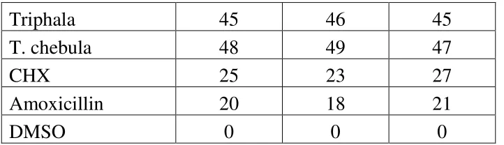
SUSCEPTIBILITY TEST BY WELL DIFFUSION
100µm of the inoculum mixed with 20ml of Mueller-Hinton agar
Held at 40 for 30 mins to aid in diffusion of the substances
5 wells of 6mm diameter were punched.100µl of the antimicrobial agents & control were transferred to it.
2% CHX 30µgm of amoxicillin DMSO T. chebula
Triphala
Incubated at 370c for 24 hrs
ASSESSMENT OF ANTIMICROBIAL ACTIVITY
AGAINST E.FAECALIS BIOFILM IN TEETH SAMPLES
28 single rooted human teeth were taken.
Smear layer removed with 5% NaOCl for 5 mins,
followed by 17% EDTA for 5 mins, 5% NaOCl for 5mins in an ultrasonic bath and then kept in saline for 5mins in an ultrasonic bath.
Decoronated below CEJ & apical portion cut to obtain 6mm of middle third of the root.
All specimens were autoclaved at 1210 C for 15mins Cementum removed to standardize the external diameter to 4 mm & internal diameter standardize to GG no. 3.
Blocks immersed in 1ml of TS broth in individual microcentrifuge tube & subjected to second cycle of sterilization
24 samples were contaminated with E. faecalis (ATCC 29212)
50µl of the inoculum transferred to individual
Dentine blocks transferred to fresh broth containing E. faecalis every second day for 21 days
After 21 days, they were irrigated with saline & assigned to the following groups.
Group 1: Positive control (n=6)
All were incubated at 370C for 7 days.
Antimicrobial assessment was done at the end of 7days, after irrigation with 5ml of saline.
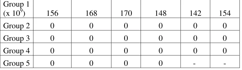
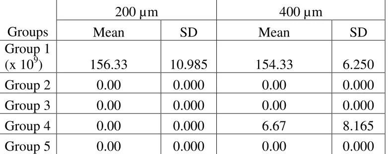
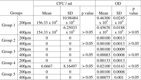
Dentine debris was harvested at 2 depths (200 & 400µm) by using GG no. 4 & 5 respectively & collected in 1 ml of sterile TS broth and incubated for 24 hrs.
Antimicrobial assessment was done by measuring the OD value and recorded.
The broth was serially diluted , plated & colonies were counted & recorded (CFU/ml).
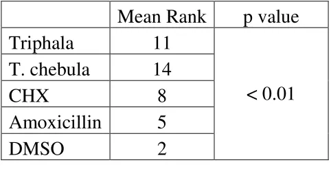
Test Groups Group2 : Triphala (n=6) Group3: T. chebula (n=6)
Group4: CHX (n=6)
Group 5:
METHODOLOGY
E. faecalis (ATCC 29212) maintained in stock culture in Institute of
Microbiology, Madras Medical College was used in this study. 24 hr growth
of E. faecalis grown on Trypticase Soy (TS) agar slope was suspended in
5ml of TS broth and incubated for 4 hours at 370C. The culture suspension
was adjusted to match the turbidity equivalent to 0.5 McFarland Standard.44
This was used as the standardized inoculum for all the procedures.
HERBAL EXTRACTS:
Alcoholic extract of Triphala and T. chebula was procured (Elles
Aromatics, India). To find the activity of the extract against E. faecalis, the
extracts were dissolved in Dimethyl Sulfoxide (DMSO), which is an inert
organic solvent and does not have any antibacterial action on its own.14
Initially to find the Minimum Inhibitory Concentration (MIC), serial
ten-fold dilutions were made to obtain a concentration range from 0.01 to
1000 µg / ml for each extract in TS broth. Then 0.02 ml of the 0.5
McFarland culture suspension was added to them. Two control tubes were
maintained. These included a positive control (the tube containing the
growth medium, physiological saline and the inoculum) and a negative
medium from each tube was subcultured on fresh TS agar plates and
incubated for 24 hours. All the plates had growth of microorganisms. This
revealed that the concentration tried was insufficient to exhibit antimicrobial
activity. Then several attempts were made with various concentration ranges
(upto 15mg/ml), to find the MIC.
Finally, a stock solution was prepared by dissolving 1500mg of
extract in 7.5ml of DMSO. The extracts were incorporated in the TS agar
and culture plates were prepared with concentration ranging from 20mg/ml
to 80mg/ml of the extracts by proportioning as in the table.
Extract Agar Net concentration of Extract present
0.5 ml + 4.5 ml = 20 mg/ml
1 ml + 4 ml = 40 mg/ml
1.5 ml + 3.5 ml = 60 mg/ml
2 ml + 3 ml = 80 mg/ml
Then the culture was spot inoculated on the plates. Positive control
was maintained by inoculating a plain TS agar plate without any
antibacterial agent. Negative controls were obtained by maintaining an
extract incorporated plate and DMSO control (TS agar plate with DMSO)
without inoculation of culture over it. The plates were incubated at 370C for
The plates were assessed for presence or absence of microbial growth.
When growth was present, the organism was confirmed as E. faecalis by
colony morphology, gram straining, heat test and standard biochemical
tests.17
Colony Morphology – It formed small (0.5 to 1 mm) clear colonies on
TS agar.
Gram Staining – It was seen as gram positive cocci, occurring in
ovoid pairs or in short chains.
Biochemical Tests – Biochemical reactions of it was assessed and it
showed :
Arabinose -
Mannitol +
Raffinose -
Sorbitol +
Urease +
NH3 produced from arginine +
Bile Esculin Test +
Heat Test – It showed growth, even after heating the culture
All of these features confirmed that the growth that was present in
some of the plates was pure culture of E. faecalis.
To confirm the bactericidal nature of that particular concentration,
samples were taken from the spotted region with a platinum loop and
subcultured on a TS agar plate and incubated for 24 hrs at 370C and
observed for the presence or absence of growth.
The concentration in which growth was present or absent was noted.
The readings were as follows:
Concentration (mg/ml) Triphala T. chebula
of extract
20 NG G
40 NG NG
60 NG NG
80 NG NG
At that point of the experiment, no growth was seen from 20 mg/ml of
extract concentration for Triphala and 40 mg/ml extract concentration for T.
chebula. To explore the possibility of activity at even a slightly lower
concentration, the extract with concentration of 100 mg/ml was used to
Triphala
Extract Agar Net concentration of extract
1 ml + 4 ml = 20 mg
0.5 ml + 4.5 ml = 10 mg
0.25 ml + 4.75 ml = 5 mg
0.125 ml + 4.875 ml = 2.5 mg
T. chebula
Extract Agar Net concentration of extract
1 ml + 4 ml = 20 mg
1.25 ml + 3.75 ml = 25 mg
1.5 ml + 3.5 ml = 30 mg
1.75 ml + 3.25 ml = 35 mg
The culture plates were spot inoculated, incubated, assessed for
growth of organism and subcultured for confirmation of bactericidal activity
and the growth when present was assessed for purity of the culture, as done
MIC was taken as the lowest concentration in which no growth was
detected. The readings were as follows.
Triphala T. chebula
20 mg / ml NG 20 mg / ml G
10 mg / ml G 25 mg / ml G
5 mg / ml G 30 mg / ml NG
2.5 mg / ml G 35 mg / ml NG
So, the MIC of Triphala was found to be 20 mg / ml and for T.
chebula 30 mg / ml.
SUSCEPTIBILITY TEST BY WELL DIFFUSION METHOD
100 µl of inoculum adjusted to 0.5 McFarland was mixed in 20 ml of
Mueller-Hinton agar and shaken. 0.5µg/ml of methylene blue was added to
it, to impart colour to the medium, which may aid in the ease of assessment
of the readings. Then the media was poured in a sterilized petridish . 5 wells
of 6 mm diameter were punched into the agar medium with sterile metal
cylinder. 100 µl of the extracts dissolved in DMSO, adjusted to the
concentration of MIC was transferred to the wells. 100 µl of 2 %
chlorhexidine and 30 µg of amoxicillin in the wells served as positive
control and DMSO served as negative control. After holding the plates at
medium,2 they were incubated at 370C for 24 hours and then the diameter of
zone of microbial growth inhibition around the wells were measured in
millimeters and recorded.
ASSESSMENT OF ANTIMICROBIAL ACTIVITY AGAINST
E. FAECALIS BIOFILM IN THE TEETH SPECIMENS:
The dentin block model used in the experiment was modified from the
one developed by Haapasalo and Orstavik (1987). In the present study,
extracted human teeth were used instead of bovine incisors (Safavi et al,
1990; Weiger et al, 2002; Lui et al, 2004; Saleh et al, 2004)89. Freshly
extracted single rooted human teeth stored in 10 % formalin were used.
Preparation of The Dentin Blocks:
A rotary diamond disk was used to decoronate the teeth below the
cemento-enamel junction. The remaining root was then sectioned such that 6
mm of the middle third of the root was obtained. Cementum was removed
from the root surface for standardizing the external diameter to 4 mm. The
internal diameter was standardized to Gates Glidden 3 (Mani Inc, Tachigi –
Ken, Japan) in a slow speed handpiece (NSK, Tokyo, Japan).44 Organic and
inorganic debris was removed by treating the blocks in an ultrasonic bath
tetra-acetic acid for 5 mins and again with 5% sodium hypochlorite for 5
mins. The blocks were immersed in the ultrasonic bath of saline for 5 mins
to remove all the traces of the chemicals used and sterilized in an autoclave
at 1210C for 15 mins. The blocks were subjected to a second cycle of
sterilization with the blocks immersed in 1 ml of Trypticase Soy (TS) broth
in individual microcentrifuge tubes. This allows better penetration of the
broth into the dentinal tubules.44
Infecting the Dentin Blocks:
50 µl of the 0.5 McFarland inoculum of E. faecalis was transferred to
presterilized individual microcentrifuge tubes contating 1 ml of the TS broth
and dentin block. The dentin blocks were transferred to the fresh broth
containing E. faecalis every second day. All the procedures were carried out
under laminar flow. The purity of the culture was checked by subculturing 5
µl of the broth from the incubated dentin block in TS broth on TS agar
plates. The dentin blocks were infected for a period of 21 days.44
Antimicrobial Assessment:
After the incubation period, the blocks were irrigated with 5 ml of
sterile saline to remove the incubation broth. Then the dentin blocks were
assigned to the following groups, each containing 6 blocks for the first 4


![Crystal structure of bis(μ {2 [(5 bromo 2 oxidobenzylidene)amino]ethyl}sulfanido κ3N,O,S){2,2′ [(3,4 dithiahexane 1,6 diyl)bis(nitrilomethanylylidene)]bis(4 bromophenolato) κ4O,N,N′,O′}dicobalt(III) dimethylformamide monosolvate](data:image/gif;base64,R0lGODlhAQABAIAAAP///wAAACH5BAEAAAAALAAAAAABAAEAAAICRAEAOw==)