HALLUM, YOUNGNER, AND ARNOLD
8
7
D-J
U,
-j
6
5
4
4.5 5.0 55 6.0 6.5 7.0
pH (INITIAL)
FIG. 2. Effect ofpHrange oninterferonactioninL cells. Cultures wereincubatedfor 5 hrwith 400 units of interferon in PBSateach pH. After removalof the interferon, the cultures were washed and challenged with VSV. Virus yieldswere determinedinfluids har-vested after 6 hrof incubation of the culturesingrowth medium.
tively inhibited; atpH 6.5 apartial inhibition of the development of resistance was observed. At
pH 6.0no resistance developed. The virus yields incontrol cultures in PBS6 that lacked interferon were not significantly different from the yields from controls in PBS7 and PBS8. By contrast,
the decrease in virus yield in cultures exposed
toPBSbelowpH 6.0, withorwithoutinterferon,
resulted from the increasing toxicity for L cells of the acidified PBS solutions.
To determine whether the effects described could bereproduced withaninterferonother than that induced inL cells by NDV, similar studies
were conductedusing the inhibitor released into the circulation of mice after injection of E. coli endotoxin (endotoxin interferon). Duplicate
cul-tures were treated with 100 units of endotoxin interferon diluted in PBS6 or in PBS7 as
pre-viously described. After incubationat37Cfor 1, 3, and5hr, the cultureswerewashed, challenged with VSV, and the yield of progeny virus was determined. Control values were obtained from cultures treated with PBS at the same pH but without interferon. The results of these studies
(Table 1) showed that endotoxin interferon
be-havedsimilarlyto interferonfrom NDV-infected L cells. Resistance to challenge with VSV did not develop incultures treated with interferon in
PBS6.
TABLE 1. Comparison ofeffect of PBS6 andPBS7 on the protective
actioni
ofendotoxin-stimulatedmouse interferon in L cellsa
LogioVSV yield (PFU/ml) Diluentadded Interferon 6 hr after challenge at
toLcells dose
Ihr 3 hr 5hr
PBS-6 None 7.45 7.70 7.65
PBS-6 100 units 7.65 7.25 7.18
PBS-7 None 7.42 7.70 7.75
PBS-7 100 units 7.20 6.65 5.70
aDuplicate cultures were exposedto 100 units
of endotoxin interferon in PBS6 or in PBS7; control cultures were exposed to diluent only.
After 1,3, or5 hr at 37 C, the solutions were re-moved, the cultures were washed with growth
medium, and they were then challenged with VSV.Afterincubation for 6 hr in growthmedium at 37 C, the released virus was harvested and assayed.
The possibility that the data obtained above were the result of inactivation of interferon in PBS6 at 37 C waseliminatedbydilutingtheL-cell interferon in PBS6, PBS7, or Eagle's medium, andincubating it at 37 C or at4C for 5 hr. No
significant change
in thetiterof the inhibitor wasobserved underany of these conditions.
Influence of pH
6.0 on the initial interactionoj
interferon with L cells. A second
explanation
for the lack ofdevelopment
of resistance atpH 6.0 is,possibly, that the initial reaction between inter-feron andLcellswasblockedat thispH.
Totestthis
possibility,
culturesexposed
to 400 units ofinterferon from NDV-infected L cells in cold PBS6, PBS7, or Eagle's medium were incubated for 15 hr at 4C. After this
time,
the interferon wasremoved and thecultures were washedthree times with coldEagle's
medium and transferred to 37 C. The resistance tochallenge
virus wasmeasured bythe
yield
inhibition method at0, 2, 4, 6, and8hrafterthetransfer of the culturesto37 C.Theresultssummarizedin
Fig.
3show that resistance to the viruschallengedeveloped
atthesame rate
regardless
ofthepH
atwhich the inter-feron solution wasapplied.
These data indicate that the initial interaction of interferon with Lcells takesplaceat
pH
6.0,eventhough
resistancedoes not
develop
under these conditions.Maintenance of cell resistance at pH 6.0.
Another
possible
explanation
for the failure of cell resistance todevelop
atpH
6.0 is that the metabolic productsrequired
toexpress theanti-viral action of interferon
(13)
were unstable atthispH. Ifinterferon
protection
weredependent
upon the so-called "second
protein"
and if this material were unstable in cellsexposed
topH 6.0,
--- CONTROLS-NO INTERFERON _- a INTERFERON TREATED
_ m _
F
I
774 J. VIROL.
on November 11, 2019 by guest
http://jvi.asm.org/
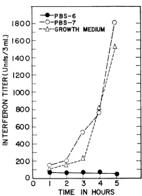
pH AND INTERFERON ACTION
E ~
N ---PBS-6
N -0-- PBS- 7
LL
0. ---A---GROWTHMEDIUM
z_ \
0
z
O \
-0~ ~ ~ 0
2
2 1
0 2 4 6 8
Time(HourAInGrowth MediumFollowing 4°Incubation
FIG.3.Lackof effectofpH6.0oninitialinteraction ofinterferonand L cells. L cellswereincubatedfor15 hr at 4 C withinterferon in PBS6, PBS7, or inEagle's
medium. After this period, the cultures were washed three timeswith coldmedium,3mlofEagle'smedium wereadded,and thecultureswere transferredto37 C. At the timesshown, thecultures were challengedwith VSVbytheyieldinhibition method. Virusyieldin con-trol cultures withoutinterferonwas2.1 X 107 PFU/ml.
cells in which interferon
protection
had been established would become moresusceptible
toinfectionwhenheldatthis
pH.
Totestthispossi-bility,
L-cell cultures were inoculated with 270unitsof L-cell NDV interferon in
Eagle's
medium and incubated for 15 hr at 37 C. The cultures were then washed withPBS6, PBS7,
orEagle's
medium, covered with3 mlof thesame
solution,
andincubated
again
at37C.After 0, 4, 8, and24 hr of additional incubation at 37C,
thecultureswere
challenged
with VSV and theyield
ofprog-enyviruswasdeterminedasdescribed
previously.
Theresults of this
experiment, plotted
inFig.
4, show thatinterferonprotection,
onceestablished,
is
equally
stable in cellsexposed
toPBS6 orPBS7.The results from cultures
exposed
toEagle's
me-diumwereidenticalto
thedataobtained
with PBS6 andPBS7.Influence of PBS6 onmacromolecularsynthesis in L cells. Another
possible
explanation
for the failure of cells treated with interferon at pH 6.0 to develop resistance to virus is that macro-molecularsynthesis might be inhibited at this pH, thus preventing the formation of significant amounts of "second protein." This possibility was testedby measuringthedifferencein the rateof
incorporation
of'4C-leucine
and 3H-uridineinto proteinand RNA,respectively, as a function of the pH of the PBS to which the cells were exposed. Replicatecultures of L cells were treated
E
D
a-0
-i
w
0
0 -J
8
7
0 4 8 t 24
Time(hours)After Tronsferto PBS-6 orPBS-7 FIG. 4. Maintenance ofcell resistance atpH 6.0. Cultures treatedfor 15 hr at 37 C with 270 units ofinter-feron in Eagle'smedium were washed andtransferred
toPBS6, PBS7, or tofreshEagle'smedium,and incu-bation was continued.Aftervarioustimes inthesemedia,
thecultures werechallengedwith VSVaccording to the
yield inhibitionmethod. TheresultswithEagle's medium
weresimilar to those in the otherdiluentsand are not shown.
0 Io
80
gr 60
<oz
,_r-ZI 40
20
az
coOUL
0 1 2 3 4
TIME (HOURS)IN PBS-6
FIG. 5. Influence ofexposureofLcellstoPBS6on proteinand RNA synthesis. Culturesweretreated with
PBS6 or PBS7 for the intervals shown, then pulsed with 14C-leucine or 3H-uridine. At each time interval,
the trichloroacetic acid-precipitable countsin the cells exposedtoPBS6werecomparedtothecountsincells in PBS7. (Average ofcountsincorporated into PBS7 controlcultures: "4C-leucine, 1,717countsperminper culture; 3H-uridine, 524countsperminperculture.)
with eitherPBS6orPBS7 and after intervalsof 10
mi to 5 hr they were pulsed with radioactive
precursorsasdescribed above.Theresultsof these experiments, summarized in Fig.5,clearly demon-stratedthattherates of both 14C-leucineand
3H-CONTROLS: -0-PBS-6
---
PBS-7-6
-- INTERFERON TREATED:
--_-
PBS-6-5 -
PBS-7-
-->K_/
-/
-4I
-e *--C1LEUCINE INCORPORATION -0- H3URIDINE INCORPORATION
O . -0 --. -O
- *
5
775
VOL. 2, 1968
on November 11, 2019 by guest
http://jvi.asm.org/
[image:2.490.267.412.80.280.2]HALLUM, YOUNGNER, AND ARNOLD
(I 100 0--0
990 I
I
z 60_
t-50
o
R 80 5 .
cr~ ~ ~~p
0 0) 3
50C4 EUIN
FI 40 3
at37-CUttepAl o rIDINen usdwt
O0 II
0
0 50 6 U65 70
p H
FIG. 6. Influence ofPBS atdifferent pHonl macro-molecularsynthesisin L cells. Cultureswere incubated
at 37 C at the pH shiownfior 5 hr, then pulsed with
either '4C-leucine or 3H-uridine. The trichloroacetic acid-precipitable counts werecomparedtocultures in-cubatedfbr 5 hr in PBS7. (Counts incorporatedinto PBS7 control cultures: 14C-leucine, 1,247 coun2ts per
minperculture; 3H-uridine, 1,801 counitsper miii per culture.)
uridine incorporation were decreased by about 70%0 within 20min ofexposureto PBS6.
Additional incorporation studies were carried
out to determine the effect on macromolecular
synthesis of exposure to a range ofpH. These
experiments at pH values from 4.5 to 7.0 were conducted as described for PBS6, except that a single 5-hrincubation periodat37 Cwasapplied
ateach pH. The results of these studies (Fig. 6)
show that the incorporation of both "4C-leucine and 3H-uridine was progressively reduced at
decreasing pH. In PBS5 and PBS5.5, no
signifi-cant synthesis of either RNA or protein was
observed.
DISCUSSION
The data presented show that exposure of L
cells to PBS at pH 6.0 results in a rapid and reversible inhibition of both RNA and protein synthesis. Control of macromolecular synthesis byexposuretoPBSatpH6.0 offers certain advan-tages over the useofthe usual chemical or anti-biotic inhibitors. In addition to the speed and
reversibility of the inhibition of synthesis, the treatment doesnot involve entry into the cell of
molecules orions thatare notordinarily present. The mechanism by which exposure of L cells to PBS6 depresses RNA and protein synthesis is under investigation; preliminary studies indicate
that exposure of L cells for 20 min to PBS6
causes a disaggregation of polysomes. Parallel
studies of chick embryo cell cultures showedthat
these cells are more resistant to the effects of
lowered pH than are L cells. The basis for the species and cell differences is being explored.
The results confirmed that interferon can interact fully with Lcells even though resistance
does not develop (3, 9). We used a lowered range ofpH to demonstrate a model system for separat-ing the initial interferon-cell interaction fromthe
synthetic processes essential for the development of resistance.
Previous studies of the effect of change of pH ontheaction of interferon have been reported by Gifford(6) and by De Maeyer and De Somer (1). The pH range covered in these reports was from 6.8 to 7.6. Gifford (6) concluded that in chick embryo cells the plating efficiency of the vaccinia virus challenge was altered by changes in pH, but that the action of interferon was not signi-ficantly affected. De Maeyer and De Somer (1), using a continuous line of rat tumor cells, re-ported an increase in interferon titer at pH 6.8 compared to pH 7.2. However, since both papers covered only a narrow physiological range of pH, theinhibition of interferon activity at the low pH usedinthis study was not observed.
ACKNOWLEDGMENT
This investigation wassupportedby Public Health Service research grant AI-06264 from the National Institute of Allergy and Infectious Diseases.
LITERATURE CITED
1. DeMaeyer, E., andP. DeSomer. 1962.Influence of pH on interferon production and activity.
Nature194:1252-1253.
2. Friedman,R. M.1967.Interferonbinding:thefirst step in establishment of anti-viral activity. Science 156:1760-1761.
3. Friedman, R. M., and J. A. Sonnabend. 1964. Inhibition of interferon action by p-fluoro-phenylalanine. Nature203:366-367.
4. Friedman, R. M., and J. A. Sonnabend. 1965. Inhibition of interferon by puromycin. J. Immunol. 95:696-703.
5. Fujioka, M., M. Koga, and 1. Lieberman. 1963. Metabolism of ribonucleic acid after partial
hepatectomy.J.Biol. Chem. 238:3401-3406. 6. Gifford, G. E. 1963. Effect of environmental
changes upon antiviral action of interferon. Proc. Soc. Exptl. Biol. Med. 114:644-649. 7. Hallum,J.V., andJ.S.Youngner. 1966.
Quantita-tiveaspectsofinhibition of virusreplication by
interferon in chick embryo cell cultures. J. Bacteriol. 92:1047-1050.
8. Levine, S. 1964. Effect of actinomycin D and
puromycin dihydrochloride on action of inter-feron.Virology24:586-588.
776 J. VIROL.
on November 11, 2019 by guest
http://jvi.asm.org/
pH AND INTERFERON ACIION
9. Levine,S. 1966. Persistenceofactive interferon in cells washed after treatment with interferon. Proc. Soc. Exptl. Biol. Med. 121:1041-1045. 10. Lockart,R. Z., Jr.1964.The necessityforcellular
RNA and protein synthesis forviral inhibition resulting from interferon. Biochem. Biophys. Res.Commun. 15:513-518.
11. Merchant,D.J.,R.H.Kahn, andW. H.Murphy. 1960. Handbook of cell and organ cultures.
BurgessPublishing Co., Minneapolis, Minn. 12. Stinebring, W. R., and J. S. Youngner. 1964.
Patterns of interferon appearance in mice
in-jected with bacteria or bacterial endotoxin.
Nature 204:712.
13. Taylor, J. 1964. Inhibition of action ofinterferon by actinomycin. Biochem. Biophys. Res.
Com-mun.14:447-451.
14. Youngner, J. S., A. W. Scott, J. V. Hallum, and W. R. Stinebring. 1966. Interferon production by inactivated Newcastle disease virus in cell cultures and in mice. J. Bacteriol. 92:862-868.
VOL. 2, 1968
777
on November 11, 2019 by guest
http://jvi.asm.org/
JOURNALOFVIROLOGY, Aug.1968,p.778-786
Copyright @ 1968 American Society for Microbiology Printed in U.S.A.Vol. 2, No. 8
Protective
Effects
of Specific Immunity
to
Viral
Neuraminidase
onInfluenza
Virus
Infection
of
Mice
JEROME L. SCHIJLMAN, MANIJEH KHAKPOUR, AND EDWIN D. KILBOURNE Division of Virus Research, Department of Public Health, Cornell University Medical College, New York,
New York 10021
Received forpublication29April 1968
Antibody specific for viral neuraminidasecanbe demonstratedin micefollowing
(i) pulmonary infection with influenza virus, (ii) immunization with
ultraviolet-in-activated influenza virus, (iii) immunization with isolated neuraminidase of
in-fluenzaA2 virus, and (iv) passive immunization withseraofrabbitsimmunized with
isolated A2 neuraminidase. Neuraminidase antibody produced by any of these
methodsexertsaprofound inhibiting effectonvirus replication in the lungs of mice
challenged with strains of virus having homologousneuraminidase protein,evenin
the absence of hemagglutinatinginhibiting antibody tothechallenge virus, and
re-sults in markedly decreased pulmonary virus titers and diminished lung lesions.
These observationssuggestthat antineuraminidase immunitymayplayasignificant
role inthe protection against influenza virus challenge observed inmice after infec-tionor artificial immunization.
It is now well established that
hemagglutinin
and neuraminidase are
antigenically
distinct proteinsof
theenvelope
of influenza virus, andthat
by
genetic
recombinationhybrid
(recom-binant) virusescanbe producedin which hemag-glutinin is derived from one parental virus and neuraminidase from the other
(7, 8,
11).
Suchantigenically
hybrid
viruses in whichhemag-glutinin and neuraminidase
proteins
from differ-ent subtypes have beensegregated
haveproved
useful in the
isolation
of neuraminidase free of demonstrablehemagglutinin protein (11)
and inthe
production
ofspecific antibody
to viralneuraminidase
(7).
In
aneuploid
cellculture,
in ovo, and inchickshell-membranesystems, neuraminidaseantibody isnon-neutralizingexcept in
high concentrations,
but it
partially
inhibits virusreplication by
its effect on therelease andyield
of influenza virus from cells(7, 14,16;R. G.Webster,W. G.Laver, and E. D. Kilbourne, submittedfor publication). In earlier studies ofmice immunizedby
infec-tion withantigenically
hybrid
influenzaAviruses,
equivalent
protection
was found afterchallenge
with viruses which contained either the same
hemagglutinin or the same "minor"
(neuramini-dase) antigen
possessed by
theimmunizing
virus (9).We now report a further
comparison
of therelativeeffectiveness of specific immunitytoviral neuraminidase and
immunity
to viral hemag-glutinininprotecting mice against challengewith influenza virus infection. In these experiments, antibodytoneuraminidase wasproduced in mice (i) by infectionwithrecombinantviruses in which hemagglutinin and neuraminidase were derived from parents ofdifferent subtypes, (ii) by injec-tion of ultraviolet-inactivated preparations of thesame
viruses, (iii) by injection
ofpurified
A2neuraminidase,
or (iv) by passive immunizationwith rabbit
antibody
tothe enzyme.MATERIALS AND METHODS
Cellsandplaqueassay. Clone1-5C4,derivedfrom theWong-Kilbournevariant ofthehumananeuploid
Chang conjunctival cell line, was used for plaque-reductiontests(15).Themethods for assay ofplaques
and for plaque inhibition with antisera have been
publishedin detail(5, 15).
Viruses. Mostof the viruses employed have been described in earlier reports (9). These include Aol
NWS andA2/Jap. 305(mouseadapted) and the
fol-lowingrecombinants: unadapted andmouse-adapted
strains ofX-7, X-3,
X-lL,
and X-9 (17 passages inmice),X-7(F1),andX-15 (hybridofA/equine1
con-taining A2neuraminidase) (6).Theantigenic
designa-tionof theseviruses(Table 1)wasmade in accordance withasystemdescribedpreviously (7, 8).
Ultraviolet inactivation ofvirus. Insomeinstances,
allantoic fluidseedviruses wereinactivatedby ultra-778
on November 11, 2019 by guest
http://jvi.asm.org/
INFLUENZA VIRUS INFECTION OF MICE
violet(UV) irradiationpriortoparenteral inoculation into mice. Fluids werecentrifuged at 8,000rev/min, dialyzed atpH 7.2overnight, and then subjectedto UVlight from a 7.5-w GElampat adistanceof 17.8 cm for 2 min. Afterthisprocedure,residualinfectivity
for eggs could be demonstrated only with undiluted
fluidsin the presence ofcortisone.Thefluidsthen were
adjustedto identical hemagglutinationtiters priorto
inoculationof mice.
Mice. Specific pathogen-free male Manor Farm mice (MF-1) 8 to 16 weeks of age wereused.
Eggs. White Leghorn chick embryos 10 to 12 days
of age were used to assess the infectivity titer of all
tissues.
Hemagglutination-inhibition tests.Hemagglutination
tests with mouse andrabbitsera were performed by
methods describedpreviously (17).
Enzyme-inhibition tests. Enzyme-inhibition tests
with mouse and rabbit sera were carried outwith a fetuin substrateaccording tomethods described pre-viously (7, 11). Measurements of inhibition of A2
neuraminidase (E) were made with X-7(FI) virus adjustedtogive optical density readings of300 to 800 at 549 nm in a Bausch and Lomb Spectronic-20 colorimeter or Beckman DU-2 spectophotometer.
Measurements ofinhibition ofAoneuraminidase (e) were made with the same substrate using an 18-hr
incubation period of X-9 (A2e) virus and substrate. Titers of antisera are expressed as the dilution at
which 50% inhibition of neuraminidase activity
occurred.
Isolation ofA2neuraminidase. Thepurificationand
disruption of X-7(F1) virus and the isolation and
elution of the neuraminidase protein from cellulose
acetatestrips were carried out bymethods described previously (11).
Virus titrations. Lungswere removed atdesignated periods afterinfection andgroundinglass tubes
ac-cording to techniques described previously (12).
Serial 10-fold dilutions of ground lung suspensions
wereinoculated into eggs, andafter40 hrof
incuba-tion allantoic fluids were harvested and were tested
for hemagglutinationwith human "O"redcells at a 1:4 dilution. Titrationendpoints were calculated in
terms ofthedilution oflung tissue infecting 50% of
thechickembryos.
Scoringofpulmonarylesions.Amodification ofthe
maximal scoremethod wasused, inwhichthe extent ofpulmonary lesions was expressed as a percentage
ofthetotallung surface (4).
Aerosolinfection ofmice. The apparatus and pro-cedures usedto generateaerosols ofinfectious virus
have been described previously (12). During the-periods in which they were inside the aerosol chamber, mice wereexposed to an estimated 10 to 100 mouse infective doses of thechallenge viruses employed.
Intranasal inoculation. In someexperiments,rabbit antiserum was delivered intranasally to mice. Mice werelightly anesthetized with ether, and three drops (0.05 ml) of the appropriate serum was delivered into the nostrilsthrough a 26-gauge needle.
Bronchial washings. Mice were killed by cervical
fracture, and the trachea was dissected free of the esophagus and surrounding connective tissue. A
TABLE 1. Antigenicdesignation ofvirusesemployed
in the presentstudy
Virus Hemagglutinin Neuraminidase Antigenic
subtype subtype designation
X-7... Ao A2
AoE-X-7(F1)... Ao A2 AoE
X-3 ... Ao Ao Aoeb
X-1L... A2 A2 A2E
X-9 ... A2
Ao
A2eX-15... A/equine A2 Eq E
1/56
NWS... A
Ao
Aoe
Jap.305... A2 A2 A2E
a E = A2 neuraminidase.
b e = Ao neuraminidase.
no. 20adapter wasinsertedintothetracheathrougha
small holeandtied inplace bythread. Anamountof
1 mlofsterile saline (0.1% gelatin) wasinjectedinto the trachea andlungsand then wasaspiratedbackinto asterile syringe (0.4to0.8 ml wasrecovered).These
fluids were treated as undiluted bronchial washings,
and all subsequent dilutions were expressed accord-ingly.
RESULTS
Demonstration of neuraminidase antibody in mice after influenza virus infection. Mice were
infected with 100 mouse infective doses of
Ao/NWS
or A2/Jap. 305 virus or were exposedto an aerosol of saline. Serum
specimens
and bronchialwashings from 10 mice in each group wereseparately pooled4weeks later forantibody
determination. Hemagglutinating-inhibiting
(HI)
activity was measured against the two
infecting
virusesandagainst recombinantX-15 (equine E) virus [a virus inhibitable in
hemagglutination-inhibition
tests withantibody
toA2
neuramini-dase
(E)
(6)].
Enzyme-inhibiting
antibody
was measuredagainst X-7 (Fl)(AoE)
andX-9 (A2e) recombinantviruses,
andplaque
size-reducing activity was titrated in human conjunctival cells infected with X-9 (A2e) or X-7(AoE)
viruses underantibody
in agaroverlays.
The results (Table 2) demonstrate that, following influenza virus infectionof
mice,
hemagglutinating-inhibit-ingantibody appeared in bronchial secretions, as well as in the serum, and that antibody to the neuraminidase component ofthe infecting virus waspresentintheserum.Thisneuraminidase an-tibodywasdemonstrated by inhibition of enzy-matic activity of intact X-9 (A2e) or X-7
(Fl)
(AoE)
virusandby plaque size reductionof virusescontaining
neuraminidasehomologousto theneur-aminidase antibody. The HI activity against X-1 5 virus in sera of mice previously infected with A2/Jap. 305 virus is additional evidence of A2
VOL.