0022-538X/96/$04.0010
Copyrightq1996, American Society for Microbiology
Interaction of Human Immunodeficiency Virus Type 1 Tat with
a Unique Site of TFIID Inhibits Negative Cofactor Dr1 and
Stabilizes the TFIID-TFIIA Complex
FATAH KASHANCHI,
1SAMIR N. KHLEIF,
2JANET F. DUVALL,
1M. REZA SADAIE,
3MICHAEL F. RADONOVICH,
1MICHAEL CHO,
4MALCOLM A. MARTIN,
4SEI-YU CHEN,
5ROBERTO WEINMANN,
5ANDJOHN N. BRADY
1*
Laboratory of Molecular Virology
1and NCI-Navy Medical Oncology Branch,
2National Cancer Institute,
and Laboratory of Molecular Microbiology, National Institute of Allergy and Infectious Diseases,
4Bethesda, Maryland 20892; Division of Transfusion Transmitted Diseases, Center for Biologics Evaluation
and Research, Food and Drug Administration, Rockville, Maryland 20859-1448
3; and Department of
Molecular Oncology and Drug Development, Bristol-Myers Squibb, Princeton, New Jersey 08543
5Received 26 September 1995/Accepted 18 April 1996
We have previously reported the direct physical interaction between the human immunodeficiency virus
(HIV) type 1 Tat protein and the basal transcription factor TBP/TFIID. Affinity chromatography demonstrated
that wild-type Tat, but not a transactivation mutant of Tat, was capable of depleting TBP/TFIID from cell
extracts. These experiments represented the first demonstration of a basal transcription factor that binds, in
an activation-dependent manner, to Tat. We now report that the Tat-TBP interaction can be detected in HIV
type 1-infected cells. The domain of TBP interacting with Tat has been mapped from amino acids 163 to 196
by using deletion and site-specific mutants of TBP. This domain of TBP, which includes the H1 and S2
domains, is distinct from the H2 binding site for other activator proteins, such as E1A. The interaction of Tat
with TFIID regulates the binding of accessory proteins to TFIID. Tat stabilizes the interaction of TFIID with
TFIIA in a gel shift assay. In addition, Tat competes for Dr1 interaction with TBP. Our results suggest that the
basal transcription factor TBP/TFIID represents an important regulatory molecule in HIV transcription.
The Tat protein of human immunodeficiency virus type 1
(HIV-1) plays a key role in virus replication and
transforma-tion. Tat-defective proviral clones do not replicate efficiently
(5, 6). Transgenic mice harboring the HIV Tat gene develop
Kaposi’s sarcoma-like lesions, and Tat has recently been shown
to synergize with basic fibroblast growth factor in inducing
angiogenic Kaposi’s sarcoma-like lesions in mice (12, 53).
Transcriptionally, Tat has been shown to be a potent
transac-tivator of the HIV promoter. Tat transactivation of the viral
long terminal repeat (LTR) is dependent upon the presence of
upstream transcription factors and the TAR RNA regulatory
element. Tat stimulates both transcription initiation and
elon-gation in vivo (13, 32). In vitro studies of Tat transactivation
also support a role for Tat in initiation and elongation (1, 28,
32, 34, 39). Bohan et al. (1) have demonstrated that Tat
facil-itates the formation of the HIV preinitiation complex.
Subse-quently, Kashanchi et al. (28) reported that Tat interacts
di-rectly with the TATA-binding protein (TBP) subunit of TFIID.
In agreement with these findings, Veschambre et al. (52) have
recently reported a functional interaction between Tat and
human TFIID. In other in vitro studies, Marciniak et al. have
shown that Tat stimulates the formation of more-processive
elongation complexes (38, 39). Further, Kato et al. (32) have
demonstrated that TFIIF, an elongation transcription factor,
decreased the requirement for Tat transactivation by
increas-ing polymerase processivity.
The ability of Tat to participate in transcriptional initiation
and elongation is perhaps not surprising, given the ability of
other transcriptional activators, such as VP16, to function
dur-ing multiple steps of preinitiation complex assembly and
elon-gation. The physical interaction of VP16 with basal
transcrip-tion machinery factors such as TBP, TFIIB, TAFII40, and
TFIIH has been demonstrated by several independent
labora-tories (15, 35, 50, 55). VP16 facilitates the recruitment of
TFIIB and then, in a second step, facilitates transcriptional
initiation through interaction with TBP-associated factors
(TAFs) (8). Interestingly, Yankulov et al. (56) have shown that
activators such as VP16 and E1A also stimulate RNA
poly-merase processivity, and they have suggested that this function
is as important as the stimulation of transcriptional initiation.
Those authors further suggest that the competence of RNA
polymerase II to elongate is an integral part of the initiation
step that is controlled by activators interacting with the general
transcription factors. In this light, it is of interest that the VP16
transcription activation domain is functional when targeted to
a promoter-proximal RNA sequence (21).
The functions of the human TBP are complex. This 38-kDa
protein was originally identified as a polymerase II
transcrip-tion factor which interacted specifically with the
2
25 TATA
promoter region (21, 26, 43). Indeed, the purified 38-kDa
protein can reconstitute basal TATA-dependent transcription
in vitro. More recently, however, it has been demonstrated that
the polymerase II TBP complex (TFIID) has a molecular mass
of approximately 750 kDa and is composed of TBP and
mul-tiple polypeptides referred to as TAFs. Interestingly, while
TBP can support basal transcription, TFIID is required for
activator-dependent transcription (20, 21, 24, 43, 44, 48). Quite
remarkably, in addition to TBP’s role in polymerase II
tran-scription, the 38 kDa protein has also been shown to be
in-volved in polymerase I and polymerase III transcription (18).
In polymerase I transcription, TBP is found in the SL1
com-plex, consisting of TBP in tight association with three TAFs of
* Corresponding author. Mailing address: Laboratory of MolecularVirology, Building 41/Room B403, NCI, NIH, Bethesda, MD 20892.
5503
on November 9, 2019 by guest
http://jvi.asm.org/
48, 63, and 110 kDa (9). In polymerase III transcription, TBP
is part of the transcription factor TFIIIB. In Saccharomyces
cerevisiae, TFIIIB consists of three proteins of 69 kDa (yeast
BRF1), 38 kDa (TBP), and 90 kDa (31). At this point, it
appears that most TBP in a cell is found either in TFIID, SL1,
or TFIIIB complexes (18). TBP is also the target of positive
transcription factors (TFIIA), viral activators (E1A and T
an-tigen), and negative cofactors (Dr1) (2, 14, 16, 33, 54).
Obvi-ously, this basal transcription factor, through complex
interac-tions with a number of cellular proteins, plays a pivotal role in
eukaryotic gene regulation.
We have previously reported that Tat interacts with the
basal transcription factor TFIID (28). Using peptide
competi-tion and binding analysis, we demonstrated that Tat amino
acids 36 to 50 were important for this interaction. Importantly,
a mutation at lysine 41 which abolishes Tat transactivation also
abolished interaction with TFIID. In this report, we extend the
initial observations on the Tat-TFIID interaction by showing
that (i) the Tat-TFIID complex can be seen in vivo by
immu-noprecipitation from HIV-1-infected cells; (ii) the domain of
TBP required for interaction with Tat includes the H1-S2
do-main, which is distinct from the E1A activator binding site; (iii)
Tat stabilizes the TFIID-TFIIA complex on the HIV-1 TATA
box; and (iv) Tat can compete for Dr1 binding to TBP.
IIIB
with HIVIIIB (H9/IIIB cells) as previously described (47) with some minor modifications. Both infected and uninfected cells were grown in RPMI 1640 supplemented with 10% fetal bovine serum at an initial density of 53105
cells per ml 2 days prior to infection. For infection, 53106
H9/IIIB cells were mixed with 23107
H9 cells and cocultivated in fresh medium at a density of 106 /ml in T75 flasks. At 0, 6, 12, 24, and 48 h postinfection, a total of 53107
cells per time point were harvested by centrifugation. For the mock infection, the same number of H9 cells were harvested. Pelleted cells were washed once with cold phosphate-buffered saline and once with buffer A (10 mM HEPES [N-2-hydroxyethylpipera-zine-N9-2-ethanesulfonic acid] [pH 7.9 at 48C], 1.5 mM MgCl2, 10 mM KCl, and 0.5 mM dithiothreitol [DTT]) and resuspended in 0.5 ml of buffer A containing 0.1% Nonidet P-40 (NP-40). After incubation on ice for 10 min, the cell lysate was vortexed briefly and nuclei were pelleted by centrifugation in a Sorvall tabletop centrifuge (MC 12V; Dupont) at 48C.
Extracts from nuclei were prepared as described by Dignam et al. (10), and one-third of the extract (250 ng) was used for immunoprecipitation. The immu-noprecipitation with anti-Tat (polyclonal, 100mg) was done at 48C overnight in the binding buffer (10 mM HEPES [pH 7.9], 50 mM KCl, 2.5 mM MgCl2, 50mM ZnCl2, 50mM EDTA, 1 mM DTT, and 8.5% glycerol). Immune complexes were precipitated with protein A-Sepharose, washed with binding buffer–0.1% NP-40, and subjected to sodium dodecyl sulfate-polyacrylamide gel electrophoresis (SDS-PAGE) (4 to 20% polyacrylamide) and to Western blotting (immunoblot-ting) with a monoclonal anti-Tat or anti-TBP antibody.
Preparation of glutathioneS-transferase (GST) proteins.Tat of HIV-1 was subcloned from the pSynTat vector (27) by using PCR. A 59oligonucleotide containing a BamHI site and a 39oligonucleotide containing an XhoI site were used for PCR with Tat72 and Tat86. The fragments were then cut with restriction enzymes BamHI and XhoI, purified, and subcloned into the polyclonal site of the pGEX-20T vector. The polyclonal site contained a thrombin-cut site followed by
BamHI, EcoRI, XhoI, ClaI, SpeI, and XbaI restriction sites. Fragments were
ligated and transformed into Escherichia coli XA-90. Tat86 dl 2-36, GST-Tat72, GST-Tat72 cys22, GST-Tat48, and GST-Tat48 cys22 were generous gifts of A. Rice, Baylor College of Medicine. Recombinant proteins were grown in 1 liter of NZCYM (28) to an A600of 0.6 and induced with fresh 1 mM IPTG (isopropyl-b-D-thiogalactopyranoside). Cells were harvested 3 h later and washed with phosphate-buffered saline without Mg21and Ca21. The pellet was resuspended in 10 ml of extraction buffer containing 50 mM HEPES (pH 7.4), 250 mM NaCl, 1 mM EDTA, 5 mM NaF, 1 mM DTT, 5mg each of aprotinin, leupeptin, and pepstatin A per ml, 100mg of phenylmethylsulfonyl fluoride per ml, and 1% NP-40 and sonicated to break the cells.
Cell lysates were centrifuged in a Sorvall RT6000D centrifuge (5,000 rpm) for 10 min at 48C. The supernatant was mixed with one ml of glutathione-agarose beads (50% slurry) and kept at 48C for 2 h on a rotator. The beads were centrifuged and washed five times with the extraction buffer, and GST proteins were eluted with 25 mM reduced glutathione (in extraction buffer). Peak frac-tions containing the proteins were dialyzed against 20 mM HEPES (pH 7.4)–150 mM NaCl–1 mM EDTA–10% glycerol–0.1% NP-40. Aliquots of 1 mg/ml were stored at2708C.
GST binding assays.The amounts of all GST proteins were normalized ac-cording to protein concentration, and the amount bound to beads was confirmed by SDS-PAGE followed by Coomassie staining. GST proteins (approximately 5 mg) and 2.5 to 10ml of the TNT lysate (Promega) containing35S-labeled Tat or TBP were incubated in either TNE100 (100 mM Tris [pH 8.0], 100 mM NaCl, 1 mM EDTA, and 0.1% NP-40) or buffer D (20 mM HEPES [pH 7.9], 100 mM KCl, 12.5 mM MgCl2, 0.1 mM EDTA, 17% glycerol, 0.1 mM DTT, and 0.2% NP-40) in a final volume of 200ml and with a total of 50ml of beads. Binding reaction mixtures were incubated for 2 h at 48C with rotation. The complexes were centrifuged for 30 s and washed twice with 1 ml of buffer containing 50 mM Tris (pH 7.9), 150 mM NaCl, and 0.2% NP-40. The bound labeled proteins were denatured, subjected to SDS-PAGE (4 to 20% polyacrylamide), dried, and autoradiographed.
For Tat and Dr1 bindings, GST-TBP (wild type) or the version containing amino acids 1 to 163 (GST-TBP 1-163) was used at 10mg. These proteins were mixed with 100ml of whole-cell extract (WCE) (described in “In vitro transcrip-tion” below), incubated overnight on a rotator at 48C, and washed three times the next day with binding buffer (used for immunoprecipitation) containing 0.1% NP-40. Bound complexes were denatured and subjected to SDS-PAGE (4 to 20% polyacrylamide) and to Western blotting to detect the presence of Tat or Dr1.
[image:2.612.105.245.74.330.2]In vitro transcription.HeLa (WCEs) were prepared according to a previously established procedure (37) and added (7.5ml) to the in vitro transcription reaction mixture at a concentration of 12 mg/ml. pAdML and a HIV-LTR chloramphenicol acetyltransferase (CAT) plasmid were linearized with BamHI and EcoRI, respectively, and added to the in vitro transcription mixture at a concentration of 30 ng per reaction mixture. The in vitro transcription buffer contained 10 mM HEPES (pH 7.9), 50 mM KCl, 0.5 mM EDTA, 1.5 mM DTT, 6.25 mM MgCl2, and 8.5% glycerol. Each reaction mixture also contained nu-cleoside triphosphates ATP, GTP, and CTP at a final concentration of 50mM FIG. 1. Immunoprecipitation of Tat-TFIID complex from HIV-1-infected
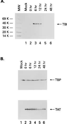
cells by using anti-Tat polyclonal antiserum. Infected cells were cocultivated with uninfected H9 cells at a 1:4 ratio. They were then processed at various times (0, 6, 12, 24, and 48 h) postinfection for immunoprecipitation with anti-Tat immu-noglobulin G purified polyclonal serum or anti-orf-1 (30). Samples (100mg of nuclear preparation) were incubated overnight at 48C, and immune complexes were collected the next day and subjected to SDS-PAGE (4 to 20% polyacryl-amide) and Western blotting with anti-TBP monoclonal serum (Promega). Panel A and the bottom of panel B represent immunoprecipitates fractionated on SDS-PAGE and then subjected to Western blotting with anti-TBP (Promega) (A) or anti-Tat (American Biotechnology) (bottom of panel B) monoclonal antibody. The TBP portion of panel B is a straight Western blot of nuclear extracts (used for immunoprecipitation) with anti-TBP monoclonal antibody. Mock, uninfected H9 cells; MW, molecular weight markers (in thousands [K]).
on November 9, 2019 by guest
http://jvi.asm.org/
each and 20mCi (2ml) of [32
P]UTP (400 Ci/mmol; Amersham). Transcription reactions were terminated by the addition of 20 mM Tris-HCl (pH 7.8)–150 mM NaCl–0.2% SDS. The quenched reactions were extracted with equal volumes of phenol-chloroform and precipitated with 2.5 volumes of ethanol and 0.1 volume of 3.0 M sodium acetate. Following centrifugation, RNA pellets were resus-pended in 12ml of a formamide denaturation mixture containing xylene cyanol and bromophenol blue, heated at 908C for 3 min, and electrophoresed at 400 V in a 4% polyacrylamide (acrylamide/bisacrylamide ratio, 19:1) gel containing 7 M urea (prerun at 200 V for 30 min) in 13Tris-borate-EDTA. Gels were exposed to Kodak X-Omat XR5 film at2708C with intensifying screens for autoradiog-raphy.
Gel shift assay.The gel shift assay conditions and TFIID-TFIIA purification have been described previously (28, 29). Purified Tat (200 ng) was added to the gel shift assay mixtures as indicated in the legend to Fig. 4A. Sarkosyl was precipitated three times in 95% chilled ethanol to remove impurities. After the third precipitation, the pellet was dried to remove the alcohol and kept as a powder at room temperature. A 10% stock was made fresh and filtered before use. A final concentration of 0.03% Sarkosyl was used to dissociate the TFIID-TFIIA complex. Binding was done at room temperature according to previously published procedure (28).
RESULTS
Tat-TBP interaction occurs both in vitro and in vivo.
We
initially tested whether an interaction between HIV-1 Tat and
TFIID could be detected in HIV-1-infected cells. When
chron-ically infected H9 cells are cocultivated with uninfected H9
cells, a synchronized infection cycle lasting approximately 60 to
72 h is readily established (47). In this system, viral DNA
synthesis is detectable by Southern blot analysis between 2 and
4 h postinfection, viral protein production is detected between
8 and 12 h by immunoblotting, and release of progeny virus
into the medium, as measured by a reverse transcriptase assay,
is detected between 16 and 20 h postcocultivation. Fresh,
un-infected H9 cells were cocultivated with un-infected H9/IIIB cells
(4:1 ratio), and nuclear extracts were prepared at 0, 6, 12, 24
and 48 h postinfection. Following immunoprecipitation with
either anti-Tat polyclonal serum or a control serum (30), the
complexes were fractionated by SDS-PAGE and TBP was
de-tected with anti-human TBP antibody by Western blotting.
TBP was detected in the anti-Tat immunoprecipitates from
HIV-infected cells at 6 and 12 h postinfection (Fig. 1A, lanes
3 and 4). In contrast, TBP was not detected in anti-Tat
immu-noprecipitates from the 12- and 24-h infected-cell extracts (Fig.
1A, lanes 5 and 6) or from mock-infected cells (Fig. 1A, lane 1)
or in immunoprecipitates from either HIV-infected or
mock-infected cells with a control immune serum (data not shown).
The inability to detect the Tat-TBP complex at the 24- and
48-h time points is not due to a problem with
immunoprecipi-tation of Tat protein from the extracts. Equivalent amounts of
the Tat protein were immunoprecipitated at each time point
(Fig. 1B, lower panel, lanes 2 to 6). Furthermore, we
demon-strated by Western blot analysis that equivalent amounts of
TBP were present in the cell extracts at each time point. These
results demonstrate that the Tat-TBP complex is present in
infected cells.
Mapping of the domain of TBP required for interaction with
Tat.
To identify the domain of TBP necessary for interaction
with Tat, GST or GST-Tat86 was produced in E. coli XA-90
and purified by binding to a glutathione bead matrix. After
extensive washes, a portion of coupled beads was loaded onto
a gradient SDS-PAGE gel (4 to 20% polyacrylamide) to
de-termine the purity and concentration of bound GST or
GST-Tat86 protein. As can be seen in Fig. 2A, the GST-GST-Tat86 fusion
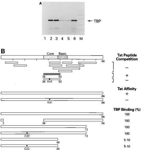
FIG. 2. Mapping of TBP domain involved in binding to Tat. (A) GST and GST-Tat86 were raised in E. coli, induced, purified, and subjected to SDS-PAGE (4 to 20% polyacrylamide). Lanes 1 and 2, 100 ng of GST and GST-Tat protein, respectively, stained with Coomassie blue. Lane MW, molecular weight markers (in thousands [K]). (B) Amounts of GST proteins were normalized according to protein concentration, and the amount bound to beads was confirmed by SDS-PAGE followed by Coomassie staining. GST proteins (approximately 5mg) and 5ml of the TNT lysate (Promega) containing35S-labeled TBP were incubated in TNE100 (100 mM Tris [pH 8.0], 100 mM NaCl, 1 mM EDTA, and 0.1% NP-40) in a final volume of 200ml and with a total of 50ml of beads. Binding reaction mixtures were incubated for 2 h at 48C with rotation. The complexes were centrifuged for 30 s and washed twice with 1 ml of buffer containing 50 mM Tris [pH 7.9], 150 mM NaCl, and 0.2% NP-40. The bound labeled proteins were denatured, electrophoresed on an SDS-PAGE gel (4 to 20% polyacrylamide), dried, and autoradiographed.35S-labeled TBP was incubated with GST (lane 1) or GST-Tat (lane 2). The arrow represents where hTBP migrates. (C) Results of TBP mapping with various deletion mutants. All bindings were performed as described in Materials and Methods. The percent TBP bound was determined from analyses presented panels D and E. (D)35
S-labeled wild-type TBP and deletion mutants (5ml) were incubated with GST-Tat as described in Materials and Methods. Following incubation, complexes were centrifuged, washed, ana-lyzed by SDS-PAGE (4 to 20% polyacrylamide), dried, and autoradiographed. (E) Five-microliter portions of35
S-labeled TBPs were electrophoresed on an
SDS-PAGE gel (4 to 20% polyacrylamide), dried, and autoradiographed.
on November 9, 2019 by guest
http://jvi.asm.org/
protein is greater than 95% pure and migrates with an
appar-ent molecular mass of 35 kDa. Importantly, when the
GST-Tat86 protein was eluted from the Sepharose column, it
re-tained transcriptional activity following electroporation with a
HIV-CAT reporter plasmid into CEM cells (data not shown).
Upon incubation of
35S-labeled TBP with GST-Tat, we
ob-served a specific binding in the presence of 0.2% NP-40 and 0.2
M NaCl (Fig. 2B, lane 2). The GST protein, in contrast, failed
to bind the
35S-TBP under these conditions (Fig. 2B, lane 1).
Utilizing a series of N- or C-terminal deletion mutants, we
have mapped the TBP domain involved in binding to Tat from
amino acid 163 to 196. The quantitative results of the binding
assays are presented in Fig. 2C, and an example of the results
of a typical TBP binding assay is seen in Fig. 2D. GST-Tat
binds to wild-type hTBP (Fig. 2D, lane 1) but not to a TBP
mutant with a deletion of amino acids 101 to 335 or 163 to 335
(dl 101-335 and dl 163-337, respectively) (Fig. 2D, lanes 4 and
5). Tat binding was observed, however, with a TBP mutant
containing the first 196 amino acids (dl 196-337) (Fig. 2D, lane
6). Consistent with the results presented above, wild-type or
mutant TBP failed to bind to the GST control protein. The
data shown in Fig. 2E demonstrate that equal amounts of
wild-type and mutant TBPs were added to the binding assay
mixtures.
efficiently to mutants dl 214-227, dl 225-241, and dl 238-252
(Fig. 2C). These mutants cover the S5 and H2 domains of TBP
and decrease binding with other activator proteins, such as
adenovirus E1A (22) (unpublished results). TBP mutant dl
251-268, which overlaps the S1
9
domain, did not affect binding
of TBP to either Tat or E1A.
In an effort to more directly localize the TBP binding site, we
have analyzed the interaction of a mutant with a
three-amino-acid H1 substitution (amino three-amino-acids 169 to 171) with Tat. As a
control for the binding studies, we also included a mutant with
a three-amino-acid substitution in the TBP H2 domain. The
results of these studies demonstrate that the H1, but not the
H2, mutation abolished the interaction with Tat. Interestingly,
the H1 mutation also abolished the interaction of TBP with
other proteins, such as p53, which are known to bind to the H2
or H2
9
domain of TBP. The most straightforward
interpreta-tion of these results is that the H1 domain may play an
impor-tant role in the structural conformation of TBP. Mutation
within the H1 domain might alter the overall structure of TBP
such that protein binding sites in distinct domains of the
pro-tein would be altered.
Interaction of Tat mutants with TBP.
Utilizing peptides
which cover the length of the Tat protein, we have previously
shown by peptide binding and competition assays that amino
acids 36 to 50 contain the TBP interaction domain (28).
Fur-thermore, we demonstrated that mutation of Lys-41 to Thr-41,
which inactivates the transactivation function of Tat, abolished
the interaction of Tat protein and peptide with TBP. The
results presented in Fig. 3A support those of our peptide
bind-ing studies and demonstrate, in the context of a larger protein,
that the core domain of Tat is important for interaction with
TBP. For example, compared with wild-type Tat, deletion of
amino acids 2 to 36 did not affect TBP binding (Fig. 3A, lanes
2 and 3). Similarly, deletion of carboxy-terminal amino acids 73
to 86 did not decrease TBP binding (data not shown). Our
results further demonstrate that amino acids 1 to 48 are
suffi-cient for interaction with TBP (Fig. 3A, lanes 4 and 5). The
binding efficiency of Tat 1-48 is decreased compared with that
of wild-type Tat (Fig. 3A, lanes 2 and 4), a conclusion that was
not evident from the peptide competition and binding assays.
Lane 6 of Fig. 3A represents one-fifth of input TBP.
Interest-ingly, Veschambre et al. (52) have recently reported that Tat
1-49 interacts with TBP to a level comparable to that of
wild-type Tat. These observations may suggest that Arg-49, which is
positioned as a transition amino acid between the conserved
Tat core domain and the basic domain, may be important for
Tat interaction with TBP. Our results are also consistent with
earlier results reported by Southgate and Green (49) which
demonstrated that a GAL4-Tat construct containing Tat
amino acids 1 to 48 was less active than Tat 1-86. Mutation of
Cys-22 did not diminish the interaction of Tat with TBP. In
view of the fact that the Cys-22 mutation abolished the
activa-tion funcactiva-tion of Tat and GAL4-Tat, we suggest that Cys-22
may be required for interaction with another protein important
for the transcription activation function. The fact that
GAL4-Tat constructs containing either amino acids 1 to 36 or 36 to 50
were not active is consistent with this conclusion. A summary
of the results of Tat peptide competition, peptide binding, Tat
affinity, and GST-Tat binding assays is presented in Fig. 3B.
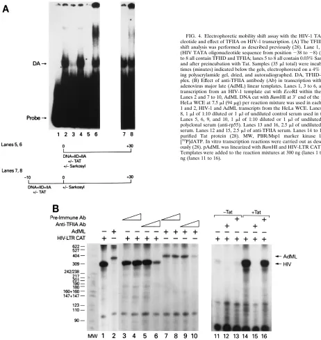
Effect of Tat on stabilization of TFIID-TFIIA complex.
[image:4.612.59.296.70.322.2]Given that the interaction between Tat and TFIID was
ob-served in vitro and in vivo, we were interested in determining
the functional consequence of the Tat-TBP interaction with
FIG. 3. Interaction of TBP with Tat mutants. (A) Amounts of GST proteinswere normalized according to protein concentration, and the amount bound to beads was confirmed by SDS-PAGE followed by Coomassie staining. GST pro-teins (approximately 5mg) and 5ml of the TNT lysate (Promega) containing 35S-labeled TBP were incubated in buffer D (20 mM HEPES [pH 7.9], 100 mM KCl, 12.5 mM MgCl2, 0.1 mM EDTA, 17% glycerol, 0.1 mM DTT) with 0.2% NP-40 in a final volume of 200ml and with a total of 50ml of beads. Binding reaction mixtures were incubated for 2 h at 48C with rotation. The complexes were centrifuged for 30 s and washed twice with 1 ml of buffer containing 50 mM Tris (pH 7.9), 150 mM NaCl, and 0.2% NP-40. The bound labeled proteins were denatured, electrophoresed on an SDS-PAGE gel (4 to 20% polyacrylamide), dried, and autoradiographed. Lane 1, GST (4ml); lane 2, GST-Tat86 (20ml); lane 3, GST-Tat86 dl 2-36 (2ml); lane 4, GST-Tat48 (10ml); lane 5, GST-Tat48 cys22 (10ml); lane 6, one-fifth of input TBP; lane M, protein molecular weight markers. (B) Schematic representation of Tat wild-type and mutant peptides and their abilities to bind TBP. Results of the peptide competition and Tat affinity column assays were previously published (28), and results of GST binding assays are presented as percent TBP binding to wild-type Tat.
on November 9, 2019 by guest
http://jvi.asm.org/
respect to TFIID binding. The results presented in Fig. 4A
demonstrate that Tat facilitates the formation of the
TFIID-TFIIA complex in the presence of Sarkosyl. Low
concentra-tions of Sarkosyl (0.03%) prevent the assembly of, but do not
dissociate, the preinitiation TFIID-TFIIA complex (1, 3, 4, 17,
36, 45, 46). When an oligonucleotide containing the HIV-1
TATA sequence (positions
2
38 to
2
8) was incubated with
purified TFIID and TFIIA, the TFIID-TFIIA complex was
observed (Fig. 4A, lane 2). Incubation of the probe with either
TFIID or TFIIA alone did not result in the formation of the
gel shift complex (data not shown) (28). The TFIID-TFIIA gel
shift complex could be specifically inhibited by excess wild-type
oligonucleotide but not a TATA mutant oligonucleotide (Fig.
4A, lanes 3 and 4). The TFIID-TFIIA complex was sensitive to
Sarkosyl if the detergent was added to the incubation mixture
at zero time but not if the DNA and transcription factors were
preincubated for 10 min prior to addition of Sarkosyl (Fig. 4A,
lanes 5 and 7). Upon addition of purified Tat to the incubation
mixture, the TFIID-TFIIA complex was formed even when
Sarkosyl was present in the incubation mixture at time zero
(Fig. 4A, lane 6). In the gel shift reaction mixtures containing
Tat, we have observed a higher-molecular-weight complex
mi-grating above the TFIID-TFIIA complex in approximately
50% of our experiments. The composition of this unstable gel
shift complex is presently under investigation. To date, we have
not been able to supershift either complex with Tat antibody.
These results raise the possibility that Tat functions
catalyti-FIG. 4. Electrophoretic mobility shift assay with the HIV-1 TATA oligonu-cleotide and effect of TFIIA on HIV-1 transcription. (A) The TFIID-TFIIA gel shift analysis was performed as described previously (28). Lane 1, probe alone (HIV TATA oligonucleotide sequence from position238 to28) (28). Lanes 2 to 8 all contain TFIID and TFIIA; lanes 5 to 8 all contain 0.03% Sarkosyl during and after preincubation with Tat. Samples (35ml total) were incubated for the times (minutes) indicated below the gels, electrophoresed on a 4% nondenatur-ing polyacrylamide gel, dried, and autoradiographed. DA, TFIID-TFIIA com-plex. (B) Effect of anti-TFIIA antibody (Ab) in transcription with HIV-1 and adenovirus major late (AdML) linear templates. Lanes 1, 3 to 6, and 11 to 16, transcription from an HIV-1 template cut with EcoRI within the CAT gene. Lanes 2 and 7 to 10, AdML DNA cut with BamHI at 39end of the promoter. A HeLa WCE at 7.5ml (94mg) per reaction mixture was used in each lane. Lanes 1 and 2, HIV-1 and AdML transcripts from the HeLa WCE. Lanes 3, 4, 7, and 8, 1ml of 1:10 diluted or 1ml of undiluted control serum used in the reaction. Lanes 5, 6, 9, and 10, 1ml of 1:10 diluted or 1ml of undiluted anti-TFIIA polyclonal serum (anti-rp55). Lanes 13 and 16, 2.5ml of undiluted preimmune serum. Lanes 12 and 15, 2.5ml of anti-TFIIA serum. Lanes 14 to 16, 300 ng of purified Tat protein (28). MW, PBR/Msp1 marker kinase labeled with [32P]dATP. In vitro transcription reactions were carried out as described previ-ously (28). pAdML was linearized with BamHI and HIV-LTR CAT with EcoRI. Templates were added to the reaction mixtures at 300 ng (lanes 1 to 10) or 100 ng (lanes 11 to 16).
on November 9, 2019 by guest
http://jvi.asm.org/
[image:5.612.56.518.71.560.2]cally, in that it may stabilize the TFIID-TFIIA complex without
remaining stably bound.
Stabilization of the TFIID-TFIIA complex is important for
HIV transcription, since both TFIID and TFIIA are required
for HIV transcription. We have previously shown that heat
inactivation or affinity depletion of TFIID in HeLa WCEs
abolishes HIV transcription (28). The results presented in Fig.
4B demonstrate that TFIIA is also required for HIV basal and
Tat-transactivated transcription. When a HeLa WCE was
pre-cleared with increasing amounts of anti-TFIIA antibody, but
not preimmune serum, a significant decrease in HIV-1
tran-scription was observed (Fig. 4B, lanes 1 and 3 to 6). As
ex-pected, a similar decrease in transcription was observed with
the adenovirus major late template (Fig. 4B, lanes 7 to 10).
Similarly, when the anti-TFIIA antibody, but not preimmune
serum, was added to a transcription reaction mixture
contain-ing the Tat protein, the transactivation was inhibited (Fig. 4B,
lanes 11 to 16). The decrease in transcription is specific to
TFIIA, since the inhibition can be overcome by the addition of
purified TFIIA to the depleted extract (11).
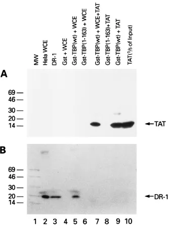
HIV Tat interferes with binding of the negative cofactor Dr1
to TBP.
In addition to stabilizing the TFIID-TFIIA complex,
the interaction of Tat with TFIID may have other functions.
Inostroza et al. have identified an activity, designated Dr1,
which inhibits the activity of TBP (23). Dr1 inhibition involves
a direct physical interaction with TBP which blocks association
with other components of the transcriptional machinery,
in-cluding TFIIA and TFIIB. In preliminary binding assays, the
TBP binding domain for Dr1 was mapped to a region
down-stream of amino acid 163, potentially overlapping the Tat
bind-ing site (56a). To determine if Dr1 and Tat competed for
binding to TBP, we performed a Tat-Dr1 competition
experi-presence or absence of purified Tat protein. Subsequently,
glutathione beads were added to the incubation mixture, and
the protein complexes were pelleted by centrifugation. The
complexes were separated on SDS gels and subjected to
West-ern blotting to detect the presence of Dr1 and Tat. The results
presented in Fig. 5A demonstrate that Tat specifically interacts
with GST-TBP wild type but not GST-TBP 1-163 (Fig. 5A,
lanes 8 and 9). The Tat-TBP interaction was also observed in
the presence of the HeLa WCE (Fig. 5A, lane 7). Similarly,
GST-TBP, but not GST-TBP 1-163, specifically interacted with
Dr1 from the HeLa WCE (Fig. 5B, lanes 5 and 6).
Interest-ingly, when Tat was added to the incubation mixture, the
in-teraction between Dr1 and TBP was inhibited (Fig. 5B lanes 5
and 7). These results provide direct evidence that Tat can
inhibit the interaction of Dr1 with TBP.
DISCUSSION
We have demonstrated a physical interaction between Tat
and TFIID in HIV-infected cells. This is an important
obser-vation since it directly demonstrates the presence of the
pro-tein-protein interaction in the infected cell. Tat has been
re-ported to interact with other transcription factors, including
Sp1, TAP, a cellular kinase (TAK), and other, less well
char-acterized proteins or cofactors. Only the interactions with Sp1
and TFIID have been confirmed to occur in an HIV-infected
cell (19, 25, 41, 57). It is interesting that Tat interaction with
TFIID is detectable at 6 and 12 h postinfection. In contrast, the
Tat-TFIID complex was not observed at the 24- and 48-h time
points. Several control experiments demonstrate the specificity
of this interaction. First, we demonstrated that the polyclonal
Tat antibody used in these experiments immunoprecipitates
Tat protein from the infected-cell extracts. Further, equivalent
amounts of Tat are immunoprecipitated in the 24- and 48-h
samples, but no TBP is detected. Second, we demonstrated
that the anti-Tat antibody does not cross-react with and
im-munoprecipitate TBP from uninfected cells. Third, we
demon-strated that the level of TBP does not vary at the different time
points. Thus, the inability to coimmunoprecipitate TBP is not
due to a change in the cellular protein level. Perhaps factors
like TAR RNA or a viral or cellular protein may physically
sequester Tat’s function as a transactivator at later stages of
viral infection.
[image:6.612.93.263.71.294.2]Using in vitro methods, we have mapped the TBP binding
domain for Tat from amino acid 163 to amino acid 196, which
contains the H1 and S2 domains of TBP. This observation may
be of particular significance. First, this is a unique site for the
interaction of regulatory or activator proteins with TBP. Other
activator proteins, such as TFIIA, p53, VP16, and c-Rel,
inter-act primarily with the H2-S1
9
domain of TBP. Second, Tat
effectively inhibits binding of a negative cofactor of
TBP-me-diated transcription, Dr1. Thus, part of Tat’s ability to regulate
transcription might revolve around its ability to compete with
Dr1 binding to TBP. Importantly, in cotransfection
experi-ments we have observed that HIV-1 Tat transactivation is
repressed by Dr1 in human lymphocytes (data not shown).
Moreover, we have demonstrated that overexpression of TBP
in eukaryotic cells will, in fact, reverse the transcription
inhi-bition of Dr1. If Tat interferes with binding of Dr1 to TFIID,
why does Tat not activate transcription of many other genes
under the control of Dr1? Removal of the negative cofactor is
just one step in the transcriptional activation pathway. If other
FIG. 5. Competition of Tat for Dr1 binding to TBP. Panels A (Western blotwith anti-Tat serum) and B (Western blot with anti-Dr1 serum) represent sam-ples that were split in half and loaded on two SDS-PAGE gels (4 to 20% polyacrylamide). Lane 1, molecular weight markers (in thousands); lane 2, 5ml (12.5mg/ml) of HeLa WCE; lane 3,;50 ng of purified recombinant Dr1; lane 4, GST and HeLa WCE; lane 5, GST-TBP and HeLa WCE; lane 6, GST-TBP 1-163 and HeLa WCE; lane 7, TBP, HeLa WCE, and Tat protein; lane 8, GST-TBP 1-163 and Tat protein; lane 9, GST-GST-TBP and Tat protein; lane 10, Tat protein. Following incubation, GST complexes were pelleted and washed three times, and proteins were separated by SDS-PAGE (4 to 20% polyacrylamide).
on November 9, 2019 by guest
http://jvi.asm.org/
promoter-specific upstream activators or cofactors are not
in-duced, subsequent steps in the activation pathway would be
blocked.
We have demonstrated that Tat stabilizes the TFIIA-TFIID
interaction and that TFIIA is required for HIV transcription.
This function may be significant, since formation of the
TFIID-TFIIA complex is a rate-limiting step in formation of the
preinitiation complex. Interestingly, it has also recently been
suggested that TFIIA has a direct role in the activation
pro-cess, stimulating the activity of a TAF(s) in a reconstituted
transcription assay (51), and may function as a regulatable
TAF (42). It remains to be established whether Tat is an
integral part of the TFIID-TFIIA complex. To date, we have
not been able to supershift either complex with Tat antibody,
opening the possibility that Tat functions catalytically, in that it
can stabilize the TFIID-TFIIA complex without remaining
sta-bly bound. Given the results of the Dr1 binding and Tat
com-petition analyses, Tat may effectively compete for Dr1 binding
to TFIID and stabilize the interaction between TFIID and
TFIIA. This model does not rule out the possibility that Tat
may first bind to TAR RNA and then interact selectively with
TFIID in the initiation complex to modify or facilitate the
formation of a distinct, more processive, complex.
Finally, several interesting observations regarding Tat
inter-action with TBP and TFIID have recently been made. First,
using a protein overlay assay, Chiang and Roeder (7) have
determined that Tat physically interacts with TAF
II55 of
TFIID. This observation is consistent with our earlier report
that the species of TFIID interacting with Tat contains
TAF
II55 (28). TAF
II55 interacts with a variety of mammalian
activators with different activation domains. This interaction
may explain the common activation properties of Tat and
VP16, which have previously been reported (49). The
interac-tions of Tat with multiple components of the TFIID complex,
TBP and TAF
II55, are not mutually exclusive, but they may
represent a synergistic pathway involving interaction with
mul-tiple domains of Tat or mulmul-tiple bound Tat activators. The
observation that Dr1, which may negatively regulate HIV
tran-scription, and Tat compete for a common binding site adds
additional complexity to understanding the biochemical
func-tion of Tat in transactivafunc-tion. Finally, Zhou and Sharp (58)
have reported that in vitro Tat transactivation, primarily a
transcriptional elongation effect in their system, could be
sup-ported by TBP. Although there are no reports which
demon-strate the existence of free TBP in eukaryotic mammalian cells,
these data may suggest a unique Tat activation pathway
inde-pendent of TAFs and negative factors. These multiple
inter-actions may represent the sequential or alternative interinter-actions
of a single activator with several transcription factors in the
transcriptional initiation and elongation complexes. The
com-bination of the interactions may be responsible for the potent
transactivation function of Tat. Our current understanding of
the mechanism of Tat transactivation awaits future
experi-ments using purified and reconstituted transcription factors to
determine the requirements and roles of these distinct
tran-scription factors in Tat transactivation.
ACKNOWLEDGMENTS
We thank members of the Laboratory of Molecular Virology for helpful comments. We also thank K. C. Yeung, Fred Mermelstein, and Danny Reinberg for communication of results of Dr1-TBP binding studies and for the purified Dr1 antibody.
REFERENCES
1. Bohan, C. A., F. Kashanchi, B. Ensoli, L. Buonaguro, K. A. Boris-Lawrie,
and J. N. Brady.1992. Analysis of Tat transactivation of human immuno-deficiency virus transcription in vitro. Gene Exp. 2:391–407.
2. Boyer, T. G., and A. J. Berk. 1995. Functional interaction of adenovirus E1A with holo-TFIID. Genes Dev. 7:1810–1823.
3. Buratowski, S., S. Hahn, L. Guarente, and P. A. Sharp. 1989. Five interme-diate complexes in transcription initiation by RNA polymerase II. Cell 59: 549–561.
4. Cai, H., and D. S. Luse. 1987. Transcription initiation by RNA polymerase II in vitro. Properties of preinitiation, initiation, and elongation complexes. J. Biol. Chem. 262:298–304.
5. Chang, L. J., E. McNulty, and M. Martin. 1993. Human immunodeficiency viruses containing heterologous enhancer/promoters are replication compe-tent and exhibit different lymphocyte tropisms. J. Virol. 67:743–752. 6. Chang-Mayer, C., T. Shioda, and J. Levy. 1991. Host range, replicative, and
cytopathic properties of human immunodeficiency virus type 1 are deter-mined by very few amino acid changes in Tat and gp120. J. Virol. 65:6931– 6941.
7. Chiang, C.-M., and R. G. Roeder. 1995. Cloning of an intrinsic human TFIID subunit that interacts with multiple transcriptional activators. Science 267: 531–534.
8. Choy, B., and M. R. Green. 1993. Eukaryotic activators function during multiple steps of preinitiation complex assembly. Nature (London) 366:531– 536.
9. Comai, L., N. Tanese, and R. Tjian. 1992. The TATA-binding protein and associated factors are integral components of the RNA polymerase I tran-scription factor, SL1. Cell 68:965–976.
10. Dignam, J. D., P. L. Martin, B. S. Shastry, and R. G. Roeder. 1983. Eukary-otic gene transcription with purified components. Methods Enzymol. 101: 582–599.
11. Duvall, J. F., F. Kashanchi, A. Cvekl, M. F. Radonovich, G. Piras, and J. N.
Brady.1995. Transactivation of the human T-cell lymphotropic virus type 1 Tax1-responsive 21-base-pair repeats requires Holo-TFIID and TFIIA. J. Vi-rol. 69:5077–5086.
12. Ensoli, B., R. Gendelman, P. Markham, V. Fiorelli, S. Columbini, M.
Raffeld, A. Cafaro, H.-S. Chang, J. N. Brady, and R. C. Gallo.1994. Synergy between basic fibroblast growth factor and HIV-1 Tat protein in induction of Kaposi’s sarcoma. Nature (London) 371:674–680.
13. Feinberg, M. B., D. Baltimore, and A. D. Frankel. 1991. The role of Tat in the human immunodeficiency virus life cycle indicates a primary effect on transcriptional elongation. Proc. Natl. Acad. Sci. USA 88:4045–4049. 14. Geisberg, J. V., W. S. Lee, A. J. Berk, and R. P. Ricciardi. 1994. The zinc
finger region of adenovirus E1A transactivating domain complexes with the TATA box binding protein. Proc. Natl. Acad. Sci. USA 91:2488–2492. 15. Goodrich, J. A., T. Hoey, C. J. Thut, A. Admon, and R. Tjian. 1993.
Dro-sophila TAFII40 interacts with both a VP16 activation domain and the basal transcription factor TFIIB. Cell 75:519–530.
16. Gruda, M. C., J. M. Zabolotny, J. H. Xiao, I. Davidson, and J. C. Alwine. 1993. Transcriptional activation by simian virus 40 large T antigen: interac-tions with multiple components of the transcription complex. Mol. Cell. Biol.
13:961–969.
17. Hawley, D. K., and R. G. Roeder. 1987. Functional steps in transcription initiation and reinitiation from the major late promoter in a HeLa nuclear extract. J. Biol. Chem. 262:3452–3461.
18. Hernandez, N. 1993. TBP, a universal eukaryotic transcription factor? Genes Dev. 7:1291–1308.
19. Herrmann, C. H., and A. P. Rice. 1995. Lentivirus Tat proteins specifically associate with a cellular protein kinase, TAK, that hyperphosphorylates the carboxy-terminal domain of the large subunit of RNA polymerase II: can-didate for a Tat cofactor. J. Virol. 69:1612–1620.
20. Hoey, T., B. D. Dynlacht, M. G. Peterson, B. F. Pugh, and R. Tjian. 1990. Isolation and characterization of the Drosophila gene encoding the TATA box binding factor, TFIID. Cell 61:1179–1186.
21. Hoffman, A., E. Sinn, T. Yamamoto, J. Wang, A. Roy, M. Horikoshi, and
R. G. Roeder.1990. Highly conserved core domain and unique N terminus with presumptive regulatory motifs in a human TATA factor (TFIID). Na-ture (London) 346:387–390.
22. Horikoshi, N., A. Usheva, J. Chen, A. J. Levine, R. Weinmann, and T. Shenk. 1995. Two domains of p53 interact with the TATA-binding protein, and the adenovirus 13S E1A protein disrupts the association, relieving p53-mediated transcriptional repression. Mol. Cell. Biol. 15:227–234.
23. Inostroza, J. A., F. H. Mermelstein, I. Ha, W. S. Lane, and D. Reinberg. 1992. Dr1, a TATA-binding protein-associated phosphoprotein and inhibitor of class II gene transcription. Cell 7:477–489.
24. Jacq, X., C. Brou, Y. Lutz, I. Davidson, P. Chambon, and L. Tora. 1994. Human TAFII30 is present in a distinct TFIID complex and is required for transcriptional activation by the estrogen receptor. Cell 79:107–117. 25. Jeang, K.-T., R. Chun, N. H. Lin, A. Gatignol, C. G. Glabe, and H. Fan. 1993.
In vitro and in vivo binding of human immunodeficiency virus type 1 Tat protein and Sp1 transcription factor. J. Virol. 67:6224–6233.
26. Kao, C. C., P. M. Lieberman, M. C. Schmidt, Q. Zhou, R. Pei, and A. J. Berk. 1990. Cloning of a transcriptionally active human TATA binding factor. Science 248:1646–1650.
27. Kashanchi, F., J. F. Duvall, and J. N. Brady. 1992. Electroporation of viral
on November 9, 2019 by guest
http://jvi.asm.org/
with the HIV-1 transactivator Tat. Nature (London) 367:295–299. 29. Kashanchi, F., R. I. Shibata, E. K. Ross, J. N. Brady, and M. A. Martin.
1994. Second-site long terminal repeat (LTR) revertants of replication-defective human immunodeficiency virus: effects of revertant TATA box motifs on virus infectivity, LTR-directed expression, in vitro RNA synthesis, and binding of basal transcription factors TFIID and TFIIA. J. Virol. 68: 3298–3307.
30. Kashanchi, F., J. Thompson, M. R. Sadaie, J. Doniger, J. Duvall, J. N.
Brady, and L. J. Rosenthal. 1994. Transcriptional activation of minimal HIV-1 promoter by ORF-1 protein expressed from the SalI-L fragment of human herpesvirus 6. Virology 201:95–106.
31. Kassavetis, G. A., C. A. P. Joazeiro, M. Pisano, E. P. Geiduschek, T. Colbert,
S. Hahn, and J. A. Blanco.1992. The role of the TATA-binding protein in the assembly and function of the multisubunit yeast RNA polymerase III transcription factor, TFIIB. Cell 71:1055–1064.
32. Kato, H., H. Sumimoto, P. Pognonec, C. Chen, C. A. Rosen, and R. G.
Roeder.1992. HIV-1 Tat acts as a processivity factor in vitro in conjunction with cellular elongation factors. Genes Dev. 6:655–666.
33. Kraus, V. B., J. A. Inostroza, K. Yeung, D. Reinberg, and J. R. Nevins. 1994. Interaction of the Dr1 inhibitory factor with the TATA binding protein is disrupted by adenovirus E1A. Proc. Natl. Acad. Sci. USA 91:6279–6282. 34. Laspia, M. F., A. P. Rice, and M. B. Mathews. 1989. HIV-1 Tat protein
increases transcriptional initiation and stabilizes elongation. Cell 59:283– 292.
35. Lin, Y.-S., and M. R. Green. 1991. Mechanism of action of an acidic tran-scriptional activator in vitro. Cell 64:971–981.
36. Luse, D. S., T. Kochel, E. D. Kuempel, J. A. Coppola, and H. Cai. 1987. Transcription initiation by RNA polymerase II in vitro. At least two nucle-otides must be added to form a stable ternary complex. J. Biol. Chem.
262:289–297.
37. Manley, J. L., A. Fire, A. Cano, P. A. Sharp, and M. L. Gefter. 1980. DNA-dependent transcription of adenovirus genes in a soluble whole-cell extract. Proc. Natl. Acad. Sci. USA 77:3855–3859.
38. Marciniak, R. A., B. J. Calnan, A. D. Frankel, and P. A. Sharp. 1990. HIV-1 Tat protein trans-activates transcription in vitro. Cell 63:791–802. 39. Marciniak, R. A., and P. A. Sharp. 1991. HIV-1 Tat protein promotes
formation of more-processive elongation complexes. EMBO J. 10:4189– 4196.
40. Meisterernst, M., A. L. Roy, H. M. Lieu, and R. G. Roeder. 1991. Activation of class II gene transcription by regulatory factors is potentiated by a novel activity. Cell 66:981–993.
41. Ohana, B., P. A. Moore, S. M. Ruben, C. D. Southgate, M. R. Green, and
C. A. Rosen.1993. The type 1 human immunodeficiency virus Tat binding protein is a transcriptional activator belonging to an additional family of evolutionarily conserved genes. Proc. Natl. Acad. Sci. USA 90:138–142. 42. Ozer, J., P. A. Moore, A. H. Bolden, A. Lee, C. A. Rosen, and P. M.
Lieber-man.1994. Molecular cloning of the small (gamma) subunit of human TFIIA
protein. Science 248:1625–1630.
44. Pugh, B. F., and R. Tjian. 1990. Mechanism of transcriptional activation by Sp1: evidence for coactivators. Cell 61:1187–1197.
45. Reinberg, D., and R. G. Roeder. 1987. Factors involved in specific transcrip-tion by mammalian RNA polymerase II. Purificatranscrip-tion and functranscrip-tional analysis of initiation factors IIB and IIE. J. Biol. Chem. 262:3310–3321.
46. Saltzman, A. G., and R. Weinmann. 1989. Promoter specificity and modu-lation of RNA polymerase II transcription. FASEB J. 3:1723–1733. 47. Sato, H., J. Orenstein, D. Dimitrov, and M. Martin. 1992. Cell-to-cell spread
of HIV-1 occurs within minutes and may not involve the participation of virus particles. Virology 186:712–724.
48. Smale, S. T., M. C. Schmidt, A. J. Berk, and D. Baltimore. 1990. Transcrip-tional activation by Sp1 as directed through TATA or initiator: specific requirements for mammalian transcription. Proc. Natl. Acad. Sci. USA 87: 4509–4513.
49. Southgate, C. D., and M. A. Green. 1991. The HIV-1 Tat protein activates transcription from an upstream DNA-binding site: implications for Tat func-tion. Genes Dev. 5:2496–2507.
50. Stringer, K. F., C. J. Ingles, and J. Greenblatt. 1990. Direct and selective binding of an acidic transcriptional activation domain to the TATA-box factor TFIID. Nature (London) 345:783–786.
51. Sun, X., D. Ma, M. Sheldon, K. Yeung, and D. Reinberg. 1994. Reconstitu-tion of human TFIIA activity from recombinant polypeptides: a role in TFIID-mediated transcription. Genes Dev. 8:2336–2348.
52. Veschambre, P., P. Simard, and P. Jalinot. 1995. Evidence for functional interaction between the HIV-1 Tat transactivator and the TATA box binding protein in vivo. J. Mol. Biol. 250:169–180.
53. Vogel, J., S. H. Hinrichs, R. K. Reynolds, P. A. Luciw, and G. Jay. 1988. The HIV Tat gene induces dermal lesions resembling Kaposi’s sarcoma in trans-genic mice. Nature (London) 335:606–611.
54. White, R. J., B. C. Khoo, J. A. Inostroza, D. Reinberg, and S. P. Jackson. 1994. Differential regulation of RNA polymerases I, II, and III by the TBP-binding repressor Dr1. Proc. Natl. Acad. Sci. USA 266:448–450. 55. Xiao, H., A. Pearson, B. Coulombe, and R. Truant. 1994. Binding of basal
transcription factor TFIIH to the acidic activation domains of VP16 and p53. Mol. Cell. Biol. 14:7013–7024.
56. Yankulov, K., J. Blau, T. Purton, S. Roberts, and D. L. Bentley. 1994. Transcriptional elongation by RNA polymerase II is stimulated by transac-tivators. Cell 77:749–759.
56a.Yeung, K. C., and D. Reinberg. Personal communication.
57. Yu, L., P. M. Lowenstein, Z. Zhang, and M. Green. 1995. In vitro interaction of the human immunodeficiency virus type 1 Tat transactivator and the general transcription factor TFIIB with the cellular protein TAP. J. Virol.
69:3017–3023.
58. Zhou, Q., and P. A. Sharp. 1995. Novel mechanism and factor for regulation by HIV-1 Tat. EMBO J. 14:321–328.