A COMPREHENSIVE STUDY ON ILEAL
PERFORATION
Dissertation submitted to
The Tamil Nadu Dr. M.G.R. Medical University,
Chennai – 600032
With partial fulfillment of the regulations
for the award of Degree
M.S. GENERAL SURGERY
BRANCH – I
DEPARTMENT OF SURGERY,
K.A.P.V. GOVT. MEDICAL COLLEGE, TRICHY.
THE TAMIL NADU DR. M.G.R. MEDICAL UNIVERSITY,
CHENNAI, INDIA
CERTIFICATE
This is to certify that this dissertation titled
“A COMPREHENSIVE STUDY ON ILEAL PERFORATION”
is a bonafide work of Dr.Arunan.T., Post Graduate in M.S. General Surgery, Department of General Surgery, K.A.P.V. Government Medical College, Trichy and has been prepared by him under our guidance. This has been submitted in partial fulfillment of regulations of The Tamil Nadu Dr. M.G.R. Medical University, Chennai -32 for the award of M.S. Degree in General Surgery (Branch- I).
Prof.Dr.A.Kanaka Sundaram,M.S., Dr.A.Thulasi,.M.S.,D.G.O.,
Professor and Head of Department Unit Chief,
Department of Surgery, Department of Surgery, K.A.P.V. Govt. Medical College, K.A.P.V. Govt. Medical College,
Trichy . Trichy.
Prof.Dr.A.Karthikeyan,M.D.,(FM)
Dean,
DECLARATION
I Dr.ARUNAN.T solemnly declare that dissertation titled, “A
COMPREHENSIVE STUDY ON ILEAL PERFORATION” is a bonafide work done by me at K.A.P.V. Government Medical College,
during 2010-2013 under the guidance and supervision of Prof. Dr.A. Kanakasundarram,M.S., Head of Department, General Surgery.
The dissertation is submitted to the Tamilnadu Dr. M.G.R. Medical
University, towards the partial fulfillment of requirement for the award of
M.S. Degree (Branch – I) in General Surgery.
Place: Trichy
Date :
ACKNOWLEDGEMENT
I whole heartedly thank with gratitude the DEAN of K.A.P.V. Government Medical College, Trichy Prof..Dr.A.Karthikeyan,M.D.(FM).,
for permitting me to conduct the study in K.A.P.V. Government Medical College,Trichy.
I thank Prof. Dr.A. Kanakasundarram,M.S., Head of Department, General Surgery, for helping me and guiding me during this study.
It is my privileged duty to thank my teacher, guide and mentor
Dr..A.Thulasi, M.S.,D.G.O., for her esteemed guidance and valuable suggestions under whom I have the great honour to work as a Post graduate student.
I cannot forget the co-operation, guidance and encouragement of my Assistant Professors,Dr.G.Rajendran.,M.S.,F.I.C.S.,
Dr.S.Senthilvel.,M.S., and other Assistant professors.
I am also thankful to Asst.Prof.Dr.P.Rajendran,M.Ch.,(Uro), Dr.SriHari.,M.S., Dr.Mohan Das.,M.S.,D.A., Dr.Sumathi Ravikumar., M.S., DGO., Dr.P.Prabhakaran.,M.ch(Uro) for their valuable guidance.
I sincerely acknowledge the timely help rendered by my fellow Post Graduates and CRRI‟s.
Last but not the least, I gratefully thank the patients without whom this study could not been completed.
ABSTRACT
Background & Objectives
: Ileal perforation is one of the
commonest occurrence in our hospital setup, with the majority of
cases having an etiology of trauma. The aim is to study the various
causes of ileal perforation and its presentation and various surgical
procedure and its complications and factors affecting the outcome.
Methods
: Seventy cases of ileal perforation were included in
this study from the period between October 2010 and October
2012. Factors were tabulated and statistically analyzed to study
their contribution.
Results:
Trauma was the most common cause of ileal perforation
in this study followed by non-specific perforations. Patients
presented primarily in the third and fourth decades of life with a
male preponderance. Many patients had air under diaphragm in
X-rays and underwent surgery within 24hrs of onset . 70% of patients
underwent 2 layer closure with complication rate of 67% and
Conclusion
: We found trauma as the most common etiology for
ileal perforation. Incidence of typhoid induced perforations seems
to have significantly reduced. Fecal peritonitis, age , shock, lag
period were found to be significant in contributing to mortality and
TABLE OF CONTENTS
S.NO CONTENTS PAGE NO.
1 INTRODUCTION 1
2 AIM AND OBJECTIVES 2
3 REVIEW OF LITERATURE 3
4 METHODOLOGY 42
5 RESULTS 47
6 DISCUSSION 62
7 SUMMARY AND CONCLUSION 70
8 FIGURES 73
9 BIBLIOGRAPHY 80
[image:10.612.86.530.95.695.2]S.NO
LIST OF TABLES
PAGE NO.
1 CAUSES OF ILEAL PERFORATION 47
2 TRAUMATIC CAUSES OF ILEAL PERFORATION 48
3 SEX INCIDENCE IN ILEAL PERFORATION 49
4 AGE INCIDENCE IN ILEAL PERFORATION 49
5 SYMPTOMS OF ILEAL PERFORATION 51
6 SIGNS IN ILEAL PERFORATION 51
7 AIR UNDER DIAPHRAGM 52
8 LAG PERIOD IN ILEAL PERFORATION 53
9 SURGICAL PROCEDUREs DONE IN ILEAL PERFORATION 54
10 NUMBER OF PERFORATIONS 55
11 DISTANCE OF PERFORATION SITE FROM ILEO CAECAL JUNCTION 55
12 COMPLICATION IN RELATION TO CAUSES 56
13 COMPLICATION IN RELATION TO SURGICAL PROCEDURE 57
14 DURATION OF HOSPITAL STAY 57
15 MORBIDITY AND MORTALITY IN RELATION TO DISEASE 58
16 MORBIDITY & MORTALITY IN RELATION TO SURGICAL
PROCEDURE 58
17 RELATION OF LAG PERIOD WITH COMPLICATIONS 59
18 RISK FACTOR FOR MORBIDITY 60
INTRODUCTION
Ileal perforation is one of the commonest problem seen in tropical
countries. The commonest cause being trauma. In western countries the
causes are malignancy, trauma and mechanical etiology, in the order of
frequency (1,2,3).
Over the years a definite changing trend was observed in ileal
perforations both in terms of causes, treatment and prognosis.Early and
Better antibiotics, aggressive surgery and the elimination of conservative
treatment, good preoperative and postoperative care have all significantly
contributed to the improvement in patient outcome (4,5).
It is true that outcomes were improved but still cases of ileal
perforation cause a significant morbidity and mortality that persists despite
of significant changes in health care over the years .
Our dissertation aims to study the etiology, presentation, management
outcome and the factors influencing prognosis and outcome in ileal
perforations The present study includes 70 patients of ileal perforation with
emphasis on trauma, nonspecific and tubercular and typhoid perforations
AIM AND OBJECTIVES
The aims and objectives of this study are
To study the various causes of ileal perforation.
To study the presentation of ileal perforation.
To study the different modes of surgical management of patients
admitted with ileal perforation.
To study the various complications and outcome of these patients.
REVIEW OF LITERATURE
HISTORICAL REVIEW
Ileal perforation is one of the commonest surgical emergency
encountered in a day to day practice. Its history goes back to the period of
SUSHRUTHA where perforation of intestines by sharp objects, fish bones
and thorns has been described. There was also available a record of
perforated intestines being drawn out and the severed edges were held close
by the large black ants with their jaws prior to the clipping of the bodies of
ants ,following which the bowel was put inside and abdomen closed(6).
In 1776 William Cullen was the one to coin the term
PERITONITIS(7). The first successful closure of perforated intestine was
performed by BENJAMIN TRAVERS(8). The introduction of LEMPERT
SUTURES(7) had begun a new era in restoring the perforated bowel
integrity.
The term TYPHUS (gr.cloudy) was first used by Hippocrates(9) in 460
B.C. The term typhoidea was first used by Louis in 1829 and about 150
cases with splenomegaly, rose spots(10), fever,intestinal perforation, cervical
lymphadenopathy were described by him. In 1973 Budd explained a theory
1880 - Discovery of typhoid bacilli by Karl Joseph Ebereth(10)
1884 - Isolation & culture of Salmonella typhi by Gaffkey(10)
1896 - Test for agglutination detection in serum of typhoid patients by
Widal .
First typhoid vaccine in humans was introduced by Pfeiffer and Kalle
(10)
.Before reviewing the aetiology , first we will try to summarise briefly
the anatomy of ileum which helps us in understanding the later pages .
ANATOMY OF ILEUM
The small intestine is a tubular structure extending from the pylorus to
the cecum and the estimated length of this structure is thought to measure
about 4 to 6 m in the living(72). It consists of three segments in series to each
other : the duodenum, jejunum, and ileum. The duodenum, the most
proximal part, lies partly in the retroperitoneum and adjacent to the head and
inferior border of the body of the pancreas. It is being demarcated from the
stomach by the pyloric antrum and from the jejunum by the ligament of
Treitz.
The jejunum and ileum are intraperitoneal and are attached to the
retroperitoneum by a broad-based mesentery. No anatomic landmark
jejunoileal segment is arbitrarily defined as the jejunum and the distal 60%
as the ileum. The ileum is demarcated from the cecum by the ileocecal
valve(72).
The jejunum and ileum are being suspended from the posterior portion
of the peritoneal cavity by a long mesentery that has a oblique travel from
the left upper quadrant to the right lower quadrant of the abdomen(71) .
Features differentiating jejunum from ileum are as follows (71,72,73)
FEATURE JEJUNUM ILEUM
Vasa recta Longer , straight Shorter with more arterial arcades
Diameter Larger Smaller
Colour Pink Pale
Wall thickness Thicker Thinner
Fatty mesentery Less More
Speciality Plica circularis or valvulae conniventes
Paeyer‟s patches
BLOOD SUPPLY(71)
The blood supply, as well as fat and lymphatics, resides within the
Mesentry.The blood supply of the jejunum and ileum is derived from the
Superior mesenteric artery, which has an extensive anastomosis (the vasa
recta) adjacent to the mesenteric border of the bowel named the marginal
artery. This artery travels along the entire length of the bowel. Near the ileal
branch of the ileocolic artery, this artery breaks up into the anastomotic
network of the vasa recta which increases in its complexity. Venous
LYMPHATIC DRAINAGE
Lymphatic drainage occurs through the lymphatic vessels travelling
parallel to the corresponding arteries. This lymph drains through the
mesenteric lymph nodes into the cisterna chyli, then through the thoracic
duct, and ultimately drains into the left subclavian vein (72).
This lymphatic drainage constitutes a major route for the transport of
absorbed lipid into the systemic circulation and likewise plays a major
pathway in immune defense mechanism and also in the spread of metastasis
NERVE SUPPLY
The innervation is provided by both sympathetic and parasympathetic
divisions of the autonomic nervous system . Parasympathetic innervation is
derived from the vagus . They traverse the celiac ganglion and synapse in
the submucosal (Meissner's) and myenteric (Auerbach's) plexus.The
myenteric plexus is responsible for the basal electrical activity of the gut.
Stimulation of the parasympathetic system prepares for the activity of
intestine by increasing blood flow, contractility, and its secretions.
The sympathetic fibers are from three sets of splanchnic nerves and
their ganglion cells being situated usually in a plexus which is around the
base of the superior mesenteric artery. Motor impulses affect the gut
secretion and motility. Afferents from the intestine is carried through the
general visceral afferent fibers of the sympathetic nervous system.
HISTOLOGY
The wall of small intestine is made up of 4 distinct layers ,
Mucosa
Submucosa
Muscularis propria
SEROSA
It is the outermost layer of the small intestine consisting of visceral
peritoneum, which is a single layer of flattened mesoepithelial cells
encircling the jejunoileum, and anterior surface of the duodenum.
MUSCULARIS PROPRIA
The muscularis propria has two muscle layers, an outer thin longitudinal
layer, and a inner thicker circular layer of smooth muscles. Ganglion cells
from the myenteric (Auerbach) plexus are being scattered between these two
muscle layers and send nerve fibers into both layers, thus permitting
conduction through the muscle layer(72).
SUBMUCOSA
It has a layer of fibroelastic connective tissue which contains blood
vessels and nerve fibres. It is the strongest component of the bowel wall and
therefore should be included in anastomotic sutures (73). It contains extensive
networks of lymphatics, arterioles, and venules and plexus of nerve fibers
and ganglion cells (Meissner plexus). The nerve fibres from the mucosa &
submucosa muscle layers are interconnected by small nerves, and cross
connections between cholinergic and adrenergic elements have been
MUCOSA
It is divided into three layers:
Muscularis mucosa
Lamina propria
Epithelial layer
The muscularis mucosa is a thin muscle layer separating the mucosa
from the submucosa.The lamina propria is a layer of connective tissue
between the epithelium and the muscularis mucosa containing plasma cells,
mast cells, eosinophils ,lymphocytes,macrophages, fibroblasts, smooth
muscle cells, and noncellular connective tissue. These plasma cells
synthesize immunoglobulins. Other immune cells in the lamina propria
release various inflammatory mediators (e.g., cytokines,histamines and
arachidonic acid metabolites) that modulates various cellular functions of
the overlying epithelium. The epithelial layer is a continuous sheet of
epithelial cells covering the villi and the crypts. The functions of the crypt
epithelium are cell renewal and endocrine,exocrine, water, and ion secretion
Four main cell types are found in the mucosal layer:
Goblet cells secreting mucus
Paneth cells secreting lysozyme, tumor necrosis factor (TNF),
and the cryptidins.
Absorptive enterocytes
Enteroendocrine cells produce the gastrointestinal hormones .
AETIOLOGY
Ileal perforation is a serious and common complication of many
systemic illness.
According to literature , in developed countries the common causes are
Foreign bodies
Crohns
Drugs
Radiotherapy
Congenital malformations
Malignancies
Diverticula
Ischemic enteritis
Non specific
Karmakar et al(1), the causes of ileal perforation were as follows (1)
The results of the karmakar et al study can be summarised as follows :
Most common cause -- Typhoid followed by perforation due to non
specific. Nil case reported due to malignancy
Indian study by Bhalerao(11) et al.
Nonspecific 18 Typhoid 8
TB 3
Trauma 3
Diverticulitis 2
In the study by Bhalero ---- Non specific > Typhoid
Series by Wani et al(41).
Typhoid 49
Non specific 21 Obstruction 5
TB 3
Radiation enteritis
1
Here Typhoid > Non specific perforation
Hence overall the common cause of ileal perforation in tropics ----
TYPHOID PERFORATION :- INCIDENCE
The documented rate of enteric perforation ranges from 0.5% to
78.6% (14,16,17,18,19,20).
The following were the incidences obtained in various studies AUTHOR YEAR COUNTRY %
Purohit 1976 India 0.5 Thakkar 1979 India 3.77 Arigbabu 1980 Nigeria 78.6 Hadley 1984 SouthAfrica 4 Santillana 1991 Peru 7.8
AGE AND SEX :-
Typhoid perforation has a preponderance towards male sex & younger
age groups especially in the second and third decades of life (4,5,26).
AUTHOR
YEAR
M:F AGE in yrs
MEAN AGE
Thakkar 1979 5.2:1 6-54 - Eggelston 1979 3.1:1 4-65 28 Tarpley 1989 2.3:1 - 27.1 Santillana 1991 6:1 4-56 - Mock 1993 2.4:1 2-72 16.7 Tacyildiz 1996 2.4:1 3-70 27
PATHOLOGY :-
The causative organism of typhoid fever is SALMONELLA TYPHI , a
gram negative bacillus. The bacillus passes through the peyers patches and
multiplies in the reticuloendothelial system for about 10-14 days. The onset
of clinical symptoms correlate with the seeding of the organism into the
blood stream.After 14 days ie 2 weeks the bacillus reaches the gut via the
blood stream and localises in the peyers patches. Ulceration and necrotic
mesenteric lymphadenitis occurs which in course of time leading on to
perforation of peyer‟s patches(17,27,28)
commonly encountered in the 2 nd
week of the onset of illness (17,24,25).
LIZZARALDE et al (25) studied the timing of perforation in a series of 59
children which is as follows ;
TIMING in wks No. %
First 8 13.5
Second 32 54.2
Third 13 22
MACROSCOPY :-
The most consistent feature is a mesenteric lymphadenopathy.
Longitudinal ulcers which are characteristic of enteric perforation are seen in
the terminal part of the ileum and caecum. The diameter of the perforations
varies between 5-8 mm. Hadley in his study found a large number of
perforations less than 5mm (17).
MICROSCOPY :
There will be both local and systemic marked proliferation of the
lymphoid follicles of reticuloendothelial cells. There also occurs
accumulation of mononuclear cells and these macrophages are found as
aggregates of small nodules filled in the center with red blood cells which is
commonly referred to as Erythrophagocytosis. Rarely typhoid bacilli may
also be seen within the aggregates(29. CLINICAL FEATURES :-
The symptoms of typhoid fever include fever , G.I. symptoms ,
respiratory symptoms ,headache . Signs like rose spots , splenomegaly , and
relative bradycardia. If untreated or there is a delay in diagnosis the patients
can enter into the stage of complications like intestinal perforation ,
pneumonia, cholecystitis, myocarditis, bleeding (28,30). Vomiting , sudden
perforation.These signs and symptoms will be masked in a toxic patient
making a delay in diagnosis (18).
DIAGNOSIS :-
In endemic areas diagnosis of typhoid fever is mainly centered around
clinical suspicion (17,18,19,31). The diagnosis of complication like intestinal
perforation can be made out by the presence of free gas under the
diaphragm, pneumoperitoneum , pus in abdominal paracentesis and
peritoneal lavage to detect pus or bile (11).
The laboratory diagnosis of typhoid fever is usually by blood culture in the
first week , serology by widal test in the second week , stool & urine culture
in the third and fourth week of illness respectively.
SEROLOGY :-
Widal test being the most commonly performed test which tend to
measure the capsular and flagellar antibodies against the antigens of S.typhi
& S.paratyphi. the test is positive between 7 – 10 days of onset of illness. In
endemic regions this test is of limited value. Rising titres are considered
significant(33) esp. four fold rise in titres(38) have a higher positive predictive
value of diagnosing this fever. Patients with history of typhoid fever could
have a transient rise in titres which is unrelated to fever is mainly due to the
CULTURE OF ORGANISM :-
The specimens like blood , urine, stool , and bone marrow aspirate can
be used to isolate the organism.In the absence of illness blood culture
positivity can be seen in about 80% during the first week of illness which
decreases to about 25% in the consequent weeks
By the third week of illness about in 75% of the patients stool cultures
show a positive result which were previously negative in the first week.
Duodenal aspirates, biopsy from rose spots , sputum ,pus in suppurative
lesions, CSF constitute the various other sources of culture(35).
HISTOPATHOLOGY :
The presence of mononuclear macrophage cells and RBC s within the phagocytes (Erythrophagocytosis) confirms the diagnosis of enteric fever(29).
NEWER DIAGNOSTIC MODES :-
Nowadays several newer diagnostic modes have reached the labs. Some of them are Indirect fluorescent Vi antibody, Indirect hemagglutination
,ELISA which have higher sensitivity and specificity than widal test(33)
.Much more rapid detection can now be obtained with the monoclonal
antibody use against Salmonella typhi flagellin .For detection of salmonella
TREATMENT :-
Appropriate treatment of typhoid perforation was undercontroversy till 1957. Many surgeons advocated surgical treatment but with the advent of
chloramphenicol into the market this trend has changed towards the
conservative line of management.
In 1959 Huckstep insisted on conservative management on the basis of
Oschner – Scherren regimen and pointed out the reasons which were as
follows ;
Cons :- The friability of the terminal ileum and the liability for multiple
perforations makes it bad for holding the sutures.
Pros :- The sterilisation of the bowel contents by the chloramphenicol
therapy and the localisation of the perforation by the adjacent bowel loops
itself(36).
Absence of localisation in patients have led Hook et al (37) to recommend
surgical treatment in those patients.Nowadays surgical treatment (38 ,39) is the
best option available as conservative management is associated with
SURGICAL TREATMENT :-
Preoperative resuscitation of patients by improving the hydration status
with intravenous fluids , appropriate antibiotics , and total parenteral
nutrition has considerably reduced the mortality rate from 28% to 10% in
one study conducted by Tacyildiz et al (20).
The various surgical options available are ;
Drainage of the peritoneal cavity
Simple closure
Wedge resection and closure
Resection anastomosis
Ileotransverse colostomy
Ileostomy
DRAINAGE OF PERITONEAL CAVITY :-
It is mainly indicated during the period of resuscitation and preparation before surgery in moribund patients (40). Here the flank drains are
inserted under local anesthesia. As a sole procedure it has an unacceptably
higher mortality rate.So it is mainly used as a bridging measure or as a
SIMPLE CLOSURE :-
Archampong(31) recommended simple freshening of edges and primary closure . A significant rate of mortality of about 17.3% was associated with
this procedure.The recommendations by Talwar et al (41) is primary simple
closure and limited surgery.
WEDGE RESECTION AND CLOSURE :-
Here a wedge resection of ileal tissue is done around the perforation and
the ileal defect is closed in two layers transversely . A mean mortality rates
of about 4% has been reported with this procedure . Reports by Ameh et al
(42)
advocated an association of very high mortality rate with this procedure.
RESECTION ANASTOMOSIS :-
Here resection of the affected segment and primary anastomosis has
been advocated by some authors .Studies by Ameh et al recommends
primary resection anastomosis over wedge resection & simple closure (42) .
Jarret & Gibbney recommends resection only for multiple ileal
perforations(28,40).
ILEOTRANSVERSE COLOSTOMY :-
closure and end to side ileo transverse colostomy which decreases the
incidence of complications (43).
This anastomosis helps by diverting the faecal matter from reaching the
diseased ileum and this reduces the risk of complications (25)
TUBE ILEOSTOMY
It was first introduced in 1940 by Lozoya . It was carried by passing a 24F foley catheter through the least edematous parts of the ileum. It is
versatile ,simple , quick in decompressing the bowel and also prevents
further decontamination of peritoneum by the faecal matter .Maloney in
Vietnam , Kaul, Ardhanari , Rangabashyam also reported a reduced rate of
complications with this procedure.Bhalero et al and Santallina recommends
exterioration of suture line preventing further bowel contamination in case
of reperforation (10,18).If there is a confirmed preoperative diagnosis of
perforation, Talwar et al recommends a Rutherford Morrison incision which
is associated with a reduced rate of complications (45).Here the laprostomy
comes into play in case of fulminant abdominal sepsis due to faecal fistula
formation. It helps in draining the pus out of peritoneal cavity thus
preventing further rise in abdominal pressure . The disadvantage of this
procedure is the perforation of the exposed bowel and incisional hernia
MEDICAL THERAPY :-
The gold standard drug for the treatment of typhoid fever was Chloramphenicol used in the dose of 3-4 g /day or about 50 mg/kg for
paediatrics which may be reduced to 2g /day or 30mg / kg in children once
the fever subsides. Rapid response to the drug occurs in 24-48 hrs and
treatment duration shouldn‟t be less than 2 weeks.Chloramphenicol resitance
has emerged as a challenge and so Quinolones has replaced chloramphenicol
as drug of choice(47,48). Ciprofloxacin in a dose of 250 – 750 mg BD is used . The alternative drug used is Ceftriaxone(47). Amoxicillin and Co trimoxazole
has also been tried(46) .In patients with perforation peritonitis , Metronidazole
and gentamicin should also be added to typhoid drugs(47).Vaidyanathan et al
found that metronidazole greatly reduced mortality when added to
anti-typhoid drugs (49).
COMPLICATIONS :-
Typhoid perforation has a very high complication rates of 28% - 80% (17,18,24,26).
Keenan et al reports in 1981
COMPLICATIONS % Chest infection 55
Sepsis 46
Wound infections 33 Recurrent typhoid 11
Reperforation 5
G.I. fistula 5
Wound dehiscence 5
Santillana et al reports in 1991
Wound infections 40.6 Respiratory 10.4 Renal failure 2.3 G.I. fistula 2.1
Melena 2
Icterus 2.1
Sepsis 3.1
Parotid abscess 0.7
MORTALITY :-
A significantly high mortality rates have been found in association
with typhoid perforation and this is elevated much more in patients managed
PROGNOSIS :-
Factors known to affect survival include(28)
Female sex
Multiple perforations
Late presentations
Age over 40 yrs
Archampong et al(19) reported following factors to affect survival :-
Duration of illness
Urine output before surgery
Serum potassium
Perforation – operation interval
Blood urea
Talwar et al in his study found out that the following factors increases the
mortality :-
Perforation – surgery time interval
Faeculent peritonitis
Ameh et al reported the relationship between the type of procedure and
mortality :-
PROCEDURES MORTALITY (% )
Simple closure 50
Wedge resection 62
Resection anastomosis 36
TRAUMATIC PERFORATION
HISTORICAL BACK GROUND
Aristotle was the first to describe visceral injury from non penetrating
injury by noting that the intestine of the deer was so delicate that it might be
ruptured by a slight external blow without injuring the skin.
In 1836 Baudens published the results of his experiences in the
French-Algerian war and suggested “Bold operations” in some cases of gun
shot wounds of the abdomen.He was probably the first to have performed
Laprotomy for gunshot wounds of the abdomen having operated on two
patients in 1830,one of whom survived.
Sims was the first in the United States to advocate surgical
intervention for penetrating organ injury.Walter,in 1859,and Kinlock in
1863,were the first to perform abdominal operations for nonpenetrating and
ETIOLOGICAL CLASSIFICATION OF ABDOMINAL INJURIES
Abdominal injuries can be classified as
(i) PENETRATING
(ii) NON-PENETRATING
PENETRATING
a. Stab wounds
b. Gun shot wounds (Velocity)
c. Shot gun wounds(Range)
d. Other(Sharpnel,Picket,Stake,Glass)
NON PENETRATING
a. Blunt injury
b. Crush injury
c. Blast injury
d. Seatbelt injury
Knives,Screw drivers, scissors,Pencil,Glass bottles are commonly
used to inflict stab wounds in our country.Contrary to the west countries gun
Today the incidence of non penetrating abdominal trauma,is
increasing primarily because of the soaring automobile
accidents.Automobile is responsible for atleast 50% of non penetrating
abdominal injuries.
According to DiVincenti and associates report:
Road Traffic Accidents - 74%
Blow to the abdomen - 14%
Falls - 9%
Other causes - 3%
Non penetrating abdominal trauma is associated with 20 to 30 percent
mortality rate, much of which is attributed to associated injuries of head and
chest and fractures of the extremities.
MECHANISM OF INJURY
PENETRATING INJURIES
Gun Shot Wound –Civilian gun shot wounds are usually caused by
low velocity pistols,whereas,military bullet wounds are of the high
velocity type and result in extreme tissue destruction that requires wide and
thorough debridement.
Physical factors involve the Kinetic energy imparted to the body by
Kinetic energy of a missile is expressed by the formula
E = 0.5 x mv2 x g x 7000
m = mass of missile
v = Velocity of missile
g = gravitational acceleration
The amount of energy imparted to the body is the difference between
the kinetic energy of the missile entering minus that when leaving thebody.
This energy is dissipated by the movement of the tissues in a perpendicular
direction from the trajectory of the bullet.
Characteristics of the tissue determine the extent of
destruction.Fascia,skin and Lung reveal little devitalisation when struck by
high velocity missile,whereas solid tissues such as muscle,bone, liver and
spleen are violently disorganized and devitalized.
The common low velocity injury is stabbing.In this injury the kinetic
energy is low. Thus deep penetration of the abdominal cavity is statistically
the exception rather than the rule.
The shape,size and length of the instruments are important in
NON-PENETRATING INJURIES
Mechanism of nonpenetrating visceral injuries include crushing,
shearing and bursting forces.
First is the crushing of the organ against the posterior abdominal
wall,especially the anterior ridge in the midline produced by the vertebral
bodies.
A sharp shearing force may suddenly be applied to both solid and
hollow organs resulting in tears with perforation or haemorrhage or both.
Thirdly an intra-abdominal hollow viscus can be burst open by asudden
increase in its intra-luminal pressure.
A sudden application of pressure is more apt to rupture solid than
hollow viscera ,thus accounting for the greater incidence of solid organ
injury.
The more elastic tissue of the young tolerate, trauma better than the
resilient tissues of the aged.A strong firmly muscled abdominal wall
constitutes a better barrier than the flaccid ,relaxed abdomen of the old or
Seatbelt injuries
Since Kulowski and Rost in 1956 first attributed a case of intestinal
obstruction to a previously ocurred seatbelt injury.Numerous cases of the
seatbelt Syndrome have appeared in the literature.
The method of injury to the Bowel in this syndrome involves direct
trauma that results in seromuscular tears and closed-loop obstructions that
temporarily increases intra luminal pressure,resulting in intestinal rupture
.Shearing and torsion forces are probably also active.Besides the intestines
and mesentry,practically all abdominal structures,including the gravid
uterus,have been injuried by seat belts.The terminal ileum is the most
common location for the intestinal injuries.
SMALL BOWEL INJURY SCALE: (BY OIS COMMITTEE OF AAST, 1990)
Grade Type of injury Description of injury
I
Haematoma Contusion or Haematoma without devascularization. Laceration Partial thickness, No perforation
II Laceration Laceration < 50% of circumference
III Laceration Laceration> 50% of circumference without transection
IV Laceration Transection of small Bowel
V Laceration Transection of small Bowel, with segmental tissue loss
Incidence of ileal perforations in a series of traumatic perforations stabs
or gunshot wounds.
AUTHORS No. of traumatic perforations
No. of ileal perforations
Kaul et al(52) 24 10
Karmakar(1) 30 13
Scully et al(53) 20 2
Paran(55) - 2
DIAGNOSIS :-
Based on
Clinical examination
X- ray
Four quadrant needle aspiration
X-ray chest or abdomen can reveal free air under the diaphragm.
Abdominal paracentesis was positive in 85 % of patients with small
bowel perforation in a study by Koul et al(52).
General guides for treatment
1. Short single tear (not more than 4 cm) are repaired in the transverse
axis of the gut.
2. Longer single tear may be repaired in the long axis.
Resection is called for
1. When associated mesenteric lesion has devitalized the damaged
section
2. When the injury has mangled the intestine
3. When several perforations are grouped close together
TUBERCULOSIS :-
Tuberculosis is usually an under diagnosed cause for ileal perforation.
AUTHORS
TOTAL NO. OF ILEAL SPECIMENS
NO. OF SPECIMENS POSITIVE FOR TB
Gera et al(57) 113 9
Karmakar et al(1) 30 1
PATHOGENESIS :-
In the past it was thought to be caused by the ingestion of
Mycobacterium bovis infected milk.
Bowel is being affected by ingestion of infected sputum or saliva in
patients following pulmonary tuberculosis. But nowadays the incidence of
intestinal tuberculosis without the evidence of significant pulmonary lesion
is on the increasing trend owing to the increase in the incidence of HIV
Free intestinal perforation accounts for about 7 % of abdominal TB
patients and associated with a high mortality rate of about 30% and a poor
prognosis .
Two types of lesion
Ulcerative
Hypertrophic
In hypertrophic variety the segment of ileum proximal to strictures is the one most commonly to perforate(58) .
Wig et al(59) reported 10 cases of perforation through the ulcerative
variety.
Incidence of perforation varies between 1- 10% .
Kakar et al found multiple perforations in 36% of cases and strictures
in 72.6%(58) .
DIAGNOSIS :-
The diagnosis is usually made by the visualisation of tubercles in the
resected segment of intestine and in mesenteric lymph nodes .Wig et al
showed acid-fast bacilli in about 40% of resected specimens. Makanguola et
al shown that CT can help in diagnosing intestinal TB in about 80 % of
patients.Chest radiography may reveal air under the diaphragm and it may
TREATMENT :-
Resection-anastomosis is the technique of choice .
Resection of the affected segment with ileo-transverse anastomosis is
an alternative procedure .
MECHANICAL FACTORS :-
Mechanical etiology of perforation is one in which perforation occurs
secondary to a obstruction in the distal part .
CAUSES :-
Obstructing growth
Hernias
Intussusception
Volvulus
Bands
PATHOGENESIS :-
The initial insult is obstruction either by a band or hernia which in
course of time leads onto venous outflow obstruction causing odema of the
proximal intestinal wall and subsequently gangrene . Increased intraluminal
pressure of the proximal gangrenous bowel leads onto rupture and
Chaikof et al(12) in his series of study , TOTAL NO. OF SMALL
BOWEL PERFORATION 76
NO. OF MECHANICAL CAUSES 18
Adhesions - 12 Hernia - 4 Carcinoma - 2
Dixon et al in their reports
TOTAL NO OF SMALL BOWEL PERFORATION 54
NO. OF MECHANICAL CAUSES 13
Adhesions - 8
Malignancy - 2 (2)Volvulus - 1
MALIGNANCY :-
Small bowel malignancies are very rare causes of all gastrointestinal
malignancies accounting for only about 3%. Reported tumors in order of
frequency is as follows ;
Adenocarcinoma
Carcinoid
Lymphoma
Sarcoma
Most common site being Ileum(61).
AUTHORS TOTAL NO OF SMALL BOWEL
PERFORATIONS
NO. OF PATIENTS WITH LYMPHOMA(13)
Rajagopalan & Pickleman
16 2
PATHOGENESIS :-
Lymphomas has a propensity to grow centrifugally in the bowel
wall causing partial or distal obstruction(13) and this being the common site
to perforate.
TREATMENT :-
Resection of the involved segment and adjoining mesentery(61) .
INFLAMMATORY BOWEL DISEASE :
It constitutes crohns disease and ulcerative colitis . In crohn‟s free
wall perforation is a rare complication.Most common site being Ileum.
Steinberg et al(62) in their study. PATHOGENESIS :-
During an acute exacerbation in crohns disease ,there occurs a
TREATMENT :-
Simple primary closure is an inappropriate treatment associated with a
poor prognosis.
Menguy et al (63) recommends primary excision of the affected
segment & formation of a double-barreled ileocolostomy with closure of
stoma in later sitting.
NON SPECIFIC PERFORATION :-
When the ileal perforation is due to an unknown cause , its called non
specific perforation .
Authors
Total no. of ileal perforation
No. of non specific perforation
Dixon et al(2) 54 30
Karmakar (1) 14 7
Recent studies are giving promising results on the pathogenesis of non specific perforation as sub mucosal vascular emboli(64) and potassium tablets
as the factors causing ulceration and subsequent perforation .
DIVERTICULITIS :-
Perforation of diverticula per se is one of the rare cause of small
bowel perforation.
Authors Total no. of ileal perforation
No. of perforation due to diverticula
Huttunen et al (66) 24 4
Ileal diverticulum - 1 Diverticulitis - 2 Meckel‟s - 1
Bhalero et al(11) 32 2
The incidence of meckel‟ s diverticulum in general population is
about 0.4 – 2.5 %. Ectopic gastric mucosa occurs in about 38 % of meckels
diverticula (67) which might ulcerate leading on to perforation (66) .The
recommended treatment of choice is resection of the diverticulum along with
the adjacent ileum .(12 ,67)
ISCHEMIC ENTERITIS :-
Ischemic enteritis is another rare cause of ileal perforation.
Author No of ileal
perforation
No of ischemic enteritis
Dixon (2) 54 3
Macroscopically the lesion can be divided into 4 stages :-
A. Segmental bluish mucosal discolouration , edema and ulceration
B. Purple circular bands with bowel wall edema
C. Rigid pipe like , lengthier intestinal segment
D. Subsequently the segment appears thin and papery.
Perforation has higher incidence of occurence in the fourth stage.
Histologically the specimen shows areas of necrosis, severity of which
varies proportionally with stage of the disease (68).
MISCELLANEOUS :-
The causes (1,2,3 ) are
Radiation enteritis
AIDS
Polyarteritis nodosa
Steroid intake
Remine et al reports as follows :-
At the occurrence of perforation about 79 patient on Steroids
A mortality rate of 85.1% was seen in patients taking prednisolone >
20mg /day
Medical conditions warranting steroids present in 62 % Of patients
like Metastatic cancer ,Connective tissue disorders ,
Myeloproliferative syndrome.
Perforation risk greatest in the period of first 3 weeks following the
initiation of steroid therapy (69).
Sunke et al in his case series shown a patient of AIDS with cytomegalo virus
METHODOLOGY
Source of Data:
This study consists of patients admitted from October 2010 to
October 2012. 70 patients of ileal perforation who were admitted in
Mahatma Gandhi Memorial Government Hospital during this period have
been included in the study.
Inclusion Criteria:
- All cases of ileal perforation of age > 14 years.
- Traumatic Ileal perforation associated with solid organ injuries
- Ileal perforation associated with Extra-Abdominal injuries
Exclusion Criteria:
- Jejunal perforation,
- Gastric & duodenal perforation,
- Appendicular , Caecal , Colonic perforation
- All cases of ileal perforation of age < 14 years
- Ileal perforation treated conservatively
Study Method:
Clinical features, investigations, operative procedures done,
postoperative morbidity, mortality and its outcome were studied. History of
mode of injury, time of injury and special reference to presence of pain,
fever, vomiting, abdominal distension, constipation were recorded. Vitals,
hydration status, abdominal distension, tenderness, guarding & rigidity,
Liver dullness obliteration and presence of free fluid were noted.
Systemic examination of cardiovascular system, respiratory system
and central nervous system has been done.
The following investigations had been done as a routine
Haemoglobin
TC, DC , ESR
Bleeding and Clotting time
Blood sugar ,urea and Serum creatinine & electrolytes
Chest X-Ray PA view, X-Ray Abdomen Erect view
Electrocardiogram
4 Quadrant Aspiration was done in some cases.
USG Abdomen was taken for whom haemoperitonium was suspected.
CT Abdomen was taken for few cases.
CT Brain was taken for some patients to rule out head injury.
X- Ray of the local region was taken where fracture was suspected.
In all non-traumatic perforations the following additional investigations
were done
1. Widal test
2. Blood Culture
When perforation due to non-traumatic causes like TB, Typhoid was
suspected lymph node and omentum was taken for biopsy and peritoneal
fluid was taken for Culture & Sensitivity and AFB staining. All patients had
been resuscitated preoperatively with intravenous fluids and blood
transfusion and iv antibiotics. For patients with medical problems Physician
opinion was obtained and treated accordingly.
Most cases received Third Generation Cephalosporins like
cefotaxime, Ceftriaxone or ciprofloxacin with metronidazole. In the event of
Nature of disease , procedure to be done and its prognosis were
explained to patient‟s relatives and written high risk consent was obtained
for all cases. All patients underwent laparotomy under Regional or general
anaesthesia. Most of the surgeries have been performed by trainee surgeons
under supervision of Duty Assistant surgeon. Either Midline or Para median
incision was employed. The type of peritoneal contamination, amount of
peritoneal fluid aspirated was noted. Thorough laprotomy was done.
Number, site and size of perforations and procedure done were noted. The
choice of surgical procedure was based on surgeon‟s preference & unit
policy.
The following procedures were done.
1. Simple two layer closure
2. Resection and anastomosis
3. Closure with free or pedicled omental patch
For both 2 layer closure and resection anastamosis , the inner all-coats
layer was sutured with polyglactin 910 and outer layer with silk. Thorough
peritoneal wash was given routinely, DT was kept in flank region,
Post-Operatively , antibiotics were routinely given for 5-8 days unless the
Diagnosis of typhoid was made only if Widal test was Strongly
positive, or Salmonella typhi was isolated from blood or urine and also if
any histopathological evidence of typhoid perforation has been found. When
the etiology of perforation was not found, it has been termed as non-specific.
Routine dressing done during post operative period. Postoperative
complications if any had been noted and treatment given for that. DT
removal and suture removal were done depending upon individual cases.
The factors influencing morbidity, mortality and outcome were . patients
with traumatic causes of perforation associated with any extra-abdominal
injuries were treated with appropriative method.
The various parameters had been recorded in a proforma and
tabulated. Analysis of result of study was done. Most of the result given in
the form of percentage, „p‟ value significance were used wherever
RESULTS
Seventy patients of Ileal Perforation admitted between October 2010
and October 2012 were included in this study. Patients have been
categorised into etiological groups namely, Traumatic , Non specific, G.I
tuberculosis and typhoid .
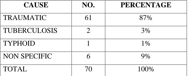
Etiology
The commonest cause of ileal perforation in my study was Trauma
accounting for 87% of the total. 6 patients had non-specific perforations and
is the second most common .Two patients were diagnosed to have
gastrointestinal tuberculosis with ileal perforation. One patient had history of
typhoid fever.
[image:63.612.127.487.503.649.2]The distribution is shown in Table 1
Table 1-CAUSES OF ILEAL PERFORATION
CAUSE NO. PERCENTAGE
TRAUMATIC 61 87%
TUBERCULOSIS 2 3%
TYPHOID 1 1%
NON SPECIFIC 6 9%
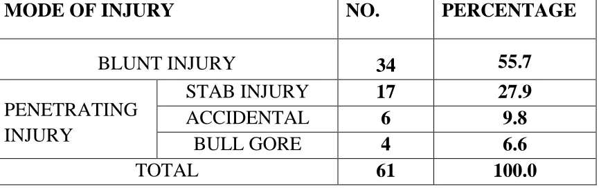
Of the traumatic perforation 34 patients were admitted with history of
blunt injury and 27 patients admitted for penetrating injury.Blunt injury
results from RTA and fall from height. 27.9% accounts for stab injury , 9.8
% accidental injury and 6.6% accounts for Bull Gore injury. The
[image:64.612.87.521.269.405.2]Distribution of Traumatic perforation is shown in table 2:
Table 2-TRAUMATIC CAUSES OF ILEAL PERFORATION
MODE OF INJURY NO. PERCENTAGE
BLUNT INJURY 34 55.7
PENETRATING INJURY
STAB INJURY 17 27.9
ACCIDENTAL 6 9.8
BULL GORE 4 6.6
TOTAL 61 100.0
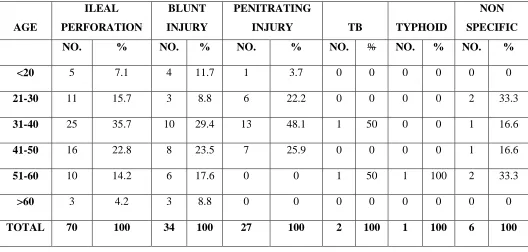
Age and Sex Incidence
The age of patients ranged from 16 to 65. Perforation commonly
occurred in the third and fourth decade of life with 58% of patients between
the ages of 30 and 50. The male to female ratio was 6.7:1.
Traumatic perforation commonly occurred in the third decades
with35.57% of cases in that age group. Male to female ratio was 5.7:1.
Non-Specific perforations occurred commonly in the second and fifth decades of
male . The distributions of age and sex of all cases and etiology specific
[image:65.612.78.578.170.334.2]distributions are shown in tables 3 and 4
Table 3-SEX INCIDENCE IN ILEAL PERFORATION
TRAUMATIC NON TRAUMATIC TOTAL NO. % NO. % NO. % MALE 52 85 9 100 61 87
FEMALE 9 15 0 0 9 13
[image:65.612.42.570.392.644.2]TOTAL 61 100 9 100 70 100
TABLE 4- AGE INCIDENCE IN ILEAL PERFORATION
AGE ILEAL PERFORATION BLUNT INJURY PENITRATING
INJURY TB TYPHOID
NON
SPECIFIC
NO. % NO. % NO. % NO. % NO. % NO. %
<20 5 7.1 4 11.7 1 3.7 0 0 0 0 0 0
21-30 11 15.7 3 8.8 6 22.2 0 0 0 0 2 33.3
31-40 25 35.7 10 29.4 13 48.1 1 50 0 0 1 16.6
41-50 16 22.8 8 23.5 7 25.9 0 0 0 0 1 16.6
51-60 10 14.2 6 17.6 0 0 1 50 1 100 2 33.3
>60 3 4.2 3 8.8 0 0 0 0 0 0 0 0
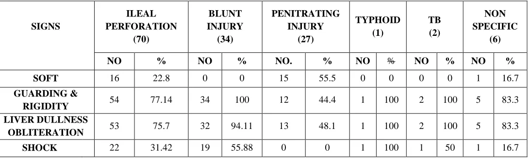
Symptoms and Signs
Majority of the patients presented with symptoms and signs of
peritonitis. The commonest symptoms encountered were abdominal pain,
fever and altered bowel habits. Almost all patient presented with abdominal
pain and altered bowel habits was seen in 25.7% of patients.
The commonest signs were guarding and rigidity, obliteration of liver
dullness and dehydration. 55% of Patients came with History of penetrating
injury presented with soft Abdomen. Typhoid patient gave a history of fever.
31.42 % of patients were in shock . No patients of penetrating injury
presented with Shock. 10% of Patients presented with Omenatal Prolapse
and 5.71% of patient presented with Bowel Prolapse. Symptoms and signs
Table 5-Symptoms of Ileal Perforation
Table 6-SIGNS IN ILEAL PERFORATION
SIGNS ILEAL PERFORATION (70) BLUNT INJURY (34) PENITRATING INJURY (27) TYPHOID (1) TB (2) NON SPECIFIC (6)
NO % NO % NO. % NO % NO % NO %
SOFT 16 22.8 0 0 15 55.5 0 0 0 0 1 16.7
GUARDING &
RIGIDITY 54 77.14 34 100 12 44.4 1 100 2 100 5 83.3
LIVER DULLNESS
OBLITERATION 53 75.7 32 94.11 13 48.1 1 100 2 100 5 83.3
SHOCK 22 31.42 19 55.88 0 0 1 100 1 50 1 16.7
SYMPTOMS ILEAL PERFORAT ION BLUNT INJURY PENETRATING
INJURY TB TYPHOID
NON SPECIFIC
NO % NO % NO. % NO % NO % NO %
ABDOMINAL
PAIN 70 100 34 100 27 100 2 100 1 100 6 100
FEVER 15 21.42 9 26.47 0 0 2 100 1 100 3 50
ALT BOWEL HABITS 18 25.71 9 26.47 0 0 2 100 1 100 6 100
VISERA EVISCE
RATED
OME 7 10 0 0 7 25.9 0 0 0 0 0 0
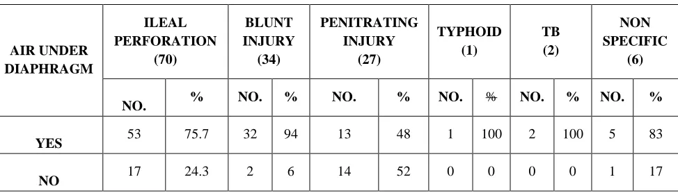
[image:67.612.36.583.363.526.2]Investigations
X-Ray: Pneumoperitoneum in chest and erect abdominal x-rays was
seen in 75.7% of patients. Features of intestinal obstruction, including
dilated bowel loops with air-fluid levels in erect abdominal x-ray were also
seen in a significant percentage of patients.Ultrasound abdomen taken for
some blunt injury abdomen patients which revealed Haemoperitoneum and
solid organ injury. 4 Quadrant aspiration done for some patients.Biopsy was
taken from Omentum and nodes of two patients who showed features of
[image:68.612.64.551.391.530.2]Tuberculosis.
Table 7-AIR UNDER DIAPHRAGM
AIR UNDER DIAPHRAGM ILEAL PERFORATION (70) BLUNT INJURY (34) PENITRATING INJURY (27) TYPHOID (1) TB (2) NON SPECIFIC (6)
NO. % NO. % NO. % NO. % NO. % NO. %
YES 53 75.7 32 94 13 48 1 100 2 100 5 83
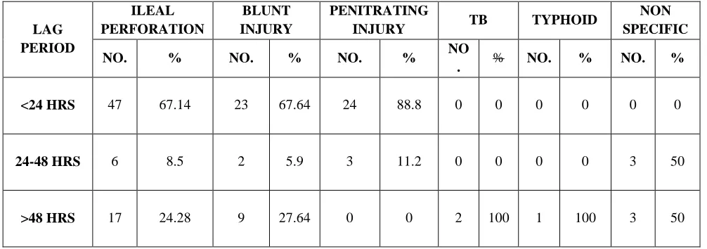
Lag Period
The time between the onset of trauma and the surgical intervention
was within 24 hrs in about 67.14% of patients. There was no significant
difference in the mean lag periods of patients of typhoid or non-specific
perforations. Traumatic perforations were found to have a significantly
reduced lag period because of early diagnosis.Pattern of lag period is shown
[image:69.612.56.559.361.538.2]in table 8.
Table 8-LAG PERIOD IN ILEAL PERFORATION
LAG PERIOD
ILEAL PERFORATION
BLUNT INJURY
PENITRATING
INJURY TB TYPHOID
NON SPECIFIC
NO. % NO. % NO. % NO
. % NO. % NO. %
<24 HRS 47 67.14 23 67.64 24 88.8 0 0 0 0 0 0
24-48 HRS 6 8.5 2 5.9 3 11.2 0 0 0 0 3 50
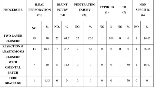
Surgical Procedures
Simple 2-layer closure was the commonest procedure done in about
70% of patients. Closure with omental patch was done in 10% and resection
anastomosis in 18.57% of patients. Different types of surgical procedure
[image:70.612.54.562.332.619.2]done in ileal perforation is shown in table 9.
Table 9- SURGICAL PROCEDURES DONE IN ILEAL PERFORATION PROCEDURE ILEAL PERFORATION (70) BLUNT INJURY (34) PENITRATING INJURY (27) TYPHOID (1) TB (2) NON SPECIFIC (6)
NO % NO. % NO. % NO % NO % NO %
TWO LAYER
CLOSURE 49 70 22 64.7 25 92.6 1 100 0 0 1 16.67
RESECTION &
ANASTOMOSIS 13 18.57 7 20.5 2 7.4 0 0 0 0 4 66.66
CLOSURE
WITH
OMENTAL
PATCH
7 10 5 14.5 0 0 0 0 1 50 1 16.67
TUBE
DRAINAGE
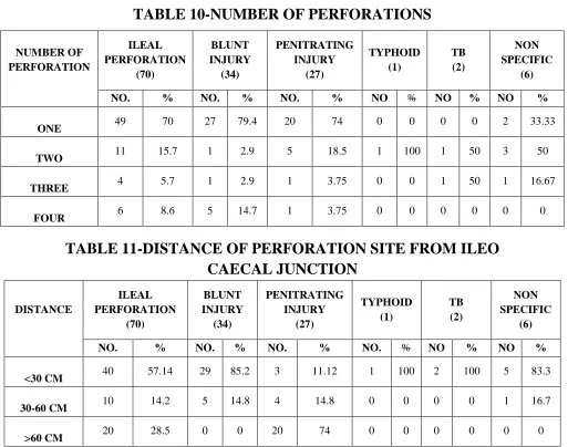
Number and Site of Perforation
About 70% of patients presented with a single perforation .
Multiple perforations occurred only in 8.6% of patients.
Over 72% of perforations were within 2 feet (60 cms) from the
ileocaecal junction and 57.14% within 30 cms. Most of the patients had
perforation of size < 1cm . Number and site of perforation is shown in table
[image:71.612.51.564.293.697.2]10 and 11.
TABLE 10-NUMBER OF PERFORATIONS
NUMBER OF PERFORATION ILEAL PERFORATION (70) BLUNT INJURY (34) PENITRATING INJURY (27) TYPHOID (1) TB (2) NON SPECIFIC (6)
NO. % NO. % NO. % NO % NO % NO %
ONE 49 70 27 79.4 20 74 0 0 0 0 2 33.33
TWO 11 15.7 1 2.9 5 18.5 1 100 1 50 3 50
THREE 4 5.7 1 2.9 1 3.75 0 0 1 50 1 16.67
FOUR 6 8.6 5 14.7 1 3.75 0 0 0 0 0 0
TABLE 11-DISTANCE OF PERFORATION SITE FROM ILEO CAECAL JUNCTION DISTANCE ILEAL PERFORATION (70) BLUNT INJURY (34) PENITRATING INJURY (27) TYPHOID (1) TB (2) NON SPECIFIC (6)
NO. % NO. % NO. % NO. % NO % NO %
<30 CM 40 57.14 29 85.2 3 11.12 1 100 2 100 5 83.3
30-60 CM 10 14.2 5 14.8 4 14.8 0 0 0 0 1 16.7
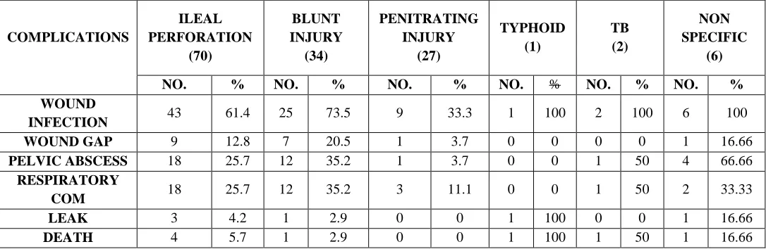
Complications
Complications occurred in majority of cases . The common
complications noted were wound infection, wound dehiscence, pelvic
abscess and respiratory complications.The commonest complication
encountered was wound infection which is seen in about 61.4% of patients
with no significant difference with regards to their etiology .The highest
complication rate of wound infection of about 92.3% was seen with
resection anastomosis and in simple closure it was about 47%.
Complication in relation to causes and modes of surgery are shown in table
[image:72.612.35.578.424.601.2]12 and 13.
TABLE 12-COMPLICATION IN RELATION TO CAUSES
COMPLICATIONS ILEAL PERFORATION (70) BLUNT INJURY (34) PENITRATING INJURY (27) TYPHOID (1) TB (2) NON SPECIFIC (6)
NO. % NO. % NO. % NO. % NO. % NO. %
WOUND
INFECTION 43 61.4 25 73.5 9 33.3 1 100 2 100 6 100
WOUND GAP 9 12.8 7 20.5 1 3.7 0 0 0 0 1 16.66
PELVIC ABSCESS 18 25.7 12 35.2 1 3.7 0 0 1 50 4 66.66
RESPIRATORY
COM 18 25.7 12 35.2 3 11.1 0 0 1 50 2 33.33
LEAK 3 4.2 1 2.9 0 0 1 100 0 0 1 16.66
TABLE 13-COMPLICATION IN RELATION TO SURGICAL PROCEDURE
COMPLICATIONS TWO LAYER
CLOSURE (49) RESECTION & ANASTOMOSIS (13) CLOSURE WITH OMENTAL PATCH (7) TUBE DRAINAGE
(1) TOTAL
(70)
NO. % NO. % NO. % NO. % NO %
WOUND INFECTION 23 47 12 92.3 7 100 1 100 43 61.4
WOUND GAP 3 6.1 5 38.4 1 14.2 0 0 9 12.9
PELVIC ABSCESS 6 12 6 46.1 5 71.4 1 100 18 25.7
RESPIRATORY
COMPLICATON 10 20 4 30.7 3 42.8 1 100 18 25.7
LEAK 2 4 1 7.6 0 0 0 0 3 4.2
DEATH 2 4 1 7.6 0 0 1 100 4 5.7
Hospital Stay
Resection and anastamosis took a longer time than the other
procedures but the difference was not statistically significant.
Median hospital stay was 18.2 days in patients undergone two layer
closure and 20.1 days in resection anastomosis .There was no significant
difference in the hospital stay of patients undergoing different surgical
procedures.
TABLE 14-DURATION OF HOSPITAL STAY
PROCEDURE DONE MEAN DURATION IN DAYS
TWO LAYER CLOSURE 18.2
CLOSURE WITH OMENTAL
PATCH 19.8
Mortality and Morbidity :-
The total mortality rate was 14%. Mortality rates in patients of blunt
injury and non-specific perforations were 2.9% and 16% respectively. No
patients of penetrating traumatic perforation died and 1 patient of tubercular
perforation expired. The differences in mortality were not found to be
statistically significant. Septicemia, respiratory Complications were the
commonest causes of death. Morbidity and Mortality in relation to disease
and surgical procedure are shown in Table 15 and 16.
TABLE 15-MORBIDITY AND MORTALITY IN RELATION TO DISEASE ILEAL PERFORATION (70) BLUNT INJURY (34) PENITRATING INJURY (27) TYPHOID (1) TB (2) NON SPECIFIC (6)
NO. % NO. % NO. % NO. % NO. % NO. %
MORTALITY 4 5.7 1 2.9 0 0 1 100 1 50 1 16.6
[image:74.612.61.555.563.665.2]MORBIDITY 47 67 25 73.5 13 48.1 1 100 2 100 6 100
TABLE 16-MORBIDITY & MORTALITY IN RELATION TO SURGICAL PROCEDURE TWO LAYER CLOSURE (49) RESECTION & ANASTOMOSIS (13) CLOSURE WITH OMENTAL PATCH (7) TUBE DRAINAGE
(1) TOTAL
(70)
NO. % NO. % NO. % NO. % NO. %
MORTALITY 2 4.08 1 7.7 0 0 1 100 4 5.7
LAG PERIOD AND ITS COMPLICATIONS
Most patients presented with peritonitis >24 hrs were associated with
increased morbidity and mortality. 88.23% of patients having lag period of
[image:75.612.64.551.243.464.2]>48hrs developed complications.
TABLE 17-RELATION OF LAG PERIOD WITH COMPLICATIONS
LAG PERIOD TOTAL (70)
NO. OF PATIENTS HAVING COMPLICATIONS
(47)
DEATH (4)
NO. NO. % NO. %
<24 HRS 47 28 59.5 0 0
24-48 HRS 6 4 66.66 0 0
>48 HRS 17 15 88.23 4 23.5
Prognostic factors
The type of surgical procedure did not affect the mortality and
morbidity in ileal perforations and also in etiology specific analysis. Risk
factors influencing the mortality were found to be age >50 yrs , faecal
peritonitis , and a lag period of >48 hrs and all the mentioned factors had a
Morbidity rates were found to be influenced by factors such as age > 50 yrs ,
preoperative shock , faecal peritonitis and lag period of >48 hrs .Of which,
Faecal periotinitis shown very significant “p” value <0.001
In patients of non-specific perforations lag period showed only a trend
towards significance but was not that much statistically significant.
Prognostic factors affecting morbidity and mortality are shown in Table 18
[image:76.612.83.563.359.648.2]and 19.
TABLE 18-RISK FACTOR FOR MORBIDITY FACTORS MORBIDITY
(47)
NO MORBIDITY
(23) P VALUE
AGE > 50 9 0 0.05
MALES 40 21
0.7
FEMALES 7 2
SHOCK 19 0 < 0.05
MULTIPLE
PERFORATIONS 12 9 > 0.05
FAECAL
PERITONITIS 9 0 < 0.05
LAG PERIOD > 48
TABLE 19-RISK FACTORS FOR MORTALITY
FACTORS MORTALITY (4)
NO MORTALITY
(66)
P VALUE
AGE > 50 3 6 0.02 <0.05
MALES 4 57 1.00 >0.05
FEMALES 0 9 1.00 >0.05
SHOCK 4 15 0.06 >0.05
MULTIPLE
PERFORATIONS 4 17 0.0793 >0.05
FAECAL
PERITONITIS 4 5 0.0004 <0.001
LAG PERIOD >
DISCUSSION
The commonest cause of ileal perforation in this series was traumatic
perforation accounting for about 87% of cases categorised separately as
blunt injury contributing to about 55.7% of total traumatic perforation .
8.25% of ileal perforations reported by Karmakar were due to trauma (1). The
increasing rates of road traffic accidents and civil violence have contributed
to this rising incidence of traumatic perforations.
When the etiology of the perfor