1
“AN OBSERVATIONAL STUDY ON THE FOLLOW UP OF
PATIENTS WHO HAD INSERTION OF MIRENA “
DISSERTATION SUBMITTED IN PARTIAL FULFILMENT OF THE REQUIREMENTS OF TAMIL NADU DR.M.G.R. MEDICAL UNIVERSITY FOR THE DEGREE OF M.S. BRANCH II (OBSTETRICS AND GYNAECOLOGY)
EXAMINATION TO BE HELD IN APRIL 2013
2
CERTIFICATE
This is to certify that the dissertation entitled ‘AN OBSERVATIONAL STUDY ON THE FOLLOW UP OF PATIENTS WHO HAD INSERTION OF MIRENA’ is the
original work of Dr. Latha Lakshmi.K, done under my guidance towards the M.S.
Branch II (Obstetrics and Gynaecology) Degree Examination of The Tamil Nadu Dr. M.G.R Medical University, Chennai to be held in April 2013.
Signature
Guide: Dr. Alice George. Professor,
Obstetrics and Gynaecology Unit I, Christian Medical College,
Vellore – 632 004.
Co-Guide
3
CERTIFICATE
This is to certify that the dissertation entitled ‘AN OBSERVATIONAL STUDY ON THE FOLLOW UP OF PATIENTS WHO HAD INSERTION OF MIRENA’ is the
original work of Dr. Latha Lakshmi.K, done towards the M.S. Branch II (Obstetrics and
Gynaecology) Degree Examination of The Tamil Nadu Dr. M.G.R Medical University, Chennai to be held in April 2013.
The Principal Professor and Head, Christian Medical College, Department of Obstetrics
Vellore - 632 004. and Gynaecology, Christian Medical College,
4
ACKNOWLEDGEMENTS
I am greatly indebted to Dr. Alice George, Professor, Department of Obstetrics and Gynaecology, Christian Medical College and Hospital, Vellore, for her guidance,
supervision and support during various stages of this study.
I also thank Dr. Abraham Peedicayil, Dr. Aruna Kekre and Dr. Annie Regi,
professors for permitting me to recruit patients for this study and for their constant encouragement.
I also want to thank my coguide Dr. Anita Thomas, for her assistance.
I am grateful to Mrs. Grace Rebekah, for her valuable help in statistical analysis of data.
I want to specially thank Mrs. Payal Surender, my friend who helped me in all the telephonic interviews with the Bengali and Hindi patients.
My sincere thanks to all the patients who consented to be part of this study.
I also thank the FLUID Research Committee for their financial assistance.
5
TABLE OF CONTENTS
S.No
CONTENT
PAGE No.
1. INTRODUCTION 2
2. AIMS AND OBJECTIVES 5
3. REVIEW OF LITERATURE 6
4. MATERIALS AND METHODS 48
5. RESULTS 53
6. DISCUSSION 74
7. LIMITATIONS 82
8. CONCLUSIONS 83
9. BIBLIOGRAPHY & ABBREVIATIONS 85
ABSTRACT
TITLE OF THE ABSTRACT : ‘An Observational study on the follow up of patients who had inserton of Mirena’
DEPARTMENT :Department of Obstetrics and Gynaecology
NAME OF THE CANDIDATE : Latha Lakshmi.K
DEGREE AND SUBJECT : M.S Branch II (Obstetrics and Gynaecology)
NAME OF THE GUIDE : Dr. Alice George, Professor, Department of O & G, UnitI
OBJECTIVES:
To follow up the patients, who had insertion of Mirena (Levonorgesterel intrauterine System) from 2007 and 2011, and to determine the effectiveness in decreasing the menstrual blood loss and pain after one year of insertion of Mirena ; To assess the quality of life at the end of one year after insertion and satisfaction among the patients.
METHODS:
The qualitative improvement in menorrhagia was assessed using the Uterine Fibroid
Symptom and Health related Quality of life questionnaire (UFS-QoL); The general quality of life at the end of one year was followed up by the SF 36 questionnaire; The improvement in pain was assessed by the Visual analogue scale; The level of satisfaction was evaluated using the CGI- (global improvement item). Survival analysis was also done and the reason for removal compared by the Tarone-Ware method.
RESULTS:
The total number of insertions from 2007 to 2011 were 353, out of which 177 patient could only be followed up. The rate of voluntary removal of Mirena was 10.7% and the expulsion rate was 20%. 68.9% of patients showed symptomatic improvement; the symptom severity and the health related quality of life before and one year after insertion of Mirena, improved and was
statistically significant (p <0.01). Similarly the SF 36 showed high scores showing the better quality of life one year after insertion of Mirena.
There was statistically significant reduction in pain one year after insertion of Mirena (p <0.01). 83.5% of patients were satisfied with Mirena; However the quantitative analysis of haemoglobin, before and one year after insertion of mirena, though showed some improvement, was not
6
INTRODUCTION
Abnormal uterine bleeding (AUB) is one of the most common reasons why women of the reproductive age group seek medical advice. Dysfunctional uterine
bleeding, in the absence of a medical illness or pelvic pathology is responsible for almost half the cases of abnormal bleeding.
In the first place any organic cause should be ruled out and pregnancy should be excluded. Structural abnormalities such as leiomyoma, polyp or endometrial hyperplasia, can present as heavy menstrual bleeding. Adenomyosis and endometriosis predominantly
present with dysmenorrhea, but can also present with menorrhagia.
There are various treatment modalities for abnormal uterine bleeding which includes the medical management with NSAIDs, tranexamic acid, progesterones, oral
contraceptives, Danazol, GnRh analogues and Levonorgestrel-releasing intrauterine system.
Levonorgestrel-releasing intrauterine system(Mirena), was initially developed as a device for contraception , which does not suppress ovulation. It is a T shaped intrauterine
7
Mirena was found to be superior to all other medical therapy and the blood loss reduced by almost 97% at the end of one year.
Compared to transcervical resection of the endometrium (TCRE), and balloon ablation, the success rate was higher in the resection and ablation group, but however the
rate of satisfaction and change in the quality of life were similar. 64% of patients waiting for hysterectomy, cancelled hysterectomy when on levonorgestrel-releasing intrauterine system. Mirena was also found to be cost effective compared to hysterectomy. The
surgical morbidity associated with hysterectomy is avoided, by using this hormone releasing intrauterine device.
As amenorrhea is expected in patients on Mirena, counselling should always include, about the advantages, side effects, the chances of expulsion of Mirena. The need for continuing some other mode of medical therapy along with Mirena, for the first few
months should also be explained prior to insertion.
Mirena has a high efficacy in reducing the menstrual blood loss, without
disturbing the fertility, and hence can be offered as a first line therapy for abnormal uterine bleeding. On the other hand hysterectomy is associated with a 100% success rate , in treating heavy menstrual bleeding and high patient satisfaction upto 95%, though there
can be greater morbidity due to surgery itself.
Mirena can also be used in patients with adenomyosis and endometriosis, and the
8
progesterones. It is successful in the patients presenting with menorrhagia of unknown reason.
As quantitative assessment is not very practical , it is possible to analyse the qualitative improvement in the life of patients on Mirena. So the different methods used
to assess the qualitative improvement includes, the fibroid related symptom improvement and health related quality of life improvement assessment , the SF36 scoring for the improvement in the general health after one year of insertion, the
improvement in the pain scale and also the level of satisfaction scale.
The above aspects were followed up in the patients who had insertion of Mirena
from 2007 to 2011, our hospital, in the department of obstetrics and gynaecology, Christian Medical College and Hospital, Vellore.
9
AIMS AND OBJECTIVES
1. To follow up patients who had Levonorgestrel releasing intrauterine system (Mirena) inserted from 2007 to 2011 (5 years), in the Department of Obstetrics
and Gynaecology, Christian Medical College and Hospital, Vellore.
2. To determine the effectiveness of Levonorgestrel releasing intrauterine system
(Mirena), in decreasing the menstrual blood loss, at the end of one year after insertion.
3. To determine the effectiveness of Levonorgestrel releasing intrauterine system (Mirena) in those with dysmenorrhea and dyspareunia, at the end of one year
after insertion.
4. To study the health related quality of life , at the end of one year after insertion.
5. To assess the level of satisfaction among those using Levenorgestrel releasing
intrauterine system.
10
REVIEW OF LITERATURE
Abnormal uterine bleeding affects 10 – 30% of women of the reproductive age group and about 50% of women in the perimenopausal age group. It accounts for 15% of office visits and almost 25% of gynecologic operations.
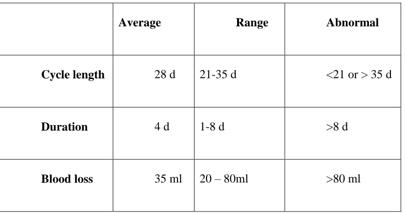
The length of one menstrual cycle is 28 days and the average duration is 4 days and an average blood loss of 35 ml per cycle.
Table 1: The characteristics of the normal menstrual cycle(1)
Average Range Abnormal
Cycle length 28 d 21-35 d <21 or > 35 d
Duration 4 d 1-8 d >8 d
Blood loss 35 ml 20 – 80ml >80 ml
[image:12.612.103.507.355.568.2]11
PATTERNS OF ABNORMAL UTERINE BLEEDING
MENORRHAGIA
The Royal college of Obstetricians and gynaecologists, defines menorrhagia as “heavy cyclical bleeding over several consecutive cycles” (2). Blood loss greater than 80
ml per cycle, is the accepted definition as far as heavy menstrual bleeding is concerned.
Menometrorrhagia is prolonged and irregular bleeding.
Polymenorrhea is bleeding at < 21 - day intervals
POSTCOITAL BLEEDING
It is genital tract bleeding after intercourse. Cervical cancer, polyps, ectropion or genital tract infection, can clinically present as bleeding . All women with postcoital
bleeding should be referred for colposcopy, inspite of a recent negative smear test.
INTERMENSTRUAL BLEEDING
12 POSTMENOPAUSAL BLEEDING
This is defined as genital tract bleeding occurring more than 12 months after the
last menstrual period. The term menorrhagia generally points to the uterine source of bleeding .
The terms postcoital bleeding, intermentstual bleeding and postmenopausal
bleeding describe only clinical terms used for the presentation and not refer to a specific site, from where the bleeding occurs.
POSSIBLE SOURCES OF BLEEDING TO BE CONSIDERED ARE:
1. Whole of the Genital tract 2. Gastrointestinal tract
3. Urinary tract
THE MECHANISMS OF MENSTRUATION -THE CURRENT CONCEPTS :
Menstruation is the process whereby the superficial or functionalis layer of the endometrium lining the uterine cavity disintegrates and is removed from the uterine
lumen towards the end of the luteal phase of a non- pregnant cycle.
The 90th percentile for blood loss during menstruation was found to be 80 ml, by
13
women who lost greater than 60 ml. Loss of more than 80 ml per menstrual episode, on the other hand was defined as menorrhagia.
When the patient perception of the quantity of loss is compared to objective
measurement, there are often significant discrepancies. 25% of women whose measured
blood loss was normal , considered it to be “heavy”, and 40% of those with documented excessive flow(i.e. > 80 ml) described their loss as “light”.(3)
Table 2: Menstrual blood loss- Random population studies
Hallberg et al (1966) Cole et al (1971)(4)
Mean blood loss(ml)
43.4 ± 2.3 37.5±3.3
Median loss(ml) 30.0
27.6
Range 10.4 – 83.9
0.1 – 280
Loss > 60 ml 19.0%
20.7%
Loss > 80 ml 11.0%
14
MECHANISMS OF MENSTRUATION:
Menstruation is the culmination of the changes in both the cellular and vascular
architecture of the endometrium which follow the withdrawal of progesterone (and oestrogen) at the end of the secretory phase.
The major cellular components that contribute to menstruation are the stromal and
vascular components. The week prior to menstruation stromal edema accompanies decidualization, growth of the large blood vessels and intense coiling of the spiral
arterioles.
Two to six days before bleeding begins the stromal oedema shrinks, with associated increase in spiral arteriolar coiling and vascular stasis. This is followed by a
period of vasodilation and perivascular bleeding from the wall of a capillary or arteriole, and then some 24 hours later by intense vasoconstriction and tissue necrosis. 70%
approximately is lost through the vessel wall, 5% by diapedisis and 25% by reflux from veins through the areas where there were previous breaks.
There are different hypothesis that explains menstruation.
1. The Vasoconstrictor theory: The Vasoconstrictor hypothesis
This suggest
(a) the existence of pressor agents which play a role in vascular stasis, (b) protection against excessive blood loss
Arachidonic acids is derived from the phospholipids present in the cell
15 Prostaglandin (PG) :
Not stored in tissues
Released locally and act locally
Rapid metabolism, and therefore a short half life
Have powerful stimulating properties of the vascular and smooth muscle .
Thromboxane A2 is a vasoconstrictor and inhibitor of platelet aggregation.
PGI2 is a vasodilator and inhibitor of aggregation
PGF2 alpha is a vasoconstrictor and smooth muscle stimulator.
PGE is a vasodilator
The role of PGs in menstruation was summarized by Baird et al (5) in 1996. PG occurs in the menstrual fluid and endometrium in high concentrations, and oestrogens and progestogens influence their synthesis. PGF2alpha caused menses and an increase
in the uterine contractility; COX-2 inhibitors reduces blood loss during menstruation and inhibits increased uterine contractility associated with dysmenorrhoea.
The Inflammatory hypothesis
Inflammation is characterized by:
1. Tissue oedema
16
Finn, postulated that menstruation could be regarded as an inflammatory process(6), as there is a dramatic increase in the numbers of lymphomyeloid cells
identified in the endometrium, prior to menstruation.
The endometrial granular lymphocytes increase from proliferative to secretory
phase. Other inflammatory cell found in substantial numbers includes eosinophils, macrophages and neutrophils, whereas T and B cells present, are in low numbers. Steroid hormones possibly modulate migratory cell influx via the chemokine action.
Neither oestrogen receptors (ER) nor progesterone receptor (PR), have been found in the leucocytes , in the human endometrium, and so their effects on leukocytes
may be only indirect.
TISSUE REPAIR
As early as 36 hours after the onset of bleeding due to menstruation, repair of the
endometrium begins. However the desquamation is still in progress, due to the very focal nature of the degradative and repair processes (7).Regeneration is complete by 140 hours.
The menstrual fluid is composed of autolysed endometrium rich in inflammatory exudates, RBCs and proteolytic enzymes(8). Plasminogen has fibrinolytic action, that prevents clotting of menstrual fluid and facilitates the expulsion of the degenerated tissue.
17
Table 3: CAUSES OF ABNORMAL UTERINE BLEEDING
Pelvic Uterine fibroids
Adenomyosis
Endometrial polyps
Pelvic infection
Endometrial hyperplasia
Endometrial adenocarcinoma
Intra uterine device
Uterine vascular malformations
Myometrial hypertrophy
Systemic Coagulation disorders(thrombocytopenia, Von Willebrand disease)
Hypothyroidism,
SLE, Chronic liver failure
18
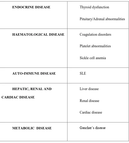
Table 4: MEDICAL DISEASE ASSOCIATED WITH MENORRHAGIA
ENDOCRINE DISEASE Thyroid dysfunction
Pituitary/Adrenal abnormalities
HAEMATOLOGICAL DISEASE Coagulation disorders
Platelet abnormalities
Sickle cell anemia
AUTO-IMMUNE DISEASE SLE
HEPATIC, RENAL AND CARDIAC DISEASE
Liver disease
Renal disease
Cardiac disease
19
GENITAL TRACT BLEEDING- NON – UTERINE CAUSES:
1. Atrophic vaginitis
2. Vulval causes
i. Vulval intra epithelial neoplasia
ii. Carcinoma vulva iii. Skin lesions 3. Lesions in the vagina
4. Trauma 5. Foreign body
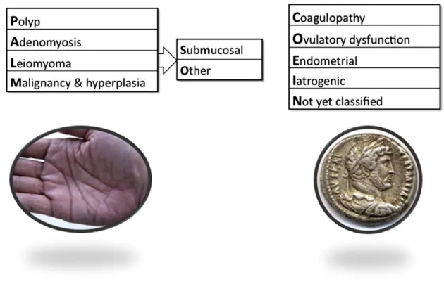
FIGO CLASSIFICATION SYSTEM (PALM-COEIN) FOR ABNORMAL UTERINE BLEEDING (9)
This is an universally accepted system of classification . This new classification including contributions from an international group of clinician-investigators from six
continents and over 17 countries, proposes a new system for the classification of abnormal uterine bleeding.
There are nine main categories, are arranged according to the acronym
20 Figure 1: FIGO CLASSIFICATION OF AUB
The PALM entities are structural, discrete, measured visually by imaging or by
histopathology; The COEIN group is non structural; related to those not defined by imaging or histopathology.
The term “DUB”, used earlier, for diagnosis in the absence of other systemic or
locally definable structural cause, should be abandoned and should not be included in the
system.
POLYP(AUB-P)
21 ADENOMYOSIS (AUB-A)
Prevalence varies from 5% to 70% (12). The relationship between adenomyosis
and the genesis of abnormal bleeding is unclear. It is proposed that sonographic criteria for adenomyosis comprises the minimum requirements for assigning an individual the
diagnosis of adenomyosis in the PALM-COINE classification(13). The sonographic appearance is partly related to the presence of endometrial tissue in the myometrium and hypertrophy.
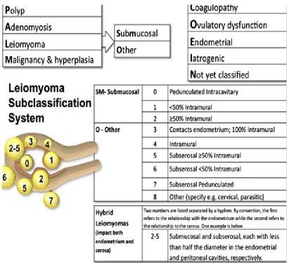
LEIOMYOMA (AUB-L)
The prevalence is upto 70% in Caucasians. Due to the spectrum of size and
location and the variable number of lesions in a given uterus it has a separate categorization in the system. Many leiomyomas are asymptomatic. Several issues like, the relationship of the leiomyoma to the endometrium and serosa; the uterine location of
the leiomyoma (upper segment, lower segment; cervix, anterior, posterior, lateral); the size of the lesions; the number, were considered in the classification system.(14)
The primary classification system reflects only the presence or absence of one or more leiomyomas; The seconday system distinguishes leiomyomas involving the endometrial cavity(submucousal) from others, as submucousal is considered most likely
to contribute to the genesis of AUB.
The tertiary classification system is a design for subendometrial or submucosal
22 Figure 2 : Leiomyoma subclassification
The size of the uterus, single longest measurement, the location and estimated number of
leiomyomas are not included in the classification.
MALIGNANCY AND HYPERPLASIA(AUB-M)
23 COAGULOPATHY(AUB-C)
Von Willebrand disease(15), is the systemic disorders of hemostasis, diagnosed
in approximately 13% of women with HMB. Some women may require chronic
anticoagulation as an intervention, required to preserve life; these patients on these drugs
present with AUB, which may or may not respond to oral medications.
OVULATORY DYSFUNCTION (AUB-O)
Any disorder of ovulation, can present as menstrual abnormalities – which could
range from amenorrhea, to extremely HMB requiring intervention. Ovulatory disorders (endocrinopathies) like polycystic ovary syndrome, thyroid disorder which includes
hypothyroidism, hyperprolactinemia, mental stress, obesity, anorexia, weight loss or extreme exercise .
ENDOMETRIAL (AUB-E)
There may be a primary disorder of “hemostasis”, where evidence has
demonstrated deficiencies in local production of vasoconstrictors such as endothein-1 and
prostaglandin F2α. Accelerated lysis of endometrial clot because of excessive production of plasminogen activator has also been demonstrated (16).
Increased production of substances that promote vasodilatation, e.g. PGE2 and
Prostacyclin I₂ has been known to cause increased uterine bleeding (17).
Retrospective evaluation does not reveal any relationship of endometritis with AUB, but there are data indicating a relationship between subclinical Chlamydia
24 IATROGENIC (AUB-I)
This includes
a. Intrauterine systems, either medicated or inert.
b. Pharmacologic agents that directly impact the endometrium.
c. Interference with blood coagulation mechanisms. d. Those influencing the systemic control of ovulation.
Episodes of break through bleeding are related to reduced circulating gonadal steroid levels, due to compliance issues, such as missed, delayed or erratic use of pills. Reduced
levels of estrogens and progestins , due to use of anticonvulsants and antibiotics (e.g. rifampicin/griseofulvin), can also cause AUB(19).
Women on Levonorgestrel-releasing intrauterine system (LNG – IUS), experience
unscheduled vaginal spotting in the first 3 -6 months (20).
In a UK study, 10% of new users, of the LNG-IUS, ceased use by the end of the
first year because of bleeding complaints (21).
Any agent that impacts serotonin uptake is a candidate for causing ovulatory dysfunction and that result in irregular bleeding (e.g. Tricyclic antidepressants)
25 NOT YET CLASSIFIED (AUB-N)
Entities such as endometritis, arteriovenous malformations, myometrial
hypertrophy need further evidence for classification, into a particular category.
EVALUATION OF ABNORMAL UTERINE BLEEDING
There may be multiple factors contributing to the AUB, and hence, a patient presenting with the symptoms should be diligently investigated.
a. General assessment b. Rule out pregnancy
c. Evaluate anaemia
d. Rule out disorders of ovulation
e. Screening for systemic disorders if required
f. Rule out anticoagulation therapy.
g. Adequate endometrial sampling, if suspicious of atypical hyperplasia or
carcinoma.
h. Abdominal examination
i. Palpate to rule out fibroid uterus
A. Examination of the vulva and vagina for any gross pathology B. Speculum examination – To exclude cervical polyp, tumour and to
take pap smear.
26 INVESTIGATIONS
The latest guidance from the National Collaborating Centre for Women’s and Children’s health states that, “ if the history is suggestive of menorrhagia, without
suspicion of any structural or histological abnormality, treatment can be commenced;
whereas if the treatment option is levonorgestrel-releasing intrauterine system, other investigations should be performed” (22).
The initial assessment requires the patient to have a combination of
unpredictability, excessive duration, abnormal volume, or abnormal frequency of menses, for atleast the previous three cycles.
A. Blood tests –
Full blood count , which includes platelets.
Serum ferritin level if any doubt about iron deficiency anaemia (23).
Thyroid function tests – if history/examination suggestive Coagulation profile if history of bleeding disorder
( puberty menorrhagia or family history if present) B. Assessment of Uterine Cavity –
a. Ultrasound
This is the first line diagnostic tool to identify structural abnormality
Helps to assess size/number/location of fibroids. Transvaginal ultrasound
27
Saline infusion sonography detects submucous fibroids. Endometrial
thickness <5 mm in postmenopausal and Endometrial thickness between 10 – 12 mm in premenopausal needs further evaluation(25).
b. Endometrial biopsy
Endometrial sampling is considered for all women with ultrasonic
abnormality and those with persistent menorrhagia(26).
Outpatient biopsy has high accuracy and the Pipelle endometrial sampler
is a preferred device.
Majority of women investigated for Menorrhagia have ‘normal’
endometrium, and so been labeled to have a functional disorder. c. Dilatation and curettage
No longer has a role.
d. Hysteroscopy
Better than ultrasound , at identifying polyps.
Performed in the outpatient room, only if ultrasound scan is inconclusive.
Normally more painful than transvaginal ultrasound.
e. Magnetic resonance imaging
No advantage over ultrasound and hence not done routinely.
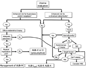
30 Figure 4: Uterine Evaluation in AUB
The uterine evaluation is in part, guided by a good history, other elements of clinical situation, such as age, chronic ovulatory disorder or presence of other risk factors for endometrial hyperplasia or malignancy. Evaluation of the uterus should include
imaging, atleast with a screening transvaginal ultrasound examination(TVUS).
Even in ideal circumstances, TVUS is not 100% sensitive, as polyps and other
31
MANAGEMENT OF ABNORMAL UTERINE BLEEDING
The treatment of menorrhagia may be medical or surgical. In one review 60% of women
referred to the hospital with menorrhagia, had hysterectomy within 5 years(28)
MEDICAL MANAGEMENT:
More than 90% of blood loss usually occurs in the first 3 days of menstruation. So treatment protocol can be either the first few days, which has an advantage of restricting medications only to those days of heavy flow. Anemia should also be adequately treated.
The three treatment regimen which fit into this are prostaglandin synthetase inhibitors, tranexamic acid and ethamsylate.
a. Hormonal treatments: Synthetic progestogens
Combined oral contraceptive pill
Danazol
Gonadotrophin- releasing hormone(GnRH) analogue.
Prostaglandin synthetase inhibitors
Inhibitors of fibrinolysis
Reducers of platelet fragility.
SYNTHETIC PROGESTERONES
This is the most popular drug prescribed in the United Kingdom and New
32
The first report of its use was in 13 women, by Bishop and de Almeida, 1960, and they reported subjective improvement in menstrual blood loss in 34 out of 52 treatment
cycles, and then this regimen was universally adopted.
A route of administration included, intermittent luteal phase oral administration
to intramuscular injection to continuous local administration via intrauterine device. Each of these have different efficacy.
Cyclical administration
Administered usually in the luteal phase of the cycle. Study of norethindrone 10 mg per day 15 to 25 , showed no significant reduction in menstrual blood loss (30).
British investigators evaluated norethindrone in a dose of 10 mg/day from day 19 to 26 and found an increase in bleeding volume by 20% compared to decrease of 45% in the Tranexemic group(31).
The Cochrane meta-analysis concluded that luteal phase progestins were less effective than tranexamic acid, danazol, progestin- releasing IUD(32). A randomized clinical trial ,
compared use of progestin releasing intrauterine device with oral progesterone from day5 to day 26 and found a 87% reduction in blood loss in the oral group, but the reduction in the volume was less, compared to the ID group(33). In another RCT comparing the same,
both were effective in the treatment of menorrhagia, but intrauterine levonorgesterel , had higher satisfaction and continuation of treatment (76% vs 22%) and hence an effective
alternative(34).
33 CONTINUOUS SYSTEMIC ADMINISTRATION
Studies of depoprovera demonstrate that by 1 year, 80% become amenorrheic(35).
There are no published data evaluating Depovera in patients with menorrhagia.
CONTINUOUS LOCAL ADMINISTRATION
Progestin-impregnated intra uterine device is in use for nearly 3 decades(36). For women in the reproductive age group, Levonorgestrel intra-uterine system is one of the most acceptable medical treatments for menorrhagia
Reduces referrals to specialists and decreases the need for operative gynaecological surgery.
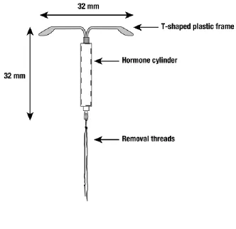
MIRENA ( LEVONORGESTREL – RELEASING INTRAUTERINE SYSTEM)
Mirena LNG-IUS (levonorgestrel-releasing intrauterine system) consists of a T-shaped polyethylene frame (T-body) with a steroid reservoir (hormone elastomer core)
around the vertical stem.
The reservoir consists of a white or almost white cylinder, made of a mixture of
levonorgestrel and silicone , containing a total of 52 mg levonorgestrel. The reservoir is covered by a semi-opaque silicone (polydimethylsiloxane) membrane. The T-body is 32 mm in both the horizontal and vertical directions. The polyethylene of the T-body is
compounded with barium sulfate, which makes it radiopaque. A monofilament brown polyethylene removal thread is attached to a loop at the end of the vertical stem of the
34 Figure 5 : Mirena
INSERTER
It is in a sterile pack which is discarded after insertion. Mirena is packaged sterile within an inserter. The inserter, which is used for insertion of Mirena into the uterine cavity, consists of a symmetric two-sided body and slider that are integrated with flange,
35 Figure 6: Diagram of Inserter
Levonorgestrel USP, (-)-13-Ethyl-17-hydroxy-18,19-dinor-17α-pregn-4-en-20-yn-3-one, the active ingredient in Mirena, has a molecular weight of 312.4, a molecular
formula of C21H28O2, and the following structural formula:
36 Pharmacology:
The initial release is 20µ gms per day till 5 years. This rate decreases to half after
5 years (37).
There is a rapid absorption of locally released levonorgestel from the uterine
cavity, via the capillary network in the basal layer of the endometrium, into the systemic circulation.
Levonorgestrel can be detected within 15 minutes after insertion, reaching
maximum plasma levels within a few hours. The intrauterine concentration are 1000 times higher than with a levonorgestrel subdermal implant(38).
Mirena has mainly local progestogenic effects in the uterine cavity.
Morphological changes of the endometrium, include
a)Stromal pseudodecidualization,
b) Glandular atrophy,
c) Leukocytic infiltration, and
d) Decrease in glandular and stromal mitoses.
e) Alteration in the cerivical mucus and uterotubal fluid
37
g) Adverse effect on the sperm motility an function inside the genital tract, preventing fertilization.
h) Vascular changes – Decrease in mean vascular density and increase in mean vessel area(39).
i) Ovulation is suppressed by reducing the pre-ovulatory surge in luteinizing hormone. 45% of the cycles were ovulatory at the end of one year. Most cycles become ovulatory from the second year onwards, to reach 75% at the end of 4 years. Presence or
absence of menstrual bleeding does not reflect ovarian activity.
POST INSERTION EFFECTS:
Women inserted with IUS, frequently complain of menstrual disturbance. Prolonged bleeding for >8 days is seen in 17% of women in the first month of use, falling
to 3% at the end of 3 months. It takes 6 months on an average to settle in women with heavy flow(40).
38 ADVERSE EFFECT(42,43).
1. Uterine and vaginal bleeding (including spotting, irregular bleeding, heavy
bleeding, oligomenorrhea and amenorrhea)
2. Ovarian cysts
3. Abdominal/pelvic pain
4. Vaginal discharge
5. Nausea/ Headache/ Nervousness.
6. Vulvovaginitis
7. Dysmenorrhea/ Back pain
8. Weight increase
9. Breast pain/tenderness
10. Acne
11. Decreased libido /Depressed mood
39 REMOVAL OF MIRENA:
Indications for removal of mirena include-
• Acquired immune deficiency syndrome (AIDS)
• Sexually transmitted disease
• Pelvic infection; endometritis
• Symptomatic genital actinomycosis
• Intractable pelvic pain
• Severe dyspareunia
• Pregnancy
• Endometrial or cervical malignancy
• Uterine or cervical perforation
40
CLINICAL STUDIES ON INTRA UTERINE LEVONORGESTREL DEVICE
1) Menorrhagia (Including Adenomyosis and Fibroid related)
In 1982, Heikkila et al showed that serum ferritin levels improved in IUS users, with normal menstrual blood loss(45).
Haemoglobin was also found to increase in women with menorrhagia(46).
In 1990, 20 women with menorrhagia, on IUS, showed a reduction in blood loss by 86% in 3 months and 97% in 12 months(47).
Table 5: In 1991, Milsom et al(48), compared the reduction of menorrhagia and found
At 3 months NSAID Antifibrinolytic IUS
Reduction in
blood loss
20.7% 44.4% 81.6%
At the end of one year, there was a 95.8% reduction in menstrual blood .Amenorrhea was
found in 35% of women, at the end of 24 months(49).
Compared to Mefenemic acid(NSAID), LNG reduced the blood loss to just 5 ml
41
Table 6: Comparison between the IUS and norethisterone (51), group showed:
In 3 cycles IUS group Norethisterone group
Reduction in menstrual blood flow
94% 87%
Wish to continue treatment 76% 22%
Table 7: Cancellation of major surgery was noticed in 6 months, after insertion(52):
At 6 months IUS Controls
Cancellation of surgery 64.3% 14.3%
In a 5 year follow of patients, with IUS, 42% of patients on IUS ultimately had hysterectomy. However, use of IUS was cheaper than hysterectomy(53). Health related
42
Table 8: Minimally invasive surgery like Endometrial resection or ablation was compared with IUS use(54).
At 12 months of use IUS Endometrial resection
Pictorial blood loss assessment chart(PBAC)
79% reduction 89% reduction
Amenorrhea/infrequent bleeding
65% 71%
Satisfaction High High
Between Thermal balloon endometrial ablation and IUS, both were found to be equally
effective(55).
Cochrane systematic review concluded hysterectomy reduced menstrual bleeding
at one year, more than the other medical treatments, but IUS, was found to be equally effective in improving the quality of life(56).
IUS has an effect on the haemostatic and fibrinolytic inhibitor systems in women
with menorrhagia and demonstrates high expression of fibrinolytic inhibitors and urokinase-type plasminogen activator in the endometrium, without altering the systemic
43 In women with fibroid related problems(58), LNG-IUS
Reduces the need for surgery
Reduces blood loss
Causes rise in serum ferritin
Causes rise in haemoglobin
No significant effect on the reduction in uterine size and fibroid volume.
In women with Adenomyosis related menorrhagia, IUS may reduce menstrual blood loss significantly, but more studies are required(59).
2. Endometriosis
About 21% of women being investigated for infertility, are found to have endometriosis. However atleast 50% of women in the United Kingdom complain of
moderate dysmenorrhea.
Table 9: On following up the insertions of IUS(60):
In 36 months Before insertion of IUS After insertion of IUS
44
IUS improved the disease staging(61), and reduced pain in those with Rectovaginal endometriosis(62).
Table 10: In those women, who had IUS inserted post operatively, after conservative surgery for endometriosis(63),
Comparing women with IUS and depot gonadotrophin-releasing hormone analogue, for controlling pain related to endometriosis, there was a rapid improvement in those with severe endometriosis, but women with IUS had a higher bleeding score
throughout 6 months of the study, but the quality of life was similar, in both the groups(64).
How does IUS improve the endometriosis- related pain? (65).
Causes alteration in the production of local tissue factors
Blockage of endometrial DNA synthesis
12 months post op. Inserted IUS Expectant treatment
Recurrence of moderate to severe dysmenorrhea
45 Blockage of mitotic activity
Endometrial atrophy
Higher level of peritoneal fluid levels of levonorgestrel
Anovulation at 3 months, till atleast 12 months, after which it becomes
ovulatory.
Long term randomized controlled trials are required to prove the additional advantages of
a treatment lasting for 5 years, with one device.
3. Endometrial Hyperplasia
Studies show that IUS would cause regression of endometrial hyperplasia. First study was published in 2003, where IUS caused complete regression of endometrial hyperplasia(66). Cell apoptosis and downregulation of oestrogen and the progesterone
receptors was found to be the mechanism of action in women with endometrial hyperplasia(67).
46 4. Contraception
Mirena is an effective contraceptive for 5 years. It is a very acceptable and
immediately reversible contraceptive, with a 3 year continuation rates of 75-82%(69).
However, if its effectiveness fails, there is chance of ectopic pregnancy in 1 in 20
women. Overall incidence of ectopic is one in 1000 over a 5 year period.
COMPLICATIONS
a) Expulsion- Partial or Complete . Occurs in 1 in 20 women in 5 years(70)
b) Uterine perforation
c) Ovarian cysts- Surgical treatment is rarely needed. 94% resolves spontaneously, in 6 months(71).
d) Risk of pelvic inflammatory disease is low, less that 1 per 100 women.
Risk is high only in the first 20 days after insertion.
COSTS
47 OTHER FORMS OF MEDICAL MANAGEMENT
Cyclical Hormonal Regimens- This is prescribed by 11% of general practitioners. Overall reduction in mean blood loss was form 158 ml to 75 ml. The fear of
thromboembolic disease in older women makes it unpopular. The third generation OCPs have increased risk of venous thromboembolism. OCPs act by making the endometrium inactive, reducing the endometrial prostaglandin synthesis and altering uterine
fibrinolysis. The common side effects are, headache, migraine, nausea, acne, weight gain, breast discomfort, hypertension, thrombosis, stroke and jaundice.
Prostaglandins – NSAIDs reduces blood loss by inhibiting endometrial prostaglandin synthesis. The reduction ranges from 22% to 46%. It is usually prescribed with oral contraceptives. The additional benefit is that NSAIDs reduces dysmenorrheal and
headaches during menstruation. The side effects include nausea, vomiting, gastric discomfort, diarrhea, headache, bronchospasm, thrombocytopenia and hemolytic
anaemia.
Antifibrinolytic agents: Tranexamic acid, an inhibitor of fibrinolysis, and has showed a mean reduction in blood loss from 295 ml to 155 ml. The reduction in blood loss was
44% with tranexamic acid. The beneficial effect lasts only during the treatment cycle. The side effects include, nausea, vomiting, diarrhea, dizziness, visual disturbances,
48 Androgens:
Danazol is a synthetic androgen with anti-oestrogenic and antiprogestogenic activity. It acts by inhibiting pituitary gonadotrophins and by local endometrial effects.
The endometrium becomes thinned out, and the menstruation decreases, leading to amenorrhoea in large doses, whereas small doses causes only decrease in bleeding during menstruation. Overall there was a reduction of MBL of 60%, when compared to NSAIDs
with a 20% reduction in blood loss.
Androgenic side effects are weight gain, muscle cramp , skin rashes, voice
change, bloating and reduction in breast size.
GnRH agonists: Causes hypo-oestrogenic state and endometrial atrophy. MBL reduced from 131 ml to 11 ml after 3 treatment cycles., 80% of patients being satisfied.
Side effects include menopause like symptoms and vaginal dryness. Long term treatments carry risks of osteoporosis.
Patients who are suffering from abnormal uterine bleeding also usually have anaemia, and hence all patients, should be prescribed, oral iron, folic acid, protein and vitamin supplementation.
49
This is performed early in the proliferative phase of menstruation, when the endometrial linning is thin. A sharp or suction curettage is done immediately before ablation.
Pretreatment with GnRH, danazol, progesterones, or combined oral contraceptive pills, makes the endometrium thin, and has the advantage of good hysteroscopic view.
Endometrial laser ablation- Normal saline is used as the distending fluid of choice. There are two techniques; The dragging technique and the blanching technique. It is important that the tip of the laser fibre is in view and it should move rapidly, to avoid
excessive coagulation and thermal necrosis. The disadvantages are that it is expensive, slowest of all techniques, greater risk of fluid overload and need for safety guidelines.
Roller ball endometrial ablation- The energy used for this is electrosurgical. It is available in 2.5 mm and 5 mm diameter and electrocoagulation is the principle behind the procedure. It is easy to perform, has shorter operating time, less risk of perforation
and haemorrhage. The most important disadvantage is , that there is no sample for endometrial histology.
Loop endometrial resection- Transcervical resection of the endometrium is an effective method of treating patients with dysfunctional uterine bleeding. It can be combined with hysteroscopic myomectomy in case of menorrhagia with submucous
50 Second generation endometrial ablation includes :
a) Cryotherapy- This is a non hysterscopic technique which uses cold temperatures for the purpose of destruction of the endometrium. The cryoprobe is cooled to -90.C, using liquid nitrogen or a gas mixture to create ice balls.
b) Thermal ballon ablation – Endometrial destruction is by conducted heat. It is quick to perform and has significant reduction in the menstrual loss.
c) Microwave endometrial ablation- the local heating effect causes coagulation and the depth of the endometrium should be more than or equal to 10 mm. Pain scores and bleeding significantly reduced.
d) Electrode:mesh – NovaSure- This consists of a disposable hand piece and a computerized generator. The amenorrhea rate is around 65%.
e) Hydrothermablation- In this procedure, heated saline circulates freely within the cavity, introduced through the hysteroscope. The patients were discharged after 2 hours of the procedure.
f) Laser interstitial hyperthermy- This is a blind procedure, where the temperature within the uterine cavity reaches 102.C.
51 Hysterectomy-
It is 100% successful in treating abnormal uterine bleeding, and the satisfaction rates after hysterectomy are very high. Abdominal compared to laparoscopic surgery
demonstrated no difference in the quality of life. Whenever possible, vaginal hysterectomy should be performed.
52
METHODOLOGY
This study was approved by the Research Committee and Institutional Review Board (Ethics Committee) of Christian Medical College, Vellore. The study was
reviewed in detail by the research committee and was accepted for follow up of patients
for whom Mirena, was inserted between 2007 to 2011.
Sample size
Studies have proved that the menstrual blood loss, is reduced by 95% by 6 months and 97% at the end of one year, after insertion of Mirena. Hence the sample size was
calculated, with a precision of 3%, and desired confidence level of 95%, as 124.
Confidence Interval(%) Sample size(n)
90 87
95 124
53
Recruitment
The records on insertion of Mirena, were obtained from the procedure room, in
the Out patient Department , Obstetrics and Gynaecology, and the hospital numbers of the patients , who had insertion of Mirena, from the year 2007 to 2011 were collected.
a. Participants:
INCLUSION CRITERIA
1. Married women
2. Age group (18-50 yrs)
3. With Mirena inserted for Menorrhagia , Endometriosis, Adenomyosis ,
Firboid and Endometrial hyperplasia.
EXCLUSION CRITERIA
1. Age above 50 yrs
2. With history of malignancy
54
Those patients who had a contact number were contacted over phone and enquired about insertion of Mirena, the symptoms they presented with, reason for
insertion, number of days of bleeding, whether excessive, and whether associated with pain. The survival analysis was done and patient asked whether Mirena was insitu ,
removed or expelled.
Those patients who had inserted Mirena, and continued to use for one year and above, and were willing to be followed up, were sent by registered post, questionnaires
translated to hindi, and the patients allowed to fill the questionnaire, after a written consent.
Those patients who had inserted Mirena, and continued to use for one year and above, and were willing for a telephonic interview, were explained about the study and an oral consent obtained, for follow up.
The general proforma (Annexure II), which includes general information about the patient, including the indication, bleeding pattern, associated symptoms, haemoglobin
before and after one year of insertion, associated comorbidities, whether ultrasound and endometrial biopsy were done, were analysed.
Menorrhagia was assessed by using the Uterine Fibroid Symptom and Health-Related Quality of Life Questionnaire (UFS-QOL) (Annexure III) , which includes a set of 37 questions. This proforma contains questions which assesses the following:
55
b) Concern (5 questions)
c) Activities (7 questions)
d) Energy/Mood ( 7 questions)
e) Control (5 questions)
f) Self Consciousness ( 3 questions)
g) Sexual function (2 questions)
UFS-QOL proforma before and after one year of insertion were filled separately,
and the health related quality of life compared. Scoring ranged from 1 to 5; a score of 5
was given to the most distressing symptom, and the total score calculated.
SF-36 questionnaire (Annexure IV)
The SF-36 questionnaire contains a set of 36 questions. The questionnaire was
used to assess the quality of life in all patients inserted with Mirena, unlike UFS-QOL,
which was used in patients only with menorrhagia. The SF-36 questionnaire was used to
assess the following:
a) Physical functioning
b) Role limitations due to physical health
c) Role limitations due to emotional problems
d) Energy/ Fatigue
e) Emotional well being
56
g) Pain
h) General health
The positive answers were given a maximum score of ‘100’, and the most negative answer a score of ‘0’.
Visual analogue scale (VAS) (Annexure V)
The improvement in pain for Endometriosis, was assessed by the Visual analogue scale (VAS) , which was used for Pain analysis, in all patients complaining of pain.
Scores of pain before insertion of Mirena and one year afte Mirena, were obtained. Both these percentages were compared and the improvement calculated, and
significance derived at.
Clininical global impressions scale (CGI SCALE – GLOBAL IMPROVEMENT ITEM)
(Annexure VI)
The degree of satisfaction was followed up using the CGI Scale. The maximum level of satisfaction was scored 7 and who were very much dissatisfied, were scored 1.
Statisical Analysis of data:
Paired t test was used for continuous variables with a 95% confidence interval
and significance were calculated .
57
RESULTS
The total number of patients who had insertion of Mirena from 2007 to 2011 were
353 patients, out of which only 177 patients were followed up.
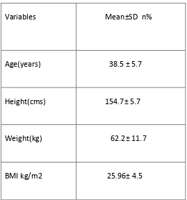
Baseline characteristics of patients followed up after insertion of Mirena.
The age of the patients who had insertion of Mirena were between 25 to 50 years of age, the mean age being 38.57 yrs.
[image:59.612.190.459.407.695.2]The mean BMI of patients who had insertion of Mirena was 25.96.
Table 11 : Baseline characteristics
Variables Mean±SD n%
Age(years) 38.5 ± 5.7
Height(cms) 154.7± 5.7
Weight(kg) 62.2± 11.7
58 Table 12: Year of Insertion of Mirena
Parity in 49% of patients who had insertion of Mirena was 2.
Number of women who had 2 children were 51%. Year of
insertion
Number of patients inserted Mirena
Number of patients followed up
2007
23 8
2008
32 19
2009
67 20
2010
123 64
2011
108 66
Total
59
Table 13: Number of living children for people with mirena insertion.
No. of children n %
O 8 4.6
1 51 29.1
2 90 51.4
3 18 10.3
4 5 2.9
5 3 1.7
Figure 8 : Place distribution
60
Table 14: The number of patients who completed the study were 110.
Completed study 110
Expelled 36
Removed 19
Improved but not contactable
09
Died 03
Total 177
Table 15: Type of Interview conducted for follow up
n %
Telephonic 117 74.1
Direct 17 10.8
Postal 24 15.2
61 Table 16: Reason for insertion of Mirena
Reason for insertion
n %
DUB 89 50.3
Endometriosis 38 21.5
Adenomyosis 29 16.4
Fibroid uterus 21 11.9
Figure 9: Reason for insertion of Mirena.
0 50 100 150 200 250 300
62
Symptoms prior to insertion of mirena:
88% of patients had menorrhagia, prior to insertion.
Table 17: Primary symptom before insertion of Mirena
Indication n %
Menorrhagia 142 80.2
Dysmenorrhea 35 19.8
Table 18:Secondary symptom before insertion of Mirena
Secondary indication n %
Menorrhagia 16 16.8
Dysmenorrhea 75 78.9
Dyspareunia 4 4.2
40.8% of Women who had insertion of Mirena were Sterilised.
Table 19: Pattern of bleeding before Mirena- Regular
Regular cycles
n %
Yes 138 78
63
89.3% of patients had excessive flow during their menstrual period, and the
[image:65.612.204.398.525.643.2]number of days of bleeding ranged between 1 to 15 days, the average being 7.5 days.
Table 20: Patients who had dysmenorrhea , before insertion of Mirena
Dysmenorrhea n %
Yes 109 61.9
No 67 38.1
53.1 % of patients had their haemoglobin checked prior to insertion of Mirena; whereas only 13.7% of patients had their haemoglobin checked, after one year of insertion of
Mirena.
Table 21: Haemoglobin before and one year after insertion of Mirena.
Mean±SD
Preinsertion Hb(gm%)
10.4±2.4
Hb after 1 year(gm%)
11.5±2.3
27.4% of patients were treated for anaemia; 29.1% received iron supplementation;
64
Figure 10: The comorbidities seen among the patients who had insertion of Mirena.
Figure 11: Number of comorbidities seen among the patient who had insertion of Mirena 29.7 39.1 15.6 3.1 3.1 9.4 100
0 20 40 60 80 100 120 Diabetes Hypertension RHD CVT Renal disease Others Total Percentage Frequency 64 8 3
0 20 40 60 80 One
Two Three
[image:66.612.146.396.420.638.2]65
[image:67.612.234.407.268.394.2]Ultrasound – 75.6% of patients who had insertion of Mirena had a screening ultrasound. Endometrial biopsy- Only 23.3% of patients had an endometrial biopsy done, for evaluation.
Table 22: Number of patients with a histopathological diagnosis of Endometrial Hyperplasia
Figure 12: Patients with histological diagnosis of endometrial hyperplasia
Yes No Total 0
5 10 15 20 25 30 35 40 45
Endometrial Hyperplasia
Endometrial Hyperplasia
Endometrial Hyperplasia
n
Yes 3
No 38
[image:67.612.146.470.460.672.2]66
Table 23: Patients on Mirena who were prescribed additional Medical therapy
Medical therapy n %
Progesterone 33 18.6
NSAIDs/Tranex 25 14.1
OCP 67 37.9
Danazol 03 1.7
None 49 27.7
Figure 13: Patients who received additional therapy
0 50 100 150 200 Progesterone
NSAID/Tranex OCP Danazol None Total
67
Table 24: Women who had Improvement of symptoms after one year of insertion of Mirena
Improvement n %
Yes 118 68.6
No 54 31.4
Number of months in which improvement occurred was between 3 to 12 months, the average being 5.36 months.
Table 25: Pattern of menstruation, after one year of insertion of Mirena
Figure 14: Pattern of bleeding after Mirena
0 50 100 150 Regular, reduced
Irregular reduced Amenorrhea Total
Percentage Frequency
Pattern of bleeding n %
Regular, reduced 28 41.8
Irregular reduced 07 10.4
68
[image:70.612.142.394.356.453.2]Table 26: Side effects reported by women after insertion of Mirena.
Table 27: Patients who had Voluntary Removal of Mirena
Voluntary removal
n %
Yes 25 14.3
No 150 85.7
Figure 15: Patients who had voluntary removal of Mirena
Yes No Total 0
50 100 150 200
Voluntary removal
Voluntary removal
Side effects n %
Spotting 6 31.6
Weight gain 11 57.9
Breast tenderness 01 5.3
69
Table 28-. Number of months within which Mirena was removed and expelled
No. of months Mean±SD
Removal 16.68±14.88
Expulsion 6.55±7.39
Of all the patients who voluntarily removed Mirena, 76% of the patients removed within one year of insertion.
Figure -16: Reason for removal of Mirena
20.8
54.2 25
100
0 20 40 60 80 100 120 Side effects
No improvement Hysterectomy Total
[image:71.612.145.527.371.624.2]70
Table 29: Number of women who spontaneously expelled Mirena
Expulsion n %
Yes 41 23.4
[image:72.612.164.439.324.408.2]No 134 76.6
Table 30: Number of Women who had reinsertion ofMirena after expulsion or removal
Reinserted n %
Yes 04 6.3
No 59 93.7
Table 31: If Mirena was not reinserted what was the treatment taken
Treatment n %
Medical management 33 55
[image:72.612.134.463.536.619.2]71
Table 32 : CGI SCALE FOR SATISFACTION AFTER MIRENA
Satisfaction n %
Very much dissatisfied 1 0.9
Much dissatisfied 4 3.7
Neither satis/ Nor dissatisfied 1 0.9
Minimally satisfied 12 11.2
Much satisfied 16 15.0
Very much satisfied 73 68.2
Table 33: Final inference on satisfaction
Satisfaction n %
Satisfied 91 83.5
Minimally satisfied 12 11.0
[image:73.612.144.457.438.548.2]72
Table 34: VISUAL ANALOGUE SCALE FOR PAIN ANALYSIS:
Comparing pain before insertion of Mirena with one year after insertion of Mirena, the mean pain reduced from 86.62% to 14.22%.
VAS Mean
Before insertion of Mirena
86.62%±20.31
One year after insertion of Mirena
[image:74.612.96.548.392.482.2]14.22%±20.10
Table 35: Visual analogue scale – Paired ‘ t’ test
VAS Mean± SD Mean Change 95% CI P value
Before 86.2±20.3
72.40 66.10 to 78.70 <0.01
After 14.2±20.1
Table 36: UFS –QoL Symptom severity (Menstrual symptoms), before Mirena and one year after Mirena, shows a significant reduction of symptoms.
Symptom severity
Mean ±SD Mean change 95% CI P value
Before 25.43 ± 4.55 13.87 12.67 to 15.06 < 0.01
73
Table 37: UFS – HRQL( Uterine fibroid symptoms, health related quality of life)
UFS-HRQL Mean ±SD Mean
change
95% CI P value
Concern Before After
17.53 ± 4.49
6.44 ± 3.30 11.09 10.01 to 12.76 < 0.01
Activity Before After
25.98 ± 4.75 9.80 ± 5.36
16.18 14.68 to 17.67 < 0.01
Energy/Mood Before
After
26.42 ± 4.77 10.72 ± 6.56
15.70 14.11 to 17.28 < 0.01
Control Before After
19.26 ± 3.76 7.48 ± 4.78
11.78 10.51 to 13.04 < 0.01
Self conscious Before
After
7.67 ± 2.50 5.15 ± 2.30
2.52 1.98 to 3.05 < 0.01
Sexual function Before
After
7.01 ± 2.29 3.51 ± 1.97
3.49 2.92 to 4.06 < 0.01
HRQL Before After
1.03 ± 16.70 43.07±20.92
74
Table 38 : UFS –QoL analysis in toto including analysis of menstrual severity and health related quality of life
UFS total Mean ± SD Mean change 95% CI P value
Before Mirena
One year after Mirena
129.23 ± 19.00
54.63 ± 25.86 74.60
68.11 to 81.08
< 0.01
Table 39: Haemoglobin values before and after one year of insertion of Mirena
Mean ± SD Mean Change 95% CI P value
Before Mirena 9.80 ± 2.39 -1.55 -3.09 to -0.03 0.049
One year after Mirena
75
Table 40: SF 36- Scoring of quality of life after one year after insertion of Mirena.
SF 36 – Scoring (Percentage) Mean ± SD Median (IQR)
Physical 88.5 ± 17.86 100 (80,100)
Role- Physical health 86.57 ± 28.68 100 (100,100)
Role- Emotional 86.72 ± 30.20 100 (100,100)
Energy 73.98 ± 20.29 80 (65,90)
Emotional 77.07 ± 19.42 84 (72,92)
Social 89.0 ± 21.32 100 (87.5,100)
Pain 83.63 ± 25.59 95 (77.5,100)
General health 78.10 ± 23.16 90 (70,95)
Table 41: Overall survival time (Time of discontinuation of Mirena)
Median (Months) 95% CI
6.00 4.0 to 7.9
76 Figure 17: Survival Analysis
Table 42: Reason for Voluntary Removal of Mirena
Reason for Removal Median (Months) 95% CI P value
Side effects 24.0 5.28 to 42.71
0.059
No improvement 9.0 5.56 to 12.43
77
The reason for Mirena Removal were Side effects, No improvement and
Hysterectomy and the duration were 24, 9 and 6 months respectively; yet the difference is
not found to be statistically significant.
Figure 18 : Survival Functions (Reason for removal)
[image:79.612.164.515.279.584.2]78
DISCUSSION
The term abnormal uterine bleeding is used for both ovulatory and anovulatory
bleeding. Abnormal uterine bleeding affects 10 – 30% of women of the reproductive age group, and about 50% of women in the perimenopausal age group.
The average length of the cycle is 28 days, the average duration is 4 days and the
average blood loss during menstrual cycle is between 30 to 40 ml per cycle. The 90th percentile for blood loss during menstruation was 80 ml, and anaemia was significantly
increased in women with a loss of greater than 60 ml. Loss of more than 80 ml per menstrual episode is defined as menorrhagia.
However 20% of women who have documented normal flow, complained of
menorrhagia, and seeked treatment(2).
The physician may not be able to assess the volume of blood lost during menstrual
cycle from the history. The different methods by which the menstrual blood loss can be assessed are as follows:
Subjective assessment-
Direct measurement of the menstrual blood loss is the only reliable way to diagnose menorrhagia. The other way of assessment is by the rate of change of sanitary pads during the
flow and the total number of pads and tampons used (75).
It is an inaccurate method and there was no correlation between self assessment,
79
blood loss (7). Only 44% of women complaining of menorrhagia, had a menstrual blood loss of more than 80 ml.
Objective measurement-
The exact amount of haemoglobin lost is measured by the alkaline hematin method. The modifications of the alkaline hematin method for the diagnosis of heavy menstrual blood loss improves the recovery rate from 89% to 98%, and so it can used as a reliable
method(74). Menstrual blood is extracted by soaking the sanitary pads in 5% sodium hydroxide and haemoglobin converted to alkaline hematin; it is centrifuged and the optical density measured by spectrophotometry, and compared with the patient’s venous blood. But
this test is laborious, costly and causes inconvenience to the patient, though it is accurate.
When there was a fall in haemoglobin, with increase in menstrual blood loss, a rise in
the ferritin levels were noticed, but was relevant only if the menstrual blood loss was more than 120 ml/cycle(76).
Indirect (semi-objective) measurement –
Clinical features associated strongly with excessive blood loss are frequency of changing sanitary pads, passage and the size of clots and flooding. Low ferritin level picks up
80
The menstrual pictogram provides a simple way of measuring the blood loss. Pictorial blood assessment charts may not accurately reflect the hygiene products used (73).
Evaluation of this chart by further studies, debated its validity.
The NICE guidelines (National Institute for Health and Clinical Excellence), does not
routinely recommend assessment of blood loss in menorrhagia(78).
The ACOG does not recommend blood count or testing for thyroid hormones for women with excessive bleeding(79). Evidence based guidelines from the Royal College of
Obstetricians and Gynaecologists, recommend,” these tests, and thyoid and bleeding disorder evaluation should be done if there are clinical or historical features”(80).
Menorrhagia can result in severe anaemia. 58% reported a history of anaemia, and 89% of them received oral iron therapy(81).
Treatment of menorrhagia results in substantial improvement in the quality of life.
Medical therapy available for menorrhagia are Non steroidal anti inflammatory drugs, antifibrinolytic drugs, progestogens, oral contraceptives and danazol. All these therapies are
partially effective in reducing the menstrual blood loss. The choice of medical treatment is the choice of the individual. Local LNG-IUS ranks much higher than all other medical treatments, when the effectiveness, side effect and acceptability are taken into account.
The other forms of treatment include endometrial resection/ablation and hysterectomy. Hysterectomy is a definitive treatment , but the risk of surgical morbidity and the cost