0022-538X/94/$04.00+0
Copyright C) 1994, American Society forMicrobiology
Expression and
Characterization of
Virus-Like Particles
Containing Rubella Virus Structural
Proteins
ZHIYONG QIU,DAWEI OU, HE WU,TOM C. HOBMAN,t ANDSHIRLEY GILLAM* Department of Pathology, Research Centre, University of British Columbia,
Vancouver,
British
Columbia,
CanadaV5Z
4H4Received 29 November 1993/Accepted 21 March 1994
Rubella virus (RV) virions contain two envelope glycoproteins (El and E2) and a capsid protein (C). NoninfectiousRV-likeparticles (VLPs) containingthree structuralproteinswereexpressedin a BHK cellline (BHK-24S) by using an inducible promoter. These VLPs were foundtoresembleRVvirions in termsoftheirsize, theirmorphology,and somebiological activities.Inimmunoblottingstudies,VLPs werefoundtobindsimilarlyto native RVvirions with 10 of a panel of12RV-specific murine monoclonalantibodies. Immunization ofmicewith VLPs induced specific antibody responses against RV structural proteins as well as virus-neutralizing and hemagglutination-inhibiting antibodies. After immunization ofmice with VLPs, in vitro challenge of isolated lymphocytes with inactivated RVand individual RV structural proteins stimulated proliferation. Our data suggest thepossibility ofusingVLPs asimmunogensforserodiagnostic assaysandRV vaccines.
Rubella virus (RV), a member of the Togavirus family(20), is theetiological agent of German measles in humans, the only known host. RV infection during pregnancy may result in spontaneous abortionandcongenital defects (1).Vaccination with live, attenuated virus has been successful in reducing the incidence of congenital rubella syndrome (29). However, ru-bella-associated arthritis and the consequence of viral persis-tenceinvaccineesresulting from RV vaccination remain major medical concerns(3). A new approach using RV-like particles (VLPs) is an alternative to conventional attenuated RV vac-cines, since these particles contain all of the RV structural proteins and are nonreplicating.
RVcontainsthree major structural proteins,capsid protein
(C, 33 kDa) and the E2 (42 to 47 kDa) and El (57 kDa)
glycoproteins (22), that are proteolytically cleaved from a polyprotein precursor translated froma24SsubgenomicRNA inRV-infected cells(23).E2andEl are membrane glycopro-teins located on the virion exterior (22, 38), and the capsid protein is associated with the genomic RNA, forming the nucleocapsid (22). Glycoprotein El is the dominant surface molecule ofRV (5, 40). Virus-neutralizing (VN) and hemag-glutination (HA) epitopes have been allocated toEl by using murine monoclonal antibodies (MAbs) (4, 11, 35, 39, 40). Studies of the immune response to RV infection in humans haveindicated thatEl is the major target antigen as compared withE2and C (5, 25, 42). Genetically engineered RV struc-tural proteins have been produced inEscherichia coli (4, 36,
41), insect cells (33), and mammalian cells (2, 13, 30, 32).
However, the antigenicityofexpressed proteins is eitherlost or greatly reduced after purification (17, 36). Furthermore, the capacity of expressedElprotein to elicit antibody responses in animals is dependent on the native conformation of the El
protein (30, 36).
Although it is known that RV-immune individuals have T cells that proliferate in vitro in the presence of RV, only recently have the specific determinants ofthis response come
*Correspondingauthor.Mailing address: Department of Pathology,
Universityof BritishColumbia, Research Centre, 950 W. 28th Ave., Vancouver,BritishColumbia,Canada V5Z4H4.
tPresent address: Division of Cellular and Molecular Medicine, UniversityofCalifornia, SanDiego,LaJolla,CA 92093-0651.
under study. Cell-mediated immunodominant domains have been identified within all three structuralproteins (5, 6, 18, 19, 24-26). These domains stimulate major histocompatibility complex class II-restricted CD4+ helper T cells (24, 26, 27) or class I-restrictedCD8+ cytotoxic T cells (18) in human popu-lations. It appears that a practical approach to produce an efficacious rubella vaccine istopreparegenetically engineered VLPs that contain both linear and conformation-dependent
epitopes ofRVstructuralproteins(C,E2, and El).
Ithas beenproposed thatimmunogenicity can be achieved bypresenting antigensonapolyvalent particlestructure.This concept ledto thedevelopment of chimeric virus (7, 15, 21), VLPs (12), or immunostimulating complexes (34), in which multiplecopies ofantigenareintegrated inaparticulate form. Theseparticles have been foundto induce both humoral and cell-mediated immune responses,includingVNantibodies (12, 15,21),Thelper cells,orcytotoxicTlymphocytes (12,15,34) inanimals.Inthisstudy,wereporthigh-levelexpression ofRV structural proteins in stable, transformed BHK cell lines. Pseudovirions have been found tobe assembled and released from cells containing cDNA prepared from 24S subgenomic RNA.Virologicalandimmunologicalproperties of theseVLPs have been characterized.
Isolation of BHK cell lines expressing RV structural pro-teins.ThreeRV cDNAs wereconstructed(Fig. 1)and usedin the isolation of stable transformed BHK cell lines. These cDNAs encode the capsid protein (C), E2E1 polyprotein precursor(E2E1),orpolyprotein precursor for all three struc-turalproteinsof RV(24S) (8, 13).ThecDNAs weresubcloned into the SmaI site of transfer vector pNUT (28), under the control of the metallothioneinIpromoter(Fig.1).The result-antrecombinantplasmidswere transfected intoBHKcellsby
thecalcium phosphate method (10). Twenty-four hours after
transfection,methotrexate (2.5 mM)wasaddedtothe culture medium and cells wereincubatedwith this selection medium for10 days. Methotrexate-resistant colonieswere picked and screened fortheintegration ofRV cDNAsinto their chromo-somesby PCR (31) and for the expression of RVstructural proteins by Western immunoblot analysis (37). Isolated cell lines were stable under normal growth conditions, as they retained thecapacitytoexpressRVstructuralproteinsafter4 months of continuousculturing. Cell lineswerenamed accord-4086
on November 9, 2019 by guest
http://jvi.asm.org/
NOTES 4087 SV40
DHFRcDNA
mMT-1
HBV3'
RVcDNA
hGH 3'
pUC 18
RVcDNAs
C ATGis
f I
C AG
I
C E2
ATG
II
E2E1
I
24SC Et El
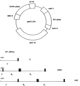
FIG. 1. Schematicdiagramof the modified pNUT vector and RV cDNAs. (Top) Plasmids used for construction of stable transformed BHK cells. RV cDNAs were clonedintothepNUT vector byusingthe SmaIsite which isflankedby the mousemetallothioneinpromoter(mMT-1)and the 3'polyadenylationsequenceofthe humangrowthhormone (hGH3').The 3' sequence ofhepatitis B virus (HBV3')allowsthe integration ofplasmidDNAinto chromosomes of transfected cells, and the presence of the dihydrofolate reductase (DHFR) cDNA permits the selection of transformed BHK cells withhighconcentrations of methotrexate. SV 40, simian virus 40early promoter. (Bottom)Diagrammaticrepresentation of RV cDNAs used in theconstruction of recombinantplasmids. The translation initiation site(ATG)from RV capsid protein was usedin all
constructs.Theputative signal peptidesandmembrane anchor domains ofE2 andEl areindicated EJ and l,respectively).
ing to the RV cDNAs used for transfection: BHK-C, BHK-E2E1, and BHK-24S.
Expression of RV structural proteinsandassemblyof VLPs instable transformed cells. The expression ofRV structural proteins from stable transformed cell lines was analyzed by immunoblotting (37). Monolayers of stable transformed cells wereincubated with mediumcontaining30 ,uMzinc sulfate for 12 h toinduce the expressionof RV structuralproteinsfrom the promoter. Samples from medium and cell lysates were collected and directly subjected to sodium dodecyl sulfate-polyacrylamide gel electrophoresis (SDS-PAGE). Separated proteins were transferred to nitrocellulose membranes and probed with human anti-RV serum. In BHK-C cells, an intracellularproteinspecieswith molecular size of 34 kDawas observed (Fig. 2A, lanes C). This protein may represent the
capsid protein of RV. In BHK-E2E1 cells, protein species
correspondingto theendoplasmic reticulum andGolgiforms ofRV E2 (13) and El glycoproteins were found in the cell lysate butnotinthe medium(Fig.2A, lanesE2E1), indicating
that the E2E1 polyprotein precursor was synthesized and
proteolytically processedtogive risetoE2andEl proteins.In BHK-24S cells, protein species corresponding to the C, E2, andEl proteinsofRV werepresent inthe celllysateaswellas in the medium (Fig. 2A, lanes 24S), suggesting that the integrated cDNA of 24S RNA was active in directing the
synthesisof RVstructural proteins and these structural pro-teins were released from the cells. The secretion of RV structuralproteinsfrom BHK-24S increased with time andwas linearover aperiod of24hunderZnSO4induction(Fig. 2C).
The secretion ofRV structural proteins into the medium was
found
tobedependentonthecoexpression of C, E2, and El, suggestingthat theseproteins areassembled into subviral particles priortotheir release from thecells. To examine this possibility, medium from BHK-24S and RV-infected cellswassubjectedtoultracentrifugation(350,000 xgfor 20
min)
inthe presence or absence of 1% of nonionic detergent Nonidet P-40. Resuspended pellets were subjected to SDS-PAGE,transferred to nitrocellulose membranes, and
probed
with humananti-RVserum.Inthe absence of NonidetP-40,C,E2,
and El were detected in the pellets from BHK-24S and
VOL.68, 1994
on November 9, 2019 by guest
http://jvi.asm.org/
[image:2.612.151.489.71.434.2]4088 NOTES
A
medium lysate
_-0 c _ cr
97
68-43- *v
29-B
C
0 0.5 1 2 4 7 10 14 18 24 RV (hour)
medium
E2
IC
1 2 3
lysate
97--68
-4Et 43--
0
*<E2
.C
29-Ei
E.
FIG. 2. (A)Immunoblotanalysis ofproteins expressedintransformedBHKcells.Monolayers ofBHK-C,BHK-E2E1,orBHK-24S cellswere
incubated with serum-free mediumin the presenceof30
pVM
zincsulfatefor 12 h.Culturemediawerecollected, and cell monolayerswerelysed withRIPAbuffer(50mMTris-HCl,10 mM EDTA, 150 mMNaCl,0.1% SDS, 1% TritonX-100, 1%Nadeoxycholate). Samplesweredirectly subjectedtoSDS-PAGEandimmunoblotting.(B) Immunoblot analysis of proteins sedimented byultracentrifugation. Samples from mediumof induced BHK-24S (lanes1and2)orRV-infectedBHKcells(lane 3)werecentrifugedat350,000xgfor 20 minin the absence(lanes 2 and3) orpresence (lane 1)of1% nonionic detergent Nonidet P-40. Thepelletswere resuspended in RIPA buffer and analyzed by SDS-PAGE and immunoblotting.Thepositions ofRVstructural proteinsareindicated.Themolecularweight markers(in thousands)areincluded for reference. (C) TimecourseofVLPssecretionfromBHK-24S cells.Expression ofRVstructuralproteinswasinducedby the addition ofZnSO4(30 p.M in theculturemedium). Culturemediumwascollected,and cells werelysedattheindicated times(hourspostinduction).Samples frommediumweresubjectedtocentrifugationat350,000xgfor 20 min andresuspendedin RIPAbuffer.Theresuspended pellets and the cell lysateswereanalyzed bySDS-PAGE andimmunoblotting. Thepositionsof RVstructuralproteinsareindicated.
RV-infected cells (Fig. 2B, lanes 2 and 3). In the presence of Nonidet P-40, El and E2 glycoproteins remained in the supernatant after ultracentrifugation (not shown), although traceamountsof Cwerefound in thepellet (Fig. 2B, lane1).
Thus, theassembled viral proteins are secretedasparticlesthat sedimentin agravitationalfield.Toconfirm thatproteinsEl, E2, and Cassembled into VLPs, samples frompelletedVLPs were centrifuged for 16 h at 90,000 x g through a density gradient of 20 to 50% sucrose. VLPs were recovered in fractions withdensities of1.17 to 1.19g/ml,similarto that of native RV virion (1.175 to 1.20g/ml) (14).
The morphology of the VLPs was analyzed by employing
conventional electron microscopic techniques with routine Epon embedding of fixed BHK-24S cells. The VLPs found in BHK-24S cells were comparable in size to RV particles (60
nm) (Fig. 3A) and indistinguishable in appearance, with an electron-dense core surrounded by an envelope (Fig. 3B, C, andD).Theseparticleswerelocatedpredominantly within the vacuoles in the juxtanuclear region (Fig. 3D) or cytoplasm
(Fig. 3C), which may represent the Golgi structure. Some particles were distributed in the cytoplasm (Fig. 3B), not associated with any membrane structure. Suchparticles were notobserved in BHK-E2E1 or BHK-C cells (data not shown). Takentogether, it is evident that VLPs were indeed assembled intracellularly prior to their release from the cells.
Antigenicity of the VLPs. HA activity of the VLPs was examinedandcompared with that of RV particles. Media were collected from BHK-24S cells at 12, 36, and 60 h after induction. RV virions were harvested from culture medium of RV-infected cells at an interval of 24 h over a period of 72 h
starting 48 h postinfection. Equal amounts of medium from
BHK-24S or RV-infected cells weresubjected to high-speed centrifugation,and thepelletedVLPsorRVs weresuspended
inphosphate-buffered saline.HA assaywas
performed
usinga heparin-manganous chlorideprocedure(16),
and theHAtiterA $' sd r<< Urs .S '.,,
-<-<i:-sstZ,.
i3'8*,s.i,,.g
;>''...,
;1''0}
9't '" ,jfi ,,,- <y! e * 9 o
t-i % r;
v
*,XsSV;;ffi>;¢D''8i-;
* w w t;x
u
'sjo:w,
+;"."m;'S.: f
i
a.;,;>..-.Y*<. jkitt- ;*
., 0 w;., ?. .1;a:.
<"< tF . 3 ^
9..,.~~~~~~~~~~~I
S i X *t: NsF~~~~of
*
'L+$
a~~.25Aj...i
FIG. 3. Electron microscopic analysis of the VLPs in BHK-24S cells.RV-infectedBHKcells(A)orinducedBHK-24S cells(B, C,and D) were fixedwith formaldehyde-glutaraldehyde, postfixed withos-mium tetroxide, ethanol dehydrated, and Epon embedded. Thin sectionswereanalyzed byelectronmicroscopyafterstaining. Arrows indicate VLPsorRVparticles. Nu, nucleus.
J. VIROL.
fl.
4 )E2
0.*-.,.4c
on November 9, 2019 by guest
http://jvi.asm.org/
[image:3.612.118.487.75.296.2]NOTES 4089 TABLE 1. Immunoreactivity of VLPs withRV-specificMAbs
Reactivityin ELISA
titer"
with:MAb Westernblot" with:
RV VLP RV VLP
H15C22(C) + + 128 256
H32C43(E1) - - 256 256
21B9H(E1) + + 256 256
3DSD(E1) - - 1,024 1,024
14B1F(E1) + + 256 256
3D9F(E1) + + 256 256
16AlOE(El) + + 1,024 1,024
E2-2(E2) - + 160 160
E2-4(E2) - + 320 320
E2-5(E2) + + 320 320
E2-6(E2) - - 160 160
H46C64(E2) + + 320 320
aMAbs were used at a dilution of 1:100for asciticfluidor1:5for tissueculture
supernatant.+,positive reactivity; -,negative reactivity.
Highestdilution ofantibodies yieldinganopticaldensityat405 nm two times
higherthanbackground.
was expressed as the end point of serial dilutions of sera at which erythrocyte aggregation was observed. The VLPs from BHK-24ScellsdisplayedHAactivity of 64, whileRVparticles retained HA activity when diluted to 1/32. This difference is dueto thehigher yield of VLPs from induced BHK-24S cells compared with that ofRVfrominfected cells.
Toevaluate theantigenicityof VLPs compared with that of RV,equal amountsof RVorVLPs(with respect to HA units) were used in each assay. Table 1 shows the antibody binding activities of VLPs and RV in immunoblot and enzyme-linked immunosorbent assay(ELISA)analysisusing 12 MAbs against RV El, E2, or C. Two ofthe E2 MAbs showed differences
between the VLPs andRVin Westernblotting(Table 1),and VLPs displayed a higher ELISA titer with MAb against C protein than did RV (Table 1). VLPs were also used in a solid-phase immunoassay to measure the immunoglobulin G response in humans. With 200 humanserum samples, it was found that the correlation coefficient between the VLPs and whole RV antigens was 0.96 by a nonparametric regression analysis method (data not shown). This indicated that the antigenic determinantsonthe VLPsresemble those of authen-tic RV.
Immunogenicityof the VLPs.Toevaluate theimmunogenic propertiesof theVLPs,weimmunized mice(BALB/c,four in each group) with the VLPs, RV, or soluble El protein
expressed in transfectedBHKcells. Toquantifytheamountof immunogens used inimmunization, purified particlesor solu-ble El protein was measured by ELISA using El-specific
MAbs. Thesame amountsofantigens (equivalentto 256HA units)wereemulsified in Freund'scomplete adjuvantand used toimmunizemice. Mice received three additionalinjectionsof
antigens in Freund'sincomplete adjuvantat3-week intervals. Mice were bled, and sera were collected for analysis. The presence of anti-RV antibodies was determined by
radioim-munoprecipitation. As shown inFig. 4, mice immunized with the VLPs produced antibodies against all three structural proteins of RV(Fig. 4, lanes3), as did mice immunized with RV (Fig. 4, lane 4). Mice immunized with El protein also
developedsomeanti-El antibodyresponse (Fig.4, lane2).
ELISAwasusedtoquantifytheantibodytitersagainst each of theRV structural proteins,by usingindividual purifiedRV structuralproteins expressed in SF9 cells infected with bacu-lovirus recombinants (unpublished data) as antigens. In the
1
2
3 4
[image:4.612.391.496.76.212.2].i4E2
FIG. 4. Radioimmunoprecipitation of RV structural proteins
ex-pressed in COS cells. COS cells were transfected with pCMV5-24S (13),labelledwith [35S]methionine, andlysed. RVstructural proteins
were recovered from cell lysates with mouse anti-RV antibodies prebound to Sepharose 4B-protein Abeads as previously described (13) andseparated bySDS-PAGE. Serawere from mice immunized with El protein (lane 2), VLPs (lane 3), or RV (lane 4) or from preimmunemice(lane 1).Thepositionsof RV structuralproteinsare
indicated.
sera from mice immunized with VLPs, a
significantly higher
anti-C
antibody
titer wasfound,
whereas anti-El and E2antibody
titers wereslightly
lower(Table 2).
Thebiological
functions of these antibodieswere
analyzed.
Sera from VLP-immunized micedisplayed
VN activities(Table
2)
as deter-minedby plaque
reduction assays(9).
HA-inhibiting
activitieswere also present in the serafrom mice immunized with the
VLPs, aswell as in the sera from mice immunizedwith RV
(Table 2).
These resultssuggested
thatalthough
VLPswere less active ininducing
overall anti-El and E2 antibodiescompared
with RV,they
induced theproduction
of both VN andHA-inhibiting
antibodies.We have also determined cell-mediated immune responses
against
RVin VLP-immunized mice inalymphocyte
prolifer-ationassay
(5, 26). Lymphocyte
proliferative
responses of miceweredetermined in vitro
by
direct stimulation oflymphocytes
with UV-inactivated RV orindividual RV structural
proteins
(C,
E2, and El) purified from recombinant baculovirus-in-fected insect cells.Lymphocytes
from VLP-immunized miceresponded strongly
to UV-inactivated RV as well as to the individual RV structuralproteins
inadose-dependent
manner(Fig. 5).
The
assembly
of RV virionsinvolvesatleasttwomajor
steps:encapsidation
andenvelopment
ofnucleocapsids.
In RV,TABLE 2. Comparisonofantibodytiters ofserafrommice immunized with different RVantigens
ELISA titerto
Immunogen RVprotein' VNtiterb HA-inhibitory
titere
C E2 El
El <10 10 10 <2 <8
RV 40 80 160 16 32
VLP 320 40 80 8 16
aIndividual RVstructuralproteins(C,E2,andEl)werepurifiedfrom SF9
cells infected with baculovirus recombinants expressing each RV structural
protein(unpublished data).
"Reciprocal ofthe highestantibody dilution thatshowed50% reduction in
plaqueformation.
Reciprocal
ofthehighest
antibodydilutionthatinhibited HA. VOL.68, 1994on November 9, 2019 by guest
http://jvi.asm.org/
[image:4.612.62.304.87.236.2]cpm, 1,000
40
30
20-10
0 2 4 6 8 10 12 14 16 18
proteinconcentration (pg/mi)
FIG. 5. Lymphoproliferation responses of mice immunized with
VLPs.Lymphocytesfrom VLP-immunized mice(2.5 x 10⁵perwell)
wereincubated withdifferent concentrations ofprotein El, E2,orCat
37°Cfor5daysbefore addition of[3H]thymidine (1 ,uCiperwell).All
assayswereperformedintriplicate,and resultsareexpressedas mean
values. WithUV-inactivated RV(109 focus-formingunitsperml) (9),
lymphoproliferation responses in the presence and absence of RV antigenwere23,000and2,000cpmrespectively.
encapsidation occurs in the cytoplasm as newly synthesized
capsid protein interacts with genomic RNA to form icosahe-dral nucleocapsids. The packagingofgenomic RNA into the nucleocapsid is believed to be a specific event, as the 40S genomic RNAbut notthe 24S subgenomicRNA ispackaged into the RV virion (23). Recently, a stretch of 31 nucleotides on the 5' end of the RV genome has been identified as
responsiblefor thebindingof thegenomicRNA tothecapsid protein in vitro (unpublished data). Employing reverse tran-scription combined with PCR (31), we failed to detect any
RV-specific RNA in the pseudovirion secreted from stable BHK-24S cells (data not shown), suggesting that capsid
pro-teinscaninteract with each other and formanucleocapsid-like
structure.The VLPswerefound to haveahigherELISA titer
withaC-specificMAb(Table 1)andtoelicitastrongeranti-C antibody response in mice than those from RV (Table 2), implying that the VLPs contain more C protein than RV
because of eitherits relativeamountorconformational
expo-sure in the particles. Although the pseudovirions do not
contain RV-specific RNA, we cannotrule out the possibility thatthey packagesomecellularRNAsor evenDNAs into the nucleocapsid.
Incorporationofnucleocapsidinto the membraneenvelope
to form virus particles is a poorly understood event in virus assembly. Wefound that in BHK-C and BHK-E2E1 cells,no
RV proteins were released into the medium (Fig. 2A). In BHK-24Scells,all threestructuralproteinswerepresentinthe mediumastheresult ofsubviralparticleformation andegress
(Fig. 2). These data strongly suggest that the interaction betweenglycoproteinsandthenucleocapsidis thedrivingforce for theassemblyandrelease ofthe VLPs. This interaction has been defined to occurbetween the cytoplasmictail of
glyco-proteinElandthe capsid protein.Deletion of thecytoplasmic domainof RV El abolished thedeliveryof theVLPs into the medium (12a).
Besidesbeingauseful tooltostudyRVassembly, BHK-24S cellsin whichVLPs aresteadily assembled and releasedcanbe used as a potential source for mass production of rubella antigensatlow cost underinducingconditions. BHK-24S cells continuously produce VLPs forupto5 dayswithout celllysis when 30pLM ZnSO4 is present in the medium and for upto 1 2 month inDME/F12(GIBCO) medium.VLPs canbe harvested daily from the medium, which is replaced with fresh medium afterharvesting. Dependingonthe methods usedtoquantitate theyields of theVLPsandRVfrom culturemedium, the yield of VLPswasfoundtobe2timeshigherthan that of RV in an HA assay, 5 times higher in ELISAs using human sera, and morethan 10 timeshigher by protein quantitation usingsilver staining after gel electrophoresis (datanotshown).
For immunogenicity studies, the VLPs were found to be significantlymoreactive than the soluble El proteinin induc-ing antibodyresponsesinmice, especiallyfor theproductionof VN and HA-inhibiting activity. The VLPs also evoked a cell-mediated immune response to RV and RV structural
proteins.This is believed tobeimportantinproviding protec-tive immunity against RV infection. Preliminary results have shown that CD4+ T cells may be the major effector in cell-mediated immune responses elicitedbytheVLPs inmice, although CD8+ T cells may be also involved (unpublished data). A study of the phenotype of the effector cells in proliferationassays is in progress. The VLPsare composedof all three structuralproteinsofRV,whichmakes them similar toRVregarding antigen presentation.Ourstudies suggest that theVLPsmay serve as a candidate for safe vaccine develop-ment.
WearegratefultoJ.Wolinskyand J. Saffordforprovidinganti-RV MAbs and to R. Palmiter for providing the pNUTvector. Special thankstoM.Weiss for technical assistanceonelectronmicroscopyand toC. Mauracher forperforming solid-phase immunoassay usingthe VLPs.
This workwassupported by a grantfrom the Medical Research Council of Canada (to S.G.). Z.Q. is the recipient of a British Columbia Children'sHospitalFoundationstudentship.T.C.H. is sup-portedbyaMedical Research Council of Canadapostdoctoral fellow-ship. S.G. is an investigator of the British Columbia Children's HospitalFoundation.
REFERENCES
1. Assad, F., and K.Ljungars-Esteves.1985.Rubella-worldimpact. Rev.Infect. Dis. 7:S29-S36.
2. Baron, M., andK.Forsell.1991.Oligomerizationofthe structural proteinsof rubella virus.Virology185:811-819.
3. Chantler, J. K., D. K.Ford, and A.J. Tingle. 1982. Persistent rubella infection and rubella associated arthritis. Lancet i:1323-1325.
4. Chaye, H., B. Brush,P.Chong, B.Tripet, and S. Gillam. 1992. Localizationof thevirusneutralizingandhemagglutinin epitopes ofElglycoproteinof rubellavirus.Virology 189:483-492. 5. Chaye,H.H.,C.A.Mauracher,A.J. Tingle,andS. Gillam. 1992.
Cellular and humoral immune responsestorubella virus structural proteinsEl,E2,andC. J. Clin.Microbiol.30:2323-2329. 6. Chaye, H.,D.Ou,P.Chong,andS. Gillam. 1993. Human T-and
B-cell epitopes of El glycoprotein of rubella virus. J. Clin. Immunol. 13:93-100.
7. Clarke, B. E., S. E. Newton, A. R. Carroll, M. J. Francis, G. Appleyard,A.D. Syred, P.E.Highfield, D.J. Rowlands,and F. Brown. 1987. Improved immunogenicity ofa peptide epitopeto
hepatitisBcoreprotein. Nature(London) 330:381-384. 8. Clarke, D., T. Loo, I. Hui, P. Chong, and S. Gillam. 1987.
Nucleotide sequence and in vitro expression ofrubella virus 24S subgenomicmRNAencodingthestructuralproteinsEl, E2and C. Nucleic Acids Res. 15:3041-3057.
9. Fukuda, A., M. Hissihiyama, Y. Umino, and A. Sugiura. 1987. Immunocytochemical focus assay for potency determination of 50 r
on November 9, 2019 by guest
http://jvi.asm.org/
[image:5.612.58.300.73.271.2]NOTES 4091 measles-mumps-rubella trivalent vaccine. J. Virol. Methods 15:
279-284.
10. Gorman,C.M.,L. F.Moffat,and B. H.Howard. 1982. Recombi-nant genomes which express chloramphenicol acetyltransferase in mammaliancells. Mol. Cell. Biol. 2:1044-1051.
11. Green, K. Y., and P. H. Dorsett. 1986. Rubella virus antigens: localization of epitopes involved in hemagglutination and neutral-ization by using monoclonalantibodies. J. Virol. 57:893-898. 12. Griffiths, J. C., S. J. Harris, G. T. Layton, E. L. Berrie, T. J.
French, N. R. Burns, S. E. Adams, and A.J. Kingsman. 1993. Hybrid human immunodeficiency virus Gag particles as an antigen carrier system: induction of cytotoxic T-cell and humoral re-sponsesbyaGag:V3 fusion. J. Virol. 67:3191-3198.
12a.Hobman, T. C., et al. Unpublished data.
13. Hobman,T. C., M. Lundstrom,and S. Gillam. 1990. Processing and intracellular transport of rubella virus structure proteins in COScells.Virology 178:122-133.
14. Horzinek, M. C.1981.Non-arthropod-borne togaviruses, p. 39-44. Academic PressLtd., London.
15. Li,S.,V.Polonis, H. Isobe,A.Zaghouani,R.Guinea,T.Moran, C. Bona, and P. Palese. 1993. Chimeric influenza virus induces neutralizing antibodies and cytotoxicTcells against human immu-nodeficiency virus type 1.J. Virol. 67:6659-6666.
16. Liebhaber, H. 1970. Measurement ofrubella antibody by haem-agglutination. I. Variables affecting rubella haemhaem-agglutination. J. Immunol. 104:818-825.
17. Loo,T.W., I. MacDonald, D. Clarke,M.Trudel,A.J. Tingle, and S. Gillam. 1986. Detection of antibodiestoindividual proteins of rubellavirus. J. Virol. Methods 13:149-159.
18. Lovett, A. E., C. S. Hahn, C. M. Rice, T. K. Frey, and J. S. Wolinsky. 1993. Rubella virus-specific cytotoxic T-lymphocyte responses: identification of the capsidas atargetof major histo-compatibilitycomplex class I-restrictedlysis and definition of two epitopes.J.Virol. 67:5849-5858.
19. Lovett,A. E., M.McCarthy, andJ. S.Wolinsky. 1993. Mapping cell-mediated immunodominant domains of the rubella virus structural proteins usingrecombinantproteins and synthetic pep-tides. J. Gen. Virol. 74:445-452.
20. Matthews, R. E. F. 1982. Classification and nomenclature of viruses. Fourth report of the International Committee on Taxon-omyof Viruses. Intervirology 17:1-199.
21. Michel, M.-L., M. Mancini,E. Sobczack,V. Favier, D. Guetard,
E. M. Bahraqui, and P.Tollias. 1988. Induction ofanti-human
immunodeficiency virus (HIV) neutralizing antibodies in rabbits immunized with recombinant HIV-hepatitis B surface antigen particles. Proc. Natl. Acad. Sci. USA 85:7957-7961.
22. Oker-Blom, C.,N.Kalkkinen,L.Kaariainen,and R. F.Pettersson. 1983. Rubella virus containsone capsid proteinand three enve-lopeglycoproteins, El,E2a, and E2b. J. Virol. 46:964-973. 23. Oker-Blom,C.,D.Ulmanen, L.Kaariainen,and R. F.Pettersson.
1984. Rubellavirus 40S genomicRNAspecifiesa24Ssubgenomic mRNAthatcodes foraprecursortostructuralproteins.J.Virol. 49:403-408.
24. Ou, D.,P.Chong,Y.Choi,P.McVeigh,W. A.Jefferies, G. Koloitis, A.J.Tingle,andS.Gillam.1992.Identification of T-cell epitopes onE2protein of rubella virus,asrecognized by human T-cell lines andclones. J. Virol. 66:6788-6793.
25. Ou, D., P. Chong, A. J.Tingle, and S. Gillam. 1993. Mapping
T-cellepitopes of rubella virus structural proteins El, E2 and C recognized by T-cell lines and clones derived from infected and immunized populations. J. Med. Virol. 40:175-183.
26. Ou, D., P. Chong, B. Tripet, and S. Gillam. 1992. Analysis of T-and B-cell epitopes of capsid protein of rubella virus by using synthetic peptides. J. Virol. 66:1674-1681.
27. Ou, D., P. Chong, B. Tripet, W. A.Jefferies,and S.Gillam.1992.
Characterization of thespecificity and genetic restriction of human CD4+ T-cell clones reactive to capsid antigen of rubella virus. Virology 191:680-686.
28. Palmiter, R. D., R. R. Behringer, C. J. Quaife, F. Maxwell, I.H.
Maxwell, and R. L. Brinster. 1987. Cell lineage ablation in transgenic mice by cell-specific expression of toxic gene. Cell 50:435-443.
29. Perfins, F.T. 1985.Licensed vaccines. Rev. Infect. Dis. 7:573-576. 30. Qiu, Z., F.Tufaro,andS. Gillam.Influence ofN-linked glycosy-lation on the antigenicity and immunogenicity of rubella virus El glycoprotein. Virology 182:768-772.
31. Saiki, R. K., D. H.Gelfand, S. Stoffel, S. J. Scharf, R.Higuchi,
G. T.Horn,K. B.Mullis,and H. A. Erlich. 1988.Primer-directed
enzymatic amplification of DNA with athermostable DNA poly-merase. Science 239:487-491.
32. Sanchez, A., and T. K. Frey. 1991. Vaccinia-vectoredexpression of rubella virus structuralproteins and characterization of the El and E2glycosidic linkages.Virology 183:636-646.
33. Seppanen, H.,M.-L.Huhtala,A.Vaheri,M. D.Summers,and C. Oker-Blom. 1991. Diagnostic potential ofbaculovirus-expressed rubellavirus envelope proteins. J. Clin. Microbiol. 29:1877-1882. 34. Takahashi, H., T. Takeshita,B.Morein, S. Putney,R. N.Germain, andJ.A.Berzofsky. 1990. Induction ofCD8+cytotoxicTcellsby immunization withpurified HIV-1 envelopeproteins in ISCOMs. Nature (London) 344:873-875.
35. Terry, G. M., L. Ho-Terry, P. Londesborough, and K. R. Rees. 1988.Localization of the rubella El epitopes. Arch. Virol. 98:189-197.
36. Terry, G. M., L. Ho-Terry, P. Londesborough, and K. R. Rees. 1989.Abioengineered rubella Elantigen.Arch. Virol. 104:63-75. 37. Towbin, H., Y. Staehelin, and J. Gordon. 1979. Electrophoretic transfer of proteins from polyacrylamide gels to nitrocellulose sheets: procedure and some applications. Proc. Natl. Acad. Sci. USA 76:4350-4354.
38. Vaheri, A.,and T. Hovi.1972. Structural proteinsandsubunits of rubellavirus.J. Virol. 9:10-16.
39. Waxham, M. N., and J. S. Wolinsky. 1983. Immunochemical identification of rubella virus hemagglutinin. Virology 126:194-203.
40. Waxham,M.N.,andJ.S.Wolinsky. 1985. Detailedimmunologic analysis of the structural polypeptides of rubella virus using monoclonal antibodies.Virology 143:153-165.
41. Wolinsky, J. S., M. McCarthy, 0. Allen-Cannady,W. T.Moore,R. Jin, S. Cao, A. Lovett, and D. Simmons. 1991. Monoclonal antibody-defined epitope mapofexpressedrubella virus protein domains. J. Virol. 65:3986-3994.
42. Zhang, T.,C. A.Mauracher,L. A.Mitchell,and A.J.Tingle. 1992. Detection of rubellavirus-specificimmunoglobulinG(IgG),IgM, and IgA antibodies by immunoblot assays. J. Clin. Microbiol. 30:824-830.
VOL.68, 1994