0022-538X/05/$08.00⫹0 doi:10.1128/JVI.79.19.12536–12543.2005
Copyright © 2005, American Society for Microbiology. All Rights Reserved.
Patterns of Cytokine Production in Human Immunodeficiency Virus
Type 1 (HIV-1)-Specific Human CD8
⫹
T Cells after Stimulation
with HIV-1-Infected CD4
⫹
T Cells
Mamoru Fujiwara,
1Hiroshi Takata,
1Shinichi Oka,
2Hiroko Tomiyama,
1and Masafumi Takiguchi
1*
Division of Viral Immunology, Center for AIDS Research, Kumamoto University, 2-2-1 Honjo, Kumamoto 860-0811,1and
AIDS Clinical Center, International Medical Center of Japan, 1-21-1 Toyama, Shinjuku-ku, Tokyo 162-8655,2Japan
Received 22 April 2005/Accepted 6 July 2005
Although human immunodeficiency virus type 1 (HIV-1)-specific CD8ⴙT cells can produce various cytokines that suppress HIV-1 replication or modulate anti-HIV-1 immunity, the extent to which HIV-1-specific CD8ⴙT cells produce cytokines when they recognize HIV-1-infected CD4ⴙT cells in vivo still remains unclear. We first analyzed the abilities of 10 cytotoxic T-lymphocyte (CTL) clones specific for three HIV-1 epitopes to produce gamma interferon, macrophage inflammatory protein 1, and tumor necrosis factor alpha after stimulation with epitope peptide-pulsed cells. These CTL clones produced these cytokines in various combinations within the same specificity and among the different specificities, suggesting a functional heterogeneity of HIV-1-specific effector CD8ⴙT cells in cytokine production. In contrast, the HIV-1-specific CTL clones for the most part produced a single cytokine, without heterogeneity of cytokine production among the clones, after stimu-lation with HIV-1-infected CD4ⴙT cells. The loss of heterogeneity in cytokine production may be explained by low surface expression of HLA class I–epitope peptide complexes. Freshly isolated HIV-1-specific CD8ⴙT cells with an effector/memory or memory phenotype produced much more of the cytokines than the same epitope-specific CTL clones when stimulated with HIV-1-infected CD4ⴙT cells. Cytokine production from HIV-1-specific memory/effector and memory CD8ⴙT cells might be a critical event in the eradication of HIV-1 in HIV-1-infected individuals.
Memory and effector CD8⫹T cells play an important role in viral eradication through their ability to produce cytokines involved in the suppression of viral replication (6, 10, 15, 26) as well as perforin and granzymes A and B, which are involved in the cytolysis of virus-infected cells (16, 24). The cytokines produced by these cells include gamma interferon (IFN-␥), tumor necrosis factor alpha (TNF-␣), and chemokines such as RANTES and macrophage inflammatory protein 1 (MIP-1). IFN-␥increases the surface expression of HLA molecules and can activate macrophages that predominantly synthesize MIP-1(8, 25). TNF-␣induces apoptosis of human immuno-deficiency virus type 1 (HIV-1)-infected cells (17). It has been shown that MIP-1and RANTES can suppress HIV-1 repli-cation in vitro by inhibiting the entry of HIV-1 via CCR5, while IFN-␥induces cellular proteins which suppress viral replica-tion (2, 6).
A previous study showed that HLA-A2-restricted, HCMVpp65495-503-specific CD8⫹ T cells expressed various
combinations of three cytokines, IFN-␥, TNF-␣, and interleu-kin 2 (IL-2), after peripheral blood mononuclear cells (PBMC) containing these cells from three individuals had been stimu-lated with HCMVpp65495-503 peptide, suggesting that
HCMVpp65495-503-specific CD8⫹T cells possess functionally
heterogeneous cytokine production (18). This functional het-erogeneity may be due to the heterogeneous populations in
HCMVpp65495-503-specific CD8⫹ T cells. Indeed, these cells
for the most part are CD8⫹T-cell populations with a memory/ effector or effector phenotype (9, 23). Additional studies have revealed the heterogeneity of the production of cytokines and cytolytic effector molecules in human CD8⫹T cells (21, 23).
It is well known that virus-specific CD8⫹T cells can produce cytokines when they recognize virus-infected cells (4, 13, 14). However, virus-specific CD8⫹ T cells stimulated with virus-infected cells may not produce cytokines more effectively than those stimulated with cells pulsed with an epitope peptide, because the expression of HLA class I–viral epitope peptide complexes is much lower on virus-infected cells than on pep-tide-pulsed cells. In particular, HIV-1-specific CD8⫹ T cells may not produce cytokines effectively when they recognize HIV-1-infected cells, because HLA class I molecules have been actively down-regulated, mostly, although not exclusively, by the Nef protein produced by HIV-1-infected cells (4, 7, 20). An analysis of the cytokine production from HIV-1-specific CD8⫹T cells stimulated with HIV-1-infected CD4⫹T cells is needed to elucidate whether HIV-1-specific CD8⫹T cells can recognize HIV-1-infected CD4⫹T cells in vivo and to allow the formulation of testable hypotheses on the role of the cy-tokines from HIV-1-specific CD8⫹T cells in the suppression of HIV-1 replication in vivo.
In the present study, we investigated the production patterns of three cytokines, IFN-␥, TNF-␣, and MIP-1, in HIV-1-specific effector CD8⫹T-cell clones of the same or different specificities. In addition, we investigated the production of the three cytokines in these cytotoxic T-lymphocyte (CTL) clones and freshly isolated HIV-1-specific CD8⫹T cells exposed to * Corresponding author. Mailing address: Division of Viral
Immu-nology, Center for AIDS Research, Kumamoto University, 2-2-1 Honjo, Kumamoto 860-0811, Japan. Phone: 96-373-6529. Fax: 81-96-373-6532. E-mail: [email protected].
12536
on November 8, 2019 by guest
http://jvi.asm.org/
200 U/ml recombinant human IL-2, 5⫻10 irradiated allogeneic PBMC from a healthy donor, and 1⫻105
irradiated A*2402, A*3303, or C1R-B*3501 cells prepulsed with 10⫺6M of the corresponding peptide, Env77-85
[DPNPQEVVL] [19], Gag28-36 [KYKLKHIVW] [12], or Env830-837 [EVAQ RAYR] [11]). Wells positive for growth after about 2 weeks were transferred to 48-well plates together with 1 ml of the cloning mixture. The clones were exam-ined for CTL activity by a standard51Cr release assay. One Env830-837-specific,
HLA-A*3303-restricted CTL clone (clone 1) had been generated previously (11). All CTL clones were cultured in RPMI 1640–10% FCS supplemented with 200 U of recombinant human IL-2/ml and were stimulated weekly with irradiated target cells prepulsed with the appropriate HIV-1 derived peptide.
Antibodies. The peridinin chlorophy protein-conjugated anti-human CD8 monoclonal antibody (MAb) and the ECD-conjugated anti-human CD45RA MAb were purchased from BD Biosciences (San Jose, CA) and the Immunotech Coulter Company (Marseille, France), respectively. Fluorescein isothiocyanate (FITC)-conjugated anti-human CD27, phycoerythrin (PE)-conjugated anti-hu-man IFN-␥, ECD-conjugated anti-human CD28, allophycocyanin (APC)-conju-gated anti-human TNF-␣, APC-conjugated anti-human IFN-␥, PE Cy7-conju-gated anti-human TNF-␣, and APC Cy7-conjugated anti-human CD27 MAbs were purchased from PharMingen (San Diego, CA). PE-conjugated anti-human CD28, APC-conjugated anti-human CD8, and FITC-conjugated anti-human MIP-1MAbs were purchased from DAKO (Glostrup, Denmark). FITC-con-jugated anti-HIV-1 p24 MAb KC-57 was purchased from Beckman Coulter (Miami, Fla.). A Cascade Blue-conjugated anti-human CD8 MAb was made by conjugating Cascade Blue (Molecular Probes, Eugene, OR) with the anti-CD8 MAb OKT8.
HLA-peptide tetrameric complexes. HLA class I–peptide tetrameric com-plexes were synthesized as previously described (3). Briefly, recombinant HLA class I proteins (HLA-A*2402, HLA-A*3303, and HLA-B*3501) and human2
microglobulin (2m) were produced inEscherichia colicells transformed with the
relevant expression plasmids. The heavy chain was modified by deletion of the transmembrane cytosolic tail and COOH-terminal addition of a sequence con-taining the BirA biotinylation site. Gag28-36, Env830-837, and Env77-85 pep-tides were used for refolding of the A*2402, A*3303, and HLA-B*3501 molecules, respectively. The HLA class I–peptide complexes were refolded in vitro. The 45-kDa complexes were isolated using gel filtration on a Superdex G75 column (Amersham Pharmacia Biotech UK, Ltd., Buckingham-shire, England). Purified complexes were biotinylated with the BirA enzyme (Avidity, Denver, CO). The biotinylated complexes were purified by using gel filtration first on a Superdex G75 column and then on a MonoQ column (Am-ersham Pharmacia Biotech UK, Ltd, Buckinghamshire, England). HLA class I–peptide tetrameric complexes (tetramers) were mixed with PE-labeled strepta-vidin (extrastrepta-vidin-PE; Sigma-Aldrich, St. Louis, MO) at a molar ratio of 4:1.
Identification of HIV-1-specific CTLs by flow cytometry.A total of 0.2⫻106
to 1⫻106cultured cells were mixed with the tetramers at concentrations of 0.02
to 0.04 mg/ml. After incubation at 37°C for 30 min, the cells were washed twice with RPMI 1640–10% FCS, and then an anti-CD8 MAb was added to the cell suspension. The cells were incubated at 4°C for 30 min, and then the cells were washed twice with phosphate-buffered saline (PBS)–10% FCS. The cells were analyzed using a FACSCalibur with CellQuest software (Becton Dickinson, San Jose, CA). The percentage of tetramer-positive cells was measured in CD8-positive cells.
Phenotype analysis of the CTL clones.Cells were stained for 30 min at 4°C using an FITC-conjugated anti-human CD27 MAb, a PE-conjugated anti-human CD28 MAb, an ECD-conjugated anti-human CD45RA MAb, and an APC-conjugated anti-human CD8 MAb; then they were washed twice in PBS supple-mented with 10% NCS. The CD27 CD28 CD45RA phenotype of CD8⫹cells was analyzed using FACSCalibur.
Cell surface and intracellular cytokine staining.Specific CTL clones were stimulated with stimulator cells prepulsed with the appropriate HIV-1-derived peptide at each concentration at an effector-to-stimulator ratio of 1:1. Stimulator
then were stained with a PE-conjugated anti-human IFN-␥MAb or an APC-conjugated anti-human TNF-␣MAb at room temperature for 20 min or with an FITC-conjugated anti-human MIP-1MAb for 30 min at 4°C. When the cells were stained with all three MAbs, they were first stained with the PE-conjugated anti-human IFN-␥MAb and the APC-conjugated anti-human TNF-␣MAb at room temperature for 20 min and then with the FITC-conjugated anti-human MIP-1MAb for 30 min at 4°C. Freshly isolated CD8⫹cells were stained with an APC-conjugated anti-human IFN-␥MAb or a PE Cy7-conjugated anti-human TNF-␣MAb at room temperature for 20 min or with an FITC-conjugated anti-human MIP-1MAb for 30 min at 4°C.
Finally, the cells were washed three times with permeabilizing buffer and were resuspended in 2% paraformaldehyde. The percentages of intracellular IFN-␥-, MIP-1-, and TNF-␣-positive cells among tetramer-positive CD8⫹cells were analyzed using FACS Aria (Becton Dickinson, San Jose, CA).
Infection of CD4ⴙT cells with HIV-1.Cultured CD4⫹T cells (purity,⬎98%) were incubated with an HIV-1 clone, NL-432 (1), or with the chimeric virus NL-432gagHXB2
for 4 h at 37°C with intermittent agitation. The cells were then washed once and cultured in RPMI 1640–10% FCS medium supplemented with recombinant human IL-2 (200 U/ml). On the following 2 to 7 days, the cells were harvested to determine the percentage of HIV-1-infected cells by measuring p24 antigen-positive cells using FACSCalibur. When HIV-1 p24-positive cells reached more than 40% of the cultured cells, they were used as stimulator cells.
CTL assay.Cytotoxicity was measured by a standard51
Cr release assay as previously described (19). Target cells (2⫻105) were incubated for 60 min with
100Ci of Na2 51
CrO4in saline and washed three times with RPMI 1640 medium
containing 10% NCS. Labeled target cells (2⫻103/well) were added into a
96-well round-bottom microtiter plate (Nunc) with the indicated amount of the corresponding peptide. After a 1-h incubation, effector cells were added and the mixtures were incubated for 4 h at 37°C. The supernatants were collected and analyzed with a gamma counter. The spontaneous51Cr release was determined
by measuring the cpm in the supernatant in the wells containing only target cells (cpm spn). Maximum release (cpm max) was determined by measuring the release of51
Cr from the target cells in the presence of 2.5% Triton X-100. Specific lysis was defined as (cpm exp⫺cpm spn)/(cpm max⫺cpm spn)⫻100, where “cpm exp” is the counts per minute in the supernatant in the wells containing both the target and effector cells. The activities of the CTL clones on target cells pulsed with peptide were tested at an effector-to-target (E:T) ratio of 2:1.
CTL assay for target cells infected with recombinant vaccinia virus. Recom-binant vaccinia virus containing theenvor thegag/polgene of HIV-1 SF2 was generated as described previously (19). Target cells (C1R-A*2402, C1R-A*3303, and C1R-B*3501 cells) were cultured with 10 PFU of recombinant or wild-type vaccinia virus per target cell overnight. These infected cells (2⫻105
) were incubated for 60 min with 100Ci of Na251CrO4in saline and then washed three
times with RPMI 1640 medium containing 10% NCS. Effector cells were added to the labeled target cells (5⫻103/well), and the mixtures were incubated for 4 h
at 37°C. The activities of the CTL clones on target cells infected with recombi-nant vaccinia virus expressingenvproteins were tested at an E:T ratio of 2:1.
RESULTS
Production of three cytokines in HIV-1-specific CTL clones.
Three SF2-Env77-85-specific, HLA-B*3501-restricted CTL clones, three SF2-Gag28-36-specific, HLA-A*2402-restricted CTL clones, and a further four SF2-Env830-837-specific, HLA-A*3303-restricted CTL clones were established from HIV-1-infected individuals. These CTL clones exhibited specific cyto-lytic activity in both target cells (C1R-A*2402, C1R-A*3303, or
on November 8, 2019 by guest
http://jvi.asm.org/
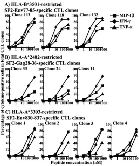
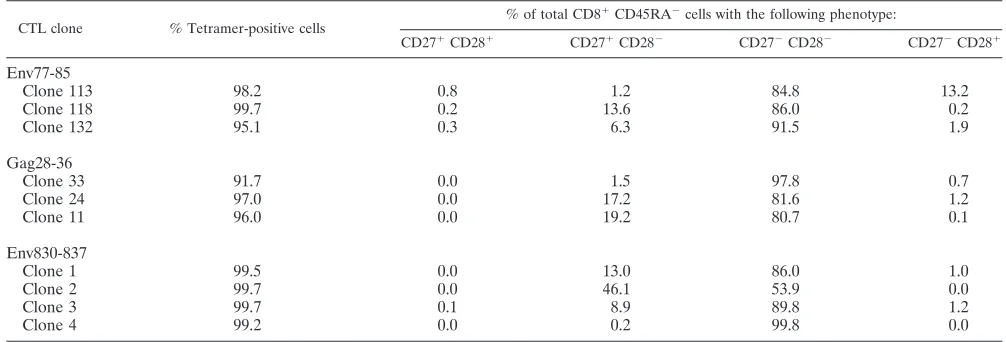
C1R-B*3501 cells) prepulsed with a peptide epitope (Fig. 1) and target cells (C1R-A*2402, C1R-A*3303, or C1R-B*3501 cells) infected with HIV-1 recombinant vaccinia virus (data not shown). The results show no significant difference in cytotoxic activity between CTL clones specific for the same epitopes. We investigated the production of three cytokines, MIP-1, IFN-␥, and TNF-␣, from these HIV-1-specific CTL clones. Intracel-lular cytokine production by the clones was measured at 6 h after stimulation with the C1R transfectants prepulsed with the epitope peptide. Almost 100% of the cells produced at least one cytokine in all three of the SF2-Env77-85-specific CTL clones, all four of the SF2-Env830-837-specific CTL clones, and all three of the SF2-Gag28-36-specific CTL clones (Fig. 2). Different cytokine production patterns were found among three SF2-Env77-85-specific CTL clones though all of the clones produced IFN-␥ more extensively than MIP-1 or TNF-␣(Fig. 2A). Different cytokine production patterns were also found for the three SF2-Gag28-36-specific CTL clones (Fig. 2B) and the four SF2-Env830-837-specific CTL clones (Fig. 2C). Clone 11 exhibited much higher production of MIP-1 than of IFN-␥and TNF-␣compared with clones 33 and 24. In addition, clones 2 and 3 exhibited much lower production of TNF-␣ than of IFN-␥and MIP-1, while the difference in production between these cytokines was at a min-imum for clones 1 and 4. These results were confirmed by performing the experiments twice. Thus, the results reveal heterogeneity in the production of these cytokines between CTL clones of the same as well as different specificities. To exclude contamination by T cells that are not epitope-specific CTLs, we stained the CTL clones with an epitope-specific HLA class I tetramer. More than 97% of the cells in nine CTL clones bound the epitope-specific tetramer, while only 92% of the cells bound to the specific tetramer in one (clone 33) of the SF2-Gag28-36-specific CTL clones (Table 1). However, this difference does not seem to be significant for the functions, since the three SF2-Gag28-36-specific CTL clones exhibited the same cytotoxic activity and IFN-␥production.
Since the T-cell clones used in the present study possess cytolytic activity (Fig. 1), they are thought to be mature effector
CD8⫹T cells. To analyze the relation between the maturation stage of the CTL clone and cytokine production, we stained these CTL clones with CD27, CD28, and anti-CD45RA MAbs. Nine CTL clones were mostly of the CD27⫺ CD28⫺ CD45RA⫺ type, while one (clone 2) of the SF2-Env830-837-specific CTL clones exhibited both CD27⫺ CD28⫺ CD45RA⫺ and CD27⫹ CD28⫺ CD45RA⫺ pheno-types (Table 1). Clone 2, stimulated with SF2-Env830-837 pep-tide, exhibited a much smaller number of TNF-␣-producing cells than of IFN-␥-producing or MIP-1-producing cells, but this is not a characteristic restricted to clone 2.
[image:3.585.309.530.66.331.2]Coproduction of three cytokines by HIV-specific CTL clones stimulated with peptide-pulsed cells.The heterogeneity of the CTL clones shown in Fig. 2 suggests that the CTL clones are actually composed of cell populations which produce different combinations of the cytokines. To clarify this, we investigated the coproduction of these three cytokines in the CTL clones by simultaneously staining multiple intracellular cytokines with anti-IFN-␥, anti-MIP-1, and anti-TNF-␣MAbs. The results of the multiple staining of the three SF2-Env77-85-specific CTL clones are shown in Fig. 3A. The SF2-Env77-85-specific CTL clones stimulated with 1,000 nM of specific peptide re-vealed different patterns of cytokine production. Clone 113 included a high number of cells producing all three of the cytokines (IFN-␥⫹ TNF-␣⫹ MIP-1⫹) (72.0%). In contrast, FIG. 1. Cytolytic activities of SF2-Env77-85-specific,
SF2-Gag28-36-specific, and SF2-Env830-837-specific CTL clones. Cytolytic activi-ties of three SF2-Env77-85-specific, HLA-B*3501-restricted CTL clones (clones 113, 118, and 132), three SF2-Gag28-36-specific, HLA-A*2402-restricted CTL clones (clones 33, 24, and 11), and four SF2-Env830-837-specific, HLA-A*3303-restricted CTL clones (clones 1, 2, 3, and 4) were tested for C1R-B*3501, C1R-A*2402, and C1R-A*3303 cells pulsed with the corresponding peptides (1 to 1,000 nM), respec-tively. They were tested at an E:T ratio of 2:1.
FIG. 2. Intracellular expression of cytokines in SF2-Env77-85-spe-cific, SF2-Gag28-36-speSF2-Env77-85-spe-cific, and SF2-Env830-837-specific CTL clones responding to epitope peptide-pulsed cells. The 10 CTL clones were incubated for 6 h in the presence of C1R-B*3501, C1R-A*2402, or C1R-A*3303 cells pulsed with specific peptide. Brefeldin A was added 2 h after stimulation, and intracellular staining for IFN-␥, MIP-1, and TNF-␣was carried out using MAbs specific for each cytokine. Cyto-kine-producing cells were analyzed by flow cytometry. The frequency of each set of cytokine-producing CD8⫹ cells was measured as the number of cytokine-producing CD8⫹cells per total CD8⫹cells.
on November 8, 2019 by guest
http://jvi.asm.org/
[image:3.585.52.276.67.185.2]clone 118 included IFN-␥⫹ TNF-␣⫹MIP-1⫹cells (58.1%) and IFN-␥⫹TNF-␣⫹cells (30.6%), while clone 132 included IFN-␥⫹ TNF-␣⫹ MIP-1⫹ cells (54.7%), IFN-␥⫹ TNF-␣⫹ cells (17.8%), and IFN-␥⫹cells (13.1%). This difference be-tween these CTL clones became even more apparent when the CTL clones were stimulated with 10 nM of specific peptide. Clone 113 included MIP-1⫹cells (16.1%) and IFN-␥⫹ MIP-1⫹cells (7.3%). In contrast, clone 118 included IFN-␥⫹cells (29.1%) and IFN-␥⫹TNF-␣⫹cells (17.0%), while clone 132 included IFN-␥⫹ cells (27.5%) and IFN-␥⫹ MIP-1⫹ cells (20.6%). This difference was consistent over two different ex-periments performed on different days.
A similar analysis was performed on the four SF2-Env830-837-specific and the three SF2-Gag28-36-specific CTL clones. The seven CTL clones were stimulated with 1,000 nM of spe-cific peptide (Fig. 3B and C). Among SF2-Gag28-36-spespe-cific CTL clones, clones 33 and 24 showed similar patterns of cyto-kine production. They included IFN-␥⫹ TNF-␣⫹ MIP-1⫹ cells (12 to 14%), IFN-␥⫹ TNF-␣⫹ cells (11 to 13%), and IFN-␥⫹ MIP-1⫹ cells (14 to 18%). In contrast, clone 11 included IFN-␥⫹ TNF-␣⫹ cells (5%) and IFN-␥⫹ MIP-1⫹ cells (34%). The difference became more complex when the clones were stimulated with a lower concentration (100 nM) of the peptide. The major populations of cytokine-producing cells were as follows: for clone 11, IFN-␥⫹cells (24.5%), IFN-␥⫹ MIP-1⫹cells (15.4%), and MIP-1⫹cells (7.7%); for clone 33, IFN-␥⫹ cells (28.0%), IFN-␥⫹ MIP-1⫹cells (9.9%), and IFN-␥⫹TNF-␣⫹cells (8.0%); and for clone 24, IFN-␥⫹cells (27.2%), IFN-␥⫹MIP-1⫹cells (9.1%), and IFN-␥⫹TNF-␣⫹ cells (7.6%). Among SF2-Env830-837-specific CTL clones, clones 1 and 4 showed similar patterns of cytokine production. These included IFN-␥⫹TNF-␣⫹MIP-1⫹cells (50 to 60%), IFN-␥⫹TNF-␣⫹cells (13 to 17%), and IFN-␥⫹MIP-1⫹cells (12 to 14%). In contrast, clones 2 and 3 included IFN-␥⫹ TNF-␣⫹ MIP-1⫹cells (40 to 42%) and IFN-␥⫹MIP-1⫹ cells (35 to 37%). The difference became more complex when the clones were stimulated with a lower concentration (100 nM) of the peptide. At this concentration the major popula-tions of cytokine-producing cells were as follows: for clone 1, MIP-1⫹cells (17.6%) and IFN-␥⫹MIP-1⫹cells (10.9%); for clone 2, IFN-␥⫹MIP-1⫹cells (19.1%), IFN-␥⫹cells (15.7%),
and MIP-1⫹ cells (12.4%); for clone 3, MIP-1⫹ cells (20.8%), IFN-␥⫹ MIP-1⫹ cells (20.6%), and IFN-␥⫹ cells (10.9%); and for clone 4, IFN-␥⫹cells (15.1%), IFN-␥⫹ MIP-1⫹ cells (12.0%), and IFN-␥⫹ TNF-␣⫹ MIP-1⫹ cells (10.1%). These differences were also confirmed by two differ-ent experimdiffer-ents performed on differdiffer-ent days.
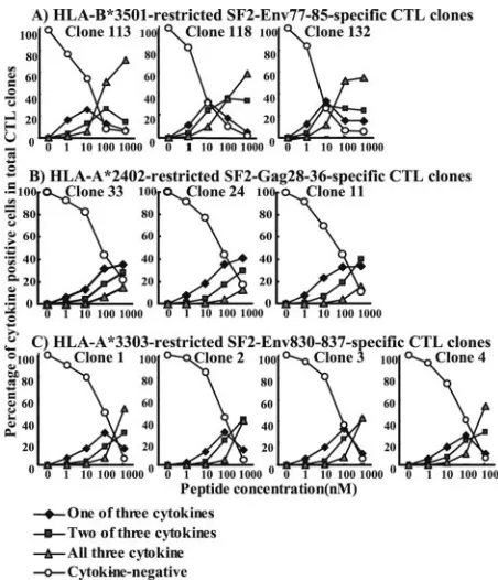
Cells expressing all three cytokines were most frequently found in the CTL clones stimulated with the stimulator cells prepulsed with 1,000 nM peptide, while cells expressing either two or all three of the cytokines were not found in the CTL clones stimulated with the stimulator cells prepulsed with 1 nM peptide (Fig. 4). The CTL clones stimulated with stimulator cells prepulsed with 1 nM peptide predominantly produced a single cytokine. These results indicate that CTL clones pro-duce multiple cytokines when they are stimulated with cells prepulsed with a high concentration of an HLA–epitope pep-tide complex but produce a single cytokine when they are stimulated with cells prepulsed with a low concentration. This implies that HIV-1-specific CTL clones produce a single cyto-kine when they are stimulated with HIV-1-infected cells be-cause a small number of HIV-1 CTL epitope peptides is pre-sented in HIV-1-infected cells.
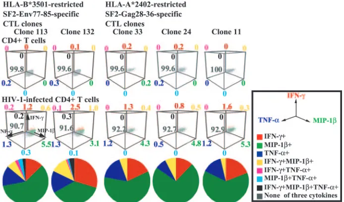
Coproduction of three cytokines by HIV-1-specific CTL clones stimulated with HIV-1-infected CD4ⴙT cells. Nef-me-diated down-regulation of HLA class I critically affects the ability of HIV-1-specific CTLs to kill HIV-1-infected cells; this could be the result of a lowered frequency of cytokine-pro-ducing cells after stimulation with HIV-1-infected CD4⫹ T cells. Indeed, our previous study showed that the lower fre-quency of HIV-1-specific CTL clones produced fewer cyto-kines by stimulation with Nef⫹HIV-1-infected CD4⫹T cells than by stimulation with Nef⫺HIV-1-infected CD4⫹ T cells (22). We investigated the coproduction of the three cytokines by HIV-1-specific CTL clones after stimulation with Nef⫹ HIV-1-infected CD4⫹T cells. Approximately 7 to 9% of the cells in five clones produced cytokines after stimulation with Nef⫹ HIV-1-infected CD4⫹ T cells (Fig. 5). Most of these cytokine-secreting cells produced only a single cytokine but were able to produce any one of the three. Thus, the results indicate that after stimulation with HIV-1-infected CD4⫹T cells, HIV-1-specific CTL clones can produce various kinds of
Clone 11 96.0 0.0 19.2 80.7 0.1
Env830-837
Clone 1 99.5 0.0 13.0 86.0 1.0
Clone 2 99.7 0.0 46.1 53.9 0.0
Clone 3 99.7 0.1 8.9 89.8 1.2
Clone 4 99.2 0.0 0.2 99.8 0.0
on November 8, 2019 by guest
http://jvi.asm.org/
[image:4.585.40.543.81.252.2]cytokines but that each CTL clone is able to produce only a single cytokine. Heterogeneity in the production of these cy-tokines among CTL clones of the same specificity is barely noticeable.
Cytokine production of freshly isolated HIV-1-specific CD8ⴙT cells stimulated with HIV-1-infected CD4ⴙT cells.
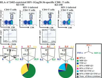
[image:5.585.306.532.66.329.2]The cytokine production results for HIV-1-specific CTL clones stimulated with HIV-1-infected CD4⫹T cells suggest the pos-sibility that when HIV-1-specific CD8⫹T cells recognize HIV-1-infected CD4⫹ T cells in HIV-1-infected individuals, they produce various combinations of cytokines but each of them produces only a single cytokine. To clarify the accuracy of this hypothetical picture, we performed ex vivo analysis of the cytokine production of HIV-1 Gag28-36-specific CD8⫹T cells after stimulation with Nef⫹HIV-1-infected CD4⫹T cells (Fig. 6). We isolated CD8⫹T cells from fresh PBMC of two HIV-1-infected individuals (KI-144 and KI-158). Analysis using a Gag28-36-specific HLA-A*2402 tetramer as well as anti-CD27 and anti-CD28 MAbs showed that approximately 0.2 to 0.3% of CD8⫹T cells were Gag28-36-specific CD8⫹T cells and that the Gag28-36-specific CD8⫹T cells predominantly have either a CD27⫹ CD28⫺ memory/effector phenotype or a CD27⫹ CD28⫹memory phenotype (Fig. 6). We then stimulated the CD8⫹T cells with NL-432-infected CD4⫹T cells. Approximately FIG. 3. Coexpression of three cytokines in HIV-1-specific CTL
clones responding to epitope peptide-pulsed cells. (A) Env77-85-spe-cific CTL clones. Intracellular cytokine production of three SF2-Env77-85-specific CTL clones was measured 6 h after stimulation with C1R-B*3501 cells prepulsed with the corresponding peptides (1,000 nM and 10 nM). Intracellular staining for the three cytokines IFN-␥, MIP-1, and TNF-␣was simultaneously carried out using three MAbs specific for these cytokines, and then the expression of these cytokines was analyzed by flow cytometry. (B) Gag28-36-specific CTL clones. Intracellular cytokine production of three SF2-Gag28-36-specific, HLA-A*2402-restricted CTL clones (clones 33, 24, and 11) was tested 6 h after stimulation with C1R-A*2402 cells prepulsed with the cor-responding peptides (1,000 nM and 100 nM). (C) Env830-837-specific CTL clones. Intracellular cytokine production of four SF2-Env830-837-specific, HLA-A*3303-restricted CTL clones (clones 1, 2, 3, and 4) was tested 6 h after stimulation with C1R-A*3303 cells prepulsed with the corresponding peptides (1,000 nM and 100 nM). Intracellular staining for the three cytokines IFN-␥, MIP-1, and TNF-␣was simul-taneously carried out using the three MAbs specific for these cyto-kines, and then the expression of these cytokines was analyzed by flow cytometry. Fluorescence-activated cell sorter data were analyzed with Paint-A-GatePRO(BD Biosciences). The frequencies of cells express-ing these cytokines are shown as percentages of the total number of cells. Cells expressing these cytokines are shown in a 3-dimensional presentation as follows: none of the three cytokines (gray), IFN-␥only (red), MIP-1only (green), TNF-␣ only (blue), IFN-␥and MIP-1 (yellow), IFN-␥and TNF-␣(violet), MIP-1and TNF-␣(cyan), and all three cytokines (black).
FIG. 4. Kinetics of multiple cytokine expression in the HIV-1-spe-cific CTL clones corresponding to epitope peptide stimulation. The frequency of CTL clones expressing a single cytokine, two cytokines, or all three cytokines was measured 6 h after stimulation with cells pre-pulsed with the corresponding peptide at different concentrations as follows: the frequency of cells expressing a single cytokine was the sum of the frequency of cells expressing IFN-␥only, MIP-1only, and TNF-␣ only in the total CD8⫹ cells, while the frequency of cells expressing two cytokines was the sum of cells expressing IFN-␥and MIP-1, IFN-␥and TNF-␣, and MIP-1and TNF-␣in total CD8⫹ cells.
on November 8, 2019 by guest
http://jvi.asm.org/
[image:5.585.52.272.67.477.2]30 and 10% of Gag28-36-specific CD8⫹T cells produced at least one cytokine in PBMC from two different HIV-1-infected individuals (Fig. 6). The frequency of cytokine-producing cells was much higher in freshly isolated Gag28-36-specific CD8⫹T cells than in Gag28-36-specific CTL clones. KI-144 cells pre-dominantly produced only MIP-1or both MIP-1and IFN-␥, while KI-158 cells predominantly produced only MIP-1 or both MIP-1and TNF-␣. This is in contrast to the finding that Gag28-36-specific CTL clones produced a single cytokine: either MIP-1, IFN-␥, or TNF-␣.
DISCUSSION
In the present study, we employed three kinds of HIV-1-specific CTL clones of the same HIV-1-specificity from the same individual. These CTL clones mostly exhibited an effector phe-notype (CD27⫺CD28⫺CD45RA⫺) and strong cytolytic activ-ity. The HIV-1-specific CTL clones exhibited functional heter-ogeneity in the production of three cytokines in clones of the same specificity as well as of different specificities, indicating that HIV-1-specific effector CD8⫹T cells, even when of the same specificity, display functional heterogeneity in cytokine production. The mechanism for this heterogeneity of function, however, is still unclear. One possibility is that these CTL clones carry different T-cell receptors (TCR), with the differ-ence in receptor signaling triggering the functional heteroge-neity, although it is not understood how different activation of TCR influences cytokine production. The fact that cells ex-pressing different cytokine production patterns do in any event exist in each clone tends to exclude this hypothesis but does support the idea that the cytokine production pattern is deter-mined by various combinations of interacting factors, such as
certain specific characteristics of the T cells, including the usage and expression level of TCR, the expression level of accessory molecules, and the relative activation status of the T cells.
Cytokine production by HIV-1-specific CTL clones stimu-lated with HIV-1-infected CD4⫹T cells reflects the response of HIV-1-specific CTLs in vivo much better than cytokine production by such clones stimulated with peptide-pulsed cells. The analysis of HIV-1-specific CTL clones stimulated with HIV-1-infected CD4⫹ T cells revealed that only 7 to 9% of cells in each CTL clone produce one of the cytokines IFN-␥, TNF-␣, and MIP-1, suggesting limited cytokine production by HIV-1-specific effector CD8⫹T cells carrying the CD27⫺ CD28⫺phenotype in vivo. This limited cytokine production may be explained by either or both of two factors: (i) the amount of HLA class I–virus peptide complexes on virus-in-fected cells is much smaller than that on peptide-pulsed cells; (ii) HLA class I–HIV-1 peptide complexes are down-regulated on the surfaces of HIV-1-infected cells. Previous studies have demonstrated that HIV-1-specific CTL clones may partially suppress HIV-1 replication, although these cells hardly kill HIV-1-infected CD4⫹ T cells because of the Nef-mediated down-regulation of the HLA-A and -B molecules (20). The partial suppression may be explained by the limited cytokine production of 1-specific CTL clones stimulated with HIV-1-infected CD4⫹T cells. Thus, it is hypothesized that cytokines produced by HIV-1-specific CD8⫹T cells play an important role in the suppression of HIV-1-replication.
[image:6.585.121.468.68.272.2]Heterogeneity in cytokine production between CTL clones of the same specificity as well as of different specificities was not found when the CTL clones were stimulated with HIV-1-infected CD4⫹T cells. This may be explained by the fact that FIG. 5. Coexpression of all three cytokines in HIV-1-specific CTL clones responding to HIV-1-infected CD4⫹T cells. Intracellular cytokine production of two SF2-Env77-85-specific CTL clones and three Gag28-36-specific CTL clones was measured 6 h after stimulation with CD4⫹T cells infected with the Nef⫹HIV-1 clone NL-432 or NL-432gagHXB2. Intracellular staining for the three cytokines IFN-␥, MIP-1, and TNF-␣was simultaneously carried out using the three MAbs specific for these cytokines, and then the expression of these cytokines was analyzed by flow cytometry. The frequency of cells expressing these cytokines is shown as a percentage of the total number of cells in a 3-dimensional presentation. The frequency of cells expressing these cytokines among total cytokine-producing cells is also shown as a pie chart. Cells expressing these cytokines are shown as follows: none of the three cytokines (gray), IFN-␥only (red), MIP-1only (green), TNF-␣only (blue), IFN-␥and MIP-1(yellow), IFN-␥and TNF-␣(violet), MIP-1and TNF-␣(cyan), and all three cytokines (black).
on November 8, 2019 by guest
http://jvi.asm.org/
heterogeneity in cytokine production between the CTL clones was not found when they were stimulated with cells pulsed with lower concentrations of HIV-1 peptides. Therefore, it is still unclear whether the heterogeneity in cytokine production be-tween CTL clones of the same specificity as well as of different specificities influences disease progression.
A previous study revealed that HLA-A2-restricted, HCMVpp65495-503-specific CD8⫹T cells are able to produce
various combinations of IFN-␥, TNF-␣, and IL-2 after PBMC from healthy individuals are stimulated with HCMVpp65495-503
peptide, suggesting that the virus-specific CD8⫹T cells possess a functional heterogeneity of cytokine production in vivo (18). Since the HCMV-specific CD8⫹ T cells are heterogeneous with regard to the surface markers CD45RO, CD45RA, CD27, CD28, CD57, and CD62L (18, 21), it is thought that they include various population types, ranging from memory to effector T cells. Therefore, the heterogeneity at maturation of CD8⫹T cells may reflect the capacity for functional heteroge-neity in cytokine production. Our recent study revealed that CD8⫹ T cells with effector phenotypes (CD27⫺ CD28⫺ CD45RA⫹/⫺) or memory/effector phenotypes (CD27low
CD28⫺CD45RA⫹/⫺) can produce IFN-␥after stimulation of
CD8⫹ T cells with an anti-CD3 MAb (23), indicating that effector and memory/effector CD8⫹T cells have a more pro-nounced ability to produce IFN-␥than memory CD8⫹T cells. Thus, the heterogeneity in maturation or differentiation of CD8⫹T cells reflects the functional heterogeneity in cytokine production capacity seen in vivo.
Freshly isolated Gag28-36-specific CD8⫹T cells produced more cytokines than CTL clones with the same specificity. Since these Gag28-36-specific CD8⫹T cells carried either a CD27⫹CD28⫺memory/effector or a CD27⫹CD28⫹memory phenotype, it is likely that they are less mature than the CTL clones. These results suggest that HIV-1-specific CD8⫹T cells with a CD27⫹CD28⫺or CD27⫹CD28⫹phenotype can pro-duce more cytokines than those with an effector phenotype. Approximately 20 to 30% of cytokine-producing CD8⫹T cells produced two or three cytokines, whereas most CTL clones of the same specificity produced a single cytokine. This indicates the ability of HIV-1-specific memory and memory/effector CD8⫹T cells to produce multiple cytokines when they recog-nize HIV-1-infected cells. Various differentiation ranges of HIV-1-specific CD8⫹ T cells, from memory to effector, are found in PBMC from 1-infected individuals (5, 22). HIV-FIG. 6. Coexpression of three cytokines in freshly isolated HIVGag28-36-specific CD8⫹T cells responding to HIV-1-infected CD4⫹T cells. Intracellular cytokine production of Gag28-36-specific CD8⫹T cells was measured 6 h after stimulation with CD4⫹T cells infected with the Nef⫹ HIV-1 clone NL-432gagHXB2. Intracellular staining for the three cytokines IFN-␥, MIP-1, and TNF-␣was simultaneously carried out using the three MAbs specific for these cytokines, and then the expression of these cytokines was analyzed by flow cytometry. The CD8⫹tetramer-positive cells are gated to determine both the frequency of cells expressing these cytokines and the CD27 and CD28 expression. The frequency is shown as a percentage of the total number of cells in a 3-dimensional presentation. The frequency of cells expressing these cytokines among total cytokine-producing cells is also shown in a pie chart. Cells expressing these cytokines are shown as follows: none of the three cytokines (gray), IFN-␥only (red), MIP-1only (green), TNF-␣only (blue), IFN-␥and MIP-1(yellow), IFN-␥and TNF-␣(violet), MIP-1and TNF-␣(cyan), and all three cytokines (black).
on November 8, 2019 by guest
http://jvi.asm.org/
[image:7.585.98.491.67.370.2]In summary, HIV-1-specific CTL clones for the most part produced a single cytokine and did not exhibit heterogeneity of cytokine production among clones after stimulation with HIV-1-infected CD4⫹T cells, although they exhibited multiple cy-tokine production and functional heterogeneity of cycy-tokine production between clones after stimulation with HIV-1 pep-tide-pulsed cells. Freshly isolated HIV-1-specific CD8⫹T cells with an effector/memory or memory phenotype produced much greater amounts of the cytokines than CTL clones with the same epitope specificity after stimulation with HIV-1-in-fected CD4⫹ T cells. HIV-1-specific CD8⫹ T cells with an effector/memory or memory phenotype might directly or indi-rectly play a crucial role in the eradication of HIV-1 via the cytokines secreted from these T cells in HIV-1-infected indi-viduals.
ACKNOWLEDGMENTS
We thank Sachiko Sakai for secretarial assistance.
This research was supported by Grants-in Aid for Scientific Re-search from the Ministry of Education, Science, Sports, and Culture (175172) and the Ministry of Health of the government of Japan and by a grant from the Japan Health Science Foundation.
REFERENCES
1.Akari, H., S. Arold, T. Fukumori, T. Okazaki, K. Strebel, and A. Adachi.
2000. Nef-induced major histocompatibility complex class I down-regulation is functionally dissociated from its virion incorporation, enhancement of viral infectivity, and CD4 down-regulation. J. Virol.74:2907–2912.
2.Alkhatib, G., C. Combadiere, C. C. Broder, Y. Feng, P. E. Kennedy, P. M. Murphy, and E. A. Berger.1996. CC CKR5: a RANTES, MIP-1␣, MIP-1
receptor as a fusion cofactor for macrophage-tropic HIV-1. Science272:
1955–1958.
3.Altman, J. D., P. A. H. Moss, P. J. R. Goulder, D. H. Barouch, M. G. McHeyzer-Williams, J. I. Bell, A. J. McMichael, and M. M. Davis.1996. Phenotype analysis of antigen-specific T lymphocytes. Science274:94–96. 4.Appay, V., D. F. Nixon, S. M. Donahoe, G. M. A. Gillespie, T. Dong, A. King,
G. S. Ogg, H. M. L. Spiegel, C. Conlon, C. A. Spina, D. V. Havlir, D. D. Richman, A. Water, P. Easterbrook, A. J. McMichael, and S. L. Rowland-Fones.2000. HIV-specific CD8⫹T cells produce antiviral cytokines but are impaired in cytolytic function. J. Exp. Med.192:63–75.
5.Appay, V., P. R. Dunbar, M. Callan, P. Klenerman, G. M. Gillespie, L. Papagno, G. S. Ogg, A. King, F. Lechner, C. A. Spina, S. Little, D. V. Havlir, D. D. Richman, N. Gruener, G. Page, A. Waters, P. Easterbrool, M. Salio, V. Cerundolo, A. J. McMichael, and S. L. Rowland-Jones.2002. Memory CD8⫹ T cells vary in differentiation phenotype in different persistent virus infec-tions. Nat. Med.8:379–385.
6.Cocchi, F., A. L. DeVico, A. Garzino-Demo, S. K. Arya, R. C. Gallo, and P.
11.Reference deleted.
12.Reference deleted.
13.Hossain, M. S., H. Tomiyama, T. Inagawa, B. Wriwanthana, S. Oka, and M. Takiguchi.2001. HLA-A*3303-restricted cytotoxic T lymphocyte recognition for novel epitopes derived from the highly variable region of the HIV-1 Env protein. AIDS15:2199–2201.
14.Ikeda-Moore, Y., H. Tomiyama, M. Ibe, S. Oka, K. Miwa, Y. Kaneko, and M. Takiguchi.1998. Identification of a novel HLA-A24-restricted cytotoxic T-lymphocyte epitope derived from HIV-1 Gag protein. AIDS12:2073–2074. 15.Kurane, I., A. Meager, and A. E. Francis.1989. Dengue virus-specific human T cell clones: serotype crossreactive proliferation, interferon-␥production, and cytotoxic activity. J. Exp. Med.170:763–775.
16.Kuwano, K., T. Kawashima, and S. Arai.1993. Antiviral effect of TNF-␣and IFN-␥secreted from a CD8⫹influenza virus-specific CTL clone. Viral Im-munol.6:1–11.
17.Levy, J. A., C. E. Mackewicx, and E. Barker.1996. Controlling HIV patho-genesis: the role of the noncytotoxic anti-HIV response of CD8⫹T cells. Immunol. Today17:217–224.
18.Liu, C. C., C. M. Walsh, and J. D. Young.1995. Perforin: structure and function. Immunol. Today16:194–201.
19.Okamoto, M., M. Makino, I. Kitajima, I. Maruyama, and M. Baba.1997. HIV-1-infected myelomonocytic cells are resistant to Fas-mediated apopto-sis: effect of tumor necrosis factor-alpha on their Fas expression and apo-ptosis. Med. Microbiol. Immunol.186:11–17.
20.Sandberg, J. K., N. M. Fast, and D. F. Nixon.2001. Functional heterogeneity of cytokines and cytolytic effector molecules in human CD8⫹T lymphocytes. J. Immunol.167:181–187.
21.Shiga, H., T. Shioda, H. Tomiyama, Y. Takamiya, S. Oka, S. Kimura, Y. Yamaguchi, T. Gojoubori, H. G. Rammensee, K. Miwa, and M. Takiguchi.
1996. Identification of multiple HIV-1 cytotoxic T-cell epitopes presented by human leukocyte antigen B35 molecules. AIDS10:1075–1083.
22.Tomiyama, H., H. Akari, A. Adachi, and M. Takiguchi.2002. Different effects of Nef-mediated HLA class I down-regulation on human immunode-ficiency virus type 1-specific CD8⫹T-cell cytolytic activity and cytokine production. J. Virol.76:7535–7543.
23.Tomiyama, H., H. Takata, T. Matsuda, and M. Takiguchi.2004. Phenotypic classification of human CD8⫹T cells reflecting their function: inverse cor-relation between quantitative expression of CD27 and cytotoxic effector function. Eur. J. Immunol.34:999–1010.
24.Tomiyama, H., S. Oka, G. S. Ogg, S. Ida, A. J. McMichael, and M. Takigu-chi.2000. Expansion of HIV-1-specific CD28⫺CD45RA⫺CD8⫹T cells in chronically HIV-1 infected individuals. AIDS14:2049–2051.
25.Tomiyama, H., T. Matsuda, and M. Takiguchi. 2002. Differentiation of human CD8⫹T cells from a memory to memory/effector phenotype. J. Im-munol.168:5538–5550.
26.Trapani, J. A., V. R. Sutton, and M. J. Smyth.1999. CTL granule: evolution of vesicles essential for combating virus infections. Immunol. Today20:351– 356.
27.Vegh, Z., P. Wang, F. Vanky, and E. Klein.1993. Increased expression of MHC class I molecules on human cells after short time IFN-␥treatment. Mol. Immunol.30:849–854.
28.Yang, O. O., S. A. Kalams, A. Trocha, H. Cao, A. Luster, R. P. Johnson, and B. D. Walker.1997. Suppression of human immunodeficiency virus type 1 replication by CD8⫹cells: evidence for HLA class-I-restricted triggering of cytolytic and noncytolytic mechanisms. J. Virol.71:3120–3128.