Hepatitis C Virus-Specific T Cell Receptor
mRNA-Engineered Human T Cells:
Impact of Antigen Specificity on
Functional Properties
Anangi Balasiddaiah,a,b*Haleh Davanian,aSoo Aleman,cAnna Pasetto,d Lars Frelin,bMatti Sällberg,bVolker Lohmann,eSarene Koh,g,h
Antonio Bertoletti,f,gMargaret Chena,b
Departments of Dental Medicine,aLaboratory Medicine,band Medicine,cKarolinska Institutet, Stockholm, Sweden; Center for Cancer Research, National Cancer Institute, National Institutes of Health, Bethesda, Maryland, USAd; Department of Infectious Diseases, Molecular Virology, University of Heidelberg, Heidelberg, Germanye; Duke-Nus Graduate Medical School, Emerging Infectious Diseases Program, Singaporef; Singapore Institute for Clinical Sciences, Agency for Science, Technology and Research (A*STAR), Singaporeg; LION TCR Pte. Ltd., Singaporeh
ABSTRACT Therapy with genetically modified autologous T cells has shown great
promise in cancer therapy. For an efficient control of hepatitis C virus (HCV) infec-tion, cytotoxic T cells (CTL) are pivotal, but persistence of activated T cells may lead to liver toxicity. Here, anti-HCV T cell receptors (TCRs) recognizing the HCV nonstruc-tural (NS) NS3 or NS5 viral peptide target were examined by mRNA transfection of human peripheral blood lymphocytes (PBLs) derived from healthy donors as well as chronically infected HCV patients. Immunological analysis shows that while the CTLs expressing the NS5-specific TCR reduced HCV RNA replication by a noncytotoxic mechanism, the NS3-specific TCR-redirected CTLs were polyfunctional and inhibited HCV RNA replication through antigen-specific cytotoxicity. Transcriptome signatures from these two types of CTL responses revealed uniquely expressed gene clusters upon encountering hepatoma target cells presenting endogenously expressed HCV proteins. The NS3 TCR induced a rapid expression of apoptotic signaling pathways and formation of embryonic gene clusters, whereas the NS5A TCR activation in-duced extended proliferative and metabolic pathways as the HCV target cells sur-vived. Our results provide detailed insights into basic HCV T cell immunology and have clinical relevance for redirecting T cells to target virally infected hepatoma cells.
IMPORTANCE Due to the protective ability of HCV-specific T cells and the
hepato-toxic potential that they possess, there is a great need for the understanding of the functional aspects of HCV-specific T cells. To circumvent the low level of precursor frequency in patients, we engineered primary CD8⫹T cells by mRNA TCR vectors to
confer HCV specificity to new T cells. HCV TCRs that differ in antigen specificity and polyfunctionality were examined. mRNA TCR engineering of peripheral blood lym-phocytes from healthy donors or chronically infected HCV patients resulted in strik-ingly high levels of HCV TCR expression and HCV-specific responses. While a cytotox-icity response from a polyfunctional T cell activation caused hepatotoxcytotox-icity and the rapid induction of apoptotic signaling pathways, the noncytotoxic T cell activation showed extended proliferative, metabolic pathways and persistence of HCV target cells. Our results provide detailed insights into basic HCV T cell immunology and have clinical relevance for immune protection of HCV-associated diseases.
KEYWORDS HCV-specific TCRs, TCR-redirected T cell therapy, hepatitis C virus
Received4 January 2017Accepted7 February 2017
Accepted manuscript posted online22 February 2017
CitationBalasiddaiah A, Davanian H, Aleman S, Pasetto A, Frelin L, Sällberg M, Lohmann V, Koh S, Bertoletti A, Chen M. 2017. Hepatitis C virus-specific T cell receptor mRNA-engineered human T cells: impact of antigen specificity on functional properties. J Virol 91:e00010-17.
https://doi.org/10.1128/JVI.00010-17.
EditorJ.-H. James Ou, University of Southern California
Copyright© 2017 American Society for Microbiology.All Rights Reserved. Address correspondence to Margaret Chen, [email protected].
*Present address: Anangi Balasiddaiah, Institute for Cancer Research, Oslo University Hospital, Oslo, Norway.
crossm
on November 7, 2019 by guest
http://jvi.asm.org/
H
epatitis C virus (HCV) is a major cause of liver cirrhosis and hepatocellular carci-noma (HCC), and approximately half a million people die each year from HCV-related liver diseases. Although new directly acting antiviral (DAA) drugs are revolu-tionary and can cure the majority of these infections, problems associated with drug resistance, compliance, medical, or regional political factors restrict the access to these drugs for the majority of infected individuals worldwide (1). In addition, despite DAA treatment, HCV patients previously treated for HCC still have a high risk of liver cancer, and the occurrence of liver cancer is unfortunately not reduced in effectively treated cirrhotic HCV patients (2, 3).Even though the majority of HCV-infected individuals develop chronic infection, it is estimated that 15 to 45% of infected people spontaneously clear the infection (4). The general consensus is that HCV elimination is associated with strong and sustained T cell responses that target multiple epitopes within the different HCV viral proteins (5–11). While CD4⫹T cells are central regulators, virus-specific CD8⫹T cells primarily function
as the key effectors. Several lines of evidence support the important role of virus-specific T cells in controlling HCV infection, as there is a clear temporal association between the onset of peripheral and intrahepatic virus-specific T cell responses and HCV clearance (5, 7, 9–13). Elegantex vivostudies of patients’ HCV-specific CD8⫹T cell
effector functions revealed that HCV-specific CD8⫹T cells exert strong antiviral effects
primarily by gamma interferon (IFN-␥) but only to a lower extent by cytolytic effector functions (14).
Despite the recruitment of HCV-specific T cells to the infected liver, the failure at the T cell level remains a great challenge for the effective control of HCV infection, as it renders the virus persistent in the majority of infected individuals (15). Several studies illustrated that a protective T cell response has signatures that feature highly polyfunc-tional HCV-specific CD8⫹ T cells, which contribute to the substantial breadth and
height of magnitude of responses to multiple viral determinants, in particular, the viral nonstructural (NS) proteins (16, 17). The impact of polyfunctional T cells on protective immunity is not restricted to HCV but is commonly shared by diseases caused by other infectious pathogens, such as HIV, yellow fever virus, Ebola virus, cytomegaloviruses, and mycobacteria, as well as by cancer (18, 19). However, in spite of its significance, the transcriptional mechanisms underlying antigen-specific T cell polyfunctionality are not completed understood.
We have previously identified HCV-specific T cell receptors (TCRs) in DNA-vaccinated HLA-A2 transgenic mice recognizing two frequently reported HLA-A2-restricted HCV epitopes (NS31073and NS51992) found in HCV patients who resolve their infection (20, 21). Among multiple cloned HCV-reactive TCR candidates generated by this approach, the NS3-H4, NS3-F8, NS5-19, and NS5-69 TCRs were selected for (i) their ability to respond to HCV NS31073 or NS51992 peptides in a CD8-independent manner with CD8-negative BW thymoma partners and (ii) their affinity to the respective HCV peptide/MHC pentamers (20, 21). In these previous studies, retroviral TCR gene transfer was used to study these TCRs, in which transduction efficiency varied substantially and was not an optimal approach for global transcriptome studies.
We report here that the syntheticin vitro-transcribed TCR mRNA can be used to redirect human T cells to transfer anti-HCV cytotoxic T lymphocyte (CTL) functions to human T cells of peripheral blood lymphocytes (PBLs), as mRNA transfection efficiency was as consistently high, e.g.,⬎90%, in T cells derived from healthy blood donors and HCV-infected individuals. This gave us a prerequisite to examining the global transcrip-tional signatures of the TCR-redirected CTLs responding to their cognate HCV RNA⫹
hepatoma target cells.
RESULTS
HCV TCR mRNA redirection of healthy human T cells.In vitro-transcribed mRNA encoding the NS31073–1081-specific TCRs (NS3-H4 and NS3-F8) and NS5A1992–2000 -specific TCRs (NS5-19 and NS5-69) were introduced into activated T cells of two HLA-A2⫹healthy donors by electroporation. Because these are murine TCRs originating
May 2017 Volume 91 Issue 9 e00010-17 jvi.asm.org 2
on November 7, 2019 by guest
http://jvi.asm.org/
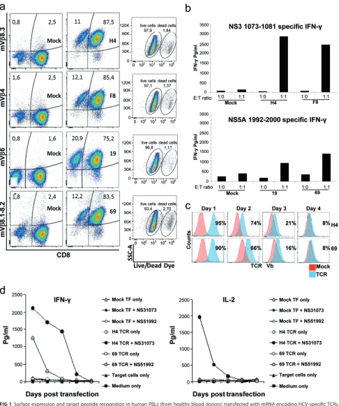
from HLA-A2-transgenic mice, they can be distinguished from human TCRs with antibodies that bind to their mouse Vchain (e.g., for NS3-H4, V8.3; for NS3-F8, V 4; for NS5-19, V 6; for NS5-69, V8.1-8.2) (Fig. 1a). Moreover, these antibodies also gave staining results comparable to those of the respective HCV peptide/major histo-compatibility complex (MHC) pentamer for each TCR (20, 21). Here we found that the cell viability after mRNA electroporation was around 95% (Fig. 1a). The surface expres-sion of the introduced TCR was detected in⬎90% of live CD3⫹T cells on the day after electroporation (Fig. 1a; Table 1), and the majority of TCR⫹cells were CD8⫹T cells with background staining (mock-transfected or TCR-transfected cells stained with isotype controls) at 1 to 3% (Fig. 1a and data not shown). HCV-specific responses, detected following coculture with NS51992–2000 or NS31073–1081 peptide-loaded T2 cells, con-firmed that NS3 TCR-redirected effector cells were superior IFN-␥producers to NS5-TCRs (Fig. 1b), which is consistent with the results obtained with retrovirus-modified T cells (21). Cell surface expression of the TCRs lasted for 3 to 4 days (Fig. 1c) and concurred with their target-specific functionality (Fig. 1d). Moreover, NS3-H4-redirected T cells secrete both IFN-␥and interleukin-2 (IL-2), whereas those of NS5-69 produce mainly IFN-␥. Because NS3-H4 and NS3-F8 (NS31073–1081specific) as well as NS5-19 and NS5-69 (NS5A1992–2000 specific) share antigen specificity and similar functional re-sponses, the NS3-H4 and the NS5-69 TCRs were chosen for further comparisons.
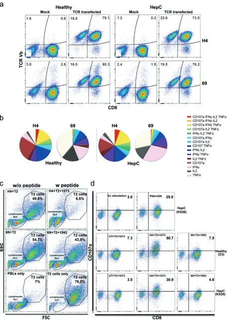
HCV TCR mRNA redirection of HCV patients’ T cells and comparative functional analysis with healthy human T cells.To examine if PBLs from chronic HCV patients can be redirected with TCR mRNAs with the same efficiency, HLA-A2⫹ PBLs from chronic HCV patients (n⫽10) and healthy donors (n ⫽9) were tested as described above. Our results showed that irrespective of the liver disease stage, HCV patients’ PBLs were as successfully redirected with HCV TCR as in healthy donors (around 90% of CD3⫹T cells), and the vast majority of CD8⫹T cells showed surface expression of the respective HCV TCRs (Fig. 2a; Table 1). Moreover, NS3-H4-redirected T cells from HCV-infected patients and healthy donors effectively eliminated T2 target cells loaded with NS3-1073 peptide as early as 5 h after the coincubation (Fig. 2c), while NS5-69-redirected cells did not. Given that T cell polyfunctionality is a hallmark of pathogen control, the two TCRs were compared at the single cell level by intracellular cytokine staining (ICS). We found that a large proportion of NS3-H4-redirected T cells, but not NS5-69-redirected cells, secreted more than one cytokine, e.g., IFN-␥, tumor necrosis factor alpha (TNF-␣), or IL-2, and had upregulated the surface expression of CD107␣ (Fig. 2b and d). Surface expression of HCV TCR was also found in the CD8-negative cell population, which showed moderate proportions of polyfunctional cells upon stimu-lation (Fig. 3). In conclusion, the data demonstrate that TCR mRNA delivery is highly effective in producing HCV TCR-redirected T cells that resemble polyfunctional/cyto-toxic T cells (NS3-H4) and monofunctional/noncytopolyfunctional/cyto-toxic T cells (NS5-69).
Antiviral efficacy and hepatotoxic potential of TCR mRNA-redirected T cells. The ability of TCR mRNA-redirected T cells to recognize endogenously processed HCV antigen was examined next. To provide a more biologically relevant condition that allows examination of the hepatotoxic potentials, the redirected T cells were coincu-bated with hepatoma target cells Huh7A2HCVRep, human hepatoma cells that persis-tently replicate the HCV genotype 1 subgenomic RNA and express HLA-A2 (22). As shown in Fig. 3a, both NS3-H4 TCR- and NS5-69 TCR-redirected T cells responded to the Huh7A2HCVRepcells by secreting IFN-␥, and these results confirmed the observations with T2 cells. Next, the hepatotoxic potential of the TCR-redirected T cells was exam-ined: the HuhA2HCVReptarget cell number was determined and the aspartate amino-transferase (AST) enzyme release was measured over 5 days of coincubation. We found a steady increase of AST, and the considerable cell detachment in the NS3-H4 T cell and HuhA2HCVReptarget cell coculture indicated a rapid hepatocellular cell injury (Fig. 4b). On the other hand, the NS5-69 T cells spared the target cells and allowed them to grow to confluence (Fig. 4b). Next, the antiviral properties of TCR mRNA-redirected T cells were tested using PBLs from healthy donors (n ⫽ 9) and HCV patients (n ⫽ 10) in overnight cocultures with Huh7A2HCVRepcells or the control Huh7 HCVRepcells lacking
on November 7, 2019 by guest
http://jvi.asm.org/
FIG 1Surface expression and target peptide recognition in human PBLs (from healthy blood donors) transfected with mRNA encoding HCV-specific TCRs. (a) Surface staining of TCRs specific for HCV NS31073–1081(H4 and F8) and NS51992–2000(19 and 69) by their Vantibody (H4, mV8.3; F8, mV4; 19, mV6; 69, mV8.1-8.2) 1 day posttransfection. Mock-transfected PBLs served as negative controls. The percentages of indicated TCR mV-expressing cells were gated on live CD3⫹T cells that were counterstained with Live/Dead Aqua fixable reagent, anti-CD3, anti-CD8, and the indicated mVantibody and
determined by FACS. (b) HCV-specific IFN-␥secretion following stimulation with T2 cells loaded with their cognate peptide (1g/ml) at the indicated E/T ratios. (c) Transient expression of HCV TCRs (H4, 69) in human PBLs following the mRNA transfection. (d) T cells redirected by the TCR H4 are capable of secreting antigen-specific (NS31073–1081) IFN-␥and IL-2, whereas TCR 69-redirected cells secrete mainly IFN-␥in response to its target peptide (NS51992–2000). Transfected T cells from healthy donors were coincubated for 16 to 18 h with T2 target cells with or without peptide from the indicated time points after the transfection, and the harvested supernatant were analyzed with cytokine ELISA. Mock-transfected PBLs were included as negative controls. Coculture experiments were done in duplicate wells and repeated two times with similar results.
May 2017 Volume 91 Issue 9 e00010-17 jvi.asm.org 4
on November 7, 2019 by guest
http://jvi.asm.org/
[image:4.585.42.528.68.650.2]TABLE 1 Clinical and viral parameters of the donors and efficiency of HCV TCR redirection with NS3-H4 and NS5-69 TCRs a Donor characteristics % of H4 TCR expression % of 69 TCR expression Overnight inhibition of HCV RNA replicon replication (%) Donor type and code Age Gender HCV genotype Viral load (IU/ml) Fibrosis stage Fibroscan score (kPa) c Transmission route Viability CD3 ⴙ CD3 ⴙ CD8 ⴙ CD3 ⴙ CD8 ⴚ Viability CD3 ⴙ CD3 ⴙ CD8 ⴙ CD3 ⴙ CD8 ⴚ H4 69 Healthy donors D1 NA b NA NA NA NA NA NA 98 98 98 97 94 97 98 95 97 76 D3 NA NA NA NA NA NA NA 96 96 96 97 93 91 91 89 93 72 BC-2 NA NA NA NA NA NA NA 97 97 97 97 98 98 99 97 97 29 BC-3 NA NA NA NA NA NA NA 98 99 100 99 98 98 98 96 99 49 BC-7 NA NA NA NA NA NA NA 90 96 97 91 93 95 95 87 100 94 BC-9 NA NA NA NA NA NA NA 99 98 98 97 99 97 98 97 98 65 BC-12 NA NA NA NA NA NA NA 98 97 98 97 98 95 95 96 99 28 BC-16 NA NA NA NA NA NA NA 93 91 93 84 92 82 84 76 99 97 BC-17 NA NA NA NA NA NA NA 98 97 96 92 98 95 91 90 99 8 Mean (SD) for group 96 (3) 97 (2) 97 (2) 95 (5) 96 (3) 94 (5) 94 (5) 91 (7) 98 (2) 58 (31) Chronic HCV patients KS21 69 F 1b 7,800,000 F0-F1 5.1 Blood transfusion 98 97 98 96 98 93 93 89 100 83 KS23 57 F 1b 3,170,000 F3 10.5 Unknown 96 96 96 96 90 97 97 97 99 78 KS25 61 M 2b 1,080,000 F4 21.6 Unknown 93 93 92 90 94 90 89 86 100 94 KS26 42 M 1a 7,290,000 F3 10.6 Blood transfusion 93 95 96 95 92 95 96 94 93 34 KS27 38 F 1b 50,400 F0-F1 7.1 Unknown 98 98 98 97 97 96 97 96 98 48 KS28 57 F 1a 8,630,000 F2 8.2 Intravenous drug use 97 97 98 97 96 96 96 96 99 58 KS32 45 F 2b 7,630,000 F0-F1 3.1 Unknown 98 97 97 95 98 96 97 94 100 66 KS35 68 F 2b 1,800,000 F1 NA Unknown 98 97 96 97 98 95 95 95 100 89 KS38 64 M 1b 8,500 F0-F1 7.3 Intravenous drug use 98 96 96 90 98 94 91 86 98 39 KS39 55 M 1b 940,000 F4 32 Blood transfusion 98 96 96 93 98 93 84 93 60 0 Mean (SD) for group 97 (2) 96 (1) 96 (2) 95 (3) 96 (3) 95 (2) 94 (4) 93 (4) 95 (12) 59 (29) aCell viability (%) and ectopic TCR expression in indicated T cell populations and antiviral efficiency against the Huh7 A2 HCV Rep target cells on day 1 after mRNA posttransfection. bNA, not available. cFibroscan scores measure liver stiffness in kilopascals.
on November 7, 2019 by guest
http://jvi.asm.org/
FIG 2TCR mRNA redirection of PBLs from chronically infected HCV patients and healthy donors. (a) HCV TCR cell surface expression on day 1 posttransfection. CD8 and mVstaining for respective TCRs is shown on live CD3-positive lymphocytes as described for Fig. 1. (b) The numbers
(Continued on next page)
May 2017 Volume 91 Issue 9 e00010-17 jvi.asm.org 6
on November 7, 2019 by guest
http://jvi.asm.org/
[image:6.585.42.498.69.712.2]HLA-A2, and the HCV replicon-driven luciferase reporter protein was quantitated. The results indicate that regardless of the age of HCV donors, HCV viral load, or liver disease status, the NS3-H4-redirected T cells strongly inhibited HCV replicon activity while the NS5-69-redirected T cells showed mainly a marginal replication inhibition (Fig. 4c; Table 1).
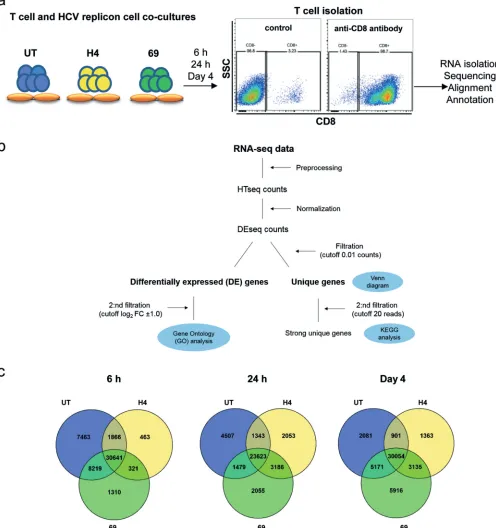
Transcriptome analysis of the TCR-redirected T cell responses against HCV replicon cells.Next, we examined the transcriptional signatures in the TCR-redirected T cells responding to HuhA2HCVReptarget cells. We chose PBLs from patient K23, as this individual had no detectable endogenous T cell responses against HCV replicon cells, and redirected T cells from this patient showed a consistent response in all experiments and was representative of the responses in the patient cohort tested. The two TCR-redirected (NS3-H4, NS5-69) T cells and undirected T cells (UT) were cocultured with HuhA2HCVReptarget cells for 6 h, 24 h, and 4 days and enriched for CD8⫹T cells by
FIG 2Legend (Continued)
of antigen-specific functions (intracellular IFN-␥, IL-2, TNF-␣, or CD107␣upregulation) in TCR mRNA-transfected CD3-gated cytokine-producing CD8⫹T cells (H4 versus 69) from healthy donors (n⫽2) versus those from hepatitis C patients (n⫽3) are summarized in pie charts. Each pie
chart represents the mean responses of the different TCRs for total CD3-gated CD8⫹T cell populations in healthy individuals and chronic HCV
disease patients. (c) Effective target-specific elimination by NS3-H4 cocultured with HCV NS31073–1081peptide-loaded T2 cells (upper panel). Representative forward scatter/side scatter (FSC/SSC) dot plots of cocultured TCR-redirected T cells (H4 in the top panels, 69 in the middle panels) cocultured with T2 cells loaded with or without the respective HCV peptides. TCR-transfected PBLs alone and T2 cells only (bottom panel) served as controls. The experiment was repeated three times using PBLs from three different HCV patient donors, all of which gave similar results. (d) Difference in degranulation capacity in NS3-H4 (H4)- and NS5-69 (69)-redirected cells. Representative dot plots gated on CD3⫹T cells and the
CD107␣surface staining in TCR-transfected T cells (healthy donor and HCV patient). All functional analyses were done 5 h after cocultivation with the target cells. PMA/ionomycin (PMA⫹ION) and mock undirected T cells were included as controls in all intracellular cytokine staining assays. FIG 3(a) Proportion of CD8⫺and CD8⫹populations of CD3-gated HCV TCR⫹live T cells. (b) Mean fluorescence intensity (MFI) of HCV TCR
Vstaining of the CD8⫹versus CD8⫺T cells; the individual points indicate the MFI of indicated HCV TCR expression in each donor (healthy
donors,n⫽8; HepC donors,n⫽9). Bars indicate the mean MFI values of the indicated HCV TCR staining in the patient group. (c) Proportions of antigen-specific cytokine-producing TCR-transfected CD3⫹CD8⫺T cells (H4 versus 69) from healthy donors versus hepatitis
C patients (Hep C).
on November 7, 2019 by guest
http://jvi.asm.org/
[image:7.585.46.473.70.386.2]FIG 4Replicon cell recognition, hepatotoxic potential, and antiviral activity of TCR-redirected T cells. (a) HCV-specific IFN-␥secretion in NS3-H4 (H4)- and NS5-69 (69)-redirected T cells from HCV patient donors (n⫽3) following 24 h of coculture with Huh7A2HCVRepcells at an E/T ratio of 10:1. (b) Hepatocellular cell injury by T cells redirected with the indicated TCRs. (Right) Percentages (left panel) of the target Huh7A2HCVRepcells that remain adherent after coincubation with the indicated T cells over 5 h of coculture. After the indicated times of cell coincubation, the suspension cells were removed and the replicon cells remaining adherent were trypsinized and counted by trypan blue exclusion. The percentages of replicon cells were determined against control target replicon cells (RepA2 only). (Right) Aspartate aminotransferase (AST) enzyme release from Huh7A2HCVRepcells after coincubation with T cells transfected with indicated TCR. Means and standard deviations (SD) are shown. nd, not detected. (c) Antiviral potential of redirected T cells from healthy donors (n⫽9) and HCV patients (n⫽10). The HCV replicon luciferase activity is measured in Huh7A2HCVRepand Huh7 HCVRepcells after 24 h of coculture with the indicated T cells at an E/T ratio of 0.5:1 in duplicate wells. Each dot represents one individual, and the bar indicates the mean value for the group. Mock undirected T cells (UT) were included as controls. The Mann-Whitney U test was used for group comparisons (*,P⬍0.05;**,P⬍0.01;***,P⬍0.001).
May 2017 Volume 91 Issue 9 e00010-17 jvi.asm.org 8
on November 7, 2019 by guest
http://jvi.asm.org/
[image:8.585.39.472.75.619.2]microbeads (Fig. 5a). TCR transfection efficiency for both TCRs was⬎95%, and 1.5⫻106 CD3⫹CD8⫹cells (⬎98% purity) were obtained and stored from each condition and
time point (Table 1 and Fig. 5a). Total RNAs were analyzed by paired-end RNA sequencing as shown in the flowchart in Fig. 5b. We obtained about 346 million mapped “reads” and found a total of 57,780 genes (both protein-coding and noncoding genes) expressed in the isolated CD8⫹ T cells. Before pursuing the analysis of the
FIG 5Transcriptome profiling by RNA-seq in NS3-H4-directed, NS5-69-directed, and undirected T cells after coculture with HCV replicon cells. (a) Schematic flow of cell and RNA isolation of TCR-redirected CD8⫹T cells; (b) sequence annotation and analysis; (c) Venn diagrams of the gene distribution in NS3-H4 (H4)- and
NS5-69 (69)-redirected and undirected (UT) T cells following coculture with HCV replicon cells at the indicated time points. The intersections of circles refer to mRNA transcripts that are shared among the different conditions.
on November 7, 2019 by guest
http://jvi.asm.org/
[image:9.585.44.540.77.605.2]sequencing data, a cutoff level of 0.01 for read counts was applied to exclude spurious transcripts.
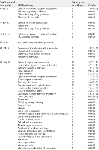
The uniquely expressed genes in NS3-H4 versus NS5-69 T cell responses.The distributions of the unique and shared expressed genes for each T cell condition and time point are illustrated by the Venn diagrams shown in Fig. 5c. They show that 6 h after the encounter with HuhA2HCVReptarget cells, 7,463 genes were uniquely ex-pressed for the mock undirected T cells (UT) whereas only 463 and 1,310 genes were unique for NS3-H4 and NS5-69, respectively. Furthermore, NS3-H4 had the lowest number of unique genes at the three time points. To further examine the protein-coding genes, we applied another threshold of 20 read counts per feature to filter away weakly expressed genes. KEGG enrichment analyses by WebGestalt were performed to study the unique protein-coding genes expressed in each T cell response. As shown in Table 2, the most significant (P ⬍ 0.01) enriched KEGG signaling pathways among uniquely expressed genes in NS3-H4 T cells were cytokine-cytokine receptor interac-tions, JAK-Stat signaling, and chemokine signaling pathways at 6 h following HCV replicon cell coculture. While the target cells were eliminated in the 24-h coculture of NS3-H4 (Fig. 4b), the responses shifted to engage steroid hormone biosynthesis, ribosome and chemokine signaling pathways, and then cytokine-cytokine receptor interaction and rheumatoid arthritis-like pathways on day 4 (Table 2). The NS5-69 T cell response, on the other hand, showed a diffuse and variable gene pathway usage: the unique genes expressed at 6 h following target encounter lacked clear enrichment of any signaling pathway, while at 24 h in the presence of confluent HCV replicon targets, the most relevant enriched pathways seen were complement/coagulation cascade, metabolic, and infection pathways, which extended to more than 30 enriched path-ways on day 4. The latter include systemic lupus erythematosus (SLE)-like, calcium signaling, adhesion, tight junction, cytoskeleton, cytokine-cytokine receptor interaction, and leukocyte migration as well as transforming growth factor beta (TGF-) signaling pathways (Table 2). All the uniquely expressed genes regulated within the significantly enriched KEGG signaling pathways for both TCRs are presented in Table S1 in the supplemental material.
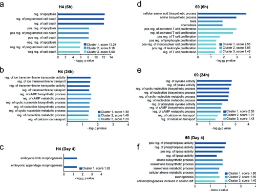
DE genes of NS3-H4 and NS5-69 T cell responses—the common genes. Differ-ential gene expression between undirected T cells (UT) and NS3-H4- and NS5-69-redirected T cells was analyzed using read counts for each gene with the DeSeq package, developed for differential expression analysis of high-throughput RNA se-quencing (RNA-seq) data. A cutoff level at ⫾1.0 log2-fold change was set for the differentially expressed (DE) genes before the downstream analysis. A complete list of all DE genes in each TCR in log2-fold change are listed in Tables S2 and S3 in the supplemental material. The upregulated genes in NS3-H4- and NS5-69-redirected T cells relative to the UT control were analyzed with the functional annotation clustering tool in the Database for Annotation, Visualization and Integrated Discovery (DAVID), which clusters related groups and orders these clusters according to their significance as determined by their EASE scores (a modified Fisher’s exactPvalue). Analysis revealed the top three clusters among all identified upregulated genes with the most significant (P⬍0.05) enriched gene ontology (GO) terms (Fig. 6). Here, the NS3-H4-redirected T cell response included mainly items related to regulation of apoptosis and pro-grammed cell death at 6 h postcoculture with HCV replicon target cells (both positive and negative regulators; n ⫽ 94 genes). At 24 h, the majority of items involved regulation of ion and membrane transporter activity, cyclic AMP (cAMP) biosynthesis, and metabolic processes (n⫽12 genes), as well as tyrosine protein kinase activity (EASE score, 2.76 [data not shown]). On day 4, only genes related to embryonic morphogen-esis (1 cluster, 5 genes) were found by this analysis. On the other hand, the NS5-69-redirected T cells at 6 h showed significant clusters of items relating to cellular amino acid biosynthetic process, chemotaxis, and positive regulation of activated T cell proliferation (n⫽13 genes), such as interleukin-2R (IL-2R) as well as chemokine (C-X-C motif) ligands (CXCL) CXCL9, CXCL10, and CXCL11, which are known to attract activated
May 2017 Volume 91 Issue 9 e00010-17 jvi.asm.org 10
on November 7, 2019 by guest
http://jvi.asm.org/
T cells, while at 24 h, activities attributed to cyclic nucleotide biosynthetic, cAMP, and metabolic processes were well represented along with calcium/metal ion transport genes (n⫽15 genes), suggesting signal transductions of second messengers. On day 4, multiple items (n⫽33 genes) resulting in lipid metabolisms (positive regulators of phospholipase and lipase) and cellular metabolic processes, such as arachidonate lipoxygenases, were found. The list of identified DE genes according to their functional clusters is presented together with Ensembl ID, gene name, and log2-fold change in Table S4 in the supplemental material. Next, GO biological process analyses (performed
TABLE 2Enriched significant KEGG signaling pathways among unique genes in NS3-H4 and NS5-69 T cells responding to Huh7A2HCVReptarget cells at indicated time pointsa
Condition and
time point KEGG pathway
No. of genes
in pathway Pvalue
H4 (6 h) Cytokine-cytokine receptor interaction 6 1.06E⫺08
JAK-Stat signaling pathway 3 0.0001
Chemokine signaling pathway 3 0.0002
Rheumatoid arthritis 2 0.0014
H4 (24 h) Steroid hormone biosynthesis 2 0.0026
Ribosome 2 0.0069
Chemokine signaling pathway 2 0.0269
H4 (day 4) Cytokine-cytokine receptor interaction 3 0.0048
Rheumatoid arthritis 2 0.0061
69 (6 h) No significantly enriched pathway
69 (24 h) Complement and coagulation cascades 5 1.05E⫺08
Glycerolipid metabolism 2 0.0013
Staphylococcus aureusinfection 2 0.0015
Metabolic pathways 5 0.0063
69 (day 4) Systemic lupus erythematosus 20 3.27E⫺13
Neuroactive ligand receptor interaction 27 4.51E⫺13
Calcium signaling pathway 17 1.57E⫺08
Focal adhesion 17 9.53E⫺08
Tight junction 13 5.72E⫺07
Cytokine-cytokine receptor interaction 18 1.13E⫺06
ECM-receptor interaction 10 2.28E⫺06
Pathways in cancer 19 5.46E⫺06
Regulation of actin cytoskeleton 15 5.61E⫺06
Hypertrophic cardiomyopathy 9 1.43E⫺05
Dilated cardiomyopathy 9 2.76E⫺05
Leukocyte transendothelial migration 10 3.67E⫺05
Axon guidance 10 9.05E⫺05
Amoebiasis 9 0.0001
TGF-signaling pathway 8 0.0001
Gap junction 8 0.0002
Malaria 6 0.0002
Long-term depression 7 0.0002
Arrhythmogenic right ventricular cardiomyopathy 7 0.0003
Long-term potentiation 6 0.0014
Gastric acid secretion 6 0.0018
Cell adhesion molecules 8 0.0023
African trypanosomiasis 4 0.0031
Arachidonic acid metabolism 5 0.0036
Vascular smooth muscle contraction 7 0.0042
Hematopoietic cell lineage 6 0.0044
Vitamin digestion and absorption 3 0.0080
Melanoma 5 0.0079
Viral myocarditis 5 0.007
Melanogenesis 6 0.0085
Maturity-onset diabetes of the young 3 0.0090
aAnalysis of pathways was performed by WebGestalt using the hypergeometric statistical method with
significance level set atPvalues of⬍0.01 and minimum number of regulated genes for each category set at 2.
on November 7, 2019 by guest
http://jvi.asm.org/
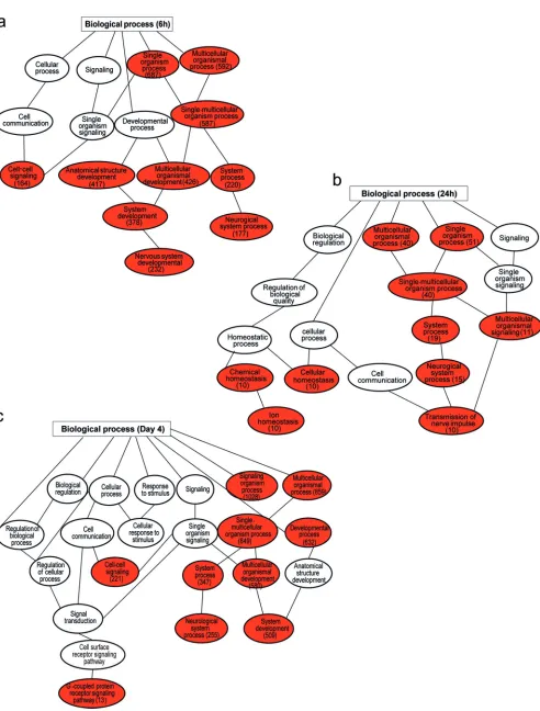
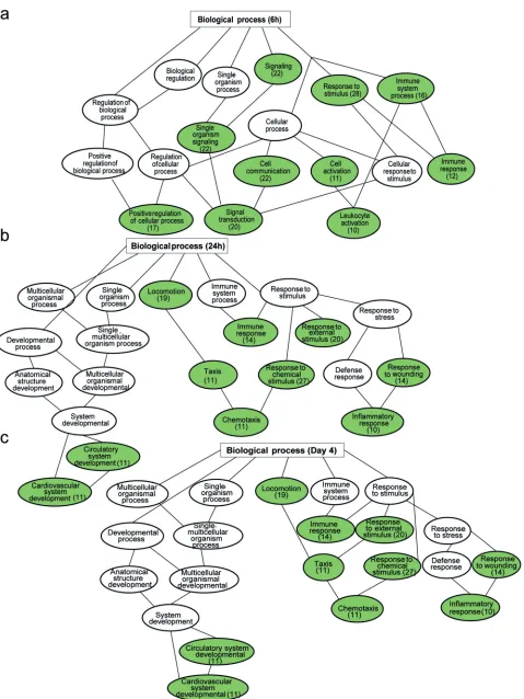
in WebGestalt) of the protein-coding differentially expressed genes between the responses of NS5-69- and NS3-H4-redirected T cells at the different time points (6 h, 24 h, day 4) were performed, with enrichment significance levelPvalues of⬍0.05 and a minimum level of 10 regulated genes in each category. The enriched GO biological process as well as the parent terms among the DE genes in NS5-69-redirected T cells are illustrated in Fig. 7 and 8, in which the number of genes regulated in each GO category are presented in parentheses. Enriched GO categories among the upregulated genes in NS5-69-redirected T cells compared to the NS3-H4-redirected T cells were indicative of mainly cell-cell signaling, multicellular organismal process, system process, and system development. As for downregulated genes, which were fewer than those that were upregulated, in NS5-69-redirected cells, they were found to be involved in GO categories of signaling, immune system processes, cell communication and activation, chemotaxis, inflammatory response, responses to external stimulus, and locomotion.
DISCUSSION
In this study, we examined a nonviral gene transfer approach to redirect human T cells with antiviral TCRs that recognize two well-characterized human CTL epitopes with clinical relevance in hepatitis C infection. The main reason for choosing these CTL
FIG 6Gene ontology (GO) categories enriched among upregulated genes in NS3-H4-redirected T cells and NS5-69-redirected T cells. Biological processes that are upregulated in NS3-H4- or NS5-69 TCR-redirected T cells relative to undirected T cells (UT) at the indicated time points following coculture with Huh7A2 HCVReptarget cells, identified using the annotation tool DAVID and related processes clustered using the functional annotation clustering tool. The top three clusters with the highest enrichment (EASE) scores and aPvalue of⬍0.05 for each GO category for NS3-H4 are presented at 6 h (a), at 24 h (b), and at day 4 (c) and for NS5-69 at 6 h (d), at 24 h (e), and at day 4 (f) after T cell and target cell coculture. The bars represent the respective GO categories, and thexaxis is the⫺log10Pvalue for enrichment level for each category. pos., positive; neg., negative; reg., regulation; diff, differentiation.
May 2017 Volume 91 Issue 9 e00010-17 jvi.asm.org 12
on November 7, 2019 by guest
http://jvi.asm.org/
[image:12.585.42.542.72.445.2]FIG 7Significantly enriched gene ontology (GO) biological process among upregulated genes in NS5-69-redirected T cells compared to NS3-H4-redirected cells at time points 6 h (a), 24 h (b), and day 4 (c). The regulated pathways are in red, and the numbers of regulated genes in the pathways are in parentheses.
on November 7, 2019 by guest
http://jvi.asm.org/
[image:13.585.49.541.59.707.2]FIG 8Significantly enriched gene ontology (GO) biological process among downregulated genes in NS5-69-redirected T cells compared to NS3-H4-redirected cells at time points 6 h (a), 24 h (b), and day 4 (c). The regulated pathways are in green, and the numbers of regulated genes in the pathways are in parentheses.
May 2017 Volume 91 Issue 9 e00010-17 jvi.asm.org 14
on November 7, 2019 by guest
http://jvi.asm.org/
[image:14.585.45.524.65.703.2]epitopes is that they are associated with spontaneous clearance of HCV in acute infection (9, 23–25). Our main findings suggest that the TCR redirection approach examined here is (i) effective in PBLs from both healthy donors and hepatitis C patients that have all four forms of liver fibrosis stages, (ii) functionally preserved and highly reproducible in regard to our previous finding with retrovirally engineered T cells (20, 21), (iii) of transient nature, reducing concerns of vector integration into host genome and long-term expression if administered, and (iv) useful in gaining a systemic insight into HCV-specific CTL responses that differ in their effector functions. These findings add an important translational significance. An immune therapy consisting of effica-cious polyfunctional cytolytic HCV-specific T cells would support the immune surveil-lance in difficult-to-treat hepatitis C patients, presuming that HCV epitopes are still unchanged and are present in the patients. This is particularly relevant given that recent reports suggest that certain HCV patient groups, especially those who are at very high risk of developing hepatocellular carcinoma, may require more-active immune surveillance of infected premalignant cells rather than curative antiviral treatment only (2, 3).
Although much effort and endeavor have been devoted to elucidating the mech-anisms that regulate CD8⫹ T cell activation, differentiation, and survival (26–31), a
fundamental examination of the genetic pathways in HCV T cell responses to hepato-cytes replicating HCV has to our knowledge not been undertaken prior to this study. This has been complicated by the seemingly low precursor frequency of HCV-specific T cells in patients and the requirement for major histocompatibility complex (MHC)-peptide tetramer enrichment, which is difficult to scale up. Single-cell sequencing could eventually surpass this challenge, but our bulk cell analysis here illuminates for the first time the dynamic of an HCV T cell response, allowing an overview of transcriptomic changes in antigen-specific T cells from the encountering to the elimination (or failure of elimination) of their target cells. Although the TCRs examined here originated from HLA-A2 transgenic mice vaccinated with DNA (20, 21), murine TCR expression on human T cells circumvents problems with TCR mispairing, e.g., the formation of nonfunctional TCR “human-human” heterodimers with endogenous host TCRs. More-over, the functionality of murine TCRs in human T cells has been demonstrated bothin vitroandin vivoin human T cell adoptive therapies (32). In spite of the fact that the ectopic expression of HCV TCR on the human T cell surface lasts for 3 to 4 days, our results indicate that the cells were biologically functional for the entire expression time, i.e., responded to the target cells in an antigen-specific manner.
The HCV RNA replicon cell model is a biologically relevant condition to examine the hepatotoxic potential of the TCRs. The observed polyfunctional profiles in response to processed antigen support the results obtained with peptide-loaded T2 cells: NS3-TCR activation stimulates IFN-␥, tumor necrosis factor alpha (TNF-␣), and granzyme B, as confirmed by RNA-seq and injury of HLA-A2-positive HCV RNA⫹hepatoma cells, while
the NS5 TCR’s response to processed antigen consists mainly of IFN-␥or TNF-␣and no hepatocellular injury. Here, human T cells conferred with NS3-H4 or NS5-69 TCR recognized the endogenously processed peptides in target HCV replicon cells. But contrary to NS5-69, the NS3-H4 TCR was consistently superior in providing polyfunc-tionality, target cell elimination, and hepatotoxicity, although the two TCRs had similar surface expression and kinetics. It is possible that the differential antiviral activities of the two different peptide-specific transfected T cell lines are due to differential antigen or differential TCR affinity. In this context, peptide titration experiments indicate that although the two TCRs behave in dose-dependent manners, the NS3 TCR is highly sensitive and requires a few log10-fold less peptide than the NS5-TCR to secrete multiple cytokines (22). Hence, the TCR affinity represents an additional important factor accompanying the antigen specificity in an antigen-specific T cell response. Given that T cell responses play a unique part in HCV infection, since CD8⫹T cells not
only contribute to viral clearance and protective immunity in those who recover from infection but also are associated with tissue injury in infected patients, e.g., hepatic flare (33), the two CD8⫹T cell responses that we report here are thus of clinical relevance
on November 7, 2019 by guest
http://jvi.asm.org/
for patients with hepatitis C. Viral clearance during HCV has been thought to reflect the destruction of infected hepatocytes by cytolytic CD8⫹ T lymphocytes, but several elegant studies have suggested that the cytolytic effector pathway is rarely utilized by HCV-specific CD8⫹T cells in patients with chronic hepatitis C (14, 34, 35), possibly to restrict liver damagein vivo.
A typical feature in hepatitis C patients who develop chronic infection is the selection of functionally impaired T cells and upregulation of suppressive cytokines and regulatory cell response such as TGF-, chemokine ligand 11 (CCL11; eotaxin), and CCL18 and CCL22, known to recruit regulatory T cells (36–38). These were also found to be upregulated here in the NS5-69 T cell response, suggesting that low-avidity CTLs may have anti-inflammatory potentials. In contrast, our data indicate that in a cytolytic CTL response, such as the NS3-H4 CTR having polyfunctional property and increased ability to eliminate HCV target cells, the response has a striking focus on cytokine-cytokine receptor interactions, chemokine signaling, and JAK-Stat signaling, lacking in the noncytolytic NS5-69 CTL response. Interestingly, our functional GO clustering analysis clearly demonstrates that NS3-H4 upregulated apoptosis and programmed cell death gene clusters, including genes encoding TNF/TRAIL, IFN-␥, and CD70, indicative of an activation-induced apoptosis early during the elimination of HCV replicon target cells. Moreover, there were, as well as antiapoptosis genes, clusters of genes encoding BCL-3, IL-2R, and CD27, among which CD27, a costimulatory molecule, is known to be required for the generation of maintenance of T cell immunity (39, 40). The early response seen here could be due to the fact that the PBLs studied here had been preactivated and were already differentiated effector cells when they encountered the HCV target cells. Furthermore, the increased ion and membrane transporter activity, cAMP biosynthesis, and metabolic processes later the following day were not totally unanticipated, since ion channels and transporters in the plasma membrane of T cells are required to modulate the cytoplasmic concentrations of diverse cations; in partic-ular, calcium channels are known to allow second messengers to regulate crucial lymphocyte effector functions, including CD8⫹T cell survival and control of activation (41, 42). The reason for the appearance of a gene cluster involved in embryonic morphogenesis on day 4 is not clear (perhaps it is a nascent process in T cell rejuvenation and memory cell formation) and deserves further examination.
Despite the fact that NS5-69-redirected T cells recognize HCV-expressing target cells by secreting a small number of cytokines, they failed to eliminate target cells. The notion that the NS5-69 response had gene clusters indicative of cellular amino acid biosynthesis suggests an active glycolysis associated with activation and effector T cell differentiation (43) at the same time, with increased taxis, and chemokine ligand genes encoding CCL9, CCL10, CCL11, and CCL22 and cell proliferation genes were upregu-lated. As the incapability to eliminate the target continues, the activities continue to include signal transduction and metabolic processes (cyclic nucleotide biosynthetic, cAMP and metabolic processes, calcium/metal ion transport), which became even more diverse to include gene cluster expressions controlling lipid metabolisms and cellular metabolic processes and pathways sharing features with an SLE-related response.
A further direct comparison of pathways based on enriched GO biological processes used by NS3-H4 and NS5-69 suggests that NS5-69 had upregulated a substantially large number of genes that are associated with cell-cell signaling, multicellular organismal process, system process, and system development. Among the downregulated genes in NS5-69 relative to NS3-H4, albeit much fewer than those that were upregulated, those in pathways involving signaling, immune system process, cell communication and activation, chemotaxis, inflammatory response, response to external stimulus, and locomotion were noted. This supports the idea that NS5-69 activated multiple default pathways as a response to the persistent HCV RNA replicating target cells. The differ-ence in function of the two HCV-specific TCRs might be related to their intrinsic TCR affinity. Alternatively, the HCV replicon cells might process and present more efficiently the NS3 epitope than the NS5 ones. Although there are currently no studies to support
May 2017 Volume 91 Issue 9 e00010-17 jvi.asm.org 16
on November 7, 2019 by guest
http://jvi.asm.org/
this hypothesis, it is in line with the previously reported hypothesis that activation of NS5-69 TCR requires more target peptide than does that of NS3-H4 (20, 21).
The questions that our study addressed were (i) whether chronic HCV patient PBLs can be effectively redirected by the mRNA approach and (ii) what gave HCV CTL effector cells the ability to eliminate or spare their target cells. The two types of effector T cell responses are considered to have important roles in viral hepatitis cure and progression (44–47). To our knowledge, this is the first study that describes the effector gene profiles distinguishing the cytolytic from the noncytolytic antiviral effector CD8 T cells with specificity to HCV viral targets. Such antiviral effector features may also be of relevance for the biology of antigen-specific polyfunctional T cells in general.
The strength of this study is that the endogenously processed HCV CTL targets and antigen-specific T cells were examined, in which the exposed effector T cells could be isolated without tetramer sorting, and transcriptomically decoded with a powerful pair-end high-throughput RNA sequencing. A limitation of this study is that the transcriptome results are merely descriptive and at the transcript level, and hence further studies are needed at the next level with proteomics and/or metabolomics analyses. Moreover, these TCRs address only HLA-A2-restricted responses and target cells, thus confining their clinical applications. However, a strong antigenic avidity and acquisition of proper effector functions have an impact on the selection of polyfunc-tional high-avidity CD8 T cells, a quality known to correlate with control of hepatitis C virus infection (48, 49). Our transcriptomic insight into the mechanisms behind a successful versus a less successful HCV CTL response has implications in understanding HCV T cell immunology and T cell biology in general.
MATERIALS AND METHODS
PBMCs from healthy blood donors and HCV-infected patients.Peripheral blood samples were collected under informed consent from healthy blood donors and HCV-infected patients at Karolinska University Hospital. Following HLA typing, HLA-A2-positive donors were included, with subsequent isolation of peripheral blood mononuclear cells (PBMCs) from 9 healthy blood donors and 10 HCV-infected patients (20, 21). Ethical permission was obtained from the Regional Ethical Review Board (EPN) in Stockholm. The Declaration of Helsinki protocols were followed, and all patients gave their written, informed consent.
Cell lines.The Lunet-HLA-A2-luc/neoET (Huh7A2HCVRep) and Lunet-luc/neoET (Huh7 HCVRep) cell lines were maintained as described previously (21, 22) in Dulbecco’s modified Eagle’s medium supple-mented with 10% fetal bovine serum (FBS), 2 mML-glutamine, 100 U ml⫺1penicillin, 100g ml⫺1 streptomycin, and 1 mM nonessential amino acids. Huh7A2HCVRepcells were selected with puromycin (1g/ml) and G418 (0.5 mg/ml) and Huh7 HCVRepcells with G418 (0.5 mg/ml). Huh7A2HCVRepcells have ectopic HLA-A2 expression and contain a selectable HCV subgenomic RNA replicon of genotype 1b (Con1-ET). The cells also express the firefly luciferase gene fused to the selectable marker by ubiquitin. Control cell line Huh7 HCVRepcontains only HCV subgenomic RNA replicon of genotype 1b and does not express HLA-A2. The T2 cell line (HLA A2.1⫹) was maintained in RPMI 1640 medium supplemented with
10% FBS, 2 mML-glutamine, 10 mM HEPES, 100 U ml⫺1penicillin, and 100g ml⫺1streptomycin. Media and supplements were purchased from Sigma-Aldrich, St. Louis, MO.
In vitrosynthesis of TCR mRNA and transfection of PBLs.Plasmids encoding NS3 TCRs or NS5
TCRs (pVAX-NS3-H4, pVAX-NS3-F8, pVAX-NS5A-19, pVAX-NS5A-69) were constructed by subcloning TCR genes from retroviral vectors described earlier (20, 21). Qiagen Endo Free Plasmid Maxi kit (Qiagen, Hilden, Germany)-purified and XbaI-linearized DNA was used as the template to synthesize TCR mRNA using the mMESSAGE mMACHINE T7 Ultra kit (Ambion, Austin, TX). T7 RNA polymerase was added to start transcription; RNA was capped with anti-reverse cap analog (ARCA) and poly(A) tailed with poly(A) polymerase and ATP. Lithium chloride-precipitated RNA was stored at⫺80°C until use. PBMCs were stimulated and prepared as described earlier (50) with 600 U/ml IL-2 (rIL-2; R&D Systems, Minneapolis, MN) and 50 ng/ml anti-CD3 (OKT-3; eBioscience, San Diego, CA) in AIM-V medium (Life Technologies, Carlsbad, CA) supplemented with 2% human AB serum for 7 days. The concentration of rIL-2 was increased to 1,000 IU/ml on day 8. Electroporations were performed with the Nucleofector device (Lonza, Cologne, Germany). Activated T cells were harvested and transfected in Cell Line Nucleofector Solution V (Lonza, Cologne, Germany) at 10⫻106cells with TCR mRNA (200g/ml) in a certified cuvette (Lonza, Cologne, Germany). Immediately after transfection, the cells were resuspended in AIM-V medium (Life Technologies, Carlsbad, CA) supplemented with 2% human AB serum and 100 IU/ml rIL-2 and incubated in a humidified 37°C and 5% CO2incubator until further analysis.
FACS antibody staining and peptides.For fluorescence-activated cell sorting (FACS), APC anti-human CD3, PerCP anti-anti-human CD8␣, fluorescein isothiocyanate (FITC)-labeled anti-mouse V 8.3, anti-mouse V8.1-8.2, anti-mouse V6, anti-mouse V4, and the Aqua fluorescent reactive dye (Life Technologies) were used for staining of the TCRs 12 to 16 h after electroporation. Intracellular cytokine staining after antigen stimulation was performed as previously described with Aqua fluorescent reactive
on November 7, 2019 by guest
http://jvi.asm.org/
dye, APC Cy7 anti-human CD8, PerCP Cy5.5 anti-human CD107␣; cells were fixed with Cytofix and Cytoperm (BD Biosciences, CA), stained with Pacific blue anti-human CD3, PE anti-human IFN-␥, APC anti-human IL-2, and PE-Cy7 anti-human TNF-␣, and run on the FACS Verse flow cytometer (BD Biosciences, CA). FlowJo version V10.07 software was used for data analysis. All antibodies were purchased from BioLegend, CA, or BD Biosciences, CA. HCV peptides NS31073-1081 gt1a (CINGVCWTV) and NS51992-2000 gt1b (VLTDFKTWL) were synthesized by EZBiolab (Carmel, IN).
Functional analysis of T cells cocultured with T2 cells or HCV replicon cells.On day 1 posttrans-fection, T cells were cocultured with HCV peptide-loaded T2 cells, Huh7A2HCVRepcells, or Huh7 HCVRep cells expressing the endogenous HCV CTL epitopes at the indicated E/T ratios. FACS analysis was done after 5 h of coculture and ICS as described above. Cytokine enzyme-linked immunosorbent assay (ELISA) was done by quantifying IFN-␥and IL-2 in the overnight (16 to 18 h) supernatant (Mabtech, Nacka Strand, Sweden).
HCV antiviral assay and AST measurement.On day 1 posttransfection, T cells were cocultured with Huh7A2HCVRepcells or Huh7 HCVRepcells in 96-well tissue culture plates as described earlier (21). After 24 h of coculture, the luciferase activity in target cells was measured using the ONE-Glo luciferase assay system (Promega, Madison, WI). Data are presented as the percentage of HCV replication in Huh7A2 HCVRepcells or Huh7 HCVRepcells coincubated with T cells by measurement of relative light unit (RLU) values compared to RLU values in Huh7A2 HCVRepcells alone or Huh7 HCVRep cells alone (without coculture). Background values of T cell control wells were subtracted (10 to 20 RLU). Aspartate amino-transferase (AST) levels in supernatants were quantitated by a validated AST assay at the Clinical Chemistry Laboratory at the Karolinska University Hospital, Huddinge, Sweden, using the Modular P apparatus (Roche Diagnostics, Mannheim, Germany).
Cell purification, RNA isolation, and library preparation for sequencing. TCR-redirected or mock-treated T cells cocultured with Huh7A2HCVRepcells at an E/T ratio of 10:1 were collected at the indicated times and enriched with anti-CD8 MicroBeads (Miltenyi Biotech) as per the manufacturer’s instructions. Enriched CD8⫹T cells (purity, 98% CD3⫹CD8⫹) at 1.5 million cells per sample were stored
at⫺80°C. Total RNA was isolated with TRIzol reagent (Thermo Fisher Scientific, MA, USA), and RNase-free reagents were used throughout the isolation. Sample quantity and purity were assessed using the NanoDrop 1000 spectrophotometer (Thermo Fisher Scientific, MA, USA). All RNA samples had RNA integrity number (RIN) values of⬎9. The RNA libraries were bar coded and prepared according to the protocol Illumina TruSeq RNA Sample Prep kit v2 (catalog number RS-122-2001) from the manufacturer (Illumina, San Diego, CA, USA).
Paired-end sequencing and sequence alignment.The nine RNA libraries were sequenced in paired ends on an Illumina HiSeq 2000 instrument, where each sample was put in two different lanes, for a total of 18 samples. The runs generated a total of 384 million reads with an average of 43 million paired-end reads per sample, of which 90% mapped to the human genome. The paired-end sequences were aligned to the human genome reference hg19, with TopHat2 version 0.9 using TopHat standard parameters. The numbers of aligned reads per gene were determined with HTSeq 0.6.1. Annotations from Ensembl Homo sapiens grch37.82 and Genome Browser were used to assign features to genomic positions. The R/Bioconductor package DESeq2 was used for differential gene expression on read counts generated by HTSeq 0.6.1.
Functional analysis of gene lists using DAVID and WebGestalt.The default parameters of the Database for Annotation, Visualization and Integrated Discovery (DAVID) were used to achieve GO analysis.Homo sapienswas used as background, and the “GOTERM_BP_FAT” option was selected with the highest classification stringency andPvalues of⬍0.05. KEGG pathway analysis was performed using the Web-based Gene Set Analysis Toolkit v3 (WebGestalt) with the following parameters: ID type, Ensembl_gene_stable; reference set,Homo sapiens; statistical method, hypergeometric; multiple test adjustment, Benjamini & Hochberg (BH); significance level,P⬍0.01; minimum number of genes for each category, 2.
SUPPLEMENTAL MATERIAL
Supplemental material for this article may be found at https://doi.org/10.1128/
JVI.00010-17.
SUPPLEMENTAL FILE 1,PDF file, 0.1 MB. SUPPLEMENTAL FILE 2,XLS file, 0.1 MB. SUPPLEMENTAL FILE 3,XLS file, 2.6 MB. SUPPLEMENTAL FILE 4,XLS file, 5.1 MB. SUPPLEMENTAL FILE 5,XLS file, 0.1 MB.
ACKNOWLEDGMENTS
We thank all patients and volunteer blood donors for making this study possible. We also thank Susann Fält for technical assistance, David Brodin at the Bioinformatics and Expression Analysis (BEA) Core Facility at Karolinska Institutet for providing biostatistics support and consultations, and Christina Barnfield for critical reading of the manuscript. This work was supported by the Swedish Cancer Society (to M.C. and M.S.), Ruth och Richard Julins funds and Karolinska Institutet funds (to M.C.), and Magtarmfonden (to
May 2017 Volume 91 Issue 9 e00010-17 jvi.asm.org 18
on November 7, 2019 by guest
http://jvi.asm.org/
S.A.). This work was also partially funded by Eradication of HBV TCR Program grant NMRC/TCR/014 NUHS/2015 and a Singapore Translational Research (STaR) Investigator Award (NMRC/STaR/013/2012) to A.B.
We declare that no conflict of interest exists.
REFERENCES
1. Pawlotsky JM, Feld JJ, Zeuzem S, Hoofnagle JH. 2015. From non-A, non-B hepatitis to hepatitis C virus cure. J Hepatol 62:S87–S99.https://doi.org/ 10.1016/j.jhep.2015.02.006.
2. Conti F, Buonfiglioli F, Scuteri A, Crespi C, Bolondi L, Caraceni P, Foschi FG, Lenzi M, Mazzella G, Verucchi G, Andreone P, Brillanti S. 24 June 2016. Early occurrence and recurrence of hepatocellular carcinoma in HCV-related cirrhosis treated with direct-acting antivirals. J Hepatol https://doi.org/10.1016/j.jhep.2016.06.015.
3. Reig M, Marino Z, Perello C, Inarrairaegui M, Ribeiro A, Lens S, Diaz A, Vilana R, Darnell A, Varela M, Sangro B, Calleja JL, Forns X, Bruix J. 13 April 2016. Unexpected high rate of early tumor recurrence in patients with HCV-related HCC undergoing interferon-free therapy. J Hepatol https://doi.org/10.1016/j.jhep.2016.04.008.
4. Lozano R, Naghavi M, Foreman K, Lim S, Shibuya K, Aboyans V, Abraham J, Adair T, Aggarwal R, Ahn SY, Alvarado M, Anderson HR, Anderson LM, Andrews KG, Atkinson C, Baddour LM, Barker-Collo S, Bartels DH, Bell ML, Benjamin EJ, Bennett D, Bhalla K, Bikbov B, Bin Abdulhak A, Birbeck G, Blyth F, Bolliger I, Boufous S, Bucello C, Burch M, Burney P, Carapetis J, Chen H, Chou D, Chugh SS, Coffeng LE, Colan SD, Colquhoun S, Colson KE, Condon J, Connor MD, Cooper LT, Corriere M, Cortinovis M, de Vaccaro KC, Couser W, Cowie BC, Criqui MH, Cross M, Dabhadkar KC, Dahodwala N, De Leo D, Degenhardt L, Delossantos A, Denenberg J, Des Jarlais DC, Dharmaratne SD, Dorsey ER, Driscoll T, Duber H, Ebel B, Erwin PJ, Espindola P, Ezzati M, Feigin V, Flaxman AD, Forouzanfar MH, Fowkes FG, Franklin R, Fransen M, Freeman MK, Gabriel SE, Gakidou E, Gaspari F, Gillum RF, Gonzalez-Medina D, Halasa YA, Haring D, Harrison JE, Hav-moeller R, Hay RJ, Hoen B, Hotez PJ, Hoy D, Jacobsen KH, James SL, Jasrasaria R, Jayaraman S, Johns N, Karthikeyan G, Kassebaum N, Keren A, Khoo JP, Knowlton LM, Kobusingye O, Koranteng A, Krishnamurthi R, Lipnick M, Lipshultz SE, Ohno SL, Mabweijano J, MacIntyre MF, Mallinger L, March L, Marks GB, Marks R, Matsumori A, Matzopoulos R, Mayosi BM, McAnulty JH, McDermott MM, McGrath J, Mensah GA, Merriman TR, Michaud C, Miller M, Miller TR, Mock C, Mocumbi AO, Mokdad AA, Moran A, Mulholland K, Nair MN, Naldi L, Narayan KM, Nasseri K, Norman P, O’Donnell M, Omer SB, Ortblad K, Osborne R, Ozgediz D, Pahari B, Pandian JD, Rivero AP, Padilla RP, Perez-Ruiz F, Perico N, Phillips D, Pierce K, Pope CA, III, Porrini E, Pourmalek F, Raju M, Ranganathan D, Rehm JT, Rein DB, Remuzzi G, Rivara FP, Roberts T, De León FR, Rosenfeld LC, Rushton L, Sacco RL, Salomon JA, Sampson U, Sanman E, Schwebel DC, Segui-Gomez M, Shepard DS, Singh D, Singleton J, Sliwa K, Smith E, Steer A, Taylor JA, Thomas B, Tleyjeh IM, Towbin JA, Truelsen T, Undurraga EA, Venketasubramanian N, Vijayakumar L, Vos T, Wagner GR, Wang M, Wang W, Watt K, Weinstock MA, Weintraub R, Wilkinson JD, Woolf AD, Wulf S, Yeh PH, Yip P, Zabetian A, Zheng ZJ, Lopez AD, Murray CJ, AlMazroa MA, Memish ZA. 2012. Global and regional mortality from 235 causes of death for 20 age groups in 1990 and 2010: a systematic analysis for the Global Burden of Disease Study 2010. Lancet 380: 2095–2128.https://doi.org/10.1016/S0140-6736(12)61728-0.
5. Thimme R, Oldach D, Chang KM, Steiger C, Ray SC, Chisari FV. 2001. Determinants of viral clearance and persistence during acute hepatitis C virus infection. J Exp Med 194:1395–1406.https://doi.org/10.1084/jem .194.10.1395.
6. Marukian S, Andrus L, Sheahan TP, Jones CT, Charles ED, Ploss A, Rice CM, Dustin LB. 2011. Hepatitis C virus induces interferon-lambda and interferon-stimulated genes in primary liver cultures. Hepatology 54: 1913–1923.https://doi.org/10.1002/hep.24580.
7. Cooper S, Erickson AL, Adams EJ, Kansopon J, Weiner AJ, Chien DY, Houghton M, Parham P, Walker CM. 1999. Analysis of a successful immune response against hepatitis C virus. Immunity 10:439 – 449. https://doi.org/10.1016/S1074-7613(00)80044-8.
8. Diepolder HM, Gruener NH, Gerlach JT, Jung MC, Wierenga EA, Pape GR. 2001. Different levels of T-cell receptor triggering induce distinct func-tions in hepatitis B and hepatitis C virus-specific human CD4(⫹) T-cell clones. J Virol 75:7803–7810. https://doi.org/10.1128/JVI.75.17.7803 -7810.2001.
9. Lechner F, Wong DK, Dunbar PR, Chapman R, Chung RT, Dohrenwend P, Robbins G, Phillips R, Klenerman P, Walker BD. 2000. Analysis of success-ful immune responses in persons infected with hepatitis C virus. J Exp Med 191:1499 –1512.https://doi.org/10.1084/jem.191.9.1499.
10. Missale G, Bertoni R, Lamonaca V, Valli A, Massari M, Mori C, Rumi MG, Houghton M, Fiaccadori F, Ferrari C. 1996. Different clinical behaviors of acute hepatitis C virus infection are associated with different vigor of the anti-viral cell-mediated immune response. J Clin Invest 98:706 –714. https://doi.org/10.1172/JCI118842.
11. Takaki A, Wiese M, Maertens G, Depla E, Seifert U, Liebetrau A, Miller JL, Manns MP, Rehermann B. 2000. Cellular immune responses persist and humoral responses decrease two decades after recovery from a single-source outbreak of hepatitis C. Nat Med 6:578 –582.https://doi.org/10 .1038/75063.
12. Thimme R, Bukh J, Spangenberg HC, Wieland S, Pemberton J, Steiger C, Govindarajan S, Purcell RH, Chisari FV. 2002. Viral and immunological determinants of hepatitis C virus clearance, persistence, and disease. Proc Natl Acad Sci U S A 99:15661–15668.https://doi.org/10.1073/pnas .202608299.
13. Diepolder HM, Gerlach JT, Zachoval R, Hoffmann RM, Jung MC, Wierenga EA, Scholz S, Santantonio T, Houghton M, Southwood S, Sette A, Pape GR. 1997. Immunodominant CD4⫹T-cell epitope within nonstructural protein 3 in acute hepatitis C virus infection. J Virol 71:6011– 6019. 14. Jo J, Bengsch B, Seigel B, Rau SJ, Schmidt J, Bisse E, Aichele P, Aichele U,
Joeckel L, Royer C, Sa Ferreira K, Borner C, Baumert TF, Blum HE, Lohmann V, Fischer R, Thimme R. 2012. Low perforin expression of early differentiated HCV-specific CD8⫹T cells limits their hepatotoxic poten-tial. J Hepatol 57:9 –16.https://doi.org/10.1016/j.jhep.2012.02.030. 15. Spangenberg HC, Viazov S, Kersting N, Neumann-Haefelin C, McKinney
D, Roggendorf M, von Weizsacker F, Blum HE, Thimme R. 2005. Intrahe-patic CD8⫹ T-cell failure during chronic hepatitis C virus infection. Hepatology 42:828 – 837.https://doi.org/10.1002/hep.20856.
16. Ciuffreda D, Comte D, Cavassini M, Giostra E, Buhler L, Perruchoud M, Heim MH, Battegay M, Genne D, Mulhaupt B, Malinverni R, Oneta C, Bernasconi E, Monnat M, Cerny A, Chuard C, Borovicka J, Mentha G, Pascual M, Gonvers JJ, Pantaleo G, Dutoit V. 2008. Polyfunctional HCV-specific T-cell responses are associated with effective control of HCV replication. Eur J Immunol 38:2665–2677. https://doi.org/10.1002/eji .200838336.
17. Abdel-Hakeem MS, Bedard N, Murphy D, Bruneau J, Shoukry NH. 2014. Signatures of protective memory immune responses during hepatitis C virus reinfection. Gastroenterology 147:870 – 881.e8.https://doi.org/10 .1053/j.gastro.2014.07.005.
18. Perreau M, Levy Y, Pantaleo G. 2013. Immune response to HIV. Curr Opin HIV AIDS 8:333–340.https://doi.org/10.1097/COH.0b013e328361faf4. 19. Ravimohan S, Tamuhla N, Nfanyana K, Steenhoff AP, Letlhogile R, Frank
I, MacGregor RR, Gross R, Weissman D, Bisson GP. 26 November 2015. Robust reconstitution of tuberculosis-specific polyfunctional CD4⫹ T-cell responses and rising systemic interleukin 6 in paradoxical tuberculosis-associated immune reconstitution inflammatory syndrome. Clin Infect Dishttps://doi.org/10.1093/cid/civ978.
20. Pasetto A, Frelin L, Brass A, Yasmeen A, Koh S, Lohmann V, Bartenschlager R, Magalhaes I, Maeurer M, Sallberg M, Chen M. 2012. Generation of T-cell receptors targeting a genetically stable and immunodominant cytotoxic T-lymphocyte epitope within hepatitis C virus non-structural protein 3. J Gen Virol 93:247–258.https://doi.org/10.1099/vir.0.037903-0.
21. Pasetto A, Frelin L, Aleman S, Holmstrom F, Brass A, Ahlen G, Brenndorfer ED, Lohmann V, Bartenschlager R, Sallberg M, Bertoletti A, Chen M. 2012. TCR-redirected human T cells inhibit hepatitis C virus replication: hepa-totoxic potential is linked to antigen specificity and functional avidity. J Immunol 189:4510 – 4519.https://doi.org/10.4049/jimmunol.1201613. 22. Lohmann V, Hoffmann S, Herian U, Penin F, Bartenschlager R. 2003. Viral
and cellular determinants of hepatitis C virus RNA replication in cell culture. J Virol 77:3007–3019.https://doi.org/10.1128/JVI.77.5.3007-3019 .2003.
on November 7, 2019 by guest
http://jvi.asm.org/
23. Chang KM, Thimme R, Melpolder JJ, Oldach D, Pemberton J, Moorhead-Loudis J, McHutchison JG, Alter HJ, Chisari FV. 2001. Differential CD4(⫹) and CD8(⫹) T-cell responsiveness in hepatitis C virus infection. Hepa-tology 33:267–276.https://doi.org/10.1053/jhep.2001.21162.
24. Urbani S, Uggeri J, Matsuura Y, Miyamura T, Penna A, Boni C, Ferrari C. 2001. Identification of immunodominant hepatitis C virus (HCV)-specific cytotoxic T-cell epitopes by stimulation with endogenously synthesized HCV antigens. Hepatology 33:1533–1543.https://doi.org/10.1053/jhep .2001.25091.
25. Shoukry NH, Grakoui A, Houghton M, Chien DY, Ghrayeb J, Reimann KA, Walker CM. 2003. Memory CD8⫹T cells are required for protection from persistent hepatitis C virus infection. J Exp Med 197:1645–1655.https:// doi.org/10.1084/jem.20030239.
26. Ariotti S, Hogenbirk MA, Dijkgraaf FE, Visser LL, Hoekstra ME, Song JY, Jacobs H, Haanen JB, Schumacher TN. 2014. T cell memory. Skin-resident memory CD8(⫹) T cells trigger a state of tissue-wide pathogen alert. Science 346:101–105.https://doi.org/10.1126/science.1254803. 27. Bottcher JP, Beyer M, Meissner F, Abdullah Z, Sander J, Hochst B, Eickhoff
S, Rieckmann JC, Russo C, Bauer T, Flecken T, Giesen D, Engel D, Jung S, Busch DH, Protzer U, Thimme R, Mann M, Kurts C, Schultze JL, Kasten-muller W, Knolle PA. 2015. Functional classification of memory CD8(⫹) T cells by CX3CR1 expression. Nat Commun 6:8306. https://doi.org/10 .1038/ncomms9306.
28. Best JA, Blair DA, Knell J, Yang E, Mayya V, Doedens A, Dustin ML, Goldrath AW. 2013. Transcriptional insights into the CD8(⫹) T cell response to infection and memory T cell formation. Nat Immunol 14: 404 – 412.https://doi.org/10.1038/ni.2536.
29. Giordano M, Henin C, Maurizio J, Imbratta C, Bourdely P, Buferne M, Baitsch L, Vanhille L, Sieweke MH, Speiser DE, Auphan-Anezin N, Schmitt-Verhulst AM, Verdeil G. 2015. Molecular profiling of CD8 T cells in autochthonous melanoma identifies Maf as driver of exhaustion. EMBO J 34:2042–2058.https://doi.org/10.15252/embj.201490786. 30. Gaiha GD, McKim KJ, Woods M, Pertel T, Rohrbach J, Barteneva N, Chin
CR, Liu D, Soghoian DZ, Cesa K, Wilton S, Waring MT, Chicoine A, Doering T, Wherry EJ, Kaufmann DE, Lichterfeld M, Brass AL, Walker BD. 2014. Dysfunctional HIV-specific CD8⫹T cell proliferation is associated with increased caspase-8 activity and mediated by necroptosis. Immunity 41:1001–1012.https://doi.org/10.1016/j.immuni.2014.12.011.
31. Chiu YL, Shan L, Huang H, Haupt C, Bessell C, Canaday DH, Zhang H, Ho YC, Powell JD, Oelke M, Margolick JB, Blankson JN, Griffin DE, Schneck JP. 2014. Sprouty-2 regulates HIV-specific T cell polyfunctionality. J Clin Invest 124:198 –208.https://doi.org/10.1172/JCI70510.
32. Hinrichs CS, Rosenberg SA. 2014. Exploiting the curative potential of adoptive T-cell therapy for cancer. Immunol Rev 257:56 –71.https://doi .org/10.1111/imr.12132.
33. Rehermann B. 2013. Pathogenesis of chronic viral hepatitis: differential roles of T cells and NK cells. Nat Med 19:859 – 868.https://doi.org/10 .1038/nm.3251.
34. Jo J, Aichele U, Kersting N, Klein R, Aichele P, Bisse E, Sewell AK, Blum HE, Bartenschlager R, Lohmann V, Thimme R. 2009. Analysis of CD8⫹ T-cell-mediated inhibition of hepatitis C virus replication using a novel immuno-logical model. Gastroenterology 136:1391–1401.https://doi.org/10.1053/j .gastro.2008.12.034.
35. Seigel B, Bengsch B, Lohmann V, Bartenschlager R, Blum HE, Thimme R. 2013. Factors that determine the antiviral efficacy of HCV-specific CD8(⫹) T cells ex vivo. Gastroenterology 144:426 – 436.https://doi.org/ 10.1053/j.gastro.2012.10.047.
36. Osburn WO, Levine JS, Chattergoon MA, Thomas DL, Cox AL. 2013.
Anti-inflammatory cytokines, pro-fibrogenic chemokines and persis-tence of acute HCV infection. J Viral Hepat 20:404 – 413.https://doi.org/ 10.1111/jvh.12052.
37. Gobert M, Treilleux I, Bendriss-Vermare N, Bachelot T, Goddard-Leon S, Arfi V, Biota C, Doffin AC, Durand I, Olive D, Perez S, Pasqual N, Faure C, Ray-Coquard I, Puisieux A, Caux C, Blay JY, Menetrier-Caux C. 2009. Regulatory T cells recruited through CCL22/CCR4 are selectively acti-vated in lymphoid infiltrates surrounding primary breast tumors and lead to an adverse clinical outcome. Cancer Res 69:2000 –2009.https:// doi.org/10.1158/0008-5472.CAN-08-2360.
38. Chenivesse C, Chang Y, Azzaoui I, Ait Yahia S, Morales O, Ple C, Foussat A, Tonnel AB, Delhem N, Yssel H, Vorng H, Wallaert B, Tsicopoulos A. 2012. Pulmonary CCL18 recruits human regulatory T cells. J Immunol 189:128 –137.https://doi.org/10.4049/jimmunol.1003616.
39. Hendriks J, Gravestein LA, Tesselaar K, van Lier RA, Schumacher TN, Borst J. 2000. CD27 is required for generation and long-term maintenance of T cell immunity. Nat Immunol 1:433– 440.https://doi.org/10.1038/80877. 40. Mitchell TC, Hildeman D, Kedl RM, Teague TK, Schaefer BC, White J, Zhu Y, Kappler J, Marrack P. 2001. Immunological adjuvants promote acti-vated T cell survival via induction of Bcl-3. Nat Immunol 2:397– 402. https://doi.org/10.1038/35073104.
41. Feske S, Skolnik EY, Prakriya M. 2012. Ion channels and transporters in lymphocyte function and immunity. Nat Rev Immunol 12:532–547. https://doi.org/10.1038/nri3233.
42. Lipskaia L, Lompre AM. 2004. Alteration in temporal kinetics of Ca2⫹ signaling and control of growth and proliferation. Biol Cell 96:55– 68. https://doi.org/10.1016/j.biolcel.2003.11.001.
43. Buck MD, O’Sullivan D, Pearce EL. 2015. T cell metabolism drives immu-nity. J Exp Med 212:1345–1360.https://doi.org/10.1084/jem.20151159. 44. Heim MH, Thimme R. 2014. Innate and adaptive immune responses in
HCV infections. J Hepatol 61:S14 –S25. https://doi.org/10.1016/j.jhep .2014.06.035.
45. Guidotti LG, Chisari FV. 2001. Noncytolytic control of viral infections by the innate and adaptive immune response. Annu Rev Immunol 19: 65–91.https://doi.org/10.1146/annurev.immunol.19.1.65.
46. Ahlen G, Frelin L, Brenndorfer ED, Brass A, Weiland O, Chen M, Sallberg M. 2013. Containing “The Great Houdini” of viruses: combining direct acting antivirals with the host immune response for the treatment of chronic hepatitis C. Drug Resist Updat 16:60 – 67. https://doi.org/10 .1016/j.drup.2013.06.001.
47. Pasetto A, Aleman S, Chen M. 2014. Functional attributes of responding T cells in HCV infection: the recent advances in engineering functional antiviral T cells. Arch Immunol Ther Exp (Warsz) 62:23–30.https://doi .org/10.1007/s00005-013-0248-8.
48. Neveu B, Debeaupuis E, Echasserieau K, le Moullac-Vaidye B, Gassin M, Jegou L, Decalf J, Albert M, Ferry N, Gournay J, Houssaint E, Bonneville M, Saulquin X. 2008. Selection of high-avidity CD8 T cells correlates with control of hepatitis C virus infection. Hepatology 48:713–722.https://doi .org/10.1002/hep.22379.
49. Ahlers JD, Belyakov IM. 2010. Memories that last forever: strategies for optimizing vaccine T-cell memory. Blood 115:1678 –1689. https://doi .org/10.1182/blood-2009-06-227546.
50. Koh S, Shimasaki N, Suwanarusk R, Ho ZZ, Chia A, Banu N, Howland SW, Ong AS, Gehring AJ, Stauss H, Renia L, Sallberg M, Campana D, Bertoletti A. 2013. A practical approach to immunotherapy of hepatocellular car-cinoma using T cells redirected against hepatitis B virus. Mol Ther Nucleic Acids 2:e114.https://doi.org/10.1038/mtna.2013.43.
May 2017 Volume 91 Issue 9 e00010-17 jvi.asm.org 20