Copyright © 2004, American Society for Microbiology. All Rights Reserved.
A
cis
-Acting Replication Element in the Sequence Encoding the
NS5B RNA-Dependent RNA Polymerase Is Required for
Hepatitis C Virus RNA Replication
Shihyun You,
1Decherd D. Stump,
2Andrea D. Branch,
2and Charles M. Rice
1*
Center for the Study of Hepatitis C, Laboratory of Virology and Infectious Disease, The Rockefeller University,New York, New York 10021,1and Division of Liver Disease, Department of Medicine,
Mount Sinai School of Medicine, New York, New York 100292
Received 23 July 2003/Accepted 10 October 2003
RNA structures play key roles in the replication of RNA viruses. Sequence alignment software, thermody-namic RNA folding programs, and classical comparative phylogenetic analysis were used to build models of six RNA elements in the coding region of the hepatitis C virus (HCV) RNA-dependent RNA polymerase, NS5B. The importance of five of these elements was evaluated by site-directed mutagenesis of a subgenomic HCV replicon. Mutations disrupting one of the predicted stem-loop structures, designated 5BSL3.2, blocked RNA replication, implicating it as an essentialcis-acting replication element (CRE). 5BSL3.2 is about 50 bases in length and is part of a larger predicted cruciform structure (5BSL3). As confirmed by RNA structure probing, 5BSL3.2 consists of an 8-bp lower helix, a 6-bp upper helix, a 12-base terminal loop, and an 8-base internal loop. Mutational analysis and structure probing were used to explore the importance of these features. Primary sequences in the loops were shown to be important for HCV RNA replication, and the upper helix appears to serve as an essential scaffold that helps maintain the overall RNA structure. Unlike certain picornavirus CREs, whose function is position independent, 5BSL3.2 function appears to be context dependent. Understanding the role of 5BSL3.2 and determining how this new CRE functions in the context of previously identified elements at the 5ⴕand 3ⴕends of the RNA genome should provide new insights into HCV RNA replication.
The first molecular clones of hepatitis C virus (HCV) were reported in 1989 (11). Comparative sequence analysis revealed that HCV is related to flavi- and pestiviruses (12), and HCV was subsequently placed in the family Flaviviridae. The genomic RNA of viruses in this family has a long open reading frame (ORF) flanked by nontranslated regions (NTRs) at the 5⬘and 3⬘ termini (48). HCV initiates translation of the ORF via an internal ribosome entry site (IRES) (25, 51). Translation of the ORF yields a polyprotein that is cleaved co- and post-translationally by host and viral proteases. The HCV polypro-tein gives rise to 10 viral propolypro-teins: core, E1, E2, P7, NS2, NS3, NS4A, NS4B, NS5A, and NS5B (3). NS5B, the viral RNA-dependent RNA polymerase, comprises the C-terminal por-tion of the polyprotein. The 5⬘ and 3⬘ NTRs of HCV have cis-acting replication elements (CREs) which are essential for the viral life cycle (14, 32, 33, 36, 70) and are known to bind cellular proteins (15, 21, 26, 46, 57, 67).
Coding regions of viral RNAs often have embedded nucleic acid signals and overlapping reading frames that encode pro-teins, as inX174 (52), hepatitis B virus (16), and Rous sar-coma virus (27). Embedded RNA signals include promoters (29), nucleation sites for encapsidation (2, 30, 35, 49), and other types of CREs (5, 18, 40, 43, 44, 53). Among the best characterized is the picornavirus CRE (17, 18, 40, 43, 45, 71). The picornavirus CRE acts as a template for the uridylylation of VPg, the protein primer for genome replication, and thus
plays a direct role in initiation of RNA replication. The exact sequence, structure, and location of these CREs vary. In po-liovirus, it is in the coding region of the nonstructural protein 2C (19, 45, 71).
Considerable sequence diversity exists among HCV ge-nomes, reflected by the many genotypes and subgenotypes (54, 55). The large number and diversity of the HCV sequences make it possible to use bioinformatics to identify regions of the main ORF that are likely to have a second function. These regions have been explored most extensively in the core-en-coding region. Bioinformatics and confirmatory antibody test-ing indicate that the core gene region is overlapped by an alternate ORF that is expressed and stimulates specific im-mune responses (62, 65, 68). Conserved RNA elements are also present (54, 64), and the initial portion of the core-encod-ing region is reported to influence IRES function (24, 25, 47, 50, 66). Frame shift signals (8a, 10) also occur in this signal-intense region of the HCV genome.
In addition to the core region, the NS5B coding region may also harbor functional RNA elements. Several stem-loop (SL) structures have been proposed in this region (23, 56, 61). How-ever, of 20 highly conserved synonymous codons present in the NS5B coding region, several cannot be accounted for by the stem-loops that have been proposed thus far (65). In this re-port, we used a combination of custom-designed sequence alignment software (65), thermodynamic folding programs, and classical comparative phylogenetic analysis to generate secondary structure models of six potential stem-loop elements in the NS5B coding region. We tested the functional signifi-cance of these structures by site-directed mutagenesis and RNA structure probing. We provide evidence for a CRE in the
* Corresponding author. Mailing address: Laboratory of Virology and Infectious Disease, Center for the Study of Hepatitis C, The Rockefeller University, 1230 York Ave., New York, NY 10021. Phone: (212) 327-7046. Fax: (212) 327-7048. E-mail: [email protected].
1352
on November 8, 2019 by guest
http://jvi.asm.org/
NS5B coding region that is essential for replication of sub-genomic replicons in cell culture. This novel CRE is a compo-nent of a larger cruciform structure located in the 3⬘end of the long ORF.
MATERIALS AND METHODS
Cell culture.Cell monolayers of the human hepatoma cell line Huh-7.5 (7) were grown in Dulbecco’s modified minimal essential medium (DMEM; Invitro-gen) supplemented with nonessential amino acids, 10% fetal bovine serum
(FBS), 100 U of penicillin per ml, and 100g of streptomycin per ml. Cells were
passaged after treatment with 0.05% trypsin–0.02% EDTA and seeded at a dilution of 1:4 to 1:5.
Comparative sequence analysis and building RNA structural models.A total of 208 sequences of HCV isolates were retrieved from GenBank and aligned with MegAlign (DNAstar, Madison, Wis.). A subset of 23 highly diverse sequences was selected and used to generate a consensus. The Windows version of Mfold was applied to a sliding window of 150 to 300 nucleotides of the consensus sequence, generating a series of potential RNA secondary structures. Structures within 10% of the energy minimum were examined to identify regions in which the consensus sequence produced a structure also present in the majority of the individual sequences. Priority was given to secondary structures located in re-gions known to have excessively conserved codons, defined as before (65). Com-parative phylogenetic analysis (28) was applied to the 208 sequences to confirm or eliminate structural models. For this analysis, the 208 sequences were divided into six groups based on genotypes containing 112 (genotype 1), 29 (genotype 2), 13 (genotype 4), 3 (genotype 5), 25 (genotypes 6, 7, 8, and 11), and 26 (genotypes 3 and 10) sequences.
Construction of plasmids for RNA synthesis: TA cloning.PCR-based site-directed mutagenesis was used to introduce silent mutations into the NS5B coding region. For PCR, KlenTaqLA DNA polymerase (kindly provided by Wayne Barnes, Washington University, St. Louis, Mo.) was used.
Mutant 5BSL1.Two separate PCRs were carried out with pCon1/SG-Neo (I)
plasmid as a template. Primers 2513 and 2516 were used to generate a 5⬘PCR
fragment. Primers 2515 and 2514 were used to generate an overlapping 3⬘PCR
fragment. For assembly, the 5⬘and 3⬘PCR fragments were mixed in an equal
molar ratio and amplified using outer primers 2513 and 2514.
Mutant 5BSL2.Primers 2752 and 2755 were used for the 5⬘PCR product, and
primers 2753 and 2754 were used for 3⬘PCR product. The 5⬘and 3⬘PCR
products were mixed and amplified with outer primers 2752 and 2753. PCR mutagenesis and assembly of mutants 5BSL3.1, 5BSL3.2, and 5BSL3.3 were performed using the same strategy. Flanking primers 2253 and 2254 were
used for the second assembly PCR. Primer sequences used to produce the 5⬘and
3⬘PCR products are listed in Table 1. Assembled mutant PCR products were
then subcloned into pCR2.1 by TA cloning (Invitrogen). Sequences were con-firmed by automated nucleotide sequencing prior to cloning into the parental plasmid pCon1/SG-Neo (I).
ThepCon1/SG-Neo (I)-5BSL1mutant was assembled in two steps. ABcl
I-Bsu36I-digested DNA fragment from the TA clone harboring the mutant 5BSL1
was subcloned intoBsu36I-BclI-digested pCon1/SG-Neo (I) without the internal
Bsu36I-Bsu36I fragment, which was then recloned into the uniqueBsu36I site in
order to restore the Con1/SG-Neo (I) backbone. The orientation of the fragment was confirmed by digestion with appropriate restriction enzymes.
ThepCon1/SG-Neo (I)-5BSL2mutant was constructed by a four-fragment
ligation. The TA-5BSL2 clone was digested withSfiI andNcoI, followed by
ligation with theNcoI-XbaI,XbaI-XhoI, andXhoI-SfiI fragments of
pCon1/SG-Neo (I).
The rest of the mutant plasmids were constructed by three-fragment ligations.
TheKpnI-SpeI fragment of each mutant TA clone was ligated with theKpn
I-XhoI andXhoI-SpeI fragments of pCon1/SG-Neo (I). Plasmids were purified by
CsCl banding prior to linearization and in vitro transcription.
Insertion of CRE sequences between the stop codon of the Neorgene and the
encephalomyocarditis virus (EMCV) IRES.To insert a 200-base sequence
con-taining the SL3 cruciform structure after the stop codon of the Neorgene in
pCon1/SG-Neo (I), aBsiW1 restriction site was engineered adjacent to aPmeI
site. Primers 3131 and 3132 were used for PCR, and the product was digested
withPmeI andHindIII. The resulting fragment was ligated withPmeI-Hin
dIII-digested pCon1/SG-Neo (I) or mutant p5BSL3.2 mutA. Primers 3133 and 3134 were used for PCR with the parental or the mutant templates. These PCR products contained the 200-base sequence of the 5BSL3 cruciform region
flanked byPmeI andBsiWI sites. The wild-type or mutantPmeI-BsiWI fragment
was then cloned into PmeI-BsiWI-digested pCon1/SG-Neo (I) or p5BSL3.2
mutA. A schematic of these constructions is depicted below in Fig. 9A.
Insertion of CRE sequences following the ORF stop codon.pCon1/SG-Neo (I)
was digested withBglII, and the staggered ends were filled in with the Klenow
fragment of DNA polymerase I. After digestion withSpeI, the 688-base fragment
was cloned into pCon1/SG-Neo (I) 3⬘AflII that had been digested withAflII
(filled in) andSpeI. pCon1/SG-Neo (I) 3⬘AflII (D. Jacobson, G. Randall, and
C. M. Rice, unpublished data) harbors two nucleotide substitutions in the 3⬘
NTR variable region that create anAflII site 17 nucleotides downstream of the
polyprotein stop codon. The final construct therefore consisted of a duplication of 440 bases, including the C-terminal NS5B coding region with the wild-type 5BSL3 cruciform structure (see Fig. 9B, construct c). To introduce the same 440-base sequence in the context of 5BSL3.2 mutB, PCR was first performed with primers 2752 and 2252 using p5BSL3.2 mutB as a template. The PCR
fragment was TA cloned, sequenced, and digested withBglII andAflII. The
resulting fragment was ligated with a 939-baseBglII-MfeI fragment of pCon1/
SG-Neo (I) and the 10,374-baseAflII-MfeI fragment of construct c to give rise to
construct e. To construct d (see Fig. 9B), p5BSL3.2 mutB was digested withBglII,
filled in, and then digested withSpeI. Plasmid e was digested withAflII, filled in,
and then digested withSpeI. The 688-baseBglII-SpeI and 11,080-baseAflII-SpeI
fragments were ligated to produce construct d.
In vitro RNA transcription.As described previously (7), plasmid DNAs were
linearized withScaI (New England Biolabs), followed by phenol-chloroform
extraction and ethanol precipitation. RNA transcripts were synthesized at 37°C
for 1.5 h in a 50-l reaction volume containing 40 mM Tris-HCl (pH 7.9), 10 mM
NaCl, 12 mM MgCl2, 2 mM spermidine, 10 mM dithiothreitol, 3 mM NTP, 100
U of RNasin (Roche), 100 U of T7 RNA polymerase (Epicentre Technologies),
and 3g of linearized DNA. At the end of the reaction, 10 U of DNase I (Roche)
was added and incubated at 37°C for 20 min to remove the DNA template. DNase-treated RNAs were extracted with acid phenol-chloroform, followed by precipitation with isopropanol. After washing the RNA pellet with 75% ethanol, the RNAs were dissolved in RNase-free distilled water. The integrity of RNA was confirmed by agarose gel electrophoresis.
Transfection of Huh-7.5 cells and selection with G418.Subconfluent Huh-7.5 cells were trypsinized, collected, and washed twice with ice-cold RNase-free phosphate-buffered saline. The cells were resuspended in phosphate-buffered
saline at 2.5⫻107cells/ml. One microgram of the in vitro-transcribed RNA was
mixed with 0.4 ml of the Huh-7.5 cell suspension, transferred to a 2-mm gap cuvette (BTX), and pulsed with a BTX ElectroSquarePorator (0.82 kV; five
pulses; 99-s pulse length, 1.1-s intervals). After 10 min at room temperature, the
cells were transferred to 9.6 ml of DMEM–10% FBS. To measure the efficiency of G418-resistant colony formation, the transfected cells were plated at a series
of densities (106, 105, and 104cells). To maintain the total number of plated cells
at 106cells per 10-mm dish, cells transfected with polymerase-defective Con1/
SG-Neo pol⫺RNA transcripts were used as feeders. After 48 h, the medium was
changed to DMEM–10% FBS supplemented with G418 (Geneticin; Gibco Life Technologies) at 1 mg/ml. The medium was replaced every 4 days. After about 3 weeks, G418-resistant colonies were stained with 1% crystal violet in 50% ethanol.
Replicon RNA analysis and quantitation.After RNA transfection as described
above, 7.5⫻105cells were plated in one well of a six-well plate. On day 2
postelectroporation, cells were passaged 1:3 for later time points. The cells were maintained without drug selection and harvested at the indicated time points. Total RNA was isolated using TRIzol reagent (Gibco-BRL) according to the manufacturer’s instructions. A 100-ng aliquot of total RNA was used to quantify HCV-specific RNA levels using an ABI Prism 7700 sequence detector (Applied Biosystems). Real-time reverse transcription (RT)-PCR amplifications were per-formed using the Platinum Quantitative RT-PCR ThermoScript One-Step
sys-tem (Invitrogen, Life Technologies) and primers specific for the HCV 5⬘NTR:
5⬘-CCTCTAGAGCCATAGTGGTCT-3⬘(sense, 10M); 5⬘-CCAAATCTCCA
GGCATTGAGC-3⬘(antisense, 10M); and 6-carboxyfluorescein-CACCGGA
ATTGCCAGGACGACCGG (probe, 10M; Applied Biosystems). RT
reac-tions were incubated for 30 min at 50°C, followed by inactivation of the reverse
transcriptase coupled with activation ofTaqpolymerase at 95°C for 5 min. After
cooling to 25°C for 2 min, 40 cycles of PCR were performed with cycling conditions of 15 s at 95°C, 40 s at 50°C, and 30 s at 72°C. The reaction mix contained the glyceraldehyde-3-phosphate dehydrogenase detection mix from Applied Biosystems (VIC-MGBNFQ) for normalization. Synthetic HCV RNA standards of known concentration were included with each set of reactions and used to calculate a standard curve. The real-time PCR signals were analyzed in a multiplex format using SDS software (version 1.6.3; Applied Biosystems).
RNA secondary structure probing: enzymatic probing.One microgram of
transcript RNA was treated with cobra venom RNase V1(Ambion) or RNase T1
on November 8, 2019 by guest
http://jvi.asm.org/
(U.S. Biochemicals) at 0°C for 30 min as described elsewhere (8). The reaction
was carried out in a 10-l reaction volume with 10 mM Tris-HCl (pH 7.0), 0.1 M
KCl, 10 mM MgCl2, 3g of yeast tRNA, and RNases. One or 0.1 U of RNase
T1and 0.1 or 0.01 U of RNase V1were used. The reaction was extracted with
acidic phenol-chloroform followed by ethanol precipitation.
Lead cleavage.Using a protocol adapted from reference 38, 1g of transcript
RNA was incubated in a 10-l reaction mixture containing 50 mM Tris-HCl (pH
7.5), 10 mM Mg-acetate, 100 mM K-acetate, 3g of yeast tRNA, and a range of
concentrations of lead acetate (0, 5, 10, 15, and 30 mM) for 5 min at 37°C. After
the reaction, the volume was adjusted to 50l with RNase-free water and
extracted with acid phenol-chloroform followed by ethanol precipitation.
Primer extension analysis.Oligonucleotides (MWG Biotechnologies) used for
primer extension analyses were primer 1 (5⬘-GAAAAAAACAGGATGGCCTA
TTG-3⬘), primer 3 (5⬘-CTATTGGCCTGGAGTGTTTAG-3⬘), and primer 4 (5⬘
-TABLE 1. Primers used in this studya
Primer Sense Enzyme or probe Sequence
2252 ⫺ AflII GCCTATTGGCCTTAAGTGTTTAGGTCCCCG
2253 ⫹ KpnI TGGGGTACCGCCCTTGCGAG
2254 ⫺ SpeI CCCACTAGTGGCGCGCCTG
2513 ⫹ BclI (5999) GGACAGGCGCCCTGATCACGCCATG
2514 ⫺ Bsu36I (6551) GAAGAGCCCATCACGGCCTGAGG
2515 ⫹ 5B SL1 (6314) TCCAGCAAAGCGGTTAACCACATCCGCTCGGTTTGGAAGGACCTCCTCGAAGACACTGAGACACC 2516 ⫺ 5B SL1 GAGGAGGTCCTTCCAAACCGAGCGGATGTGGTTAACCGCTTTGCTGGATAGGTTCCGG
2517 ⫹ 5B SL3.2 B GTTGCTGGTTACTCGGGCGGTGACATATATCACAGCCTCAGCAGGGCCAGGCCGAGGTGGTTCATGTGG 2518 ⫺ 5B SL3.2 B CCACATGAACCACCTCGGCCTGGCCCTGCTGAGGCTGTGATATATGTCACCGCCCGAGTAACCAGCAAC 2519 ⫹ 5B SL3.2 A GGTTACTCGGGCGGTGACATCTACCATTCTCTCAGCAGGGCCAGGCCGAGGTGG
2520 ⫺ 5B SL3.2 A CCACCTCGGCCTGGCCCTGCTGAGAGAATGGTAGATGTCACCGCCCGAGTAACC
2521 ⫹ 5B SL3.3 CCCCGCTGGTTCATGTGGTGTCTCCTGCTCTTGTCTGTCGGTGTTGGGATCTATCTACTCCCC 2522 ⫺ 5B SL3.3 GGGGAGTAGATAGATCCCAACACCGACAGACAAGAGCAGGAGACACCACATGAACCAGCGGGG 2621 ⫹ 5B SL3.2 D AGCGGGGGAGACATCTACCATTCTCTGTCTCGTGCC
2622 ⫺ 5B SL3.2 D GGCACGAGACAGAGAATGGTAGATGTCTCCCCCGCT
2623 ⫹ 5B SL3.2 E CACAGCCTGTCTCGGGCCAGGCCCCGCTGGTTC
2624 ⫺ 5B SL3.2 E GAACCAGCGGGGCCTGGCCCGAGACAGGCTGTG
2625 ⫹ 5B SL3.2 C AGCGGGGGAGACATCTACCATTCTCTGTCTCGGGCCAGGCCCCGCTGGTTC 2626 ⫺ 5B SL3.2 C GAACCAGCGGGGCCTGGCCCGAGACAGAGAATGGTAGAGGTCTCCCCCGCT
2752 ⫹ 6505 CTTTACGATGTGGTCTCCACC
2753 ⫺ 7394 (NcoI) GCGCTAAGGCCATGGAGTCG
2754 ⫹ SL2 GACGAGGCGAGCCTTCGCGCCTTCACCGAAGCAATGACTCGCTACTCTGCCCCC
2755 ⫺ SL2 GGGGGCAGAGTAGCGAGTCATTGCTTCGGTGAAGGCGCGAAGGCTCGCCTCGTC
2756 ⫹ SL3.1 GCTGCGTCCCAGTTGGATTCCAGCTCGTGGTTCGTTGCTG
2757 ⫺ SL3.1 CAGCAACGAACCACGAGCTGGAATCCAACTGGGACGCAGC
2937 ⫹ A68C CGGGGGAGACATCTATCACAGCCTG
2938 ⫺ A68C CAGGCTGTGATAGATGTCTCCCCCG
2939 ⫹ U71C GGGAGACATATACCACAGCCTGTCTC
2940 ⫺ U71C GAGACAGGCTGTGGTATATGTCTCCC
2941 ⫹ C74U GAGACATATATCATAGCCTGTCTCGTG
2942 ⫺ C74U CACGAGACAGGCTATGATATATGTCTC
2943 ⫹ A75U/G76C/C77U GACATATATCACTCTCTGTCTCGTGCC 2944 ⫺ A75U/G76C/C77U GGCACGACACAGAGAGTGATATATGTC
2945 ⫹ C77U CATATATCACAGTCTGTCTCGTGCC
2946 ⫺ C77U GGCACGAGACAGACTGTGATATATG
2947 ⫹ C76A/U86G CACAGCCTGTCTAGGGCCCGACCCCGC
2948 ⫺ C76A/U86G GCGGGGTCGGGCCCTAGACAGGCTGTG
2949 ⫹ C76A/U86A CACAGCCTGTCTAGAGCCCGACCCCGC
2950 ⫺ C76A/U86A GCGGGGTCGGGCTCTAGACAGGCTGTG
2951 ⫹ U86G CAGCCTGTCTCGGGCCCGACCCCGC
2952 ⫺ U86G GCGGGGTCGGGCCCGAGACAGGCTG
2953 ⫹ C90A/A92G CTGTCTCGTGCCAGGCCCCGCTGGTTC
2954 ⫺ C90A/A92G GAACCAGCGGGGCCTGGCACGAGACAG
2955 ⫹ A92G GTCTCGTGCCCGGCCCCGCTGGTTC
2956 ⫺ A92G GAACCAGCGGGGCCGGGCACGAGAC
3057 ⫺ Primer 1 GAAAAAAACAGGATGGCCTATTG
3059 ⫺ Primer 3 CTATTGGCCTGGAGTGTTTAG
3060 ⫺ Primer 4 GAGTGTTTAGGTCCCCGTTC
3131 ⫹ Pmel-BsiW1 CTTCTGAGTTTAAACACGTACGACCACAACGGTTTCCCTCTAG
3132 ⫺ HindIII GTTGTTTGTCTTCAAGAAGCTTCC
3133 ⫹ PmeI 5B SL3 AGCTTTGTTTAAACTCACTCCAATCCCGGC
3134 ⫺ BsiWI 5B SL3 CCGTTGTGGTCGTACGAGTGTTTAGGTCCCCGTTCATC
3298 ⫹ A92C GTCTCGTGCCCGCCCCCGCTGGTTC
3299 ⫺ A92G GAACCAGCGGGGGCGGGCACGAGAC
3300 ⫹ A92U GTCTCGTGCCCGTCCCCGCTGGTTC
3301 ⫺ A92U GAACCAGCGGGGACGGGCACGAGAC
3302 ⫹ C90A/A92A GTCTCGTGCCAGACCCCGCTGGTTC
3303 ⫺ C90A/A92A GAACCAGCGGGGTCTGGCACGAGAC
aMutations are indicated in boldface. Generated restriction enzyme sites are underlined.
on November 8, 2019 by guest
http://jvi.asm.org/
[image:3.603.46.544.82.620.2]GAGTGTTTAGGTCCCCGTTC-3⬘). Each oligonucleotide was gel purified and
5⬘-end labeled with 10 U of T4 polynucleotide kinase (NEB) and 100Ci of
[␥-32P]ATP (8,000 Ci/mmol; Amersham) in a reaction mixture containing 70 mM
Tris-HCl (pH 7.6), 10 mM MgCl2, 100 mM KCl, and 1 mM-mercaptoethanol
at 37°C for 30 min. The unincorporated radioisotope was removed by passing the reaction mixture through QIAquick silica gel (Qiagen). Cleaved RNAs were
mixed with the 5⬘-end-labeled oligonucleotide (5⫻104cpm as determined by
liquid scintillation counting) in a 20-l reaction volume containing 50 mM
Tris-HCl (pH 8.3), 75 mM KCl, 3 mM MgCl2, and 10 mM dithiothreitol,
pre-warmed to 40°C for 2 min, and incubated for 50 min following addition of 100 U
of Superscript II RNase H⫺reverse transcriptase (Gibco-Invitrogen). The
cleav-age sites on RNA were determined by analyzing the reverse-transcribed cDNA products separated by denaturing polyacrylamide gel (12% polyacrylamide–7 M urea) electrophoresis. Dideoxy sequencing reactions were carried out on unmod-ified RNA and run in parallel.
RESULTS
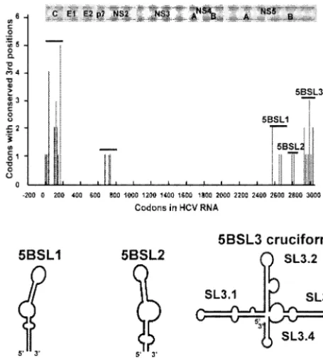
Models of RNA secondary structural elements in the HCV NS5B coding region. Previously, synonymous codons with highly conserved third-position nucleotides were identified throughout the main HCV ORF by analyzing eight diverse HCV sequences (65). Five clusters of such conserved codons were identified: two in the structural gene region and three in the NS5B coding region (Fig. 1). A cluster was defined as a region in which at least three highly conserved codons oc-curred within a 100-codon segment. Analysis revealed that the NS5B coding region did not contain a conserved alternative ORF similar to the one found in the core-encoding region (65). This suggested that the exceptionally conserved codons might
be due to RNA structural elements and RNA signals embed-ded in the polymerase-encoding region.
A combination of thermodynamic RNA folding programs and classical comparative phylogenetic analysis was used to build secondary structural models of RNA elements in the NS5B gene. In a reiterative process, a consensus sequence of 23 diverse NS5B sequences was generated. The Windows ver-sion of Mfold was then applied to a sliding window of 150 to 300 nucleotides, and a series of potential secondary structures was obtained. Structures that were also predicted to be present in the majority of the individual sequences were selected for further investigation. Comparative phylogenetic analysis of 208 sequences was used to identify compensatory base pair changes and to refine the structural models. This process yielded six predicted stem-loop elements (Fig. 1). Five have been pro-posed previously, either in whole or in part. 5BSL1 (codons 111 to 128) and 5BSL2 (codons 333 to 342) were originally noted by Hofacker and colleagues (23) and later modified by Tuplin et al. (61). 5BSL3.1, 5BSL3.3, and 5BSL3.4 were first noted by Smith and Simmonds (56).
The stem-loop element 5BSL3.2 has not been described previously. The 48 nucleotides that fold into 5BSL3.2 encode amino acids 555 to 571 of the NS5B. 5BSL3.2 is one of four stem-loop elements that may comprise a larger structure, which is depicted as a cruciform. The nucleotides comprising the cruciform encode the NS5B amino acids 539 to 591. These amino acids make up the C-terminal portion of the polymerase structure, a linker region, and a membrane anchor (52a), which are downstream of the thumb domain (37) of the protein.
Directed mutagenesis identified a potential CRE in the NS5B coding region.To examine the possible function of these RNA structures in the NS5B coding region, the subgenomic replicon system (6) was used. Con1/SG-Neo (I), the parental replicon in this study, contains an adaptive S2204I mutation in NS5A that greatly enhances replication (6). Multiple muta-tions were introduced into each element to disrupt its pre-dicted RNA structure. In all cases, mutations were chosen to preserve the amino acid sequence of NS5B and to conserve the codon usage frequencies found in the HCV ORF. Codon choice was considered to be important, since silent mutations that introduce rare codons can affect the folding of the result-ing protein (13), possibly by reducresult-ing the rate of translation and favoring the production of-strands (1).
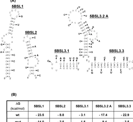
The multiple nucleotide changes designed to disrupt the predicted RNA structures are indicated in Fig. 2A. The Mfold algorithm was used to generate the lowest energy structure predicted for each mutant sequence and to determine its free energy. The predicted mutant RNA structures (data not shown) and their calculated free energies (Fig. 2B) indicated that the mutations should successfully disrupt the target struc-tures.
To assess the impact of these substitutions on HCV RNA replication, we measured the G418 transduction efficiencies of replicons bearing these mutations. In vitro-transcribed RNAs were transfected into Huh-7.5 human hepatoma cells by elec-troporation. A polymerase-defective (pol⫺) replicon was
[image:4.603.48.276.70.322.2]tested in parallel as a negative control. The initial set of mu-tations in the 5BSL3.2 structure were lethal. The 5BSL3.2 mutA replicon produced no G418-resistant colonies (Fig. 3A). In contrast, the G418 transduction efficiencies of the 5BSL1,
FIG. 1. Locations of codons with highly conserved third positions and diagrams of predicted RNA secondary structures in the NS5B coding region. Large clusters of third-base conservation were identi-fied in the core and NS5B by phylogenetic analysis (65). Shown below are models of potential secondary structures identified in the NS5B coding region with methods including the RNA folding program Mfold. The locations of these predicted RNA structures (NS5B codon number) are indicated.
on November 8, 2019 by guest
http://jvi.asm.org/
5BSL2, 5BSL3.1, and 5BSL3.3 mutants were similar to that of the parental replicon (Fig. 3A). These results indicate that 5BSL3.2 contains one or more essential RNA elements that play an important role in HCV RNA replication.
HCV RNA levels of each mutant were also quantified by real-time RT-PCR. The replication-defective subgenomic rep-licon (pol⫺) was used to follow input RNA and to distinguish
it from replicating RNA. As shown in Fig. 3B, at day 1 similar levels of RNA were present for all constructs tested, suggesting that mutations in the predicted structures did not affect RNA stability. Over time, the level of parental replicon RNA was maintained and even increased slightly, whereas that of the pol⫺mutant decreased. By day 3 postelectroporation, the level
of parental RNA was significantly higher than that of the pol⫺
mutant. By day 5, the RNA level of the parental replicon was 200-fold higher than that of the pol⫺mutant. Mutants 5BSL1,
5BSL2, 5BSL3.1, and 5BSL3.3 generated progeny RNA at about the same rate as the parent, paralleling the results of the G418 transduction assay. The RNA levels for these mutants
were about ⬃150- to 300- fold higher than the RNA of the pol⫺mutant on day 5. In contrast, RNA levels of 5BSL3.2
mutA decreased over time, falling at about the same rate as the pol⫺mutant. This indicates that 5BSL3.2 is essential for viral
RNA replication.
[image:5.603.77.510.76.472.2]We then examined the proposed stem-loop structure of 5BSL3.2 in greater detail. Figure 4 summarizes a phylogenetic analysis of 208 GenBank sequences relative to Con1 (a con-sensus sequence of genotype 1b [41]). 5BSL3.2 has a lower helix, an upper helix, a terminal loop, and an internal loop (bulge). The phylogenetic data are presented for each pre-dicted base pair and each unpaired nucleotide. All of the base pairs that are indicated in the diagram can be formed by at least 75% of the 208 sequences, except for the initial base pair, which is present in about 66% of the sequences. Fifteen of the 48 nucleotides in 5BSL3.2 are perfectly conserved among the 208 sequences. Four of the proposed base pairs are comprised of perfectly conserved bases and, thus, by the definitions used in comparative phylogenetic analysis it is not possible for them
FIG. 2. Targeting of each stem-loop structure by site-directed mutagenesis. (A) Predicted RNA secondary structures and the mutations that were introduced. (B) Lowest thermodynamic free energies of wild-type and mutant structures as determined by Mfold.
on November 8, 2019 by guest
http://jvi.asm.org/
to have supporting mutational data. The lower helix of 5BSL3.2 contains 8 bp. Three of the eight are supported by compensatory base pair changes, providing strong evidence that this helix exists in vivo. The upper helix contains six Watson-Crick base pairs. The nucleotides in three of these base pairs are perfectly conserved and thus are not amenable to comparative phylogenetic analysis. Two of the other 3 bp are supported by compensatory base pair changes. The third base pair in this group, which is located at the base of the helix, cannot be confirmed by phylogenetic analysis; however, its existence in the HCV RNA of the replicon is supported by structure probing data (see below).
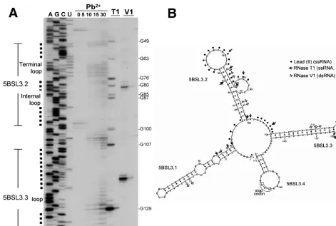
RNA structure probing supports the proposed cruciform.
The 5BSL3.2 element is predicted to be a part of a larger cruciform structure that is formed by 171 nucleotides compris-ing the C-terminal portion of the NS5B codcompris-ing region and the first part of the 3⬘ NTR. Cleavage by various RNases and lead(II) was used to identify nucleotides within this 171-base region that are part of single-stranded or double-stranded structures. To allow possible long-range interactions between nucleotides in the cruciform structure and other parts of func-tional replicon RNA to be detected, we used the same 7,987-base subgenomic RNA transcripts for the structure probing experiments that we used in the transduction assays. Optimal
FIG. 3. Effects of silent mutations in stem-loop structures on replication of the subgenomic replicons. (A) Transduction efficiencies of the mutant constructs in a G418 drug selection assay. After electroporation, 106, 105, or 104transfected cells were plated with feeders as described in
Materials and Methods. After selection for 3 weeks, G418-resistant colonies were stained with crystal violet. Approximate transduction efficiencies (in CFU per microgram of RNA) are indicated. (B) Effects of mutations on HCV RNA levels at various times after transfection. Cells were harvested on day 1, 2, 3, 4, and 5 postelectroporation, and total RNA was harvested. One hundred nanograms of total RNA was analyzed by real-time RT-PCR (see Materials and Methods). HCV RNA was normalized to glyceraldehyde-3-phosphate dehydrogenase RNA. Similar results were obtained in two independent repetitions of the experiment.
on November 8, 2019 by guest
http://jvi.asm.org/
Mg2⫹ and salt concentrations, and also oligonucleotides for
primer extension analyses, were determined using the parental replicon RNA (Table 1; see also Materials and Methods).
The RNA structure probing confirmed many features of the predicted cruciform structure, particularly those in and around 5BSL3.2 and 5BSL3.3 (Fig. 5). RNase T1, which cleaves after
guanosine residues in single-stranded regions, detected G76 in the terminal loop and G85 and G87 in the internal loop of 5BSL3.2. Lead(II), a reagent that primarily cleaves bases in single-stranded loops or in bulges, reacted with bases in both loops of 5BSL3.2 and the terminal loop of 5BSL3.3. In addi-tion, lead(II) cleavage confirmed the single-stranded character of the regions between 5BSL3.1 and 5BSL3.2 as well as be-tween 5BSL3.2 and 5BSL3. Cleavages by RNase V1, which
prefers base-paired nucleotides or stacked helical single-stranded regions, were found in two distinct areas. One was in the upper helix of 5BSL3.2. Cleavage in this region supports the predicted interaction between bases A62 and U81. The other area with a high density of RNase V1cleavages was in the
upper portion of the long helix of 5BSL3.3. Cleavage in this region supports the predicted interaction between C119 and G133. With longer exposure, additional RNase V1cleavages
were detected within the lower helix of 5BSL3.2, indicating that C56 pairs with G97 and that G57 pairs with C96. Finally,
RNase V1cleavage indicated that there might be an
interac-tion between C46 and G9 in 5BSL3.1.
In general, enzymatic and chemical mapping data strongly support the main features of the predicted cruciform structure and the model of 5BSL3.2 in particular. Most of the unex-pected cleavages were minor and mapped to bases that are near the ends of helices. RNase T1cleaved guanosine
nucleo-sides near the base of 5BSL3.1 (G48 and G49) and near the bottom of the 5BSL3.3 helix (G109 and G129). These cleav-ages may reflect breathing or the presence of conformational isomers in the RNA preparation.
Interesting minor RNase T1 cleavage sites, G80 and G63,
were found in the upper helix of 5BSL3.2. G80 and its partner, C65, as well as G63 and its partner, C82, were perfectly con-served among the 208 sequences analyzed (Fig. 4). This degree of conservation cannot be explained by the need to preserve the coding sequence of the NS5B. The genetic code offers alternatives that would preserve both the amino acid sequence and allow a compensatory base pair change at this position. It is possible that the greater stability of the C-G pair, compared to A-U and G-U pairs, constrains the sequence and helps to explain the conservation. In addition, the susceptibility of the upper helix to both single- and double-strand-specific RNases and the unusual sequence conservation of C65-G80 and
G63-FIG. 4. Analysis of conserved nucleotides and covariant base pairs in 5BSL3.2. The sequence and predicted secondary structure of the parental replicon [Con1/SG-Neo (I)] were compared to 208 sequences from GenBank. The number and identity of nucleotides in the GenBank sequences that differ from those in the wild-type replicon are indicated. Base pairs in the 5BSL3.2 structure that were confirmed by compensatory base pair changes are shaded. Nucleotides that are perfectly conserved among all 208 sequences are circled. The number of sequences that have covariant base pairs, wobble base pairs, and those that are unable to form conventional base pairs are indicated. NP (nonpaired) represents pairs of bases that cannot form conventional base pairs. Arrows identify codons whose third-position nucleotides are highly conserved, as defined before (65). On the 5⬘side, UAC is the codon for the NS5B amino acid Tyr555. C53 is the 53rd base according to the 5BSL3 cruciform numbering scheme used
below in Fig. 5, 7, and 8. On the 3⬘side, UGG is the codon for Trp571. In this analysis, the GenBank sequences were also divided into six groups:
genotype 1; genotype 2; genotype 4; genotype 5; genotypes 6 to 9 and 11; and genotypes 3 and 10 (55, 60). Each group was analyzed individually (data not shown). There was general agreement, with one exception. The base pair located at the base of the upper helix does not appear to be formed by sequences of genotype 3 and 10.
on November 8, 2019 by guest
http://jvi.asm.org/
[image:7.603.145.439.70.319.2]C82 could indicate that the upper helix undergoes conforma-tional change in solution and that these base pairs participate in as-yet-unidentified interactions that may be important for the function of 5BSL3.2.
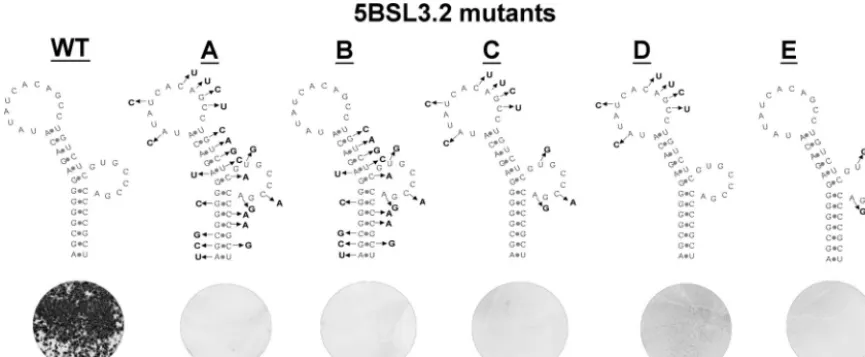
Both the structure and primary sequence of 5BSL3.2 are required for HCV RNA replication. A series of progressively more subtle mutations were introduced into Con1/SG-Neo (I) to define key features of the 5BSL3.2 element. In all cases, the amino acid sequence of NS5B and HCV codon usage were preserved. The original mutant analyzed, 5BSL3.2 mutA, con-tained 22 substitutions in both the stem and loop sequences. 5BSL3.2 mutB contained multiple substitutions within the base-paired regions and was also not viable (Fig. 6). RNA structure probing revealed that the silent changes in both mutA and mutB did indeed disrupt Watson-Crick interactions (data not shown).
To determine the impact of substitutions in loop nucleo-tides, 5BSL3.2 mutC, mutD, and mutE were produced. These three constructs separate the clusters of loop mutations present in mutA. mutC has changes in both loops and mutD has six changes within the terminal loop, whereas mutE has three changes in the internal loop. Remarkably, the G418 transduction assay revealed that all three sets of loop muta-tions were lethal. This implies that both the terminal and the
internal loops contain primary sequence elements that are crucial for HCV RNA replication.
To gain further insight into the sequence requirements of the two 5BSL3.2 loops, more subtle substitutions were made and tested in the G418 transduction assay (Fig. 7). Single point mutations made at positions 71 (U71C) and 74 (C74U) blocked HCV RNA replication. In addition, the triple muta-tion of AGC to UCU at posimuta-tions 75, 76, and 77 also blocked replication. In contrast, position 68 (A68C) could be changed without affecting HCV replication significantly. The mutant RNAs were predicted to fold into structures of similar shape and thermostability as wild-type RNA, and structural probing did not reveal any irregularities (data not shown). These results emphasize the importance of the primary sequence of the terminal loop.
The effects of substitutions in and around the internal loop suggested that the structure of this region, as well as its primary sequence, were important. Three mutant sequences contained substitutions in and around the 5⬘portion of the internal loop. These mutations involve the codon that specifies Arg566 of
[image:8.603.55.526.67.384.2]NS5B. Mutant C84A/U86G was not viable; mutant C84A/ U86A replicated at reduced levels, and mutant U86G repli-cated efficiently. The change of C84 to A would be expected to disrupt a G-C base pair at the bottom of the upper helix,
FIG. 5. RNA structure probing of the 5BSL3 cruciform. (A) Primer extension analysis of RNA treated with RNase T1(1 or 0.1 U) and RNase
V1(0.1 or 0.01 U), or lead(II) acetate (0, 5, 10, 15, and 30 mM; see Materials and Methods). cDNA products were resolved in a 12% PAGE–7
M urea denaturing gel. (B) Comparison of the predicted RNA secondary structure with the results of enzymatic and chemical probing. As indicated, the symbols mark the position of cleavage or modification by the different probing reagents.
on November 8, 2019 by guest
http://jvi.asm.org/
potentially altering the overall structure of the 5BSL3.2 ele-ment. Such a disruption could explain the phenotypes of mu-tant C84A/U86G (lethal) versus mumu-tant U86G (viable). The RNA structure probing data presented below help to account for the phenotype of mutant C84A/U86A (attenuated, but viable) by demonstrating that this replicon retained a structure similar to wild-type 5BSL3.2. Additional studies demonstrated that the sequence of the 3⬘ portion of the internal loop is critical. Mutant C90A was lethal (Fig. 7B), as might be ex-pected because C90 is a perfectly conserved nucleotide (Fig. 4). Mutants A92C and A92U were viable, although the latter produced 10-fold fewer colonies in the G418 transduction as-say. Mutant A92G was lethal, which was surprising given that G occurs at this position in many HCV sequences in GenBank.
C84A/U86G disrupts the upper helix of 5BSL3.2. RNA structure probing was carried out to examine possible alter-ations in 5BSL3.2 that might account for the nonviability of the C84A/U86G mutant compared to viable mutants C84A/U86A and U86G. The RNase T1, RNase V1, and lead(II) cleavage
patterns of mutants C84A/U86A and U86G were similar to those of the parental RNA (Fig. 8). In contrast, the RNase T1
digestion pattern of mutant C84A/U86G revealed two addi-tional intense bands (Fig. 8). These bands resulted from en-hanced cleavage after G63 and G80, two nucleotides that had not been mutated. This indicates that the C84A/U86G substi-tutions modified the structure of the 5BSL3.2 upper helix and that this alteration correlates with the failure of mutant C84A/ U86G to replicate.
The differences noted between the digested products of C84A/U86A and those of C84A/U86G suggest that the substi-tution of A for C at position 84 can be tolerated, but not if G is also present at position 86. In fact, G85 and G87 of the mutant C84A/U86A were targeted by RNase T1strongly, but
not in C84A/U86G. Having a mutation at C84A would disrupt the base pair at the bottom of the upper stem, which would enlarge the internal loop. This could increase the accessibility of G85 and G87 to RNase T1digestion. However, the C84A/
U86G mutant did not have the same cleavage pattern as the C84A/U86A mutant, despite the fact that they both contain the C84A change. In the absence of a stable base pair at the bottom of the upper helix, a G at position 86 may interact with nucleotides that would otherwise contribute to this helix, thereby disrupting the overall structure. As noted above, G80 is a minor RNase T1cleavage site in wild-type RNA, indicating
that the base pair C65-G80 most likely exists as a meta-stable interaction. Taken together, these observations suggest that base pairs in the upper helix establish an essential scaffold of 5BSL3.2. One likely possibility is that the helices hold the bases in the terminal and internal loops in an optimal orientation for interaction with other components of the viral replication com-plex.
Cruciform duplications and attempts at rescue.Some picor-navirus CREs found in the polyprotein coding region can func-tion when relocated to other genome posifunc-tions. Such con-structs, liberated from the constraint of maintaining the protein-coding sequence, can be very useful for fine mutational analysis of CREs. To examine this possibility for 5BSL3.2, we created a convenient insertion site in the replicon between the Neorgene and the EMCV IRES (Fig. 9A, construct b).
Inser-tion of an addiInser-tional wild-type or mutant (5BSL3.2 mutA) copy of 5BSL3 at this position was tolerated without any reduction in replication efficiency (Fig. 9A, constructs c and d). However, when these insertions were tested on the otherwise nonviable 5BSL3.2 mutA background, neither the mutant nor the wild-type 5BSL3 was able to restore replication (Fig. 9A, constructs e and f).
[image:9.603.76.509.71.249.2]We also tested the 3⬘NTR as a possible CRE insertion site. A 440-base sequence that included wild-type 5BSL3 (NS5B codons 447 to 591) was inserted downstream of the polyprotein stop codon (Fig. 9B; see also Materials and Methods). The presence of this duplication dramatically reduced replication efficiency even in the absence of 5BSL3.2 mutations (Fig. 9B, construct c). A construct containing the 5BSL3.2 mutB se-quence in both the polyprotein and 3⬘ NTR positions was
FIG. 6. Genetic analysis of mutant subgenomic replicons targeting the 5BSL3.2 structure. The silent mutations introduced into each construct are shown. Mutant 5BSL3.2 mutA contains mutations throughout the structure. 5BSL3.2 mutB contains the same changes as mutA except for the mutations in the terminal loop. Mutant 5BSL3.2 C has changes in both the terminal and internal loops, which are separated in mutD and mutE, respectively. The effects of these changes were examined by G418 drug selection. Plates with 106transfected cells are shown.
on November 8, 2019 by guest
http://jvi.asm.org/
nonviable, as expected (Fig. 9B, construct d). However, when the 3⬘ NTR copy was the wild-type sequence (Fig. 9B, con-struct e), a few G418-resistant colonies were obtained but in reduced numbers compared to the parental duplication con-struct (Fig. 9B, concon-struct c). Although we have only examined a limited number of constructs, it appears that the context of the HCV NS5B CRE is important for optimal function.
DISCUSSION
We used custom-designed sequence alignment software, thermodynamic RNA folding programs, and comparative phy-logenetic analysis to build models of six RNA stem-loop struc-tures in the HCV NS5B gene. Five out of the six elements identified in this study were reported previously. 5BSL1 was first identified by Hofacker et al. (23) and later by Tuplin et al., who designated it SL7730 (61). 5BSL2 is the tip of a larger structure designated SL8376 by Tuplin et al. Both Hofacker et al. and Tuplin et al. mapped a structure designated SL8828
(codons 474 to 501), which did not appear in our search, and Tuplin et al. identified an additional structure that did not meet our selection criteria, SL8926 (codons 509 to 530). 5BSL3.1 (codons 539 to 554) and 5SB3.3 (codons 575 to 585) correspond to SL9011 and SL9118, respectively (61). 5BSLSL3.1 was first identified by Smith and Simmonds, who also described 5BSL3.3 and 5BSL3.4 (56). As a group, these studies and our investigation indicate that an unusual percent-age of the nucleotides in the HCV NS5B coding region may form distinctive RNA structures. The cruciform, which in-cludes the final 171 bases of the main ORF, may be part of an even-larger structure. The NS5B codons immediately up-stream, 536, 537, and 538, have exceptionally conserved third-position nucleotides, as defined previously (65). Two stem-loop elements identified by Tuplin et al., SL8828 and SL8926, lie only slightly further upstream.
We used site-directed mutagenesis to analyze five of the elements identified in our search. In an initial screen, multiple mutations designed to disrupt the predicted RNA structures
FIG. 7. Mutational analysis of the terminal and internal loops of 5BSL3.2. (A) One-, two-, or three-nucleotide substitutions were introduced at specific positions in the 5BSL3.2 structure, as indicated. (B) Effects of these mutations on G418 transduction efficiency. Plates with 106
transfected cells are shown.
on November 8, 2019 by guest
http://jvi.asm.org/
were engineered while preserving the amino acid sequence of the NS5B protein. Uniquely, mutations in 5BSL3.2 blocked replicon replication, indicating that 5BSL3.2 is an essential CRE. Mutations in 5BSL1, 5BSL2, 5BSL3.1, and 5BSL3.3 did not markedly diminish replication in cell culture. We also tested three different mutant subgenomic replicons containing different combinations of silent mutations of the SL8926 ele-ment, proposed by Tuplin et al. None could be distinguished from the wild-type replicon in the G418 selection assay (data not shown). However, these negative results should not be taken as definitive evidence that these RNA elements lack functional importance. They may exert subtle effects, function in different cellular environments, or perform other functions in the HCV life cycle (such as packaging) that were not assayed using the current replicon systems. We did not perform di-rected mutagenesis on 5BSL3.4; however, other studies suggest that this stem-loop may not be essential. 5BSL3.4 includes 8 nucleotides of the variable region of the 3⬘ NTR, and HCV RNA transcripts lacking these 8 bases and the subsequent 16 bases were still infectious in a chimpanzee (69). Deletion of the entire variable region significantly reduced replication of a subgenomic replicon but was not lethal (14).
Essential features of the CRE 5BSL3.2.5BSL3.2 is about 50 bases in length and has an 8-bp lower helix, a 6-bp upper helix, a 12-base terminal loop, and an 8-base internal loop (bulge).
Disruption of 5BSL3.2 had a striking impact on HCV replica-tion. Many of the mutant replicons with changes in this ele-ment did not yield any G418-resistant colonies during drug selection. Directed mutagenesis and structure probing demon-strated that sequences in both loops are important for viability and indicated that the upper helix is an essential scaffold that is easily disrupted.
Primary sequence requirements in the loops of 5BSL3.2.
Analysis of a series of mutants with one, two, and three base changes revealed that many of the nucleotides in the loops of 5BSL3.2 are highly constrained and may be invariable. Point mutations at five positions (71, 74, 77, 90, and 92) were lethal (Fig. 7). Structure probing did not reveal any irregularities in the cleavage products of RNAs with mutations in these loca-tions (data not shown). These results indicate that the primary sequences of the loop regions, rather than their contributions to the global structure, are important.
In general, we found a high correlation between the results of directed mutagenesis and the results of phylogenetic se-quence analysis. Typically, mutations that were lethal in the replicon were perfectly conserved among the 208 sequences analyzed, while substitutions that retained viability were rep-resented in the set of natural sequences. For example, mutant A68C had a wild-type phenotype, and this position is variable in nature (Fig. 4). Conversely, mutant C74U is nonviable, and
FIG. 8. Destabilization of the 5BSL3.2 upper helix by C84A/U86G. RNA structure probing was used to examine the 5BSL3.2 structure for mutants C84A/U86G, C84A/U86A, and U86G. RNAs were treated with RNase T1(1 U), RNase V1(0.1 U), or lead(II) (15 and 30 mM) followed
by primer extension analysis (see Materials and Methods). cDNA products were resolved on a 12% PAGE–7 M urea denaturing gel. Unique T1
cleavages observed for mutant C84A/U86G RNA are indicated by arrows. At the right, the locations of these cleavages are also shown on the secondary structure model of 5BSL3.2.
on November 8, 2019 by guest
http://jvi.asm.org/
C74 is perfectly conserved in all 208 sequences. The discrep-ant results noted above for position 92 may indicate that a G at this site must be accompanied by a second sequence change elsewhere in the genome. By modifying our selection
conditions, we are attempting to obtain revertants with sec-ond-site mutations that restore viability. Such revertants may reveal new RNA-RNA interactions involving bases in the 5BSL3.2 loops.
FIG. 9. Attempts to rescue the 5BSL3.2 mutant subgenomic replicon by adding a sequence harboring the cruciform structure. (A) Effects of an additional copy of the cruciform between the Neorgene and the EMCV IRES. Schematic diagrams of the constructs tested are shown. The
phenotype of these mutant constructs was compared based on G418 transduction efficiency. (a) Parental subgenomic replicon, Con1/SG-Neo (I); (b) Con1/SG-Neo (I) with aBsiWI restriction site inserted after the Neo stop codon; (c) wild-type copy of the cruciform inserted after the Neor
gene; (d) mutant (mutA) copy of the cruciform on the parental background; (e) mutant (mutA) copy of the cruciform inserted in the context of 5BSL3.2 mutA; (f) wild-type copy of the cruciform inserted in the context of 5BSL3.2 mutA. (B) Effects of inserting an additional copy of the cruciform after the polyprotein stop codon. A schematic diagram and a crystal violet-stained plate indicating G418 transduction efficiency are shown for each construct. (a) Parental replicon; (b) 5BSL3.2 mutB; (c) parental replicon with an additional wild-type cruciform in the 3⬘NTR; (d) 5BSL3.2 mutB with an additional mutB cruciform in the 3⬘NTR; (e) 5BSL3.2 mutB with an additional wild-type cruciform in the 3⬘NTR.
on November 8, 2019 by guest
http://jvi.asm.org/
The upper helix is a scaffold.The importance of the upper helix is strongly supported by three mutants. Mutant C84A/ U86G was not viable. This mutant is unable to form the C-G base pair that lies at the bottom of the upper helix in wild-type sequences (Fig. 8). Elimination of this C-G base pair and the change of the loop nucleotide 86 from U to G caused misfold-ing and disrupted the upper helix, as revealed by RNA struc-ture probing data. Interestingly, mutant C84A/U86A was via-ble and maintained a similar pattern of cleavage products as wild type, providing a clear indication that preservation of the 5BSL3.2 structure correlates with viability. The contrast be-tween C84A/U86G and C84A/U86A suggested that a G at position 86 is able to alter the upper helix, while an A at position 86 finds no suitable partners within the nucleotides that make up the stem. When the upper helix is intact, as in mutant U86G, the wild-type cleavage pattern and level of rep-lication are preserved. This implies that one essential function of the upper helix may be to position the loop bases for inter-action with specific binding partners in the HCV replication complex.
Location dependence of 5BSL3.2. Certain CREs of other viruses, such as human rhinovirus (43), foot-and-mouth disease virus (42), and poliovirus (18), can be translocated and remain functional. If the function of the 5BSL3.2 CRE were similarly independent of its location, it might be possible to rescue mutant replicons by adding a wild-type version of the 5BSL3 cruciform to the mutant replicon. To test this possibility, the wild-type 5BSL3 cruciform domain was inserted (incis) into two different locations in mutant subgenomic replicons. When a wild-type 200-nucleotide-long segment was inserted after the stop codon of the Neorgene in the mutant subgenomic
repli-con, there was no rescue. In addition, there was no significant change in the level of replication in the wild-type replicon when either a wild-type or a mutant copy of the 5BSL3 cruci-form domain was inserted into the wild-type replicon at this site (Fig. 9A, constructs c and d). These results suggest this structural element has no impact on the replicon when it is placed out of context near the 5⬘terminus of the RNA. How-ever, when the wild-type cruciform domain was inserted into the variable region of the 3⬘NTR of the wild-type replicon, a significant reduction in replication was observed (Fig. 9B, con-struct c). It is not clear whether this reduction was due to the presence of two wild-type CREs competing for interacting partners when placed in close proximity or to the disruption of a higher-order RNA structure important for HCV replication. It is interesting that insertion of an IRES-Neorcassette into
the 3⬘NTR variable region also compromised replicon viability (D. Jacobson, G. Randall, and C. M. Rice, unpublished data). These observations, with two different sequences, suggest that insertions in this region may be poorly tolerated in general. Interestingly, when a wild-type copy of the cruciform was in-serted into the 3⬘NTR of a replicon with a mutated 5BSL3.2 element, a small number of G418-resistant colonies were pro-duced. While these results are promising and suggest that the CRE may be able to function outside its normal polyprotein context, further work is needed to define the positional con-straints and sequence context important for CRE 5BSL3.2 function.
Unlike CREs found in the HCV NTRs, 5BSL3.2 is in the coding region. From the standpoint of genome design, it is
interesting to consider the potential advantages and disadvan-tages of placing CREs within coding sequences. The overlap of a CRE and a coding sequence places a constraint on the evolution of escape mutants if the protein stimulates a signif-icant immune response, and it impedes the evolution of the CRE. However, the overlap offers the advantage of allowing a genome to be more compact, and it may give the CRE a unique ability to regulate translation of the coding gene. Direct com-munication with the translation machinery is known to be essential for CREs involved in the frameshift required for expression of the Rous sarcoma virus polymerase (27) and for CREs that influence protein conformation, e.g., by slowing the rate of translation (13, 59). A conserved pseudoknot in mRNAs of prion proteins may nucleate protein folding (1). However, the CRE in NS5B of HCV does not seem to act at the level of translation. Cell-free extracts from Huh-7.5 cells revealed no significant differences in the production of viral proteins between wild type and the 5BSL3.2 mutants (data not shown); however, if the CRE influences the conformation of the polymerase or translation termination, these effects would not have been detected in our assay.
Potential role of 5BSL3.2 in HCV replication.Currently, it is not clear how the 5BSL3.2 structure is involved in HCV rep-lication. 5BSL3.2 is just upstream of the polyprotein stop codon, at a site that would appear to provide the CRE with ready access to newly synthesized polymerase. The placement of the CRE in the C-terminal portion of the coding region may simultaneously promote binding to the nascent polymerase and assembly of the minus-strand initiation complex. Such CREs are present in the intergenic region of RNA3 of brome mosaic virus (58) and at the S and M sites of the bacteriophage Qgenome (44). Cheng et al. reported that the NS5B protein has a strong affinity for the 200 nucleotides upstream of the stop codon that terminates the main ORF, a region that in-cludes 5BSL3.2. The interaction between the NS5B protein and the RNA of the NS5B coding region was strong enough to withstand competition with other HCV RNA regions, such as the 5⬘and 3⬘NTRs (9).
The life cycles of many RNA viruses require long-range interactions between RNA elements. These interactions can be mediated by base pairing or by proteins. In porcine reproduc-tive and respiratory syndrome virus, a kissing interaction be-tween stem-loop structures in the ORF and the 3⬘ NTR is required for RNA replication (63). Other examples of long-distance RNA-RNA interactions important for RNA virus rep-lication have been found for flaviviruses (31, 72), flock house virus (39), RNA coliphage Q(34), and barley yellow dwarf luteovirus (20). In the case of the HCV 5BSL3.2 CRE, we did not detect any obvious interactions between essential bases in the loops and sequences elsewhere; rather, these regions were susceptible to cleavage by lead(II) and RNase T1, indicating
that they were single stranded under the experimental condi-tions. It is possible that bases in the 5BSL3.2 structure interact with CREs in other parts of the RNA genome but do so only in the presence of a viral protein(s) or host factor(s) that was not available. A protein-protein bridge between the 5⬘ clover-leaf and the 3⬘ poly(A) tail is required for poliovirus minus-strand RNA initiation (4, 22).
In summary, we have used phylogenetic modeling, site-di-rected mutagenesis, and RNA structure probing to identify a
on November 8, 2019 by guest
http://jvi.asm.org/
new CRE, 5BSL3.2, that is important for HCV RNA replica-tion. This CRE lies in the polyprotein coding sequence corre-sponding to NS5B codons 555 to 571. We are currently at-tempting to define the step(s) in HCV RNA replication that requires 5BSL3.2 and using genetic and biochemical ap-proaches to identify protein factors and other RNA elements important for 5BSL3.2 function.
ACKNOWLEDGMENTS
We thank James Fan, Jack Heitpas, Hernan Jaramillo, and Merna Torres for expert technical assistance. We are also grateful to many colleagues for helpful discussions during the course of this work and to Brett Lindenbach, Margaret MacDonald, Joseph Marcotrigiano, and Timothy Tellinghuisen for critical reading of the manuscript.
This work was supported by grants from the Public Health Service (CA57973, AI40034, DK052071, and DA16156) and the Greenberg Medical Research Institute.
REFERENCES
1. Barrette, I., G. Poisson, P. Gendron, and F. Major.2001. Pseudoknots in prion protein mRNAs confirmed by comparative sequence analysis and
pattern searching. Nucleic Acids Res.29:753–758.
2. Bartenschlager, R., M. Junker-Niepmann, and H. Schaller.1990. The P gene product of hepatitis B virus is required as a structural component for
genomic RNA encapsidation. J. Virol.64:5324–5332.
3. Bartenschlager, R., and V. Lohmann.2000. Replication of hepatitis C virus.
J. Gen. Virol.81:1631–1648.
4. Barton, D. J., B. J. O’Donnell, and J. B. Flanegan.2001. 5⬘cloverleaf in poliovirus RNA is a cis-acting replication element required for
negative-strand synthesis. EMBO J.20:1439–1448.
5. Baumstark, T., and P. Ahlquist.2001. The brome mosaic virus RNA3 in-tergenic replication enhancer folds to mimic a tRNA TpsiC-stem loop and is
modified in vivo. RNA7:1652–1670.
6. Blight, K. J., A. A. Kolykhalov, and C. M. Rice.2000. Efficient initiation of
HCV RNA replication in cell culture. Science290:1972–1974.
7. Blight, K. J., J. A. McKeating, and C. M. Rice.2002. Highly permissive cell lines for hepatitis C virus genomic and subgenomic RNA replication. J.
Vi-rol.76:13001–13014.
8. Blight, K. J., and C. M. Rice.1997. Secondary structure determination of the
conserved 98-base sequence at the 3⬘terminus of hepatitis C virus genome
RNA. J. Virol.71:7345–7352.
8a.Boulant, S., M. Becchi, F. Penin, and J. P. Lavergne.2003. Unusual multiple recoding events leading to alternative forms of hepatitis C virus core protein
from genotype 1b. J. Biol. Chem.278:45785–45792.
9. Cheng, J. C., M. F. Chang, and S. C. Chang.1999. Specific interaction
between the hepatitis C virus NS5B RNA polymerase and the 3⬘end of the
viral RNA. J. Virol.73:7044–7049.
10. Choi, J., Z. Xu, and J. H. Ou.2003. Triple decoding of hepatitis C virus RNA
by programmed translational frameshifting. Mol. Cell. Biol.23:1489–1497.
11. Choo, Q.-L., G. Kuo, A. J. Weiner, L. R. Overby, D. W. Bradley, and M. Houghton.1989. Isolation of a cDNA clone derived from a blood-borne
non-A, non-B viral hepatitis genome. Science244:359–362.
12. Choo, Q.-L., K. H. Richman, J. H. Han, K. Berger, C. Lee, C. Dong, C. Gallegos, D. Coit, A. Medina-Selby, P. J. Barr, A. J. Weiner, D. W. Bradley, G. Kuo, and M. Houghton.1991. Genetic organization and diversity of the
hepatitis C virus. Proc. Natl. Acad. Sci. USA88:2451–2455.
13. Cortazzo, P., C. Cervenansky, M. Marin, C. Reiss, R. Ehrlich, and A. Deana.
2002. Silent mutations affect in vivo protein folding in Escherichia coli.
Biochem. Biophys. Res. Commun.293:537–541.
14. Friebe, P., and R. Bartenschlager.2002. Genetic analysis of sequences in the
3⬘nontranslated region of hepatitis C virus that are important for RNA
replication. J. Virol.76:5326–5338.
15. Fukushi, S., M. Okada, T. Kageyama, F. B. Hoshino, K. Nagai, and K. Katayama.2001. Interaction of poly(rC)-binding protein 2 with the 5⬘
-ter-minal stem loop of the hepatitis C-virus genome. Virus Res.73:67–79.
16. Galibert, F., E. Mandart, F. Fitoussi, P. Tiollais, and P. Charnay.1979. Nucleotide sequence of the hepatitis B virus genome (subtype ayw) cloned in
E. coli. Nature281:646–650.
17. Gerber, K., E. Wimmer, and A. V. Paul.2001. Biochemical and genetic studies of the initiation of human rhinovirus 2 RNA replication: purification and enzymatic analysis of the RNA-dependent RNA polymerase 3D(pol).
J. Virol.75:10969–10978.
18. Goodfellow, I., Y. Chaudhry, A. Richardson, J. Meredith, J. W. Almond, W. Barclay, and D. J. Evans.2000. Identification of acis-acting replication
element within the poliovirus coding region. J. Virol.74:4590–4600.
19. Goodfellow, I. G., D. Kerrigan, and D. J. Evans.2003. Structure and function
analysis of the poliovirus cis-acting replication element (CRE). RNA9:124–
137.
20. Guo, L., E. M. Allen, and W. A. Miller.2001. Base-pairing between untrans-lated regions facilitates translation of uncapped, nonpolyadenyuntrans-lated viral
RNA. Mol. Cell7:1103–1109.
21. Hahm, B., Y. K. Kim, J. H. Kim, T. Y. Kim, and S. K. Jang.1998.
Hetero-geneous nuclear ribonucleoprotein L interacts with the 3⬘ border of the
internal ribosomal entry site of hepatitis C virus. J. Virol.72:8782–8788.
22. Herold, J., and R. Andino.2001. Poliovirus RNA replication requires
ge-nome circularization through a protein-protein bridge. Mol. Cell7:581–591.
23. Hofacker, I. L., M. Fekete, C. Flamm, M. A. Huynen, S. Rauscher, P. E. Stolorz, and P. F. Stadler.1998. Automatic detection of conserved RNA structure elements in complete RNA virus genomes. Nucleic Acids Res.
26:3825–3836.
24. Honda, M., E. A. Brown, and S. M. Lemon.1996. Stability of a stem-loop involving the initiator AUG controls the efficiency of internal initiation of
translation on hepatitis C virus RNA. RNA2:955–968.
25. Honda, M., L. H. Ping, R. C. Rijnbrand, E. Amphlett, B. Clarke, D. Row-lands, and S. M. Lemon.1996. Structural requirements for initiation of translation by internal ribosome entry within genome-length hepatitis C virus
RNA. Virology222:31–42.
26. Isoyama, T., N. Kamoshita, K. Yasui, A. Iwai, K. Shiroki, H. Toyoda, A. Yamada, Y. Takasaki, and A. Nomoto.1999. Lower concentration of La protein required for internal ribosome entry on hepatitis C virus RNA than
on poliovirus RNA. J. Gen. Virol.80:2319–2327.
27. Jacks, T., and H. E. Varmus.1985. Expression of the Rous sarcoma virus pol
gene by ribosomal frameshifting. Science230:1237–1242.
28. James, B. D., G. J. Olsen, and N. R. Pace.1989. Phylogenetic comparative
analysis of RNA secondary structure. Methods Enzymol.180:227–239.
29. Joost Haasnoot, P. C., R. C. Olsthoorn, and J. F. Bol.2002. The Brome mosaic virus subgenomic promoter hairpin is structurally similar to the iron-responsive element and functionally equivalent to the minus-strand core
promoter stem-loop C. RNA8:110–122.
30. Junker-Niepmann, M., R. Bartenschlager, and H. Schaller.1990. A short cis-acting sequence is required for hepatitis B virus pregenome
encapsida-tion and sufficient for packaging of foreign RNA. EMBO J.9:3389–3396.
31. Khromykh, A. A., P. L. Sedlak, and E. G. Westaway.2000.cis- andtrans
-acting elements in flavivirus RNA replication. J. Virol.74:3253–3263.
32. Kieft, J. S., K. Zhou, A. Grech, R. Jubin, and J. A. Doudna.2002. Crystal structure of an RNA tertiary domain essential to HCV IRES-mediated
translation initiation. Nat. Struct. Biol.9:370–374.
33. Kieft, J. S., K. Zhou, R. Jubin, and J. A. Doudna.2001. Mechanism of
ribosome recruitment by hepatitis C IRES RNA. RNA7:194–206.
34. Klovins, J., V. Berzins, and J. van Duin.1998. A long-range interaction in
QRNA that bridges the thousand nucleotides between the M-site and the
3⬘end is required for replication. RNA4:948–957.
35. Knaus, T., and M. Nassal.1993. The encapsidation signal on the hepatitis B virus RNA pregenome forms a stem-loop structure that is critical for its
function. Nucleic Acids Res.21:3967–3975.
36. Kolykhalov, A. A., S. M. Feinstone, and C. M. Rice.1996. Identification of a
highly conserved sequence element at the 3⬘terminus of hepatitis C virus
genome RNA. J. Virol.70:3363–3371.
37. Lesburg, C. A., M. B. Cable, E. Ferrari, Z. Hong, A. F. Mannarino, and P. C. Weber.1999. Crystal structure of the RNA-dependent RNA polymerase from hepatitis C virus reveals a fully encircled active site. Nat. Struct. Biol.
6:937–943.
38. Lindell, M., P. Romby, and E. G. Wagner.2002. Lead(II) as a probe for
investigating RNA structure in vivo. RNA8:534–541.
39. Lindenbach, B. D., J. Y. Sgro, and P. Ahlquist.2002. Long-distance base pairing in flock house virus RNA1 regulates subgenomic RNA3 synthesis
and RNA2 replication. J. Virol.76:3905–3919.
40. Lobert, P. E., N. Escriou, J. Ruelle, and T. Michiels.1999. A coding RNA sequence acts as a replication signal in cardioviruses. Proc. Natl. Acad. Sci.
USA96:11560–11565.
41. Lohmann, V., F. Korner, J. O. Koch, U. Herian, L. Theilmann, and R. Bartenschlager.1999. Replication of subgenomic hepatitis C virus RNAs in
a hepatoma cell line. Science285:110–113.
42. Mason, P. W., S. V. Bezborodova, and T. M. Henry.2002. Identification and
characterization of acis-acting replication element (CRE) adjacent to the
internal ribosome entry site of foot-and-mouth disease virus. J. Virol.76:
9686–9694.
43. McKnight, K. L., and S. M. Lemon.1998. The rhinovirus type 14 genome contains an internally located RNA structure that is required for viral
rep-lication. RNA4:1569–1584.
44. Miranda, G., D. Schuppli, I. Barrera, C. Hausherr, J. M. Sogo, and H. Weber.1997. Recognition of bacteriophage Qplus strand RNA as a
tem-plate by Qreplicase: role of RNA interactions mediated by ribosomal
proteins S1 and host factor. J. Mol. Biol.267:1089–1103.
45. Morasco, B. J., N. Sharma, J. Parilla, and J. B. Flanegan.2003. Poliovirus cre(2C)-dependent synthesis of VPgpUpU is required for positive- but not
negative-strand RNA synthesis. J. Virol.77:5136–5144.
46. Pudi, R., S. Abhiman, N. Srinivasan, and S. Das.2003. Hepatitis C virus internal ribosome entry site-mediated translation is stimulated by specific
![FIG. 4. Analysis of conserved nucleotides and covariant base pairs in 5BSL3.2. The sequence and predicted secondary structure of the parentalreplicon [Con1/SG-Neo (I)] were compared to 208 sequences from GenBank](https://thumb-us.123doks.com/thumbv2/123dok_us/240338.58170/7.603.145.439.70.319/analysis-conserved-nucleotides-covariant-secondary-structure-parentalreplicon-sequences.webp)