This is a repository copy of
Bayesian optimization for fitting 3D morphable models of brain
structures
.
White Rose Research Online URL for this paper:
http://eprints.whiterose.ac.uk/116581/
Version: Accepted Version
Proceedings Paper:
García, H.F., Álvarez, M.A. orcid.org/0000-0002-8980-4472 and Orozco, Á.A. (2017)
Bayesian optimization for fitting 3D morphable models of brain structures. In: Progress in
Pattern Recognition, Image Analysis, Computer Vision, and Applications. CIARP 2016.
21st Iberoamerican Congress, CIARP 2016, 08/11/2016-11/11/2016, Lima, Peru. Lecture
Notes in Computer Science, 10125 . Springer Verlag , pp. 291-299. ISBN 9783319522760
https://doi.org/10.1007/978-3-319-52277-7_36
The final publication is available at Springer via
http://dx.doi.org/10.1007/978-3-319-52277-7_36.
[email protected] https://eprints.whiterose.ac.uk/
Reuse
Unless indicated otherwise, fulltext items are protected by copyright with all rights reserved. The copyright exception in section 29 of the Copyright, Designs and Patents Act 1988 allows the making of a single copy solely for the purpose of non-commercial research or private study within the limits of fair dealing. The publisher or other rights-holder may allow further reproduction and re-use of this version - refer to the White Rose Research Online record for this item. Where records identify the publisher as the copyright holder, users can verify any specific terms of use on the publisher’s website.
Takedown
If you consider content in White Rose Research Online to be in breach of UK law, please notify us by
Models of Brain Structures
Hern´an F. Garc´ıa, Mauricio A. ´Alvarez and ´Alvaro A. Orozco
Grupo de Investigaci´on en Autom´atica, Universidad Tecnol´ogica de Pereira, Pereira, Colombia,
{hernan.garcia,malvarez,aaog}@utp.edu.co
Abstract. Localize target areas in deep brain stimulation is a difficult task, due to the shape variability that brain structures exhibit between patients. The main problem in this process is that the fitting procedure is carried out by a registra-tion method that lacks of accuracy. In this paper we proposed a novel method for 3D brain structure fitting based on Bayesian optimization. We use a morphable model in order to capture the shape variability in a given set of brain structures. Then from the trained model, we perform a Bayesian optimization task with the aim to find the best shape parameters that deform the trained model, and fits ac-curately to a given brain structure. The experimental results show that by using an optimization framework based on Bayesian optimization, the model performs an accurate fitting over cortical brain structures (thalamus, amygdala and ventricle) in comparison with common fitting methods, such as iterative closest point.
Keywords: Bayesian optimization, 3D Brain structures, Shape fitting, Morphable model.
1
Introduction
2 Hern´an F. Garc´ıa, Mauricio A. ´Alvarez and ´Alvaro A. Orozco
volumes could improve the robustness (generalizability and accuracy) of the target area localization [3].
The model-based approaches such as morphable models (MM), make use of the prior knowledge of the shape variability in a set of images (MRI volumes) and typi-cally finds the best match between the model and the new image (brain structure) [6]. In medical image analysis, the use of prior information (shape contour of the brain structures labeled by a medical specialist), combined with some model that can be able to represent a shape contour, leads to an accurate fitting of a given brain structure [3]. A commonly used method for fitting morphable models, is the Iterative Closest Point (ICP) [6]. This method establishes closest point associations from one shape to another, and finds a rigid transformation that minimizes an Euclidean error between shapes. However, due to the large deformations that the brain volumes present over different patients (i.e. different thickness for ventral brain structures), the performance of the fit-ting process becomes inaccurate [7]. The reason for this performance, is that the global minimum of the cost function that measures the fitting process (mean square error be-tween the deformed and the target brain structure), often does not corresponds to the optimal fitting (That is, a rigid transform only estimates the scale, rotation and transla-tion parameters, discarding the shape parameters that control the deformatransla-tions) [8].
Bayesian optimization (BO) provides an elegant approach for the global optimiza-tion problem, in which a given cost funcoptimiza-tion is minimized in a probabilistic way [9]. For continuous functions, Bayesian optimization assumes that the unknown function is sam-pled from a Gaussian process, and maintains a posterior distribution for this function as observations [10]. In the fitting process of the morphable model, these observations are the measure of generalization performance (matching accuracy) under different set-tings of the hyperparameters that we wish to optimize (shape parameters that control the deformations) [11]. In this paper we propose a Bayesian optimization framework for fitting 3D morphable models of brain structures. We use as morphable model, a point distribution model (PDM) in order to capture the shape variability in a set of brain volumes (brain structures related with Parkinson’s disease). From the trained model, we perform a fitting process based on Bayesian optimization in order to find the optimal match between the morphable model and a given brain structure. The main contribu-tion of our work, is based on the Bayesian optimizacontribu-tion process that finds the shape parameters that control the deformations of a morphable model in a probabilistic way.
2
Materials and Methods
2.1 Database
2.2 3D Brain Model
We use a 3D brain model based on a PDM to represent the shape of the brain structures. The shape information is captured by the vertexes information (point-cloud data in the R3
) from the mesh data that represents each brain structure. Besides, the model uses statistical information of the shape variation across the training set in order to model the brain volumetry of a given brain structure [6]. The PDM is a parametrized model,
S = γ(b), whereS = [w1,w2, . . . ,wN], withwi = [xi, yi, zi] ⊤
, wi ∈ R3×1 representing each landmark (point data in3Dspace). The vectorbholds the parameters which can be used to vary the shape across the surface andγdefines the function over the parameters. We useN landmarks representing the points of the surface related to a given brain structure, from a training set ofL brain meshes, where each shape is
Sk=
wk1,w
k
2, . . . ,w
k N
, Sk∈R3×N .
In order to eliminate the global transformations for the training shapes, we use Pro-crustes analysis [6]. The alignment process is carried out by minimizing the square-distance of each shape Sk with respect to their meanS¯ = N1 P
L
k=1Sk (S¯ is scaled such that|S| = 1). We use Principal Component Analysis (PCA) to model the shape variations of the brain structures in the training set. The model estimate these variations by computing the eigenvalues and eigenvectors of the covariance matrix of the training set defined by,C= 1
L−1
PL
k=1(sk−¯s) (sk−¯s)
⊤
, wheresk and¯sare the vectorized forms of the shapeSkand the mean shapeS¯respectively. Les us defineφiandλias the ith eigenvector and eigenvalue of the covariance matrixC. IfΦholds theteigenvectors corresponding to the largest eigenvalues, a given plausible shape (similar to the brain structures in the training set) can be estimated by
ˆ
s≈¯s+Φb, (1)
whereΦ= (φ1|φ2|. . .|φt)andbis atdimensional vector representing the shape parameters (those who controls the shape variability). The value oftis chosen such that the model represents the98%of the shape variance [6].1
2.3 Bayesian optimization with Gaussian process priors
Since we want to compute the model parameters in a probabilistic way, the goal is to find the minimum of a cost functionf(x)(i.e. Euclidean distance between the ground truth landmarks and the landmarks deformed by the 3D-MM model) on some bounded setXthat controls the shape variations. To this end, Bayesian optimization constructs a probabilistic framework forf(x)with the aim to exploit this model to make predictions of the shape parametersX evaluated in the cost function [9].2
The main components of the Bayesian optimization framework are the prior over the function being optimized and the acquisition function that will allow us to determine the next point to evaluate the cost function [10]. In this work we use Gaussian process prior,
1The variance of theith parameter,b
i, across the training set is given byλi.
2The main reason of the Bayesian optimization framework is to use all of the available
4 Hern´an F. Garc´ıa, Mauricio A. ´Alvarez and ´Alvaro A. Orozco
due to its flexibility and tractability. A Gaussian Process (GP) is an infinite collection of scalar random variables indexed by an input space such that for any finite set of inputs
X={x1,x2, . . . ,xn}, the random variablesf ,[f(x1), f(x2),· · ·, f(xn)]are dis-tributed according to a multivariate Gaussian distributionf(X)∼ GP(m(x), k(x,x′)). A GP is completely specified by a mean functionm(x) =E[f(X)](usually defined as the zero function) and a positive definite covariance function given byk(x,x′
) =
Eh(f(x)−m(x)) (f(x′
)−m(x′
))⊤i(see [12] for further details).
Let us assume thatf(x)is drawn from a Gaussian process prior and that our obser-vations are of the form{xn, yn}Nn=1, whereyn =N(f(xn), v))andvis the variance of noise introduced into the function observations. The acquisition function is denoted by u : X ∈ R+
and establish the point in X that is evaluated in the optimization process asxnext = arg maxxu(x). Since the acquisition function depends on the GP
hyperparameters,θ, and the predictive mean functionµ(x,{xn,yn},θ)(as well as the predictive variance function), the best current value is thenxbest= arg minxnf(xn).
2.4 Morphable Model Fitting Using Bayesian Optimization
Our approach is based on the Bayesian optimization process for estimating the shape parameters that fit accurately a given brain structure in a probabilistic way. In this work, we used the 3D models of the labeled anatomical structures for the ten patients of the dataset. We trained three PDM models by capturing the shape variability for the tha-lamus, amygdala and ventricles (due to their importance in the Parkinson’s disease). Since the brain structures are non-rigid shapes and their size vary along the training set, we need to decimate all the 3D shapes (with respect to the smallest brain structure) to perform the eigenvalue decomposition. We useleave-one-outvalidation (we train the 3D-MM withL−1brain structures) to measure the fitting accuracy. Besides, we initial-ize the fitting process by performing rigid iterative closest point (ICP) in order to find the scale, rotation and translation parameters between the model and the given brain structure. The main reason for this prior initialization is to let the BO process explore the shape parameters that deform the PDM model. For the BO process we use as cost function, the Euclidean distance between the landmarks of the model and the ground truth landmarks of the brain structure. We use the GP yOpt3
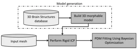
toolbox for python. In this work, we report results for the expected improvement (EI), and the probability of improvement (PI) acquisition functions [9]. Figure 1 shows the block diagram of the proposed model used in this work.
3
Results and Discussions
In the following sections we show the results for our BO framework for fitting 3D morphable models of brain Structures. On section 3.1, we show the results of the 3D shape modeling for the brain structures using a PDM as morphable model. Section 3.2 shows the results for the fitting process of the morphable model using BO.
3D Brain Structures database
Build 3D morphable model
[image:6.595.187.430.115.202.2]Input mesh Perform Rigid ICP PDM Fitting Using Bayesian Optimization Model generation
Fig. 1.Block diagram of the proposed model fitting approach based on BO.
3.1 Training results for 3D-MM
Figure 2 shows the training process results for the morphable models. From the figure, it can be noticed that for the three brain structures (thalamus, amygdala and ventricle), the morphable models capture the shape variability along the training set. Moreover, the results also show that by modeling the covariance matrix of the training dataset using PCA, the morphable model capture the relevant information in the latent space by analyzing the shape parameters that controls the deformation of a given brain struc-ture. Besides, figure 2 shows that by changing the shape parameters (eigenvalues of the PDM) the models deform a given brain structure from thin shapes (upper left corner of the figure 2(c)) to curvy shapes (lower right corner of the figure 2(c)). This shape variability can be related with the range of ages of the subjects in the database(between
35−65years), due to the fact that the brain volume decreases their mass over time in patients with Parkinson’s disease (thin shapes modeled by the first eigenvalues of the morphable models) [2].
(a) PDM for the thalamus (b) PDM for the amygdala (c) PDM for the ventricle
Fig. 2. Effects of varying the first 3 shape parameters of the PDM for the analyzed brain structures. Figures 2(a), 2(b) and 2(c) show the model variation for the most relevant eigen-values: (top) b1 =
−3√λ1, . . . ,3√λ1 ; (middle) b2 =
−3√λ2, . . . ,3√λ2 ; (bottom)
b3 =
−3√λ3, . . . ,3√λ3 . Each shape parameter (structures for each row in the subfigures)
ranges from−3√λistarting with the left column till3√λifor the right column.
3.2 Fitting Results Using Bayesian Optimization
6 Hern´an F. Garc´ıa, Mauricio A. ´Alvarez and ´Alvaro A. Orozco
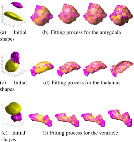
of the brain structures (amygdala, thalamus and ventricle). Figure 3 shows the results for the fitting process using BO. The results show that by using a fully Bayesian treat-ment of the optimization process, the model can estimate the shape parameters that performs accurately the fitting process between shapes. Figure 3 shows that by adding a prior registration process through rigid ICP, the BO method can explore an exploit the probabilistic search of the shape parameters that minimizes the cost function (Eu-clidean distance between the deformed model landmarks and the target shape). Besides, the results in figure 3(a) prove that the optimization step estimates the plausible shape parameters that deforms a given brain structure, even if the target shape has large curva-tures (significant changes over the shape surface, see figures 3(c) and 3(e)). Beside, the figure 4 shows the convergence of the BO process for both the EI and the PI acquisition functions. The results show that EI has better global convergence, due to the fact that by using this acquisition function the model fits more accurately the target shape and take less iteration to converge (200iterations for the EI and more than300iterations for the PI). The main reason is that the EI estimates best shape parameters in the explo-ration step than the PI acquisition function. However, both acquisition functions exploit the probabilistic search of the shape parameters with lower values in the evaluated cost function (mean square error of the Euclidean distance,52for EI and60 for the PI). Finally, table 1 shows the accuracy of the BO process compared against the common fitting method such as Rigid ICP. The results show that by using a given acquisition function the optimization process improves the selection of those values that control the deformation with high variance (shape parameters in regions not well explored) and values with high mean value (shape parameters worth exploiting that increase the fit-ting accuracy, MSE error of10.295for the amygdala) in comparison with the Rigid ICP which only removes the translation, rotation and scale between two shapes (MSE error of23.971for the amygdala).
Table 1.Accuracy of the BO process for fitting the three morphable models using EI and PI acquisition functions. The table shows the mean square error of the Euclidean distance between the deformed model and the target brain structure.
Fitting method
Brain Model Rigid ICP BO with EI BO with PI
Amygdala 23.971±4.163 10.295±2.575 13.927±4.665 Thalamus 32.486±4.169 19.907±1.340 22.351±4.170 Ventricle 26.623±4.287 15.924±2.079 16.686±4.289
4
Conclusions and Future Works
(a) Initial shapes
(b) Fitting process for the amygdala
(c) Initial shapes
(d) Fitting process for the thalamus
(e) Initial shapes
[image:8.595.196.417.120.355.2](f) Fitting process for the ventricle
Fig. 3.Fitting process for the PDM models of the amygdala, thalamus and ventricle brain struc-tures (shapes with yellow color are related to the model and those with color magenta depict the target shapes). The figures show the model to be deformed and the target brain structure. Also the figure shows the deformed models at10,50,100and200iterations.
0 50 100 150 200 250 Iteration 0 100 200 300 400 500 600 700 d(x[n], x[n-1])
Distance between consecutive x's
0 50 100 150 200 250 Iteration 50 60 70 80 90 100 110 120 Best y
Value of the best selected sample
0 50 100 150 200 250 Iteration 0 50 100 150 200 250 300
CI (centered at zero)
Predicted sd. in the next sample
(a) Experiment using EI
0 50 100 150 200 250 300 350 Iteration 0 100 200 300 400 500 600 d(x[n], x[n-1])
Distance between consecutive x's
0 50 100 150 200 250 300 350 Iteration 50 100 150 200 250 300 Best y
Value of the best selected sample
0 50 100 150 200 250 300 350 Iteration 0 20 40 60 80
CI (centered at zero)
Predicted sd. in the next sample
(b) Experiment using PI
Fig. 4.Convergence of the BO process for the EI (up) and PI (down) acquisition functions. The figure shows the distance between consecutive selectedxvalues (left column), the mean of the current model in the selected sample (middle) and the variance of the model in the selected sample (right column).
[image:8.595.155.466.428.527.2]8 Hern´an F. Garc´ıa, Mauricio A. ´Alvarez and ´Alvaro A. Orozco
As future works, we want to analyze the Bayesian optimization framework in high dimensional problems that includes the whole optimization process of a raw point-cloud data. Finally, we want to analyze a morphable model that adds both shape an appearance information in order to model tissue properties related to a given brain structure.
Acknowledgments
This research is developed under the project with codeF P44842−584−2015, financed by Colciencias and SINCLECLICK SOLUTIONS S.A.S. H.F. Garc´ıa is funded by Col-ciencias under the program:Formaci´on de alto nivel para la ciencia, la tecnolog´ıa y la innovaci´on - Convocatoria 617 de 2013.
References
1. Hill, D.: Neuroimaging to assess safety and efficacy of ad therapies. Expert Opinion on Investigational Drugs19(1) (2010) 23–26 PMID: 19947893.
2. Ibarretxe-Bilbao, N., Tolosa, E., Junque, C., Marti, M.J.: Mri and cognitive impairment in parkinson’s disease. Movement Disorders24(S2) (2009) S748–S753
3. Cabezas, M., Oliver, A., Llad´o, X., Freixenet, J., Cuadra, M.B.: A review of atlas-based segmentation for magnetic resonance brain images. Computer Methods and Programs in Biomedicine104(3) (2011) e158 – e177
4. Cosa, A., Canals, S., Valles-Lluch, A., Moratal, D.: Unsupervised segmentation of brain regions with similar microstructural properties: Application to alcoholism. In: Engineering in Medicine and Biology Society (EMBC), 2013 35th Annual International Conference of the IEEE. (July 2013) 1053–1056
5. Sotiras, A., Davatzikos, C., Paragios, N.: Deformable Medical Image Registration: A Survey. Medical Imaging, IEEE Transactions on32(7) (July 2013) 1153–1190
6. Nair, P., Cavallaro, A.: 3-d face detection, landmark localization, and registration using a point distribution model. IEEE Trans. Multimedia11(4) (2009) 611–623
7. Sj¨oberg, C., Ahnesj¨o, A.: Multi-atlas based segmentation using probabilistic label fusion with adaptive weighting of image similarity measures. Computer Methods and Programs in Biomedicine110(3) (2013) 308 – 319
8. Sj¨oberg, C., Johansson, S., Ahnesj¨o, A.: How much will linked deformable registrations decrease the quality of multi-atlas segmentation fusions? Radiation Oncology9(1) (2014) 1–8
9. Snoek, J., Larochelle, H., Adams, R.P.: Practical bayesian optimization of machine learn-ing algorithms. In: Advances in Neural Information Processlearn-ing Systems 25: 26th Annual Conference on Neural Information Processing Systems 2012. Proceedings of a meeting held December 3-6, 2012, Lake Tahoe, Nevada, United States. (2012) 2960–2968
10. Srinivas, N., Krause, A., Seeger, M., Kakade, S.M.: Gaussian process optimization in the bandit setting: No regret and experimental design. In F ˜A1
4rnkranz, J., Joachims, T., eds.:
Proceedings of the 27th International Conference on Machine Learning (ICML-10), Omni-press (2010) 1015–1022
11. Wang, Z., Zoghi, M., Hutter, F., Matheson, D., de Freitas, N.: Bayesian optimization in high dimensions via random embeddings. In: International Joint Conferences on Artificial Intelligence (IJCAI) - Distinguished Paper Award. (2013)
![Nano assemblies of cationic mPEG brush block copolymers with gadolinium polyoxotungstate [Gd(W5O18)2]9− form stable, high relaxivity MRI contrast agents](data:image/gif;base64,R0lGODlhAQABAIAAAP///wAAACH5BAEAAAAALAAAAAABAAEAAAICRAEAOw==)