0022-538X/99/$04.00⫹0
Copyright © 1999, American Society for Microbiology. All Rights Reserved.
The Human Immunodeficiency Virus Type 1
nef
Gene Can to a Large Extent Replace Simian
Immunodeficiency Virus
nef
In Vivo
FRANK KIRCHHOFF,1* JAN MU¨ NCH,1SILKE CARL,1NICOLE STOLTE,2KERSTIN MA¨ TZ-RENSING,2 DIETMAR FUCHS,2PETER TEN HAAFT,3JONATHAN L. HEENEY,3TOMEK SWIGUT,4
JACEK SKOWRONSKI,4ANDCHRISTIANE STAHL-HENNIG2
Institute for Clinical and Molecular Virology, University of Erlangen-Nuernberg, 91054 Erlangen,1and German Primate
Center, 37077 Go¨ttingen,2Germany; Biomedical Primate Research Center, Department of Virology, 2288 GJ Rijswijk,
The Netherlands3; and Cold Spring Harbor Laboratory, Cold Spring Harbor, New York 117244
Received 29 December 1998/Accepted 12 July 1999
Thenefgene of the pathogenic simian immunodeficiency virus (SIV) 239 clone was replaced with primary human immunodeficiency virus type 1 (HIV-1)nefalleles to investigate whether HIV-1 Nef can substitute for SIV Nef in vivo. Initially, two rhesus macaques were infected with the chimeric viruses (Nef-SHIVs). Most of thenefalleles obtained from both animals predicted intact open reading frames. Furthermore, forms contain-ing upstream nucleotide substitutions that enhanced expression of the inserted gene became predominant. One animal maintained high viral loads and slowly progressed to immunodeficiency. nef long terminal repeat sequences amplified from this animal were used to generate a second generation of Nef-SHIVs. Two macaques, which were subsequently infected with a mixture of cloned chimeric viruses, showed high viral loads and progressed to fatal immunodeficiency. Five macaques received a single molecular clone, named SHIV-40K6. The SHIV-40K6nefallele was active in CD4 and class I major histocompatibility complex downregulation and enhanced viral infectivity and replication. Notably, all of the macaques inoculated with SHIV-40K6 showed high levels of viral replication early in infection. During later stages, however, the course of infection was variable. Three animals maintained high viral loads and developed immunodeficiency. Of the remaining two macaques, which showed decreasing viral loads after the acute phase of infection, only one efficiently controlled viral replication and remained asymptomatic during 1.5 years of follow-up. The other animal showed an increasing viral load and developed signs of progressive infection during later stages. Our data demonstrate that HIV-1nefcan, to a large extent, functionally replace SIVmacnefin vivo.
Experimental infection of rhesus macaques with simian im-munodeficiency virus (SIV) has demonstrated that an intact nefgene is important for maintaining high virus loads and for the development of immunodeficiency (36). The observation that some long-term nonprogressors with human immunode-ficiency virus type 1 (HIV-1) infection harbor nef-defective viruses (16, 39, 59) suggests a similar importance for the patho-genicity of HIV-1 in humans. SIV Nef and HIV Nef show similar in vitro activities: downmodulation of CD4 and of class I major histocompatibility complex (MHC) cell surface expres-sion (1, 3, 7, 12, 21, 27, 46, 50, 60, 62), enhancement of virion infectivity (22, 43, 52, 69), and stimulation of replication in primary lymphocytes (10, 18, 43, 52, 69). Moreover, both are able to alter T-cell signaling pathways (2, 5, 15, 19, 25, 33, 48, 49, 67) and interact with cellular serine/threonine and tyrosine kinases (11, 19, 32, 44, 45, 54, 58, 61, 66).
Although HIV-1 Nef and SIV Nef perform similar in vitro functions, some noteworthy differences exist. In contrast to HIV-1nef, the SIVnefgene overlaps theenvgene and encodes a protein that has a higher molecular mass (33 to 36 kDa) than HIV-1 Nef (27 to 30 kDa). Sequence homology between the SIVmac and HIV-1 Nef proteins is mainly restricted to the N-terminal myristylation signal and the highly conserved cen-tral core region (53). HIV-1 Nef and SIV Nef use overlapping
but distinct target sites for CD4 downregulation (31) and may utilize distinct motifs for the interaction with cellular adapter complexes (8, 13, 24, 48, 55). A putative SH3 binding domain (PxxP)3is highly conserved in HIV-1 Nef and important for the binding of Src family tyrosine kinases Hck and Fyn (4, 44, 58). SIV Nef contains only two prolines at the corresponding po-sition (PxxP) that are dispensable for Src association and of little importance for SIV pathogenicity (37, 43). Recently, it has been shown that SIVmac Nef and HIV-2 Nef, but not HIV-1 Nef, interact with the zeta chain of the T-cell receptor (6, 30). It seems that SIV Nef and HIV-1 Nef are functionally homologous but that several functions involve different inter-actions.
Investigators have constructed a variety of SIV-HIV chi-meric viruses (SHIVs) to address pathogenicity and vaccine issues related to the incorporated HIV-1 gene sequences (63). Recently, molecular SHIV clones that replicate to high levels and induce an AIDS-like disease in macaques have been re-ported (34, 64). However, all of these pathogenic SHIV con-structs carry the SIVnef gene and the corresponding clones containing HIV-1nefusually failed to induce high viral loads and immunodeficiency (64). Thus, although functional equiv-alence between HIV-1 and SIV Nef has been demonstrated in in vitro infectivity and replication systems (66), no animal model suitable for the study of the role of the HIV-1nefgene in viral pathogenicity has been described.
Nef plays an important role in AIDS pathogenesis and may represent a key factor in the development of a live attenuated AIDS vaccine. Nef-SHIVs might allow the elucidation of which * Corresponding author. Mailing address: Institute for Clinical and
Molecular Virology, University of Erlangen-Nuernberg, Schlossgarten 4, 91054 Erlangen, Germany. Phone: 852 6483. Fax: 49-9131-852 2101. E-mail: [email protected].
8371
on November 9, 2019 by guest
http://jvi.asm.org/
in vitro functions of Nef are critical for disease progression in vivo and also provide an animal model for testing of HIV-1 Nef inhibitors. It is unknown whether HIV-1 Nef can substitute for SIV Nef in vivo. Therefore, we have replaced thenefgene of the well-characterized pathogenic SIVmac239 clone (35, 36, 57) with a pool of primary HIV-1nefalleles. In the first pas-sage, these chimeras showed little pathogenicity in infected macaques. However, a selective pressure for open functional HIV-1nefopen reading frames (ORFs) and for efficient Nef expression could be demonstrated. A second generation of molecularly cloned Nef-SHIVs showed consistently high levels of replication during acute infection and caused immunodefi-ciency in the majority of infected rhesus macaques. One chron-ically infected animal, however, efficiently controlled Nef-SHIV replication and remained asymptomatic. It remains to be elucidated if differences in Nef expression levels or in func-tional aspects are the major reason why Nef-SHIVs seem to induce disease less reproducibly than SIVmac239nef-open.
MATERIALS AND METHODS
Construction of Nef-SHIVs.The pBR⌬NU proviral construct was used for the insertion of HIV-1nefgenes into the SIV genome (Fig. 1A). This clone contains deletions of 513 bp in theneflong terminal repeat (LTR) region and mutations in the SIVnefinitiation codon and a second ATG at codon 7 of thenefORF (26). These mutations did not alter the predicted Env sequence. Two oligonu-cleotides (5⬘-CCGGACCGCGGCCGCCGCTCGCGACGCGT-3⬘and 5⬘-CCG GACGCGTCGCGAGCGGCGGCCGCGGT-3⬘) were used to insert a 5⬘-Not
I-NruI-MluI-3⬘ polylinker into the XmaI site of pBR⌬NU (pBR⌬NU-PL). Fragments spanning thenefgene were amplified from patient-derived peripheral blood mononuclear cells (PBMC) as described previously (39) and used as templates for amplification with primers pF107 (5⬘-TTTTGCGGCCGCATGGG TGGCAAGTGGTCA-3⬘) and pF103 (5⬘-GCAAGCACCGTTCAGCAGTCTT GTAGTACTCCGGATG-3⬘) (Fig. 1B). The digested, gel-purified PCR frag-ments were pooled and cloned into the pBR⌬NU-PL vector by using theNotI andMluI sites (in boldface) inserted just upstream of the HIV-1nefinitiation codon and downstream of the TGAneftermination codon (underlined). Ali-quots of transformed supercompetentEscherichia coliXL-2 (Stratagene) were plated on Luria broth-ampicillin dishes to assess transformation efficiency, and the remaining 90% of the transformed bacteria were used for direct inoculation of large-scale plasmid preparations. The percentage of the plasmid population containing an HIV-1nefinsert was estimated by restriction and PCR analysis (Fig. 2). As a control, the NL4-3nefallele was also inserted into pBR⌬NU-PL to generate SHIV-NL43nef.
DNA sequences spanning the 3⬘end of the SIVenvgene, the HIV-1nef
gene, and the SIV 3⬘LTR were amplified by one round of PCR amplification using primers pNeMu1 (5⬘-GGAGTAATACTGTTAAGAATAGTG-3⬘); 895738980) and pNeMu2 (5⬘-CTTCAAGAATTCTGCTAGGGATTTTCC-3⬘) for the construction of the second generation of Nef-SHIVs. Whole cellular DNA isolated from CEMx174 cells, cocultivated with PBMC obtained from Mm7745 at 40 weeks postinfection (wpi), was used as a template. The 1.7-kb hybridenv-nef-LTR fragments were sequenced and inserted into a modified pBR322 vector containing the full-length SIVmac239 provirus by using the uniqueNheI andEcoRI sites (in bold type) in SIVenvand the vector sequences flanking the 3⬘end of the provirus as described previously (43).
Production of virus stocks.COS cells were transfected and virus stocks were generated as described previously (14). 293T cells were transfected by the cal-cium phosphate method (17) with 10g of the proviral constructs. The medium was changed after overnight incubation, and virus was harvested 24 h later. Viral stocks were aliquoted and frozen at⫺80°C; p27 antigen concentrations of viral stocks were quantitated with a commercial HIV-1–HIV-2 enzyme-linked immu-nosorbent assay (Immunogenetics, Zwijndrecht, Belgium).
Cells, infectivity, and viral replication.COS, 293T, and sMAGI cells were grown in Dulbecco modified Eagle medium supplemented with 10% fetal calf serum (FCS). Infection of sMAGI cells was performed as described previously (9), and viral infectivity was quantitated by using the Galacto-Light Plus chemi-luminescence reporter assay kit (Tropix, Bedford, Mass.) as recommended by the manufacturer. CEMx174 cells were maintained and virus production was mea-sured by reverse transcriptase assay as previously described (56). The herpesvirus saimiri-transformed T-cell line 221 (2) was maintained in the presence of 100 U of interleukin-2 per ml (Boehringer, Heidelberg, Germany) and 20% FCS, and infections were performed in the presence of 50 U of interleukin-2 per ml and 5% FCS. Rhesus PBMC were isolated, cultured, and infected with virus stocks containing 2 ng of p27 as described previously (43). The SIVmac239nef* variant, containing a premature in-frame TAA stop signal at the 93rd codon ofnef, was used as anef-defective control (36).
Infection of rhesus macaques and clinical assessment.Juvenile rhesus ma-caques of Indian origin were experimentally infected by intravenous inoculation
of the Nef-SHIV stocks containing 10 (first experiment) or 5 (second experi-ment) ng of p27 produced by transfected COS-7 cells. The animals were healthy and seronegative for SIV, type D retrovirus, and simian T-cell lymphotropic virus type 1 at the time of infection. Serological, virological, and immunological anal-yses were performed as described previously (20, 70–72).
Nef sequence analysis.DNA sequences spanning HIV-1nefwere either am-plified directly from sequential PBMC DNA samples with a nested PCR ap-proach (38) or obtained from whole cellular DNA isolated from positive PBMC-CEMx174 bulk cocultivation followed by one round of PCR amplification. To detect alterations upstream of the inserted HIV-1nefgene and in the remaining SIV 3⬘ LTR sequences, fragments spanning the 3⬘env-nef-LTR region were amplified from virus-positive bulk cocultivations by using primers pNeMu1 and pNeMu2 and cloned and sequenced as described above.
Luciferase expression constructs. The generation of an env-deficient SIVmac239 reporter construct (pBR239⌬envLuc) has been described previously (40). AnNheI-BglI restriction fragment containing the luciferase gene was used to replace the corresponding region of pBR239 to generate a replication-com-petent reporter virus. The region between theNheI site and the inserted lucif-erase gene was replaced with the corresponding region of the Nef-SHIV-K6 clone by using standard DNA techniques.
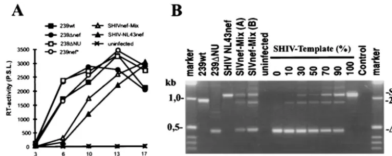
FIG. 1. Generation of chimeric SIVmac239 carrying a complex mixture of primary HIV-1 nefalleles. (A) Schematic representation of the SIVmac239 genome (top) and the cloning strategy (below). The original SIVmac239nef
initiation codon and a second ATG at amino acid position 7 of thenefORF were mutated, and thenefunique region and upstream U3 sequences were deleted to generate 239⌬NU (26). Subsequently, a polylinker was inserted just downstream of theenvgene (⌬NU-PL) and HIV-1nefgenes were cloned into the uniqueNotI andMluI restriction sites. The deletions in⌬NU are indicated by black bars, and the mutations in the ATGs are indicated by an X; underneath, the positions of the inserted polylinker and the HIV-1nefgenes are indicated. PPT, polypurine tract; IN, U3 sequences required for integration. (B) Amplification of HIV-1nef
genes from 55 HIV-1-infected individuals with different rates of disease progres-sion. PCR amplification products were digested withNotI andMluI and sepa-rated by electrophoresis through 1.5% agarose gels. Cloning into the⌬NU-PL vector was performed as described in Materials and Methods. M, marker; C, negative control. Numbers specify individual patients.
8372 KIRCHHOFF ET AL. J. VIROL.
on November 9, 2019 by guest
http://jvi.asm.org/
Functional analysis.Dose-response analysis of the effect of Nef on CD4 and MHC class I cell surface expression was performed as described previously (24, 50, 68). The effect of Nef expression on CD3-initiated signaling, as revealed by CD69 induction in response to an anti-CD3 monoclonal antibody (MAb), was assayed as previously described (33). Briefly, Jurkat T cells were electroporated with 25g of DNA containing 3g of cytomegalovirus CD20 marker plasmid and various amounts of expression plasmids and carrier DNA. At 18 to 24 h after transfection, cells were stimulated by overnight incubation with anti-CD3 MAb HIT3A (PharMingen). To reveal CD4 and MHC class I surface expression, cells were incubated for 1 h on ice with PerCP-conjugated anti-CD20 MAb Leu-16 (Becton Dickinson) for 30 to 36 h after transfection and phycoerythrin-conju-gated anti-CD4 MAb Leu3A (Becton Dickinson) together with fluorescein iso-thiocyanate-conjugated anti-HLA A, B, and C MAb G46-2.6 (PharMingen). To reveal CD69 expression, cells were incubated with fluorescein isothiocyanate-conjugated anti-CD69 MAb FN50 (PharMingen). CD4, CD20, CD69, and class I MHC surface expression was analyzed by using an Epics-Elite flow cytometer. For dose-response analysis, CD4, CD69, or class I MHC levels are represented by the peak channel number of red or green fluorescence on CD20⫹cells.
Western blot analysis.CEMx174 cells were infected with virus containing 10 ng of p27 core antigen derived from transfected 293T cells. When cytopathic effects were observed, cells were harvested and expression of Nef proteins in whole cellular lysates was analyzed by immunoblotting using rabbit anti-Nef serum (19). For detection of p27 core protein, an anti-Gag serum derived from SIVmac p27 hybridoma cells (55-2F12) was used (28). For enhanced chemilu-minescence detection, horseradish peroxidase-conjugated secondary antibodies were used as described by the manufacturer of the ECL detection system (Am-ersham, Chicago, Ill.).
RESULTS
Construction of SHIVs containing primary HIV-1 nef al-leles. Our strategy to obtain pathogenic Nef-SHIVs was to maximize the genetic information in the proviral constructs used in the initial study and to utilize the selective pressure in vivo as a tool to select for those forms that replicate and persist most efficiently in infected macaques. Therefore, the SIVmac nef gene was replaced with a complex mixture of primary HIV-1nefalleles amplified from 55 HIV-1-infected individuals (Fig. 1B). Furthermore, the proviral constructs contained two regions encompassing the polypurine tract and the U3 se-quences required for integration. One is located in the inserted HIV-1nefgene, and a second is just upstream of the SIV core enhancer elements (Fig. 1A). Based on previous observations (38), we expected that sequences not advantageous for viral replication would be efficiently deleted or mutated in vivo.
As described in Materials and Methods, the HIV-1nefPCR fragments were cloned as a pool into the pBR⌬NU-PL con-struct. Control experiments indicated that the plasmid
prepa-ration (named pBR-SHIVnef-Mix) represented approximately 80,000 transformants and that about 90% of the plasmid pop-ulation contained an insert of the expected size (data not shown). Ten colonies were randomly picked and analyzed. Nine clones contained an HIV-1nefgene, and one represented the original pBR⌬NU-PL construct. Thenefsequences were heterogeneous and likely all originated from different HIV-1-infected individuals (data not shown).
Virus stocks were generated by transient transfection of COS-1 cells with the pBR-SHIVnef-Mix DNA preparation and used for infection of CEMx174 cells. The virus population was replication competent. However, similar to infection with SHIV-NL43nef, the replication kinetics were slightly delayed compared to those of the nef-open and nef-defective SIVmac239 controls (Fig. 2A). Both the chimeric proviral se-quences and those withnefdeleted were readily detectable in infected CEMx174 cells (Fig. 2B, lane 4). PCR analysis of the plasmid population used for transient COS-1 transfection yielded a similar pattern (Fig. 2B, lane 5). The shorter⌬NU template was amplified more efficiently than the SHIV tem-plate (Fig. 2B, lanes 7 to 13). Thus, the results of the PCR analysis are in agreement with the restriction analysis and indicate that the majority of the plasmid population used for transfection and most of the proviral sequences in infected cells contained an inserted HIV-1nefgene.
Low pathogenicity of the first generation of Nef-SHIVs.Two juvenile rhesus macaques, Mm7739 and Mm7745, were in-fected intravenously with aliquots of the virus stocks obtained after transient transfection of COS-1 cells with pBR-SHIVnef-Mix. The viral inoculum contained 10 ng of p27 antigen, which corresponded to approximately 10,000 50% tissue culture-in-fective doses for CEMx174 cells. Unexpectedly, very early after infection, predominantly the 239-⌬NU-PL virus withnef de-leted could be detected (Fig. 3A). However, in both animals, the chimeras came to predominate by 2 to 4 wpi, indicating a selective pressure for HIV-1 nef-containing forms. The p27 antigen levels during the acute phase of infection were low, compared to those measured after wild-type 239 (239wt) in-fection (Table 1). Both animals developed strong humoral immune responses. Only Mm7745 showed persistently high cell-associated viral loads (Fig. 3B). The CD4⫹T-cell numbers
[image:3.612.110.493.73.226.2]were stable in Mm7739 and decreased only slightly in Mm7745 FIG. 2. Characterization of SHIVnef-Mix in vitro. (A) Replication of the indicated SIVmac239nefvariants in CEMx174 cells. Cells were infected with virus stocks containing 5 ng of p27 core antigen derived from transfected COS-1 cells. RT, reverse transcriptase; P.S.L., photostimulated light emission. (B) Amplification of the
nefregion from infected (lanes 1 to 4) and uninfected (lane 6) CEMx174 cells. Genomic DNA was extracted at 10 days postinfection. For comparison, 0.2 ng of the pBR-SHIVnef-Mix plasmid preparation was used as the template for amplification (lane 5). To assess relative amplification efficiencies, the⌬NU and SHIV templates were mixed at different molar ratios (lanes 7 to 13). The expected positions of fragments representing 239nef, the⌬NU mutant, and the chimeras are indicated. Control, no DNA added to the PCR mixture.
on November 9, 2019 by guest
http://jvi.asm.org/
(data not shown). Mm7739 developed mild (12 wpi)-to-mod-erate (40 wpi) lymphadenopathy and remained clinically healthy with low viral loads throughout the 74-wpi observation period. Mm7745 lost body weight and had to be euthanized at 84 wpi because of weakness. Postmortem examination revealed severe hyperplasia to depletion of the germinal centers of the lymph nodes, severe multiorgan vasculitis and perivasculitis with evidence of cytomegalovirus infection, and moderate ac-tivation of microglia cells. Both animals were infected with the same virus stock. Therefore, it was unexpected that the cell-associated virus load was similar to nef-deleted infection of Mm7739 but comparable to SIVmac239nef-open infection of Mm7745 (Fig. 3B).
Selective pressure for intact HIV-1 nef genes and for up-stream nucleotide substitutions. We sequenced PCR frag-ments spanning the inserted HIV-1 nef genes derived from PBMC samples and from reisolation bulk cultures to investi-gate whether (i) the HIV-1 nef genes remained intact, (ii) specific nef variants came to predominate, or (iii) consistent sequence alterations occurred upstream or downstream of the insertednefgenes. Nine of 10nef-spanning fragments ampli-fied from PBMC obtained from Mm7739 at 12 wpi and 25 of 27 clones obtained from the spleen, lymph nodes, or thymus at 74 wpi predicted an intactnefORF (Fig. 4 and data not shown). Similarly, 37 (95%) of 39 nefORFs amplified from the pro-gressing animal, Mm7745, at different time points were intact. These frequencies of intact nef alleles are similar to those observed in human HIV-1 infection (32, 65) and indicate a strong selective pressure for open functional HIV-1nefgenes, even in the animal with the low viral load.
Several previously defined conserved motifs in HIV-1 Nef (41, 65) were also conserved in the deduced Nef sequences obtained from the infected macaques: an N-terminal myristy-lation signal, a PxxP motif, a putative protein kinase C recog-nition site, a predicted-turn, and a dileucine motif (13) (Fig. 4). However, some unusual amino acid variations, that are rarely present in HIV-1 Nef sequences derived from infected humans (32, 41, 51, 65), are notable. These include alterations close to the N terminus of Nef (G3A, K4R, and W5L), muta-tion of a usually highly conserved cysteine (C55V), and the presence of a glycine residue in the central part of the acidic region (EEGEE) (Fig. 4 and data not shown). For most of these variations, it is unclear if they were already present in the
initialnefpool or occurred during in vivo selection. However, the oligonucleotide used for amplification of the HIV-1 nef genes predicted the N-terminal sequence MGGKWS. All 25 nefalleles amplified from both macaques at 12 wpi also pre-dicted MGGKWS, whereas 25 (86%) of 29 deduced Nef amino acid sequences obtained at later time points contained alter-ations in this region. Therefore, it seems likely that the changes close to the N terminus emerged during the course of infec-tion. The K4R, C55V, and EEGEE changes were not present in all of the clones obtained at late time points from the progressing animal and were also detected in the asymptomatic animal, Mm7739 (Fig. 4). Thus, these sequence variations are not linked to disease progression. Nonetheless, since they were frequently observed in both animals, these HIV-1 Nef features may be advantageous for SIV replication in macaques.
To generate Nef-SHIVs with enhanced pathogenicity and to investigate which upstream and downstream changes are se-lected, 1.7-kb fragments spanning the 500 bp upstream ofnef and the entire 3⬘ LTR were amplified from positive bulk co-cultures obtained from the progressing animal, Mm7745, at 40 wpi. Sequence analysis revealed the following consistent changes upstream of the insertednefgene: A9110G (15 of 15, predicting amino acid changes of Env R751G and Rev K41R), A9224G (12 of 15, predicting Env T789A and Rev N79C), T9276C (4 of 15, predicting Rev C97R), G9277C (9 of 15, predicting Env A807P and Rev C97S), A9442G (12 of 15), and T9276C (3 of 15, predicting Rev C97R) (Fig. 5). Numbering refers to the SIVmac239 sequence (57). Furthermore, a change of C3A was observed at theNotI site. Most of these upstream changes were already present in the majority of clones ob-tained from Mm7745, but not in those from Mm7739, at 12 wpi (data not shown). However, at the 74-week necropsy time point, changes at nucleotide positions 9276 or 9277 and 9442 and the C3A change at theNotI site were also detected in all 27 of thenef-LTR fragments amplified from lymphatic tissues obtained from the asymptomatic animal, Mm7739.
[image:4.612.62.546.71.232.2]Changes downstream of the HIV-1 nef genes were rarely observed. Three of the 15 clones obtained from Mm7745 at 40 wpi contained a deletion of 36 bp removing the SIV polypurine tract and the SIV U3 sequences required for integration (Fig. 5). This deletion was detected in fragments carrying highly divergent nef alleles (40wk1, 40wk8, and 7745-40wk10; Fig. 4). Thus, it seems likely that the deletion evolved FIG. 3. In vivo properties of SHIVnef-Mix. (A) Nef-LTR sequences were amplified from positive PBMC-CEMx174 bulk cocultivations, obtained at the indicated times (weeks postinfection), and separated on a 1.5% agarose gel. Similar results were obtained by nested PCR using DNA extracted from PBMC. M, marker, C, control. (B) Cell-associated viral load. The values shown are for Mm7739 (Œ) and Mm7745 (‚) inoculated with the SHIVnef-Mix stock, one 239wt-infected animal (■), and averages obtained from four macaques infected with⌬NU (䊐) (26).
8374 KIRCHHOFF ET AL. J. VIROL.
on November 9, 2019 by guest
http://jvi.asm.org/
independently in the progressing animal and that the intact SIV polypurine tract and U3 sequences required for integra-tion are not important for replicaintegra-tion of Nef-SHIVs in ma-caques. No changes in the mutated SIVnefATGs, the HIV-1 polypurine tract, or the HIV-1 U3 sequences required for integration were detected.
The upstream changes enhance gene expression. Some of the nucleotide substitutions observed upstream of the inserted HIV-1 nefgenes were likely to influence protein expression. For example, the change at position 9442 or 9443 removed an upstream ATG that is not in frame with the original SIVnef ORF or the inserted HIV-1nefgene. The C-to-A change at the NotI site generates higher homology to the optimal translation initiation sequence (42).
To test the influence of these changes on viral infectivity and gene expression, the luciferase gene was inserted between the NotI and MluI restriction sites in the pBR239⌬NU-PL con-struct to obtain a replication-competent reporter virus (239⌬NU-Luc). The region upstream of the inserted luciferase (NheI toSstII) gene was subsequently replaced with the cor-responding region of the Nef-SHIV-K6 clone to generate 239-K6NS-Luc (Fig. 5). Compared to SIVmac239, this fragment contains mutations of A9110G, A9224G, T9276C, and A9442G. The C-to-A change selected at theNotI site, located just upstream of the insertednefgenes, is not present in this construct. sMAGI and CEMx174-SEAP reporter cell lines were infected, and the expression of the cell-associated re-porter genes and that of the viral luciferase gene were mea-sured in parallel to assess the influence of the upstream changes selected in vivo on both infectivity and expression of the inserted gene. The-galactosidase (sMAGI) and secreted alkaline phosphatase (CEMx174) activities were only slightly higher after infection with the 239-K6NS-Luc variant than after infection with 239⌬NU-Luc (Fig. 6). The luciferase ac-tivities, however, were consistently approximately threefold higher for the virus containing the upstream changes selected in Mm7745 (Fig. 6). This result suggests that selective pressure for efficient expression of the inserted HIV-1nefgene in vivo exists. The C-to-A change at theNotI site is likely to further enhance gene expression. In agreement with this assumption, we detected larger amounts of Nef protein in CEMx174 cells infected with Nef-SHIV-40K6 than in cells infected with SHIV-NL43nef (data not shown).
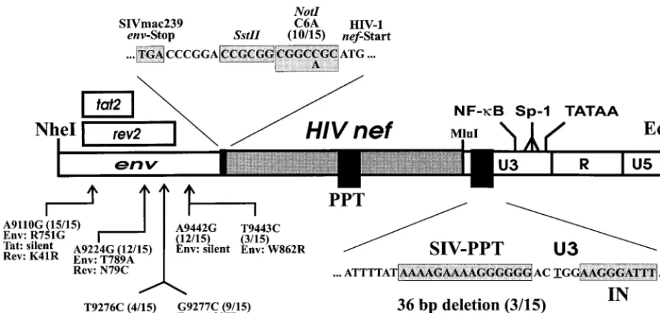
HIV-1 nef alleles derived from Mm7745 are functionally active.Sixenv-nef-LTR fragments derived from Mm7745 at 40 wpi were inserted into the pBR239wt vector by using the single NheI andEcoRI sites (Fig. 5) and used to produce virus stocks by transient transfection of 293T cells. All six Nef-SHIVs showed reduced infectivity in sMAGI cells compared to 239wt (Fig. 7A). However, the infectivity was also reduced when HIV-1 nefwas replaced with the 239wt nefallele (239K6wt) (Fig. 7A). This virus, containing a duplication of 167 bp of the
nef-envoverlapping region, showed lower steady-state levels of
Nef expression than SIVmac239wt (Fig. 7B and data not shown). It was more infectious in sMAGI cells, however, than an otherwise isogenic form containing a stop signal at the 93rd codon innef(239K6nef*) (Fig. 7A). Furthermore, a frameshift mutation in the HIV-1nefgene of SHIV-40K6 (K6fr) or mu-tation of the ATG initiation codon in SHIV-40K13 (K13x) reduced viral infectivity about fourfold (Fig. 7A). Thus, intact HIV-1nefgenes were able to enhance SIV infectivity.
The Nef-SHIVs grew with delayed kinetics in CEMx174 cells, in which we never observed a significant difference be-tween nef⫹and nef⫺variants of SIVmac239 (an example is
shown in Fig. 7C, left). However, the chimeras replicated more efficiently than nef-defective SIVmac239 in the herpesvirus
TABLE 1. Viral load and nonspecific immune activation in macaques infected with Nef-SHIVs Animal(s) a Virus used for infection Acute phase (first 6 wpi) b Postacute phase (after 12 wpi) c p27 concn (pg/ml) Viral load Neopterin RNA d p27 concn (pg/ml) Viral load Neopterin RNA d Mm7739 SHIVnef-Mix 56 66 4.9 ND e BQL f 0–0.1 1.0 ND Mm7745 SHIVnef-Mix 287 2,500 4.2 ND BQL 285–1,250 3.9 ND Mm8655 SHIV-40Mix 5,482 2,048 8.8 1.9 ⫻ 10 7 37 256–4,096 3.6 1.1 ⫻ 10 7 Mn8664 SHIV-40Mix 756 4,096 11.1 8.9 ⫻ 10 6 16,000 1,024–8,192 8.1 6.4 ⫻ 10 6 Mm8653 SHIV-40K6 795 4,096 3.4 4.6 ⫻ 10 6 86 0.0–8 1.8 1.7 ⫻ 10 3 Mm8658 SHIV-40K6 3,562 4,096 6.3 2.6 ⫻ 10 7 337 32–1,024 6.6 1.4 ⫻ 10 7 Mm7746 SHIV-40K6 384 1,028 7.0 9.4 ⫻ 10 5 BQL 2–32 4.7 2.0 ⫻ 10 5 Mm7793 SHIV-40K6 2,952 8,192 12.3 1.0 ⫻ 10 7 BQL 128–2,048 7.0 2.9 ⫻ 10 6 Mm8014 SHIV-40K6 1,208 4,096 8.4 1.0 ⫻ 10 7 579 256–4,096 9.8 8.5 ⫻ 10 6 Mm ( n ⫽ 5) SHIV-40K6 1,780 ⫾ 1,249 4,302 ⫾ 2,279 7.5 ⫾ 2.9 1.0 ⫻ 10 7 ⫾ 8.6 ⫻ 10 6 3 ⫹ /2 BQL 0.0–4,096 1.8–9.8 5.1 ⫻ 10 6 ⫾ 5.4 ⫻ 10 6 Mm ( n ⫽ 9) 239wt 4,311 ⫾ 3,049 2,786 ⫾ 1,556 10.5 ⫾ 3.1 1.1 ⫻ 10 7 ⫾ 7.5 ⫻ 10 6 3 ⫹ /6 BQL 128–4,096 1.5–10.3 1.6 ⫻ 10 6 ⫾ 1.4 ⫻ 10 6 Mm ( n ⫽ 4) ⌬ NU 67 ⫾ 39 590 2.8 ⫾ 1.1 1.8 ⫻ 10 5 ⫾ 1.2 ⫻ 10 5 BQL 0.0–0.3 0.6–1.4 3.9 ⫻ 10 3 ⫾ 8.3 ⫻ 10 3 a Animals were housed at the German Primate Center. Mm, Macaca mulatta . b The highest value observed during the first 6 weeks after infection is shown. Viral load is given as the number of infected cells per million PBMC. The neo pterin level is given as fold of baseline. The RNA level is the number of copies per milliliter of plasma. For the viral load of the ⌬ NU-infected macaques, the end point was not reached at 2 wpi. c Surviving animals were observed at least until 52 wpi. Peak values are given for p27, neopterin, and RNA load; ranges are given for the cell-associated viral load. d RNA values for SIV nef -open and nef -defective controls were derived from 10 ⌬ NU-infected and 7 SIVmac32H/1XC-infected rhesus macaques housed at the Biomedical Primate Research Center in Rijswijk, The Netherlands (72). e ND, not determined. f BQL, below quantitation limit.
on November 9, 2019 by guest
http://jvi.asm.org/
[image:5.612.325.531.80.728.2]FIG.
4.
Alignment
of
the
predicted
HIV-1
Nef
sequences
derived
from
macaques
infected
with
SHIVnef-Mix.
In
the
left
column,
the
four-digit
numbers
sp
ecify
the
animals,
the
two-digit
numbers
are
weeks
postinfection,
and
the
last
numbers
specify
the
individual
clones.
nef
-spanning
fragments
were
amplified
from
PBMC
or
from
bulk
cocultivations
obtained
at
the
time
points
indicated.
The
Nef
sequences
derived
from
the
spl
een,
thymus,
and
axillary
lymph
nodes
of
Mm7739
represent
consensus
sequences
obtained
from
the
analysis
of
nine
cloned
nef
fragments
for
each
tissue.
For
each
time
point
or
tissue,
sequences
were
obtained
from
at
least
three
independent
PCRs.
For
comparison,
HIV-1
Nef
consensus
sequences
derived
from
41
nonprogressors
(Nonpr)
and
50
progressing
HIV-1-infected
individuals
(Progr)
are
shown.
Dashes
indicate
identity
with
the
consensus
Nef
sequence,
periods
indicate
gaps
introduced
to
optimize
the
alignment,
and
asterisks
indicate
stop
signals.
/,
frameshift
mutation;
X,
nef
alleles
selected
for
functional
analysis.
The
SHIV-40K6
sequence
is
in
boldface
letters.
Some
conserved
sequence
motifs
in
Nef
are
indicated
by
shading.
8376
on November 9, 2019 by guest
http://jvi.asm.org/
[image:6.612.161.441.50.732.2]saimiri-transformed 221 cell line (Fig. 7C, middle) and in rhe-sus PBMC (Fig. 7C, right). In contrast, variants of SHIV-40K6 and SHIV-40K13 lacking the initiation codon or containing a frameshift mutation were unable to stimulate viral replication (data not shown).
Five of these HIV-1nefalleles were also tested for the ability to alter T-cell receptor signaling pathways and to downmodu-late class I MHC and CD4 cell surface expression. Expression of the 40K1, 40K6, and 40K11 nef alleles derived from Mm7745 blocked the induction of CD69 cell surface expres-sion after stimulation with the anti-CD3 MAb (Fig. 8A). Fur-thermore, transient transfection of Jurkat cells with thesenef
expression constructs resulted in a dose-dependent decrease in surface class I MHC (Fig. 8B) and CD4 (Fig. 8C) expression. Thenefalleles derived from Mm7745 were almost as active as the strong control HIV-1 NA7 nefallele. The remaining two nef alleles, 40K5 and 40K13, were nonfunctional in these in vitro assays. Relatively high frequencies of functionally defec-tive nef alleles that predict intact ORFs have also been de-scribed for human HIV-1 infection (51).
Pathogenic potential of the second generation of Nef-SHIVs. The progressive status of Mm7745 and the in vitro analysis of
theenv-nef-LTR fragments derived from this animal suggested
that the changes selected in vivo might enhance the pathoge-nicity of the chimeric viruses. To assess the pathogenic poten-tial, two rhesus macaques were infected intravenously with a mixture of cloned Nef-SHIVs, representing the six HIV-1nef alleles indicated in Fig. 4, and five animals were infected with the SHIV-40K6 clone.
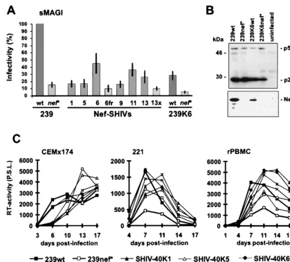
Mm8655 and Mm8664, infected with the mixture, showed high viral loads and progressed to fatal immunodeficiency. The characteristics of infection were highly similar to those of in-fection with pathogenicnef-open SIVmac239, i.e., high levels of plasma viremia and viral RNA, persistently high cell-asso-ciated viral loads, declining CD4⫹cell counts, and about
10-fold elevated levels of neopterin during the acute phase of infection (Fig. 9, Table 1, and data not shown). Both animals were euthanized at 39 (Mm8655) and 40 (Mm8664) weeks after infection, respectively. At the time of death, Mm8655 showed lymph node alterations ranging from severe hyperpla-sia to depletion of germinal centers and a malignant centro-blastic-monomorphic B-cell lymphoma. During the course of infection, this animal showed declining T4/T8 cell ratios and weight loss. Mm8664 developed anemia, thrombocytopenia, and opportunistic Campylobacter, Giardia, and Trichomonas infections. Postmortem examination revealed a generalized follicular hyperplasia together with follicular depletion,
[image:7.612.61.541.75.303.2]Pneu-mocystis cariniipneumonia, and chronic enteritis with bacterial
FIG. 5. Nucleotide substitutions and deletions upstream and downstream of the HIV-1nefgenes. The positions of changes observed in 15 clones derived from Mm7745 at 40 wpi compared to the pBR-⌬NU-PL construct are indicated; numbering refers to the SIVmac239 sequence (57). Numbers in parentheses indicate the proportion of clones containing the alteration. The corresponding amino acid changes are in the single-letter code. Only alterations observed in at least 3 of the 15 clones are indicated. Abbreviations are defined in the legend to Fig. 1.
FIG. 6. The changes upstream of the inserted HIV-1nefgenes selected in vivo enhance gene expression. Recombinant reporter viruses 239⌬NU-Luc and 239-K6NS-Luc were generated, and expression of the cellular and viral reporter genes was measured as described in Materials and Methods. Infections were performed with virus stocks containing 100 (sMAGI) or 10 (CEMx174-SEAP) ng of p27 antigen. Given are average values obtained from 12 infections performed with four independent virus stocks. c.p.s., counts per second;-Gal, -galacto-sidase; Luc, luciferase; SEAP, secreted alkaline phosphatase.
on November 9, 2019 by guest
http://jvi.asm.org/
[image:7.612.54.292.510.661.2]overgrowth. Sequence analysis from PBMC samples revealed that the SHIV-40K11 clone was the predominant form in Mm8655 (8 wpi, four of six clones analyzed; 16 wpi, four of five clones analyzed; and 28 wpi, three of four clones analyzed), although the SHIV-40K6 clone was also detected (8 wpi, two of six clones analyzed; 16 wpi, one of five clones analyzed; and 28 wpi, one of four clones analyzed). The SHIV-40K6 clone pre-dominated in Mm8664 (8 wpi, five of five clones analyzed; 16 wpi, four of five clones analyzed; and 28 wpi, three of three clones analyzed).
Five macaques were inoculated with the molecular SHIV-40K6 clone. This clone was selected because (i) the deduced Nef amino acid sequence showed 94% homology and 90% identity to the HIV-1 consensus Nef sequence, (ii) this chimera showed a phenotype similar to that of the 239nef-open virus in in vitro replication assays (Fig. 7), (iii) the 40K6nefallele was active in cell-based functional assays (Fig. 8), and (iv) this allele came to predominate in the progressing animal Mn8664.
[image:8.612.89.509.70.449.2]All five macaques became infected and developed a humoral immune response against SIV (Fig. 10A). Peak levels of p27 plasma viremia were observed at 2 wpi and ranged from 384 to 3,562 pg/ml (Fig. 10B; Table 1). On average, the detectable p27 levels were 2.5-fold reduced compared to those produced by 239wt infection but 25-fold higher than those compared by infection with SIVmac239 with nef deleted and also 10-fold higher than those in the two macaques infected in the initial study (Table 1). Both the viral RNA copy numbers and the cell-associated viral load were similar to those caused by 239 nef-open infection during the acute phase of infection (Fig. 10C and D; Table 1). With a single exception (Mm8653), the levels of neopterin, which is a marker for nonspecific immune activation, were also comparable to those observed after infec-tion with pathogenic SIVmac239 (Table 1; Fig. 10F). Thus, during the early phase of infection, the molecular SHIV-40K6 clone replicated about as efficiently in vivo as the parental 239 clone containing SIV nef. Thereafter, however, the course of FIG. 7. Infectivity and replication of Nef-SHIVs. (A) sMAGI cells were infected in triplicate with three different virus stocks. The HIV-1nefalleles were derived from Mm7745 at 40 wpi (Fig. 4). As an additional control, a frameshift mutation was inserted at position 23 of the SHIV-40K6nefgene (6fr) and the ATGnefinitiation codon was mutated in SHIV-40K13 (13⫻) by overlap extension PCR (29). As shown on the right, HIV-1nefin SHIV-40K6 was replaced with the SIVmac239wt and
nef* genes. Infectivity is shown relative to that of SIVmac239nef-open. Error bars indicate standard deviations. wt, wild type. (B) Detection of the p27 capsid antigen and Nef in CEMx174 cells infected with the mutants indicated. Immunoblotted proteins were detected with anti-Gag serum derived from SIVmac p27 hybridoma cells (55-2F12) (28) or rabbit anti-Nef serum (19). (C) Comparison of the replication kinetics in CEMx174 cells (left), 221 cells (middle), and rhesus PBMC (right). Cells were infected and cultivated as described in Materials and Methods. Similar results were obtained in two independent experiments. RT, reverse transcriptase, P.S.L., photostimulated light emission.
8378 KIRCHHOFF ET AL. J. VIROL.
on November 9, 2019 by guest
http://jvi.asm.org/
infection differed between individual animals. Three of these five animals, Mm8658, Mm7993, and Mm8014, maintained high virus loads (Fig. 10C and D) and showed declining CD4⫹
T-cell counts (Fig. 10E). Interestingly, in Mm8658, the number of CD4⫹cells increased from 385/mm3at 20 wpi to 1,317/mm3 at 32 wpi (Fig. 10D). Following partial recovery, however, the number of CD4⫹lymphocytes started to decrease again after
40 wpi (Fig. 10E). Mm8658 was alive at 80 wpi but maintained a high viral load and a low CD4⫹ lymphocyte count. This
animal developed moderate lymphadenopathy by 8 wpi and severe splenomegaly by 28 wpi. Mm7793 and Mm8014 were also alive at 52 wpi, but both showed persistently high viral
loads and developed severe lymphadenopathy, splenomegaly, and thrombocytopenia. In contrast, the RNA copy numbers and cell-associated viral loads strongly decreased after the acute phase of infection in the remaining two animals, Mm8653 and Mm7746 (Fig. 10C and D). Both animals re-mained clinically healthy and maintained CD4⫹ lymphocyte
counts of ⬎500/mm3 (Fig. 10E) after 80 (Mm8653) and 52 (Mm7746) weeks of follow-up, respectively. However, efficient control of SHIV-40K6 replication was only temporary in Mm7746 and the viral RNA load increased again by almost 2 logs from 12 to 44 wpi (Fig. 10C). The increase in viral load coincided with progressive lymphadenopathy. Similarly to the three macaques which maintained high viral loads throughout the course of infection, Mm7746 shows increasing neopterin levels, indicating immune activation and progressive infection (Fig. 10F). Of the five animals infected with SHIV-40K6, Mm8653 and Mm7746 showed the lowest levels of replication (assessed by plasma viremia and RNA load) during the acute phase of infection (Table 1). The SIV-specific antibody titers in the progressors were usually higher (204,800 to 819,200) than those observed in the two asymptomatic macaques (51,200 and 204,800), indicating that the humoral immune response did not play a major role in the control of viral replication (Fig. 10A). Nef-LTR sequences were analyzed at 16 wpi (Mm7793, Mm7746, and Mm8014) and 24 wpi (Mm8653 and Mm8658) to investigate if changes in the 3⬘env-nef-LTR region contributed to the different rates of disease. Compared to the original 40K6 Nef sequence, the alterations were as follows: Mm7793, E28K and P53S; Mm8014, none; Mm8658, I11S, R17K, K32E, N55S, K76R, P80S, and Q130H; Mm8653, K32E, A57D, and P77S; Mm7746, E67K. Unexpectedly, mutations in an otherwise highly conserved P(xxP)3motif (boldface) were present innef alleles derived from Mm8653 (PxxSxxPxxP) and Mm8658 (Pxx-PxxSxxP). For both animals, the P3S changes were predicted by all six of thenefalleles derived from two different PCRs. These alterations were not detected in clones obtained at 6 wpi (data not shown). Compared to SHIV-40K6, no consistent changes in the HIV-1nefflanking regions were detected.
DISCUSSION
[image:9.612.116.490.71.240.2]In this study, we found that SIV containing the HIV-1nef gene shows efficient replication and can induce CD4⫹T-cell FIG. 8. Functional activity ofnefalleles derived from Mm7745. (A) Jurkat cells were transfected with the Nef expression constructs and stimulated with the indicated amounts of anti-CD3 MAb to assess the effect of Nef on T-cell receptor signaling. (B) MHC class I downregulation bynefalleles derived from Mm7745. (C) nefalleles derived from Mm7745 downregulate CD4. Human CD4⫹Jurkat T cells were transiently transfected with the indicated amounts of plasmids, and analysis was performed as described in Materials and Methods.
FIG. 9. Replication of SHIV-40mix in rhesus macaques. Two rhesus ma-caques, Mm8655 and Mm8664, were infected by intravenous inoculation of medium containing a mixture of six different Nef-SHIV clones (Fig. 4). Panels: A, levels of plasma viremia; B, viral RNA load; C, number of infectious cells per million PBMC; D, absolute CD4⫹T-cell count. The limit of viral RNA detection is approximately 40 copies/ml (72) of plasma, and for plasma p27 antigen it is about 10 pg/ml of plasma. Values were determined as described in Materials and Methods.
on November 9, 2019 by guest
http://jvi.asm.org/
[image:9.612.58.293.447.659.2]depletion and immunodeficiency in rhesus macaques. How-ever, the clinical course of infection of rhesus macaques inoc-ulated with Nef-SHIVs varied. Most of the animals showed characteristics of infection similar to those of pathogenicnef -open SIV infection. One of five macaques infected with Nef-SHIV40K6, however, showed low viral loads after the acute phase and remained asymptomatic, similar to animals infected with a virus withnefdeleted. Host factors, and not the repli-cative capacity of the virus, seem to be a major determinant of the different outcome of Nef-SHIV infection, since these dif-ferent patterns were observed after inoculation with the same dose of a molecular Nef-SHIV clone. All SHIV-infected ma-caques developed an efficient humoral immune response, and it remains to be elucidated why some animals could efficiently control these chimeras.
Reversions in premature stop codons innefare selected to predominate within the first 2 weeks after infection (36). After coinfection of macaques with pathogenic SIV and an approx-imately 3,000-fold excess of anefdeletion mutant, thenef-open form came to predominate within a similar time frame (47). These previous findings show that an intactnefgene already provides a strong selective advantage very early in SIV infec-tion. Therefore, it was unexpected that in macaques inoculated with the SHIVnef-Mix stock, forms containing HIV-1nef al-leles only came to predominate at 2 to 4 wpi (Fig. 3A). In the absence of an intactnef gene, SIV variants containing large deletions have a replicative advantage in vivo (38). Our results suggest that the levels of functionally active HIV-1 Nef ex-pressed by the chimeric constructs used in the initial experi-ment were insufficient to stimulate SIV replication during the acute phase of infection. Perhaps the selective advantage of
forms expressing even low levels of Nef is greater after the onset of the cellular immune response.
[image:10.612.59.545.72.338.2]In the first animal passage, chimeras were selected that con-tained intact HIV-1 nefreading frames, as well as upstream nucleotide substitutions that enhanced Nef expression. Cloned chimeric viruses containing env-nef-LTR sequences derived from a slowly progressing Nef-SHIV-infected macaque repli-cated with almost SIVmac239nef-open efficiency during the early stages of infection and were clearly more pathogenic than the initial virus stock. Thus, the nucleotide substitutions se-lected in vivo supported Nef-SHIV replication to higher titers. The high viral load observed in most animals inoculated with the second generation of Nef-SHIVs suggests that SIV Nef and HIV Nef exert similar effects in vivo. However, some sequence variations evolved that are rarely observed innefalleles de-rived from HIV-1-infected humans, suggesting that the Nef proteins that are optimal for viral replication in simians and humans are slightly different. The N terminus of HIV-1 Nef is usually MGGKWS, whereas in SIVmac239 Nef it is MGGAIS. Thus, the W5L substitution, predicted by a number of nef alleles derived from Nef-SHIV infected macaques (Fig. 4), represents a change to an amino acid that is homologous to that usually found in SIV-Nef. Substitution of G3A, which was also frequently observed, is rarely found in both HIV-1 and SIV Nef. However, an alanine at position 3 is present in the majority of Nef sequences derived from HIV-2, which is highly related to SIVmac (53). A cysteine at position 59 was changed to valine in most Nef sequences obtained during later stages of Nef-SHIV infection. This cysteine residue is highly conserved among HIV-1 isolates (41, 65) and may be involved in forma-tion of disulfide bonds (73). Nonetheless,nefalleles predicting
FIG. 10. In vitro properties of SHIV-40K6. Five macaques were infected with virus stocks containing 5 ng of p27 produced by COS-7 cells transfected with the SHIV-40K6 proviral construct. Panels: A, SIV enzyme-linked immunosorbent assay antibody titers; B, levels of p27 viremia; C, viral RNA load; D, cell-associated viral load; E, absolute CD4⫹T-cell count; F, urinary neopterin levels in infected animals. Parameters were determined as described in Materials and Methods. The neopterin/creatine ratio is expressed for each animal as the fold increase over the mean ratios determined prior to infection. Shown are mean values of at least three samples.
8380 KIRCHHOFF ET AL. J. VIROL.
on November 9, 2019 by guest
http://jvi.asm.org/
mutation of C59 were able to enhance viral replication and also active in MHC class I and CD4 downmodulation. Notably, these changes were not present in all of thenefalleles derived from progressing macaques and additional experiments on the functional relevance of these amino acid variations in Nef are needed to understand why they may be advantageous for SIV replication in macaques.
The two major goals of our study were (i) to investigate whether HIV-1nefcan functionally replace SIVnefin vivo and (ii) to establish a pathogenic Nef-SHIV model for the study of specific HIV-1nefmutants and the evaluation of new thera-peutic agents. The first goal has been almost fully achieved. However, infection of macaques with Nef-SHIVs as a model for the study of the role of HIV-1 Nef in disease induction has some limitations. Ideally, (i) the Nef-SHIVs should consis-tently replicate with high efficiency and induce disease within a year after infection, (ii) the Nef-SHIVs should be present as well-characterized molecular clones that allow the convenient analysis ofnef, and (iii) the deduced protein sequences should contain all of the typical HIV-1 Nef features. The molecular SHIV-40K6 clone contains unique SstII andMluI restriction sites flanking the nef gene, allowing convenient mutational analysis. As discussed above, some sequence variations in 40K6 Nef may reflect adaptation to the simian host. Nonetheless, the similarity of SHIV-40K6 Nef to the consensus Nef sequence derived from HIV-1-infected individuals is high and most typ-ical HIV-1 Nef features were well conserved. However, al-though this molecular clone replicated efficiently early in in-fection, the CD4⫹lymphocyte counts remained stable in two
of five infected macaques and one of these two animals was still asymptomatic with a low viral load after 80 weeks of follow-up. Thus, to study the impact of specific mutations in the HIV-1 nefgene on viral pathogenicity, one would have to inoculate a relatively high number of animals.
It may not be an easy task to generate Nef-SHIVs that reproducibly induce disease in all infected macaques. The SHIV-40K6 nef allele was active in altering T-cell receptor signaling and in downregulation of class I MHC and CD4. Furthermore, this HIV-1 nefallele was able to enhance SIV infectivity in sMAGI cells and replication in PBMC. This find-ing is similar to the results of Sinclair et al. (66). In contrast to the first in vivo passage, no consistent nucleotide substitutions in or upstream of the HIV-1nefgene were observed in animals that progressed to immunodeficiency after infection with the SHIV-40K6 clone. Accordingly, the 40K6nefallele might be almost optimal for replication in macaques and additional an-imal passages may not further enhance the pathogenicity of these chimeras. The effect of Nef on cell surface expression of CD4 and MHC class I depends strongly on Nef expression levels. Likely, reduced Nef expression by the chimeric con-structs resulted in less efficient removal of CD4 and MHC class I from the infected-cell surface. Higher susceptibility of Nef-SHIV-infected cells to the antiviral immune response, com-pared to 239wt-infected cells, might explain why some chron-ically infected animals could efficiently control the chimeras. However, recent studies also suggest that HIV-1 and SIV Nef proteins interact in different ways with cellular proteins that are required for Nef-mediated downregulation of CD4 and class I MHC cell surface expression (13, 23, 24, 48) or are involved in signal transduction (43, 58). It remains to be elu-cidated if reduced HIV-1 Nef expression levels or functional differences between the two proteins are the major reason why HIV-1 Nef could not fully substitute for SIV Nef function in vivo.
In conclusion, we show that HIV-1 Nef expression enhances viral replication in rhesus macaques, indicating that they exert
similar functions in vivo. Infection of macaques with a mixture of cloned chimeric viruses might allow the evaluation of ther-apeutic agents that block HIV-1 Nef function. During the acute phase of infection, the SHIV-40K6 clone generated in this study replicated with much higher efficiency in rhesus macaques than did SIV with nef deleted. Thus, this chimera can be used to test the impact of specific mutations in HIV-1 nef on viral replication in vivo. However, it failed to induce disease in some infected animals. Therefore, the development of Nef-SHIVs that consistently induce simian AIDS within a reasonably short time frame would clearly increase the value of this animal model for the analysis of specific HIV-1 Nef functions.
ACKNOWLEDGMENTS
We thank Mandy Krumbiegel and Marion Hamacher for excellent technical assistance and Bernhard Fleckenstein for constant support and encouragement. We also thank Ronald C. Desrosiers for provid-ing 221 and CEMx174-SEAP cells and for helpful discussions. We are also indebted to Julie Overbaugh and Bryce Chackerian for providing sMAGI cells.
This work was supported by BMBF grant 01Ki9478, the Deutsche Forschungsgemeinschaft (SFB466), the Sander Stiftung, and PHS grant IA42561 (to J.S.).
ADDENDUM IN PROOF
After submission of the manuscript, similar findings were reported by Alexander et al. (L. Alexander, Z. Du, A. Y. M. Howe, S. Czajak, and R. C. Desrosiers, J. Virol.73:5814–5825, 1999).
REFERENCES
1.Aiken, C., J. Konner, N. R. Landau, M. E. Lenburg, and D. Trono.1994. Nef induces CD4 endocytosis: requirement for a critical dileucine motif in the membrane-proximal CD4 cytoplasmic domain. Cell76:853–864.
2.Alexander, L., Z. Du, M. Rosenzweig, J. V. Jung, and R. C. Desrosiers.1997. A role for natural simian immunodeficiency virus and human immunodefi-ciency virus type 1 Nef alleles in lymphocyte activation. J. Virol.71:6094– 6099.
3.Anderson, S., D. C. Shugars, R. Swanstrom, and J. V. Garcia.1993. Nef from primary isolates of human immunodeficiency virus type 1 suppresses surface CD4 expression in human and mouse T cells. J. Virol.67:4923–4931. 4.Arold, S., P. Franken, M. P. Strub, F. Hoh, S. Benichou, R. Benarous, and
C. Dumas.1997. The crystal structure of HIV-1 Nef protein bound to the Fyn kinase SH3 domain suggests a role for this complex in altered T cell receptor signaling. Structure5:1361–1372.
5.Baur, A. S., E. T. Sawai, P. Dazin, W. J. Fantl, C. Cheng-Mayer, and B. M. Peterlin.1994. HIV-1 Nef leads to inhibition or activation of T cells depend-ing on its intracellular localization. Immunity1:373–384.
6.Bell, J., J. Maughan, E. Hooker, F. Cook, and T. A. Reinhart.1998. Asso-ciation of simian immunodeficiency virus Nef with the T-cell receptor (TCR) chain leads to TCR downmodulation. J. Gen. Virol.79:2717–2727. 7.Benson, R. E., A. Sanfridson, J. S. Ottinger, C. Doyle, and B. R. Cullen.1993.
Downregulation of cell-surface CD4 expression by simian immunodeficiency virus Nef prevents viral super-infection. J. Exp. Med.177:1561–1566. 8.Bresnahan, P. A., W. Yonemoto, S. Ferell, D. Williams-Herman, R.
Geleziu-mas, and W. C. Greene.1998. A dileucine motif in HIV-1 Nef acts as an internalization signal for CD4 downregulation and binds the AP-1 clathrin adaptor. Curr. Biol.22:1235–1238.
9.Chackerian, B., N. L. Haigwood, and J. Overbaugh.1995. Characterization of a CD4-expressing macaque cell line that can detect virus after a single replication cycle and can be infected by diverse simian immunodeficiency virus isolates. Virology213:386–394.
10. Chowers, M. Y., C. A. Spina, T. J. Kwoh, N. J. S. Fitch, D. D. Richman, and J. C. Guatelli.1994. Optimal infectivity in vitro of human immunodeficiency virus type 1 requires an intactnefgene. J. Virol.68:2906–2914.
11. Collette, Y., H. Dutartre, A. Benziane, A. Ramos-Morales, R. Benarous, M. Harris, and D. Olive.1996. Physical and functional interaction of Nef with Lck. HIV-1 Nef-induced T-cell signaling defects. J. Biol. Chem.271:6333– 6341.
12. Collins, K. L., B. K. Chen, S. A. Kalams, B. D. Walker, and D. Baltimore.
1998. HIV-1 Nef protein protects infected primary cells against killing by cytotoxic T lymphocytes. Nature391:397–401.
13. Craig, H. M., M. W. Pandori, and J. C. Guatelli.1998. Interaction of HIV-1 Nef with cellular dileucine-based sorting pathway is required for CD4
on November 9, 2019 by guest
http://jvi.asm.org/
regulation and optimal viral infectivity. Proc. Natl. Acad. Sci. USA95:11229– 11234.
14.Cullen, B. R.1987. Use of eukaryotic expression technology in the functional analysis of cloned genes. Methods Enzymol.152:684–703.
15. De, S. K., and J. W. Marsh.1994. HIV-1 Nef inhibits a common activation pathway in NIH-3T3 cells. J. Biol. Chem.269:6656–6660.
16. Deacon, N. J., A. Tsykin, A. Solomon, K. Smith, M. Ludford-Menting, D. J. Hooker, D. A. McPhee, A. L. Greenway, A. Ellett, and C. Chatfield.1995. Genomic structure of an attenuated quasi species of HIV-1 from a blood transfusion donor and recipients. Science270:988–991.
17. Deng, H., R. Liu, W. Ellmeier, S. Choe, D. Unutmaz, M. Burkhart, P. Di Marzio, S. Marmon, R. E. Sutton, C. M. Hill, C. B. Davis, S. C. Peiper, T. J. Schall, D. R. Littman, and N. R. Landau.1996. Identification of a major co-receptor for primary isolates of HIV-1. Nature381:661–666.
18. deRonde, A., B. Klaver, W. Keulen, L. Smit, and J. Goudsmit.1992. Natural HIV-1 NEF accelerates virus replication in primary human lymphocytes. Virology188:391–395.
19. Du, Z., S. M. Lang, V. G. Sasseville, A. A. Lackner, P. O. Ilyinskii, M. D. Daniel, J. U. Jung, and R. C. Desrosiers.1995. Identification of anefallele that causes lymphocyte activation and acute disease in macaque monkeys. Cell82:665–674.
20. Fendrich, C., W. Lu¨ke, C. Stahl-Hennig, O. Herchenro¨der, D. Fuchs, H. Wachter, and G. Hunsmann.1989. Urinary neopterin concentrations in rhesus monkeys after infection with simian immunodeficiency virus (SIV-mac251). AIDS3:305–307.
21. Garcia, J. V., and A. D. Miller.1991. Serine phosphorylation-independent downregulation of cell-surface CD4 by nef. Nature350:508–511. 22. Goldsmith, M. A., M. T. Warmerdam, R. E. Atchison, M. D. Miller, and
W. C. Greene.1995. Dissociation of the CD4 downregulation and viral infectivity enhancement functions of human immunodeficiency virus type 1 Nef. J. Virol.69:4112–4121.
23. Greenberg, M., L. DeTulleo, I. Rapoport, J. Skowronski, and T. Kirch-hausen.1998. A dileucine motif in HIV-1 Nef is essential for sorting into clathrin-coated pits and for downregulation of CD4. Curr. Biol.22:1239– 1242.
24. Greenberg, M. E., A. J. Iafrate, and J. Skowronski.1998. The SH3 domain-binding surface and an acidic motif in HIV-1 Nef regulate trafficking of class I MHC complexes. EMBO J.17:2777–2789.
25. Greenway, A., A. Azad, and D. McPhee.1995. Human immunodeficiency virus type 1 Nef protein inhibits activation pathways in peripheral blood mononuclear cells and T-cell lines. J. Virol.69:1842–1850.
26. Gundlach, B. R., H. Linhart, U. Dittmer, S. Sopper, S. Reiprich, D. Fuchs, B. Fleckenstein, G. Hunsmann, C. Stahl-Hennig, and K. U¨ berla.1997. Con-struction, replication, and immunogenic properties of a simian immunode-ficiency virus expressing interleukin-2. J. Virol.71:2225–2232.
27. Guy, B., M. P. Kieny, Y. Riviere, C. Le Peuch, K. Dott, M. Girard, L. Montagnier, and J. P. Lecocq.1987. HIV F/3⬘orf encodes a phosphorylated GTP-binding protein resembling an oncogene product. Nature330:266–269. 28. Higgins, J. R., S. Sutjipto, P. A. Marx, and N. C. Pedersen.1992. Shared antigenic epitopes of the major core proteins of human and simian immu-nodeficiency virus isolates. J. Med. Primatol.21:265–269.
29. Ho, S. N., H. D. Hunt, R. M. Horton, J. K. Pullen, and L. R. Pease.1989. Site directed mutagenesis by overlap extension using the polymerase chain reac-tion. Gene77:51–59.
30. Howe, A. Y. M., J. U. Jung, and R. C. Desrosiers.1998. Zeta chain of the T-cell receptor interacts with Nef of simian immunodeficiency virus and human immunodeficiency virus type 2. J. Virol.72:9827–9834.
31. Hua, J., and B. R. Cullen.1997. Human immunodeficiency virus types 1 and 2 and simian immunodeficiency virus Nef use distinct but overlapping target sites for downregulation of cell surface CD4. J. Virol.71:6742–6748. 32. Huang, Y., L. Zhang, and D. D. Ho.1995. Characterization ofnefsequences
in long-term survivors of human immunodeficiency virus type 1 infection. J. Virol.69:93–100.
33. Iafrate, A. J., S. Bronson, and J. Skowronski.1997. Separable functions of Nef disrupt two aspects of T cell receptor machinery: CD4 expression and CD3 signaling. EMBO J.16:673–684.
34. Karlsson, G. B., M. Halloran, J. Li, I.-W. Park, R. Gomila, K. A. Reimann, M. K. Axthelm, S. A. Iliff, N. L. Letvin, and J. Sodroski.1997. Character-ization of molecularly cloned simian-human immunodeficiency viruses caus-ing rapid CD4⫹lymphocyte depletion in rhesus monkeys. J. Virol.71:4218– 4225.
35.Kestler, H. W., T. Kodama, D. J. Ringler, M. Marthas, N. Pedersen, A. Lackner, D. Regier, P. K. Sehgal, M. D. Daniel, and R. C. Desrosiers.1990. Induction of AIDS in rhesus monkeys by molecularly cloned simian immu-nodeficiency virus. Science248:1109–1112.
36.Kestler, H. W., D. J. Ringler, K. Mori, D. L. Panicali, P. K. Sehgal, M. D. Daniel, and R. C. Desrosiers.1991. Importance of thenefgene for mainte-nance of high virus loads and for development of AIDS. Cell65:651–662. 37.Khan, I. H., E. T. Sawai, E. Antonio, C. J. Weber, C. P. Mandell, P.
Mont-briand, and P. A. Luciw.1998. Role of the SH3-ligand domain of simian immunodeficiency virus Nef in interaction with Nef-associated kinase and simian AIDS in rhesus macaques. J. Virol.72:5820–5830.
38. Kirchhoff, F., H. W. Kestler III, and R. C. Desrosiers.1994. Upstream U3 sequences in simian immunodeficiency virus are selectively deleted in vivo in the absence of an intactnefgene. J. Virol.68:2031–2037.
39. Kirchhoff, F., T. C. Greenough, D. B. Brettler, J. L. Sullivan, and R. C. Desrosiers.1995. Absence of intact nef sequences in a long-term, nonpro-gressing survivor of HIV-1 infection. N. Engl. J. Med.332:228–232. 40. Kirchhoff, F., S. Po¨hlmann, M. Hamacher, R. E. Means, T. Kraus, K.
U¨ berla, and P. Di Marzio.1997. Simian immunodeficiency virus variants with differential T-cell and macrophage tropism use CCR5 and an unidentified cofactor expressed in CEMx174 cells for efficient entry. J. Virol.71:6509– 6516.
41. Kirchhoff, F., P. J. Easterbrook, N. Douglas, M. Troop, T. C. Greenough, J. Weber, S. Carl, J. L. Sullivan, and R. S. Daniels.1999. Sequence variations in human immunodeficiency virus type 1 Nef are associated with a different stages of disease. J. Virol.73:5497–5508.
42. Kozak, M. 1986. Point mutations define a sequence flanking the AUG initiator codon that modulates translation by eukaryotic ribosomes. Cell
44:283–292.
43. Lang, S. M., A. J. Iafrate, C. Stahl-Hennig, E. M. Kuhn, T. Nißlein, M. Haupt, G. Hunsmann, J. Skowronski, and F. Kirchhoff.1997. Association of simian immunodeficiency virus Nef with cellular serine/threonine kinases is dispensable for the development of AIDS in rhesus macaques. Nat. Med.
3:860–865.
44. Lee, C. H., B. Leung, M. A. Lemmon, J. Zheng, D. Cowburn, J. Kuriyan, and K. Saksela.1995. A single amino acid in the SH3 domain of Hck determines its high affinity and specificity in binding to HIV-1 Nef protein. EMBO J.
14:5006–5015.
45. Lee, C. H., K. Saksela, U. A. Mirza, B. T. Chait, and J. Kuriyan.1996. Crystal structure of the conserved core of HIV-1 Nef complexed with a Src family SH3 domain. Cell85:931–942.
46. Le Gall, S., M. C. Prevost, J. M. Heard, and O. Schwartz.1997. Human immunodeficiency virus type 1 Nef independently affects virion incorpora-tion of major histocompatibility complex class I molecules and virus infec-tivity. Virology229:295–301.
47. Linhart, H., B. R. Gundlach, S. Sopper, U. Dittmer, K. Matz-Rensing, E. M. Kuhn, J. Muller, G. Hunsmann, C. Stahl-Hennig, and K. Uberla.1997. Live attenuated SIV vaccines are not effective in a postexposure vaccination model. AIDS Res. Hum. Retroviruses13:593–599.
48. Lock, M., M. E. Greenberg, A. J. Iafrate, T. Swigut, J. Mu¨nch, F. Kirchhoff, N. Shohdy, and J. Skowronski.1999. Two elements target SIV Nef to the AP-2 clathrin adaptor complex, but only one is required for the induction of CD4 endocytosis. EMBO J.18:2722–2733.
49. Luria, S., I. Chambers, and P. Berg.1991. Expression of the type 1 human immunodeficiency virus Nef protein in T cells prevents antigen receptor-mediated induction of interleukin 2 mRNA. Proc. Natl. Acad. Sci. USA
88:5326–5330.
50. Mariani, R., and J. Skowronski.1993. CD4 down-regulation by nef alleles isolated from human immunodeficiency virus type 1-infected individuals. Proc. Natl. Acad. Sci. USA90:5549–5553.
51. Michael, N. L., G. Chang, L. A. d’Arcy, C. J. Tseng, D. L. Birx, and H. W. Sheppard.1995. Functional characterization of human immunodeficiency virus type 1nefgenes in patients with divergent rates of disease progression. J. Virol.69:6758–6769.
52. Miller, M. D., M. T. Warmerdam, I. Gaston, W. C. Greene, and M. B. Feinberg.1994. The human immunodeficiency virus-1nefgene product: a positive factor for viral infection and replication in primary lymphocytes and macrophages. J. Exp. Med.179:101–114.
53. Myers, G., B. Korber, S. Wain-Hobson, and R. F. Smith.1993. Human retroviruses and AIDS. Los Alamos National Laboratory, Los Alamos, N. Mex.
54. Nunn, M. F., and J. W. Marsh.1996. Human immunodeficiency virus type 1 Nef associates with a member of the p21-activated kinase family. J. Virol.
70:6157–6161.
55. Piguet, V., Y. L. Chen, A. Mangasarian, M. Foti, J. L. Carpentier, and D. Trono.1998. Mechanism of Nef-induced CD4 endocytosis: Nef connects CD4 with the mu chain of adaptor complexes. EMBO J.17:2472–2481. 56. Potts, B. J.1990. “Mini” reverse transcriptase (RT) assay, p. 103–106.InA.
Aldovini, and B. D. Walker (ed.), Techniques in HIV research. Stockton, New York, N.Y.
57. Regier, D. A., and R. C. Desrosiers.1989. The complete nucleotide sequence of a pathogenic molecular clone of SIV. AIDS Res. Hum. Retroviruses
6:1221–1231.
58. Saksela, K., G. Cheng, and D. Baltimore.1995. Proline-rich (PxxP) motifs in HIV-1 Nef bind to SH3 domains of a subset of Src kinases and are required for the enhanced growth of Nef⫹viruses but not for down-regulation of CD4. EMBO J.14:484–491.
59.Salvi, R., A. R. Garbuglia, A. Di Caro, S. Pulciani, F. Montella, and A. Benedetto.1998. Grossly defectivenefgene sequences in a human immuno-deficiency virus type 1-seropositive long-term nonprogressor. J. Virol.72:
3646–3657.
60.Sanfridson, A., B. R. Cullen, and C. Doyle.1994. The simian
immunodefi-8382 KIRCHHOFF ET AL. J. VIROL.