A STUDY OF
MYCOPHENOLATE AND STEROIDS
IN IGA NEPHROPATHY
A dissertation submitted to the
Tamilnadu Dr.M.G.R. Medical
University, Chennai
in partial fulfillment of
D.M. Branch II
BONAFIDE CERTIFICATE
This is to certify that the work presented in this dissertation titled
“
A STUDY OFMYCOPHENOLATE AND STEROIDS IN IGA NEPHROPATHY
”
done towards partial fulfillment of the requirements of the Tamil Nadu Dr. M.G.R. Medical University, Chennai for the D.M. (Branch II) (Nephrology) exams to be conducted in July/August 2010, is a bonafide work of the candidate Dr. Satya Vamseedhar.P , Senior Post graduate student in the Department of Nephrology, Christian Medical College, Vellore under my guidance and supervision. This dissertation has not been submitted, fully or in part to any other board or University.Dr. V. Tamilarasi, B.A, M.D. D.M.
Professor and Acting Head, Department of Nephrology,
Christian Medical College,
ACKNOWLEDGEMENTS
At the outset, I thank Almighty God for His ever present grace. I am deeply indebted to my guides: Dr. V.Tamilarasi BA MD DCH DM (Nephrology) (Professor and Ag. HOD Department of Nephrology), Dr. George T John MD DM (Nephrology)
FRACP FRCP (Ex Professor, Department of Nephrology) and Dr. Santosh Varughese MD DM (Department of Nephrology), for their valuable suggestions, continued guidance, support and encouragement in doing this study.
I would like to express my gratitude to Dr. Antonisamy B , Dr Prasanna Samuel P (Department of Clinical Biostatistics) for all the help regarding the analysis of the data, and to all patients who cooperated in doing the study.
I am grateful to the institution for providing the necessary support to do the study.
I am thankful to Dr. Chacko Korula Jacob MD DM( Nephrology) MNAMS (Retd.) for all the academic support.
CONTENTS
CONTENTS
PAGE
1. ABSTRACT
…
5
2.
REVIEW
OF
LITERATURE
…
8
3. AIMS
… 57
4. PATIENTS AND METHODS
… 60
5. PROFORMA … 64
6. MASTER CHART … 70
7. RESULTS
… 76
8.
DISCUSSION
…
86
9. CONCLUSIONS
… 90
ABSTRACT
A STUDY OF MYCOPHENOLATE AND STEROIDS IN IGA NEPHROPATHY
Department of Nephrology Dr.Vamseedhar.P.S
( D.M.) , Nephrology
Guide : Dr. V.Tamilarasi , MD,DM ( Neph.)
Aim: IgA Nephropathy (IgAN) has a rapid course in the Indian population. This case control study was performed to study mycophenolate (MPA) in high risk IgAN.
Methods: 41 IgAN cases at high risk of progression (24 hour proteinuria >3grams or e GFR(MDRD) <60ml/min/1.73m2 or mean arterial pressure (MAP) >=107mmHg or glomerulosclerosis involving >=50% of glomeruli) who were treated with MPA (dose controlled with therapeutic drug monitoring) and steroids +/- ACE-I/ARB were compared to two historical control groups, one that received steroids+/- ACE-I/ARB (control 1, n=31) and the other, without immunosuppression +/- ACE-I/ARB (control 2, n=41).
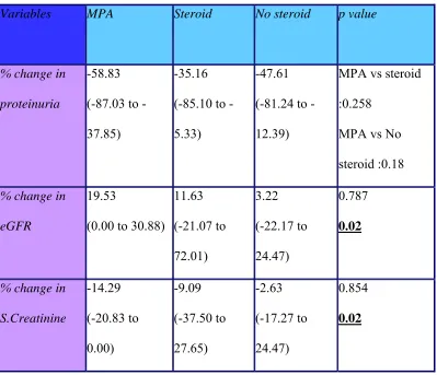
Results: Baseline characteristics of 3 groups were similar with respect to age, sex, MAP, proteinuria whereas e GFR in control 1 was higher by 15 ml/min. (p=0.03).
When cases were compared to control 2, there was a significant improvement in e GFR and s.creatinine (p=0.004 and 0.005) respectively, whereas there was a trend towards improvement in proteinuria in cases by 10% (p=0.313).On comparing cases to control 1, there was a trend towards improvement of proteinuria and e GFR in cases by 13% (p=0.548) and 5% (p=0.58) respectively. Sub group analysis of patients in the 3 groups who received ACE-I/ARB revealed similar trends.
IgA nephropathy (IgAN) is the most common form of primary glomerulonephritis in the developed world and it is an important cause of end stage kidney failure [1,2]
History of IgA Nephropathy (Berger’s Disease) –
The idea of using fluorescent-labeled specific antibodies to detect proteins in tissue was introduced by Coons and Kaplan[3] in 1950 and was first used for evaluation of diseased renal tissue by Mellors and Ortega[4] in 1957.
Percutaneous renal biopsy was initially described as a technique to diagnose kidney disease by Iverson and Brun in 1951 [5] .
Tomasi et al. had discovered the IgA immune system in 1965 [6].
Thus two techniques, immunofluorescent tagging of antibodies to detect antigens in tissues and percutaneous renal biopsy, along with the discovery of a new
immunoglobulin present in serum and in tissue secretions (IgA) all paved the way for the seminal observations , beginning in 1967 of Jean Berger and Nicole Hinglais at the Necker Hospital in Paris, France concerning a new entity they subsequently called mesangial IgA/IgG deposition . They described their novel observations in a brief paper published in 1968 which described predominant IgA mesangial deposition in some renal biopsies where the immunostaining of IgA strongly outshone the IgG reagent. This was the discovery of IgA nephropathy, also subsequently called Berger’s disease [7] . The different names of IgA nephropathy are -
1. Nephropathy with mesangial IgA-IgG deposits
4. IgA-IgG-Nephropathie
5. Glomerulites a depots d’IgA diffuse dan le mesangium 6. IgA-associated glomerulonephritis
7. IgA nephropathy
8. IgA-IgG deposits nephritis
9. Immunoglobulin A glomerulonephritis
10. Primary glomerulonephritis with mesangial deposits of IgA 11. Benign hematuria-loin pain syndrome
Epidemiology and Ancestral Difference :
Deposition of IgA is present in 4%–16% of normal, healthy living adults and
cadaveric donors , the significance of which is not completely understood . Thus, from the epidemiological perspective, the biopsy-proven IgAN cases represent a very small fraction of the total individuals with such deposits in the population as a whole [8,9] .
In 1988 Levy and Berger outlining the “Worldwide perspective of IgA nephropathy” suggested that the apparent geographic variations in the percentage of this
glomerulonephritis in kidney biopsy specimens could reflect different clinical policies for diagnostic tests more than a real ancestral difference [10] .
The systematic screening of urine samples could have influenced the higher prevalence reported both in Japan and in Singapore, the rare detection of IgAN in blacks either from the United States or from Africa was to be ascribed to the infrequent
While on one hand they were quite attentive to this primary aspect,they also got the hint that ancestral differences could suggest a possible role of genetic factors in the
etiology of IgAN.
The clinical onset of IgAN is usually found in the second and third decade of life but may occur at any age. Males are affected from two-fold in Japan, to six-fold more than females as reported in Europe and in the United States. The disease is more frequent in Whites and Asians than in Blacks from United States and South Africa, but the explana- tion of this difference is still unknown.
Most of the worldwide studies report prevalence rates as a percentage of cases of primary glomerulonephritides or as a percentage of a total series of renal biopsies, while few epidemiologic studies focused on the real incidence of primary IgAN in various populations [13].
An interesting study by Geddes et al. aimed at examining four IgAN databases from
four different countries in three continents to determine if geographic variability in long term outcome is independent of renal function, proteinuria and blood pressure at the time of diagnosis. They included patients from Glasgow (United Kingdom), Helsinki (Finland), Sydney (Australia) and Toronto (Canada). The results supported the hypothesis that the variation in the rate of progression of renal disease was probably due to variability at presentation in both duration (lead-time bias) and severity of disease in different countries. They did not exclude the possibility that true geographic variability exists mainly because of genetic variability, but also because of environmental factors, including dietary
ten year renal survival in patients with IgAN ranging between 95.7% in Helsinki and 61.6% in Toronto [14].
The role of environmental antigen triggers together with a genetic susceptibility factor determining the onset of the disease is still under debate. The higher prevalence of IgAN in Asian/Pacific Islanders could be supported by the evidence that high intake of rice and n-6 poly-unsaturated fatty acids could be associated with an increased risk of the disease as reported by Wakai et al. [161] or by the suggested role of Hemophilus parainfluenzae as a causative agent of the disease in Japanese individuals [15 , 16 ,17 ].
Worldwide Distribution
Europe :
The incidence of IgAN in Italy is 8.4 patients per million population(pmp) per year. According to the Italian Registry of Renal Biopsies (IRRB), based on data related to 32,862 renal biopsies collected during the years 1987–2000 from 128 renal units in Italy, IgAN is the most frequent disease among primary glomerulonephritides (21.5%) and its frequency is higher in males (39.3%) than in females (27.8%) (http://www.irrb.net).
The disease is also the most common primary glomerulopathy in Germany.
In a study of renal biopsies between 1970 and 1986, with a sample size of 8545 cases in adults and 1364 in children, Rivera et al. described the high incidence of IgAN and the
decrease of membranoproliferative glomerulonephritis in Spain. The mean annual inci- dence (pmp) of the disease was 7.9, making the disease the most common primary nephropathy [18].
America :
In Brazil, IgAN (29.4%) is the most frequent disease diagnosed among non-nephrotic patients. An increase in frequency of focal and segmental glomerulosclerosis has been reported in non-nephrotic patients; thus this disease became as common as IgAN (31.6% and 28.0%, respectively) from 1994–1999 [19] .
The most common primary glomerular disease in San Paolo is focal and segmental glomerulosclerosis (29.7%), followed by membranous nephropathy (20.7%) and IgAN (17.8%), confirming the above data [20] .
The United States Renal Data System (USRDS) reports that 0.8% of incident end stage renal disease patients in the USA have documented or suspected IgAN
(http://www.usrds.org.) .
In a 30-year renal biopsy study in Olmsted County (Minnesota), IgAN was present in 22% of biopsy-proven patients, thus representing the most frequent glomerulonephritis [21].
In 2006, Nair and Walker examined if IgAN still remained the most common primary glomerulopathy among young adults in the USA. In their study from a large renal biopsy referral center serving Midwestern and Southern states they grouped the patients in adults (≥ 20 years) and young adults (20–39 years). The disease was the most common primary glomerulopathy in young adult Caucasians (with a ratio IgAN/focal segmental glomerulosclerosis of 2.1:1) and also the most common cause of end-stage renal disease in this race. On the contrary, the disease was rare in African Americans in whom focal seg- mental glomerulosclerosis remains more common (focal segmental
Australia :
In a retrospective review of the pathology reports of all native renal biopsies performed in the state of Victoria, Australia, the most common glomerulonephritis in adults in 1995 and 1997 was IgAN (10.5 per 100,000 individuals). The disease accounted for 34.1% of all biopsy proven glomerulonephritides and was also the most common cause of end-stage renal disease due to glomerulonephritis [ 23 ] .
Asia :
Renal biopsy has been routinely performed in China in the last 20 years and IgAN is the most common primary glomerulonephritis occurring in the largest Asian country, representing 45.3% of the primary glomerulonephritides. The disease is also the leading cause of end stage renal disease, accounting for approximately 18% of patients [ 24 , 25 ]. It has also been reported at a frequency of 35% in Hong Kong by Lai and 34% in
Singapore by Sinniah in the 1980s [26] .
In Korea, in a study on a total of 4514 cases of renal biopsy collected over a 23-year period between 1973 and 1995, the most common primary glomerulonephritis in adults was minimal change disease (26.6%), followed by IgAN (22.1%) [27] .
The disease is listed as the primary cause of end stage renal disease in 28% of new dialysis patients as reported by Koyama et al. in a national survey of Japanese patients
The Research Group on Progressive Chronic Renal Disease, in their Nationwide and long-term survey of primary glomerulonephritis in Japan, found that 47.4% of 1045 biopsy specimens examined by immunofluorescence microscopy were IgAN [29].
In a retrospective analysis of 1592 renal biopsies received from various hospitals all over Kerala (India) over a period of two years, Chandrika observed that of the 1544 native kidney biopsies, the majority of cases (18.84%) were focal segmental glomerulosclerosis followed in frequency by IgAN (14.26%) [30] .
Genetic Contribution to IgA Nephropathy :
Several lines of evidence support a contribution by genetic factors to the development of, or susceptibility to, overt IgAN –
1. Many pedigrees have been described in which more than one individual is affected by IgAN. In some of these, genetic loci have been identified by linkage analyses[31,32,33]. 2. There are differences in prevalence in ethnically different populations which are unlikely to be fully accounted for by differences in environment and ascertainment [34]. 3. First and second degree relatives of IgAN patients have higher relative risks of developing this disease compared to the general population [35].
Using linkage analysis, several loci have been implicated in IgAN in different pedigrees - 2q36 locus, 6q22-q23 (IGAN1) and 3p24-p23 loci, 4q26-q31 (IGAN2) and 17q12-q22 (IGAN3) loci. The relative risk of IgAN is 16 times higher in first degree relatives (parents and siblings), and more than two-fold increased in second degree relatives (grandparents, grandchildren) of index cases compared to the general population [35]. Some genetic variations may contribute to more rapid progression of the disease . There is some evidence that variants in ACE (Angiotensin I converting enzyme I) [36] and AGT (Angiotensinogen) alter response to ACE inhibitors and/or angiotensin receptor blocker [37].
Histopathology, Immunofluorescence and Ultrastructural Examination :
Primary IgAN is a chronic glomerulonephritis defined by the presence or deposition in the mesangium of predominant IgA-C3 immune complexes, within which antigenic moiety remains elusive [38]. IgAN has conventionally been subdivided into primary and secondary IgA nephropathy, depending upon whether there is absence or presence of recognizable systemic conditions, respectively. These systemic diseases refer to Henoch-Schonlein purpura, dermatitis herpetiformis, liver cirrhosis, chronic hepatitis, coeliac disease,
ankylosing spondylitis, rheumatoid arthritis, Reiter’s syndrome, human immunodeficiency virus infection, mycosis fungoides and lung cancer.
Histopathology of IgA Nephropathy :
There have been many proposed classifications of renal lesions, but none of these is widely adopted. The separation of renal lesions into active and chronic is somewhat arbitrary, in that the pathogenesis of these lesions is poorly understood.
Acute Renal Lesions in IgA Nephropathy :
Mesangial hypercellularity is diagnosed when the mesangial cells exceed three per segmental area to as much as ten or 20 mesangial cells. This lesion is common, with characteristic focal and segmental distribution, particularly in early disease. These may be accompanied by expansion of mesangial matrix or sclerosis. Mesangial hypercellularity is not correlated with disease progression or renal survival [39].
Necrotizing lesion can be defined when any three of the following features are
identified in glomeruli - disruption of capillary wall, mesangiolysis, leucocytic infiltration, nuclear fragments, fibrinous deposits or cellular crescents. It is seen in about 10% of
patients with IgAN and up to 50% in the nephritis of Henoch-Schonlein purpura. While this lesion may not affect actuarial renal survival, immunosuppression is beneficial to prevent flares and progression to end-stage renal failure in some of the patients [40].
tubulo-interstitial fibrosis are all uncommon. When present, these consist generally of modest focal and segmental lesions in IgAN. These glomerular lesions have not been correlated with disease progression or renal survival.
Chronic Renal Lesions in IgA Nephropathy :
Glomerular sclerosis refers to accumulation of mesangial or basement membrane matrix. Its pathogenesis is incompletely understood, but inflammatory mediators such as TGF-β, PAI-1, and PDGFs play an important role in their development and their
progression [42]. The clinical correlation between extent of glomerular sclerosis and loss of renal function supports the notion that sclerosing process is cumulative and parallels the progressive decrease in functional nephrons.
recognized when tubular injury appears more pronounced than the extent of glomerular sclerosis, this raises the possibility of a superimposed drug-induced interstitial nephritis. Hyaline arteriosclerosis is identified in at least a third of the patients with IgAN at the time of biopsy, and is attributed to hypertension.
Immunofluorescence Examination of IgA Nephropathy :
Mesangial deposition of predominant IgA-C3 immune complexes defines the disease[45] and consists mostly of polymeric IgA1 with the absence of secretory
component and presence of J chain, regarded as the result of abnormal glycosylation of the IgA1 molecule [46]. Peripheral capillary loop deposits may or may not be found.
Ultrastructural Examination of IgA Nephropathy :
Two or more primary glomerular diseases may co-exist or be overlapping, requiring electron microscopy for diagnosis. Co existent minimal change nephropathy, membranous nephropathy, diabetic nephropathy and thin membrane nephropathy are recognized.
Tubulointerstitial Injury in IgA Nephropathy :
In IgA nephropathy, several studies have shown that the degree of tubular atrophy and interstitial fibrosis correlate better with deterioration of renal function than the extent of glomerular injury. In a study of 194 patients with a mean follow-up of ten years, tubular grade 2 defined as ≥ 25% but < 50% tubular damage (relative risk: 5.5) and grade 3 defined as > 50% tubular damage (relative risk: 28.8) were the best factors to predict chronic renal failure in multivariate analysis [49].
To et al. examined separately the degree of glomerulosclerosis, tubulointerstitial
damage, and hyaline arteriosclerosis in 126 renal biopsies of IgA nephropathy. In this study, global glomerulosclerosis represented the only independent prognostic factor for renal survival. Both glomerulosclerosis and tubulointerstitial damage, however, were significantly correlated with the degree of proteinuria, hypertension and serum cre- atinine level [39] .
More recently, Myllymaki et al.analyzed 204 renal biopsies of IgA nephropathy and
In the evidence-based Oxford classification 2008 for IgA nephropathy, tubular atrophy/interstitial fibrosis was significantly associated with renal outcome independently of glomerular filtration rate, proteinuria, and blood pressure at presentation.
Altogether these independent clinicopathological studies reveal that the degree of tubulointerstitial damage correlates with deterioration of renal function in IgA nephropathy. The principal mechanisms implicated in the progression of tubulointerstitial injury in IgA nephropathy are –
1. Glomerulo-Tubular Communication :
In recent years, glomerulotubular crosstalk has been proposed as one of the
mechanisms involved [43]. Upon deposition of IgA, mesangial cells start to proliferate and release cytokines/chemokines, among which TNF-α and IL-6. These inflammatory mediators are filtered by damaged glomeruli and stimulate downstream tubular epithelial cells(TECs), which subsequently produce proinflammatory mediators including IL-1, TNF-α, MIF, IL-8,MCP-1, RANTES, and sICAM-1 [51].
2. Proteinuria :
3. Downstream interstitial fibrosis due to reduced blood supply because of
glomerulosclerosis .
4.Misdirected filtration into the interstitium from the synechiae of
glomerulosclerotic segments.
5. Leukocytes :
Chemokines play pivotal roles in the recruitment of inflammatory cells into the kidney. The chemokine receptors CXCR3 and CCR5 are expressed on activated T cells. Interstitial CXCR3, as well as CCR5-positive T cells may play a role in the progression of renal diseases [53,54] .
PDGF is a potent chemo-attractant for macrophages. In addition,it causes the release of other cytokines among which TGF-β is predominant. Interstitial PDGFR-β is
significantly increased in IgA nephropathy and associated with monocyte/macrophage infiltration [55].
The secretion of cytokines by TECs not only leads to up-regulation of adhesion molecules including ICAM-1 and CD44 but also of MHC class II and the co-stimulatory signals CD80 and CD86,which support infiltration and activation of inflammatory cells.In a study comprising 33 cases of IgA nephropathy, CD80/CD86 expression
In contrast, little has been published about B-cell infiltration and IgA nephropathy. Heller et al. characterized B-cell infiltrates and the factors involved in B-cell recruitment
in 18 cases of IgA nephropathy. They concluded that CD20-positive B cells form a prominent part of the interstitial inflammatory infiltrate and that B-cell infiltrates are associated with increased local expression of the chemokine CXCL13 and the
corresponding receptor CXCR5 on B cells. However, no correlation could be established between B cell infiltration and the Haas classification of IgA nephropathy [57].
6. Growth Factors
Hepatocyte growth factor (HGF) has recently emerged as a potent antifibrotic and reno-protective factor that counteracts the pro-fibrotic actions of TGF-β [58]. This may represent an endogenous mechanism of repair.
The expression of TGF-β is increased in tubules and tubulointerstitial areas in IgA nephropathy and correlates significantly with the severity of histological damage [59]. TGF-β has also been shown to induce the activation of the transcription factor c-Jun in TECs that in turn regulates the expression of genes involved in proliferation and inflammation [60].
7. Myofibroblast Influx
Accumulation of myofibroblasts in the interstitium is a key event in the development of fibrosis. These cells are characterized by the expression of α-smooth muscle actin (α -SMA) and fibroblastic-specific protein-1(FSP-1) [62]. In a series of 38 patients with IgA nephropathy, the intensity of the interstitial α-SMA staining correlated with renal
functional outcome [63].
8. Matrix Metalloproteinases
Accumulation of extracellular matrix (ECM) results from an imbalance between synthesis by myofibroblasts and degradation by matrix metalloproteinases (MMP) [64]. Surprisingly, very few studies address the issue of MMP activity in IgA nephropathy. Brisk glomerular activity of MMP-9 and, to a lesser extent, MMP-2 has been reported in IgA nephropathy [65]. Furthermore, peripheral blood mononuclear cells(PBMCs) from patients with IgA nephropathy have been shown to express higher level of MMP-9 mRNA than those from patients with other forms of glomerulonephritis or from healthy controls [66].Also, MMP-9 mRNA production by PBMCs correlated significantly with the severity of histopathologic grading for IgA according to Haas.
Although these data suggest that PBMC-derived MMP are of importance during disease progression in IgA nephropathy, solid data on MMP activity in the
9. The Renin-Angiotensin System
The Renin-Angiotensin system (RAS) has been shown to play a key role in the development of renal fibrosis. While several components of the RAS exert profibrotic activity, Angiotensin II (Ang II), produced by activated macrophages and fibroblasts has been shown to be the key hormone responsible for renal fibrosis [67]. Ang II induces expression of several cytokines/chemokines, including TGF-β, TNF-α, IL-6, MCP-1 and RANTES, through interaction with the Ang II receptors type 1 and 2 (ATR1 and ATR2) [68].
Furthermore, Ang II influences renal tissue turnover by mediating TEC apoptosis (via interaction with ATR2) while at the same time mediating TEC proliferation (via
ATR1)[69]. Pharmacologic blockade of the RAS effectively slows down the progression of interstitial fibrosis [70].
In IgA nephropathy, the intrarenal RAS is activated.In a study of 20 renal biopsies from IgA nephropathy patients, mRNA levels of angiotensinogen, renin, ACE and AT1R and AT2R were significantly enhanced in both microdissected glomerular and
tubulointerstitial tissue samples [71].
10. CD44
atrophy, rarefaction of the peritubular capillary network and ultimately fibrosis [73]. Under normal conditions, CD44 is undetectable in the kidney except in passenger
leukocytes. In IgA nephropathy, a strong basolateral expression of CD44 was observed in damaged tubules as well as in the interstitium. CD44 expression paralleled the degree of tubular atrophy and interstitial fibrosis and correlated significantly with the degree of proteinuria[74].
Podocyte Pathology :
Podocytes are unique epithelial cells necessary for maintaining the selectivity and integrity of the glomerular filtration barrier. Podocytes have also been shown to fully express most or all the elements of the renin-angiotensin system (RAS) [75]. In vitro studies of podocytes cultured with either supernatant from mesangial cells cultured with polymeric IgA isolated from patients with IgA nephropathy (IgAN) or recombinant tumor necrosis factor-α (TNF-α) reveal enhanced proinflammatory changes with increased synthesis of TNF-α and interleukin-6 (IL-6) [76].
Necrosis and detachment of the podocytes from the glomerular basement membrane was observed in IgAN [77]. Complementing the histological findings of podocytopenia, patients with IgAN had increased urinary excretion of podocytes[78]. Treatment with angiotensin-converting enzyme inhibitor or angiotensin II receptor antagonist reduced the urinary excretion of podocyte in IgAN [79].
Lai et al. examined the expression of two podocyte markers (nephrin and ezrin) in
were examined as nephrin has a crucial role in the filtration barrier of the glomerular podocyte and ezrin is a glomerular epithelial cell marker of podocyte injury
(podocytopathy) [80].
Although IgA isolated from patients with IgAN exerted no apoptotic effect on podocytes, in vitro studies revealed TNF-α released from glomerular mesangial cells after IgA deposition modulates the expression of Bcl-2 by podocyte through
auto-amplification of TNF-α [81]. There is an apoptotic effect of mesangial derived-TNF-α on podocytes in chronic IgAN.
Mechanisms of Podocyte Injury in IgAN :
It is well demonstrated that a glomerulo-tubular cross-talk involving TNF-α exists in IgAN [51]. Lai et al. hypothesized that a similar glomerulo-podocytic cross-talk existed
Clinicopathologic Findings :
Pathogenesis –
IgAN arises as a consequence of circulating IgA1-containing immune complexes binding to mesangial cells. The IgA1 in these complexes has a reduced content of galactose in hinge-region O-glycans to constitute a neoantigen that is recognized by circulating IgG or IgA1 [82]. The galactose deficiency accentuates binding of the IgA1 to mesangial cells. The resulting stimulation of mesangial cells leads not only to their proliferation as the light-microscopic hallmark of this renal disease, but also to synthesis of extracellular matrix and secretion of a host of cytokines/chemokines that may
culminate in inflammation and scarring in the glomerular and tubulointerstitial compartments [83].
Prognostic Factors :
Demographic Characteristics :
Among demographic features, male gender [85], and older age [14] and perhaps obesity[86] portend a worse long-term outcome for patients with IgAN.
Clinical Features :
Patients with at least one episode of synpharyngitic hematuria fared better than those who had never experienced this degree of glomerular bleeding [87]. There is nearly universal agreement that hypertensive patients fare less well than normotensive patients. The National Kidney Foundation in the United States recommends blood pressure <130/80 mm Hg for patients excreting < 200 mg protein per g creatinine per day and a lower target for patients excreting > 1000 mg protein per g creatinine per day.
Laboratory Findings :
Even mildly impaired eGFR (45–60 ml/min/1.73 m2) at the time of diagnostic biopsy have outcomes worse than those with normal clearance function.
Proteinuria has generally correlated with prognosis, although the threshold for a deleterious effect has been less clear. Analyses found a continuous-variable effect, with an adverse influence starting at 500 mg/day. More importantly, rather than one
hypertension, and renal insufficiency (eGFR < 60 ml/min/m2) 7.5 years later than were patients with normal albuminuria at biopsy [90].
Other laboratory features associated with worse outcome include hyperlipidemia and hyperuricemia.
Light-Microscopy Histology :
A low number of podocytes per glomerulus has been associated with worse
proteinuria and a decrement in renal clearance function [91]. Other glomerular features signifying an unfavorable outcome include endothelial cellular proliferation, focal
necrosis, focal segmental scars, extracapillary cellular proliferation (a crescent if involved area exceeds 10% of the circumference of Bowman’s capsule) and global sclerosis. Another vascular feature associated with poor prognosis is thrombotic
microangiopathy [92].
Degree of interstitial damage is a better prognostic indicator than degree of injury within glomeruli [90]. Enumeration of fibroblasts after staining for
fibroblastic-specific protein 1 (FSP1) has been correlated with extent of
glomerulosclerosis and interstitial fibrosis. Other poor prognostic factors are intensity of staining for CD3+ lymphocytes, increased numbers of Mac387+
Beyond Light Microscopy :
IgG in the mesangial immune deposits, immune deposits in the capillary loops on electron microscopy are other poor prognostic factors.
Potential New Prognostic Markers :
A high serum IgA/C3 ratio was correlated with worse histological lesions in non-nephrotic patients and a ratio above 4.5 indicated a worse renal survival [93].
Urinary IL-6 or the IL-6/epidermal growth factor ratio and greater amounts of IL-8, TGF-β, and transforming growth factor-α1 have been proposed as markers of poor prognosis.
Genetic Markers :
Pathology based grading systems :
Classification
Author/Year Grade 1
Grade 2 Grade 3 Grade 4 Grade 5
SPNSG/19821
Minimal glomerular
changes Mesangial proliferation only Any focal or sclerotic lesion
Haas/19972
Minimal or no mesangial hypercellularity ; without sclerosis or crescents
Focal segmental glomerulosclerosis ;minimal increase in mesangial
hypercellularity; no crescents
Focal proliferative changes , <50% glomeruli are hypercellular
Diffuse proliferative, > 50% glomeruli are hypercellular
Advanced sclerotic changes, ≥40% glomeruli are globally sclerotic and/or ≥40% tubular atrophy or loss of cortex To/20003 Mean glomerular sclerosis <25%.Tubular atrophy and interstitial fibrosis <5% Mean glomerular sclerosis 25%-49%. Tubular atrophy and interstitial fibrosis 5%-49%
Mean glomerular sclerosis ≥50%. Tubular atrophy and interstitial fibrosis ≥50%.
Lee/20054
Normal or focal mesangial cellular proliferation
Diffuse mesangial cellular proliferation, or <25% of
glomeruli with crescents, segmental/global sclerosis
25% to 49% of glomeruli with crescents, segmental/global sclerosis
50% to 75% of glomeruli with crescents, segmental/global sclerosis
>75% of glomeruli with crescents, segmental/global sclerosis
Wakai/20065
Slight mesangial cell proliferation and increased matrix
Slight mesangial cell proliferation and increased matrix. Glomerulosclerosis, crescent formation or adhesion to
Bowman’s capsule in < 10% of glomeruli. Moderate diffuse mesangial cell proliferation and increased matrix. Glomerulosclerosis, crescent formation or adhesion to
Bowman’s capsule in 10% to 30% of glomeruli Severe diffuse mesangial cell proliferation and increased matrix. Glomerulosclerosis, crescent formation or adhesion to
Bowman’s capsule in > 30% of glomeruli
Manno/20076
Normal glomeruli or slight increase in mesangial matrix and/or cellularity
Moderate or diffuse mesangial proliferation and/or focal segmental sclerosis and/or endocapillary proliferative and/or cellular crescents in ≤50% of glomeruli
SPNSG, Southwest Pediatric Nephrology Study Group
1. A multicenter study of IgA nephropathy in children. (1982) A report of the Southwest Pediatric
Nephrology Study Group. Kidney Int 22: 643–652.
2. Haas M. (1997) Histologic subclassification of IgA nephropathy: a clinico-pathologic study of 244 cases.
Am J Kidney Dis 29: 829–842
3. To KF, Choi PC, Szeto CC, et al. (2000) Outcome of IgA nephropathy in adults graded by chronic
histological lesions. Am J Kidney Dis 35: 392–400
4. Lee HS, Lee MS, Lee SM, et al. (2005) Histological grading of IgA nephropathy predicting renal
outcome: revisiting H. S. Lee’s glomerular grading system. Nephrol Dial Transplant 20: 342–348
5. Wakai K, Kawamura T, Endoh M, et al. (2006) A scoring system to predict renal outcome in IgA
nephropathy: from a nationwide prospective study. Nephrol Dial Transplant 21: 2800–2808
6. Manno C, Strippoli GF, D’Altri C, et al. (2007) A novel simpler histological classification for renal
survival in IgA nephropathy: a retrospective study. Am J Kidney Dis 49: 763–775.
Clinical Course of Primary IgA Nephropathy :
about 80% of all cases from the secondary forms.
Comparisons between males and females showed that females had significantly less proteinuria, lower GOS, and lower serum IgA level . In general at time of diagnosis, the clinical presentation was less severe in females.
The natural history of primary IgAN is both clinical and pathological progression towards chronic renal failure (CRF) and end-stage renal failure (ESRF) [94 ,95 ]. In the literature, the cumulative incidence at 20 years of ESRF/dialysis varies from
10% to 40% depending on the policy of renal biopsy.
IgA Molecule in IgA Nephropathy :
Deposited IgA is predominantly polymeric IgA (pIgA) of the IgA1 subclass and has been shown to be differentially glycosylated. Moreover, vaccination studies have demonstrated alterations in mucosal immunity, whereas in many patients mucosal infections are associated with episodes of macroscopic hematuria.
Plasma IgA1 are elevated in about half of the IgAN patients [96]. The elevated IgA concentration seems to be the result of higher production of IgA by plasma cells in the bone marrow[97]. Next to the higher concentrations of IgA1, qualitative changes of IgA in IgAN patients have been described. IgA of IgAN patients contains a reduced
galactosylation of the O-linked glycans in the hinge region [98].
IgA concentrations, like multiple myeloma or HIV, are not associated with renal IgA depositions supports the idea that the higher IgA1 concentrations are not the only cause of mesangial IgA deposition in IgAN.
Dendritic cells (DC) might be less effective in inducing IgA production by naïve B cells. DC derived from IgAN patients showed a reduced capacity to induce IgA
production in the presence of IL-10 [99]. However, so far it not clear which factors are responsible for the disturbed DC function in IgAN patients.
Galactose deficiency of IgA1 appears to be a key pathogenetic factor contributing to the development of the disease. Circulating complexes in IgA nephropathy contain IgA1 with galactose-deficient hinge-region O-linked glycans [82]. Notably, galactose-deficient IgA1 is the predominant glycosylation variant of IgA1 in the mesangium[100]. Cells from patients with IgA nephropathy had low expression and activity of the corresponding galactosyltransferase and high expression and activity of sialyltransferase. Consequently, the O-glycans on the secreted IgA1 are galactose deficient with high proportion of GalNAc being sialylated [101]. In view of the fact that sialylation was shown to prevent galactosylation of GalNAc, it is quite possible that “premature” sialylation helps to increase the levels of galactose-deficient O-glycans in IgAN.
After alteration of the glycosylation of the IgA1, neoepitopes represented by IgA1 glycans or hinge-region glycopeptides are exposed and recognized by
resultant IgA1 complexes are relatively large. Because of their size, they are not
efficiently cleared from the circulation and thus tend to deposit in the renal mesangium. Galactose-deficient IgA1 is retained in the circulation for long periods of time. Galactose deficiency in itself should not hinder disposal of IgA1 molecules because the asialoglycoprotein receptor recognizes terminal GalNAc as well as galactose.However, if the GalNAc is linked to sialic acid or is covered by an antibody, it cannot be recognized by the hepatic asialoglycoprotein receptor and is not catabolized [103].
Immunopathogenesis of IgAN is considered to occur in three phases –
1. In the early primary phase, B cells produce polymeric IgA (pIgA) in response to variety of antigens that initiates glomerular IgA immune deposit formation.
2. In the secondary phase, continuous activation of the mesangial cells, the complement system and the innate immune system by persistent glomerular pIgA immune deposits attract macrophages that produce inflammatory mediators leading to glomerulosclerosis. 3. In the tertiary phase, interstitial infiltration by T cells cause tubular injury and sets in motion irreversible interstitial fibrosis leading to end stage renal failure in 20%–40% of the affected patients.
Antigen-Dependent Mechanism of IgA Nephropathy :
Since IgA is an important immunoglobulin in defense mechanisms against
antigens are exogenous and/or endogenous in patients with IgA nephropathy. It is speculated that there are many kinds of antigens, i.e. food, virus, bacteria and/or
fungus, in patients with IgA nephropathy as follows: (1) dietary antigens,(2) respiratory antigens, (3) intestinal antigens, (4) biliary antigens, and(5) dermal antigens. These antigens might form antigen-antibody dependent immune complexes.
Food components such as bovine albumin, ovalbumin , lactoglobulin , gliadin have been implicated.
IgA nephropathy is frequently preceded by episodes of upper respiratory tract infection that is presumed to have some viral etiology [104]. Virus-like particles and/or microtubular structures were occasionally observed in the glomerular mesangial areas by electron microscopy.
Slight deposition of adenovirus, herpes simplex, varicella zoster or parainfluenza 3 virus
was observed by immunofluorescence [106], the contributory role of which is debated. IgA nephropathy has been described as a complication of infections with Yersinia enterocolitica, Campylobactor jejuni, and Mycoplasma pneumoniae. Suzuki et al.
observed glomerular deposition of Hemophilus parainfluenzae antigens by
immunofluorescence and the presence of IgA antibody against H. parainfluenzae in sera
by enzyme-linked immunosorbent assay (ELISA) in patients with IgA nephropathy [16].
Complement Activation :
complement in disease pathogenesis [108]. Local complement production by various intrinsic renal cells and infiltrating cells may contribute to tissue injury in IgAN [109]. Moreover, proteinuria but not serum creatinine, at the time of renal biopsy correlated with C3 mRNA expression. This last phenomenon may be attributed to the finding that apical proteins stimulated basolateral C3 synthesis by cultured human proximal tubular epithelial cells, in which transferrin and apotransferrin stimulated C3 biosynthesis more intensely. Locally secreted C3 may further activate tubular cells via the C3a receptor to enhance renal injury [110]. Evidence is accumulating that complement activation via MBL and the lectin pathway is also associated with disease progression in IgAN [111]. More recent data suggest that IgA was co-deposited with MBL in about 25% of patients and that MBL deposition showed more severe renal disease as compared with MBL-negative cases, suggesting an important role for MBL in disease progression [112].
Corticosteroids :
For decades, these agents have been used with the concept of a non-specific but very potent antiinflammatory effect.
In 1986 Kobayashi et al. first reported their experience coming from a prospective,
Encouraging results were also obtained in 13 children who were treated with alternate day prednisone 60 mg/m2 for three months,reduced to 30 mg/m2 by one year and 15 mg/m2 by two years. In comparison with a historical group, Waldo et al. showed
a significant improvement in urine analysis (both proteinuria and hematuria) and a preserved normal GFR. Follow-up biopsy after two years of treatment revealed a significant fall in the activity score (from 5.2 to 4.3), without any significant increase in the chronicity score [114].
Conversely, first reports coming from small, randomized clinical trials were mostly negative. Lai et al. found no benefit of prednisone (1 mg/kg/day for two months and a
tapered dosage for another two months) in a controlled trial involving 34 patients with nephritic syndrome. However, the length of treatment (only four months) was
perhaps not long enough [115].
Julian et al. made a multicenter prospective trial in IgAN patients with proteinuria
greater than 2 g/day by comparing symptomatic therapy with alternate-day prednisone (60 mg/day tapered by 10 mg every three months to 10 mg/day over 24 months).
However, they published only preliminary data of a small set of patients not supporting a favorable effect of steroids (only a modest reduction in proteinuria was observed) [116]. However, the lack of effectiveness on renal function may be due to a too short follow-up (nearly two years).
In 1987 Pozzi etal started a large, multicenter, randomized,open-label, controlled trial
six-months’ steroid treatment (methylprednisolone 1 g intravenously for three
consecutive days at the beginning of months 1, 3 and 5, plus oral prednisone 0.5 mg/kg every other day for six months). Baseline characteristics were comparable in the two groups. After five years of follow-up, renal survival was significantly better in the steroid treated patient group than in the control group for both the primary endpoints of 50% and 100% increase from baseline plasma creatinine levels (respectively of 17% and 21%; log-rank test p < 0.048 and p < 0.005). Evaluation of renal survival after ten years of follow- up confirmed that outcomes in the steroid-treated group were better than those in the control group (97% vs. 53%, p = 0.0003). Mean urinary protein excretion also
significantly decreased in the steroid group (from 1.93 ± 0.45 g/day at baseline to 0.78 ± 0.41 g/day at one year), and this decrease persisted throughout the follow-up,whereas proteinuria remained unchanged in the control group .It is likely that treatment with ACE inhibitors or ARBs did not affect the findings, since a similar percentage of patients in both groups received these agents. Interestingly, glucocorticoids were effective in every histological class. This suggests that the main indication to steroid treatment in patients with IgAN should be proteinuria level and not indexes of activity and/or chronicity at renal biopsy examination. None of the patients in the steroid group experienced any major side effects [117].
More recently, in an open-label study, Katafuchi et al. randomized 90 IgAN
were ineffective on renal survival. However, the event rate was very low (only three patients in each group progressed to end-stage renal disease during follow-up) and the study was severely underpowdered to show any effect on end-stage renal disease [118]. No controlled, randomized studies have yet been conducted comparing intravenous pulse with oral steroid for IgAN.
There are few studies evaluating the effect of glucocorticoids on histological lesions. Yoshikawa et al.studied 78 children with IgAN and normal renal function, 40 of whom
received prednisolone, azathioprine, heparin/warfarin and dipyridamole for two years, and 38 heparin/warfarin and dipyridamole alone. Clinical and pathological data before treatment were similar in the two groups. In the second biopsy, performed after treatment, the mean percentage of glomeruli showing segmental or global sclerosis was unchanged in patients treated with glucocorticoid and azathioprine, but increased from 3.9% to 16.4% in patients who did not receive glucocorticoids [119].
There are few clinical studies testing steroid therapy in IgAN patients with advanced disease. Tamura et al. studied retrospectively 60 IgAN patients with creatinine clearance
below 70 ml/min at the time of renal biopsy (on average 58 ml/min), 20 of whom received steroids. The mean follow-up period was about 4.5 years. After one year, proteinuria decreased in the steroid group (from 2.33 to 1.02 g/day), but remained unchanged in the non-steroid group (from 1.39 to 1.28 g/day). Interpretation of results is slightly biased by the fact that before treatment, proteinuria was higher in patients
(2.51 vs. 1.79 mg/dl, respectively) [120].
Renin-Angiotensin Blockade :
In recent years, two prospective RCTs [123,124] proved that in proteinuric IgAN with rather good glomerular filtration rate, a significant effect of ACEi not only in reducing proteinuria but also in preserving renal function, and another RCT[126] proved that ARB decreases proteinuria and slows renal deterioration after adjustment for blood pressure control. The additional benefit of a combination therapy has never been adequately addressed in IgAN by properly designed RCT, and only short-term studies or RCT sub- analysis are available, which suggest a possible superior effect of the combination therapy in reducing proteinuria and possibly renal disease progression.
Treatment of IgAN with ACEi :
A more recent prospective study pointed out a significant stabilization of GFR in IgAN only in the intensive treatment group, when BP was lowered to <130/70 mmHg (often using multidrug combinations), while patients with BP > 135/75 mmHg
failed to be protected against functional decline [121].
Dillon in 2001 performed a meta-analysis of 237 IgAN patients treated with ACEi and enrolled in three short-term crossover trials, one prospective RCT and three retrospective studies, and concluded that the effect on proteinuria was clear, while the renoprotective effect remained unproved [122].
In 2003, a RCT of ACEi in IgAN was published by Praga et al. The trial was not
one single center with wide range of proteinuria (from 0.5 to 5 g/day) and various degrees of renal function impairment. Patients randomly assigned to enalapril or control group had similar baseline characteristics of GFR, proteinuria and BP. Enalapril was given at a starting dose of 5 mg/day and titrated to achieve and maintain a target BP of ≤ 140/90 mmHg until a maximal dose of 40 mg/day.After a mean follow-up of 75 months, the proportion of patients developing the primary end point (50% increase of baseline plasma creatinine) was significantly lower in ACEi group than in the control group (12% versus 57%, respectively). After four years of follow-up, renal survival was 100% in treated patients versus 70% in controls and this trend worsened after eight years when the renal survival decreased to 55% in placebo, versus 92% in treated subjects. Proteinuria significantly decreased in the treated group from 2 ± 1.3 g/day (0.5–5.3) to 0.9 ± 1.3 g/day (0.5–5.3) ( p < 0.001) while no significant changes where observed in the control group [123].
IgACE was the first placebo-controlled RCT designed to investigate the effects of ACEi in young IgAN patients (aged less than 35 years), with moderate proteinuria (> 1 and < 3.5 g/day/1.73 m2 ) and GFR >50 ml/min/1.73 m2 . Sixty-six patients, meanly 20 years old (range 9–35), were randomized to receive benazepril 0.2 mg/kg/day or placebo, and were followed for a median of 38 months. The end point of progression of IgAN was defined as >30% decrease of baseline creatinine clearance (CrCl) and also as a composite end point of >30% decrease of baseline CrCl and/or worsening of proteinuria until ≥ 3.5 g/day/1.73 m2 was considered. Secondary outcomes included proteinuria, partial
reached the end point of renal function decline. No patient on ACEi developed nephrotic syndrome, versus 20.6% on placebo group. The renal disease progression to the
composite end point of renal function decline and worsening of proteinuria above the nephrotic range resulted significantly different between the two groups of ACEi treated patients and those assuming placebo( p = 0.035). In conclusion, angiotensin antagonism was successful in limiting progression of renal damage in young IgAN patients with proteinuria between 1 and 3.5 g/day. ROC analysis identified 1 g/day/1.73 m2 at one year as a cut-off level of proteinuria protective over progression. In the IgACE trial, patients with time average proteinuria < 1 g/day/1.73 m2 had a favourable survival to 30% reduction of baseline CrCl (100% survival after 58 months of follow-up), while those with time-average proteinuria between 1 and 3 g/day/1.73 m2 had 75% survival and those over 3 g/day/1.73 m2 a 65% survival (log rank:
p = 0.01) [124].
Treatment of IgAN with ARB :
In a short term study of 12 weeks, ARB therapy (losartan 50 mg) resulted superior to the calcium channel blocker treatment (amlodipine 5 mg) in reducing proteinuria and TGF-β1 excretion, even though BP control was similar[125].
The long-term effects of ARB therapy in proteinuric IgAN have been recently demonstrated by Li et al.in a multicenter, placebo controlled RCT. They enrolled 109
(titrated up to 160 mg/d for BP control), or placebo for 104 weeks.Additional antihypertensive therapy was allowed to achieve a target BP of 140/90 mmHg. Proteinuria decreased significantly in the treatment group but did not change in the placebo group. With multiple linear regression models, ARB treatment resulted in a 33% decrease in proteinuria after adjusting for other confounding factors. There was a
significant decrease in mean rate of GFR decline in the ARB group (−5.62 ± 6.79 ml/min/year), compared with the placebo group (−6.98 ± 6.17 ml/min/year) throughout the study period after adjustment for average BP and proteinuria ( p = 0.014). This RCT demonstrated that ARB significantly decreases proteinuria and slows renal deteriora- tion in patients with IgAN after adjustment for confounding factors, notably BP [126].
Combination Therapy in Proteinuric IgAN :
In a short-term clinical investigation, doubling ACEi or ARB doses in 12 adults with IgAN did not improve the antiproteinuric effect.However, a higher effect was
observed when the two drugs were co-administered and an additional reduction in proteinuria was obtained when combined therapy doses were doubled [127].
In 31 patients with IgAN and mild proteinuria (from 0.3 to 0.7 g/day), GFR > 50 ml/min and normal BP, the dual blockade of RAS given for six months was reported to induce remission of proteinuria in 63% of the cases versus 41% in ACEi and 36% in ARB monotherapies [128].
In conclusion, the additional benefit of a combination therapy has never been
Cyclophosphamide in Combination with Corticosteroid
:In 2002, Ballardie et al. published a randomized, controlled, single center study on
38 patients with progressive loss of renal function. Patients, none of whom had crescentic glomerulonephritis, were randomized to prednisolone and low-dose cyclophosphamide followed by azathioprine or supportive therapy only. Renal survival in treated patients showed considerably better preservation of function at five years (72% compared with 6% in controls). Proteinuria and erythrocyturia reduced from 12 and six months of treatment, respectively. No side effect was reported apart from marrow suppression in one subject and pulmonary tuberculosis in another. This study may be faulted, however for suboptimal blood pressure control and insufficient use of medications that block the angiotensin system, the unusually poor survival rate of the placebo group, and the small number of patients [129].
Tumlin et al. conducted a prospective, uncontrolled, open-label trial of 12 patients
with crescentic IgAN and clinically progressive disease. Treatment consisted of three doses of methylprednisolone at 15 mg/kg/day, followed by intravenous
cyclophosphamide at 0.5 g/m2 /month for six months. Serum creatinine fell from 2.7 to 1.5 mg/dl, and proteinuria decreased from 4 to 1.3 g/day after treatment, suggesting a beneficial role of cyclophosphamide in crescentic glomerulonephritis [130].
Mitsuiki et al. retrospectively examined the outcome of 35 patients with
supportive treatment. Renal prognosis was significantly better in the treatment group [131].
Overall, these studies suggest that combined cyclophosphamide/steroid therapy may benefit IgAN patients at very high risk of renal failure, namely those with a progressive decline in GFR and/or crescentic lesions before randomization. However, more definitive evidence involving patients in sufficient numbers is required before this treatment can be widely accepted.
Cyclosporin :
Lai et al. conducted a randomized prospective single blind study of 19 patients with
proteinuria > 1.5 g/day. Patients who received cyclosporin had significant reduction of proteinuria, serum IgA, and increase of plasma albumin concentration compared with placebo. However, there was transient deterioration of renal function during treatment, despite within-range trough drug levels. The authors discouraged indiscriminate use of cyclosporin in IgAN due to nephrotoxicity [132].
Azathioprine :
Goumenos et al. performed a retrospective analysis of 74 IgAN patients followed
between 1.4–2.5mg/dl, this immunosuppressive regimen reduced the risk of doubling serum creatinine compared to controls (27% vs. 78%) and delayed progression to end-stage renal failure (17% vs. 55%) [133].
The Japanese Pediatric IgA Nephropathy Treatment Study Group randomized 78 children with newly diagnosed early IgAN to receive either prednisolone, azathioprine, heparin-warfarin, and dipyridamole or the combination of heparin-warfarin, and
dipyridamole only. After two years, there was significant reduction of proteinuria and serum IgA levels in the prednisolone/azathioprine group, while there was no difference in renal function between the two groups. The study was flawed by a lack of data on
baseline proteinuria and creatinine clearance as well as blood pressure control in both groups [134].
Mycophenolate Mofetil :
To date, four randomized clinical trials have been published on the use of MMF in IgAN,which add more controversy than consensus.
The first randomized study, published in 2002 in the Chinese literature,was
conducted in Beijing in which 62 Chinese patients with severe IgAN with Lee’s grade IV and V renal histology and urinary protein > 2.0 g/d received MMF or oral prednisone for at least 12 months. After 18 months’ follow up, the MMF group showed significant improvement in proteinuria and serum lipids than the prednisone group [135]. In 2004 Maes et al. described a prospective study in 34 Belgian patients with
13) after instituting salt restriction and angiotensin-converting enzyme inhibitor therapy in all. They only included patients with histologic unfavorable criteria and arterial hypertension, and excluded those with mild histopathologic changes despite heavy proteinuria. After three years of follow-up evaluation, inulin clearances and proteinuria did not differ between the groups [136].
In 2005 Tang et al published a prospective study in 40 Chinese patients with IgAN
and mild tubulointerstitial lesions who were randomized 1:1 to 1.5 (BW < 60 kg) or 2.0 g/day MMF for six months or continuation of contemporaneous medication only after blockade of the renin-angiotensin system failed to reduce proteinuria to < 1 g/d. Twelve months after stopping MMF, the overall remission rate (proteinuria < 0.3 g/d or 50% of baseline) was significantly higher in MMF-treated patients whose proteinuria dropped to 62% of baseline, whereas urine protein in control patients increased to 120% of baseline. Adverse events in the MMF group included anemia, urinary tract infections and cervical lymphadenitis. There were no adverse events in the control group [137].
In late 2005, Frisch et al. published a randomized controlled trial in which 32
predominantly Caucasoid North American patients with advanced IgAN (mean serum creatinine, 2.5mg/dl) were randomized to MMF or placebo. MMF was given for one year as a “salvage” therapy in 16 patients with advanced renal insufficiency. Notably, the presence of glomerulosclerosis or tubulointerstitial atrophy and fibrosis on renal
biopsy was an inclusion criterion. The study was terminated prematurely after observing a trend towards worse outcome in the MMF group [138].
account for the differences observed in these studies. Another possibility is the mild histologic grade in Tang’s study versus the moderate-to-severe grades in the studies by Maes and Frisch.Further observation and studies are needed to provide more definitive answers on the efficacy of MMF in IgAN.
Other Immunomodulatory Therapy :
Lai et al.had performed plasma exchanges for two patients with crescentic IgA in
whom corticosteroid and immunosuppressive therapy failed to control the progression of the disease. In both cases, the rapid progression of renal failure was apparently halted. Nevertheless, the long-term benefit of plasma exchange in crescentic IgA nephropathy was unsatisfactory as the renal function continued to deteriorate in the following 12 months despite an initial stabilization [139].
Lou et al. showed in 60 patients randomized to leflunomide or fosinopril that
leflunomide effectively suppressed proteinuria, however this effect was not superior to that achieved by fosinopril [140].
In a study that employed 41 historical pediatric controls who received prednisolone, warfarin, and dipyridamole therapy before 1989, the addition of mizoribine in 20 subjects after 1990 appeared to be more effective in ameliorating proteinuria and histological severity on repeat biopsy at two years [141].
Information of Rituximab (anti-CD20 antibody) for treatment of IgAN is lacking.
Fish Oil :
The rationale for using fish oil supplements in patients with IgAN is based on experimental data suggesting that omega-3 polyunsaturated fatty acids may limit the immunologic renal injury in IgAN. These omega-3 fatty acids compete with arachidonic acid to produce trienoic eicosanoids which, in turn, may slow renal disease progression by reducing glomerular and interstitial inflammation, mesangial cell contractility, platelet aggregation, and vasoconstriction in response to renal injury [143].
There have been six RCTs of fish oil (EPA and DHA) in IgAN. Two trials by the Mayo Nephrology Collaborative Group have demonstrated a renoprotective effect of fish oil in patients with persistent proteinuria[144,145].
The first of these was an RCT of 106 patients with proteinuria > 1 g/24 hours and impaired renal function at enrolment (60% also hypertensive) which found that those treated with fish oil had a slower rate of decline in GFR (but no improvement in proteinuria) at both two and five years[144].
These results have not been replicated in three other RCTs studying similar patient cohorts [146, 147, 148] and a meta-analysis (three trials, 175 patients) failed to detect a benefit of fish oils on renal outcome in IgAN [149].
At present the available evidence does not yet give unequivocal support for the use of fish oil and a further confirmatory study of fish oil in IgAN would be of great value. There is no data in published clinical trials suggesting a synergistic or otherwise effect of co-administration of ACEI/ARB and fish oil.
Tonsillectomy :
There are many experimental studies suggesting the involvement of a tonsillar autoimmune response in the pathogenesis of IgAN. Tonsillectomy is commonly
performed in Japan for IgAN, especially combined with steroid pulse therapy, whereas it is only rarely performed in European countries and the USA. The main reason for this deep controversy is the lack of evidence from randomized controlled trials (RCTs). Japanese studies show that tonsillectomy is beneficial only if done at an early stage of disease .The Okayama group in Japan followed 85 IgAN patients, 43 of whom had undergone tonsillectomy. They originally reported unfavorable results on renal survival at five years after the tonsillectomy. However, they subsequently recognized the
Similarly, the Niigata group followed 118 patients with IgAN in which 48 underwent tonsillectomy, for 192.9 ± 74.8 months, and reported a favorable renal survival rate, as assessed by Kaplan-Meier analysis, in the tonsillectomy group [151].
Effect of Tonsillectomy plus Steroid Pulse Therapy :
Tonsillectomy and steroid pulse therapy have been demonstrated to be independent contributing factors for clinical remission. The likelihood of clinical remission (i.e. disappearance of both hematuria and proteinuria) with tonsillectomy per se is less than 50% even in patients with early-stage IgAN. On the other hand, a marked increase in the likelihood of clinical remission may be expected when tonsillectomy is combined with steroid pulse therapy in patients with early stage IgAN, which is approximately 80% of patients treated [152].
While clinical remission is highly unlikely, slowing of disease progression by tonsillectomy plus steroid pulse therapy may be expected even in patients with advanced IgAN, if the serum creatinine level is under 2.0 mg/dl [153].
Indications of Tonsillectomy in Patients with IgAN :
The principle for tonsillectomy in IgAN patients is “the earlier the better.” The indications for tonsillectomy in patients with IgAN remain controversial. Some
nephrologists contend that tonsillectomy should be restricted to IgAN patients suffering from recurrent episodes of tonsillitis or showing macroscopic hematuria after an episode of pharyngitis. Matsutani et al.,however demonstrated that the remission rates of both
proteinuria and hematuria following tonsillectomy plus steroid pulse therapy were similar regardless of the history of recurrent tonsillitis, history of synpharyngitic gross hematuria, presence of pus plugs in the tonsillar lacunae, size of the tonsils, age or the results of the tonsillar provocation test [155]. Advancing histological score or clinical stage decreases the likelihood of clinical remission by tonsillectomy, regardless of whether or not it is combined with steroid therapy.
Therefore, the indications for tonsillectomy should be tentatively determined based mainly on the clinical or histological stage, with less importance attached to other parameters such as episodes of synpharyngitic gross hematuria, gross appearance of the tonsils or results of the tonsillar provocation test.
Future Prospects for IgA Nephropathy :
Diagnosis of IgA Nephropathy :
showing increased levels of under-galactosylated IgA1 in the circulation [156].Urinary proteomics also shows great promise for non-invasive diagnosis of IgAN [157].
However, even if an accurate and reliable means of non-invasively diagnosing IgA nephropathy emerges, renal biopsy will remain as an important tool for rational decision making with regards to treatment and perhaps also for prognosis.
Prognostication in IgA Nephropathy :
Use of new tissue markers of prognosis, such as those which can be obtained by special immunohistochemical studies for examining deposition of specific proteins (such as fibroblast specific protein-1), in-situ hybridization studies for cytokine expression, microdissection for mRNA and RNAi expression and studies of infiltration of renal tissue with immune cells carrying specific markers (such as GMP-17+ T cells) may overtake the conventional approach. This new wave of “modern” renal pathology will likely replace the older approaches to morphology and will offer new avenues for more accurate prognostication based on tissue biopsy,even in “early” disease before progression to renal insufficiency has occurred [158]. Urinary markers of progressive disease are also likely to be increasingly described and validated [159].
Treatment of IgA Nephropathy :
AIM :
Abbreviations used :
IgA N – Immunoglobulin A Nephropathy MPA – Mycophenolic acid
MAP – Mean arterial pressure
ACE-I – Angiotensin converting enzyme inhibitor ARB – Angiotensin receptor blocker
e GFR – Estimated glomerular filtration rate
PATIENTS AND METHODS
:Inclusion criteria :
Patients with biopsy confirmed IgA Nephropathy (based on presence of predominant mesangial IgA deposits) and who have any one of the following features were deemed to be at high risk of progression –
1. Mean arterial pressure ≥ 107 mm Hg
2. eGFR at presentation < 60 ml/min/1.73 m2 (Abb. MDRD) 3. Proteinuria > 3 gm/day
4. Glomerulosclerosis ≥ 50% on renal biopsy
Exclusion criteria : 1. Age < 12 years
2. Secondary causes of IgA Nephropathy like liver diseases etc. 3.Henoch Schonlein nephritis
identified from records whereas those enrolled from 2008-09 were prospectively followed.
Such patients who were treated with mycophenolic acid with steroids and antihypertensives and who had atleast 3 months of followup (MPA group) were compared with historical controls taken from an earlier published study from our institution [160]. Dose of mycophenolic acid was started at 30 mg per kg body weight and subsequently adjusted according to therapeutic drug monitoring(TDM) (6 hour area under curve) which is done in our Clinical pharmacology department. Regarding MPA, it is questionable whether standard dose therapy is the best way to treat a patient, given the large inter-individual variability in pharmacokinetics[162]. Several studies have shown a correlation between MPA exposure and efficacy[163]. Neumann et al. reported on the
value of measuring MPA plasma concentrations in patients with autoimmune
diseases[164]. The same may hold true in IgAN although it was not studied. Hence, we used TDM in determining MPA dose.
Steroids were started at 1mg per kg body weight on alternate days and continued for 3 months following which they were tapered gradually by 10 mg reductions on alternate days until they were stopped. Doses of antihypertensives including angiotensin
converting enzyme inhibitors/angiotensin receptor blockers wherever possible were adjusted according to the target blood pressures which is <130/80 mm Hg .
history, clinical examination and laboratory parameters were recorded as per the proforma.
All patients were analysed at the end of 3 months follow up with regard to proteinuria and renal function expressed in terms of serum creatinine and.eGFR
(estimated glomerular filtration rate by Abbrd. MDRD formula). Only patients who could complete treatment without any interruptions due to non compliance or side effects requiring dose reduction were taken for analysis .
Proforma
PROFORMA