CERTIFICATE
This is to certify that this dissertation in “NON TRAUMATIC
GASTROINTESTINAL PERFORATIONS” is a work done by
Dr.A.ANANDHI under my guidance during the period 2005 -2008. This has been submitted in partial fulfillment for the award of M.S. Degree in General Surgery (Branch-1) by The Tamilnadu Dr.M.G.R. Medical University, Chennai.
Prof. Dr.G.GUNASEELAN, M.S.,
Professor and Unit Chief, Department of Surgery,
Government Kilpauk Medical College and Hospital, Chennai.
Prof. Dr. R.N.M. FRANCIS, M.S.,
Professor and Head of the Department, Department of Surgery,
Government Kilpauk Medical College and Hospital, Chennai.
THE DEAN
Prof. Dr. M. DHANAPAL, M.D., D.M.,
ACKNOWLEDGEMENT
I thank the respected DEAN of Kilpauk Medical College and Hospital for permitting me to conduct this study in the Surgical Department of Government Kilpauk Medical College and Hospital, Chennai.
I thank Prof. Dr. R.N.M. FRANCIS, M.S., for his guidance and for the constant support and encouragement in my work.
My heartfelt gratitude to Prof. Dr. G.GUNASEELAN, M.S., under whom I have the great honour to work as a post graduate student, for his esteemed guidance and valuable suggestion. It is my privileged duty to profusely thank my chief, who is my teacher guide and mentor.
I am greatly indebted to my unit Assistant Professors
Dr.S.SURESH, M.S., Dr.B.SATHYA PRIYA, M.S., and
Dr.V.KOPPERUNDEVI, M.S., who have put in countless hours in guiding me in many aspects of this study also in honing my surgical skills.
My gratitude to the Professors and Assistant Professors of all other units. Also I thank my dad Mr. A. Amaranathan, B.Sc., B.Ed., my husband Mr. R.Ramesh, my sister Mrs. Manohary, B.Arch., and Mr.Saravanan B.Sc., for their immense help in formatting my work.
INTRODUCTION
Perforation peritonitis is the most common surgical emergency in India. Despite advances in surgical techniques, antimicrobial therapy and intensive care support, management of peritonitis continues to be highly demanding, difficult and complex. The spectrum of etiology of perforation continues to be different from that of western countries [1]
AIMS OF THE STUDY
• To study the epidemiology, seasonal trends, etiology and clinical
presentation.
• To study the incidence in perforation in different part of GIT. • To study the different management techniques used.
REVIEW OF LITERATURE
Gastro Intestinal perforation is a complete perforation of the wall of the stomach, small intestine or large bowel, resulting in intestinal contents flowing into the abdominal cavity. Perforation of the intestine results in the potential for bacterial contamination of the abdominal cavity (a condition known as peritonitis). Perforation of the stomach can lead to chemical peritonitis due to leaked gastric acid. Perforation any where along the gastro intestinal tract is a surgical emergency.
RELEVENT ANATOMY
The peritoneal cavity is divided by the transverse mesocolon. The greater omentum extends from the transverse mesocolon and from the lower pole of the stomach to line the lower peritoneal cavity.
Abdominal organs such as the pancreas, duodenum, ascending colon and descending colon are located in the anterior retroperitoneal space. The kidney, ureter and adrenal glands are found in the posterior retro peritoneal space. Other abdominal organs, the liver, stomach, gallbladder, spleen, jejunum, ileum, transverse colon, sigmoid colon, caecum and appendix are found with in the peritoneal cavity.
A small amount of fluid sufficient to allow movement of organs is usually present in the peritoneal cavity. This fluid is normally serous (Protein content of <30g/l, <300WBCs/µl). In the presence of infection the fluid becomes an exudate.
GASTRO DUODENAL PERFORATION
Gastric Perforation: Perforation 1 to 2 cms proximal to the pyloric ring.
Duodenal Perforation: Distal to pyloric ring upto duodeno jejunal flexure perforations.
• Peptic ulcer
• Drugs like NSAIDS
• Cushing’s ulcer
• Curling’s ulcer
• Malignancy
• Trauma
• Iatrogenic perforation
• Radiotherapy of cervical cancer and other intra
abdominal malignancy. The late complication of which is obstruction and perforation
• Ingestion of caustic substance
• Zollinger Ellison syndrome
INCIDENCE:
The incidence of perforated peptic ulcer is approximately 10 to 15 cases per 1,00,000 population per year. About 0.7% of the ulcers are complicated by perforation(3).
Overall and despite the wide spread use of gastric antisecretory agents and eradication therapy the incidence of perforated peptic ulcer has changed little.
AGE:
Previously more common in middle aged between 30 to 50 years.
Increasing use of NSAID have resulted in an increased incidence
in 6th and 7th decade. But this data suits only the developed countries. In developing countries like India the young adult population is most commonly affected.
SEX:
Male: Female ratio is 2:1. Now steady increase in number of females especially older females due to wide spread use of NSAIDS.
OCCUPATION:
More Common in lower socioeconomic status
SEASON:
More Common in winter season.
Perforation of an ulcer is due to sudden sloughing of base of the ulcer due to impairment of blood supply. Normally, the stomach is relatively free from bacteria & other micro organism. Presence of bacteria in the peritoneal cavity stimulates an influx of acute inflammatory cells. The omentum and viscera tend to localize the site of inflammation producing a phlegmon. (This usually occurs in perforation of large bowel). The resulting hypoxia on that area facilitates the growth of anaerobes and produces impairment of bactericidal activity of granulocytes, which leads to increased phagocytic activity of granulocytes, degradation of cells, hypertonicity of fluids forming the abscess, osmotic effects, shift of more fluid into the abscess area, and enlargement of the abdominal abscess. If untreated bacteremia, paralytic ileus, generalized sepsis, multi organ failure and shock may occur.
The infection is usually mixed with gram negative aerobes & anaerobes in addition to gram positive bacteria Escherichia coli & Bacteroides fragilis are predominant(4). 90% of all the perforated duodenal ulcer occurs in the anterior wall of the duodenal bulb 60% of perforated gastric ulcer are located on the lesser curvature(5).
ULCEROGENS
1. Helicobacter pylori infection
3. Excessive alcohol, coffee & cola consumption
4. Old age
5. COPD
6. Major Burns
7. Multi organ failure
8. Immuno suppression
9. Other drugs like cocaine & Amphetamines
ABOUT H.PYLORI IN DETAIL:
H.Pylori bacteria are small, microaerophilic, spiral-shaped
gram-negative rods. The presence of H.Pylori in the stomach and duodenum is probably the most common bacterial infection in the world. Areas with a high prevalence of H.Pylori infection have a high incidence of
duodenal ulcer.
H pylori organisms produce urease. Urease hydrolyzes urea to
ammonia and carbon dioxide. Hydroxide ions produced by equilibration of ammonia with water may damage the gastric and duodenal mucosa. H pylori produces proteins that may serve as chemotactic factors for neutrophils and monocytes, which act as proinflammatory cells. H pylori also affects the gastric and duodenal mucous layer because these organisms produce proteases that degrade the protective mucous layer. H pylori does not lead to the development of gastric and duodenal
ulcers through alteration of the bacterial flora.
Several noninvasive laboratory tests are available to aid in the diagnosis of H pylori infection.
• Urea breath test
• Enzyme-linked immunoassay (ELISA) can detect both
immunoglobulin G (IgG) and immunoglobulin A (IgA) antibodies directed against H pylori.
CLINICAL FEATURES
Symptoms:
• Severe pain abdomen
• Shoulder pain (Referred pain)
• Dyspepsia
• Vomiting
• Fever
• Haemetemesis / melena in 5% of Patients(6).
Signs:
• Diffuse abdominal tenderness
• Muscle gaurding
• Silent abdomen
• Pinched & anxious face
• Sunken eyes
• Hollow cheeks
• Rising Pulse Rate
• Low Volume Pulse
• Shallow respiration
• Increasing distention of abdomen
CLINICAL COURSE OF PERFORATION: (7)
It is divided into 3 stages.
(a) Primary stage: (Stage of peritoneal irritation)
This occurs in the first 2hrs of perforation. Also known as peritonitis. Peritoneal irritation is due to gastric juice in the peritoneal cavity (Chemical Peritonitis)
Patient may or may not vomit. Referred pain to the tip of the shoulder. There will be little change in the pulse, temperature and respiration. Tenderness and muscle guarding are constantly present over the right half of abdomen.
b) Secondary Stage: (Stage of peritoneal reaction)
This occurs at 3-6 hrs. This is a stage of peritoneal reaction. Irritant fluid becomes diluted with the peritoneal exudates. The patient feels comfortable. Symptoms are reduced but signs are still present. Muscular rigidity continues to present. Two other features like obliteration of liver dullness and air under the diaphragm in chest X-ray appears.
This occurs after 6hrs.Typical facies Hippocratic develops.
Rising pulse rate with low volume & tension, persistent vomiting, board like rigidity, increasing distention of the abdomen.
MORBIDITY AND MORTALITY PREDICTORS
• Elderly age group
• Delay in seeking medical attention for more than 24 hours.
• Coexisting medical illness
• Chronic ulcer symptoms
• Amount of peritoneal contamination
CONDITIONS MIMICKING PERITONITIS
ABDOMINAL CAUSES:
• Acute gastritis
• Acute Pancreatitis
• Acute cholecystitis, Biliary colic
• Acute intestinal obstruction
• Mesenteric thrombosis
• Torsion (eg: Overian cyst, Fibroid, Omentum, Tumor,
Diverticulum )
• Rupture (eg: Ectopic pregnancy, Corpus luteal cyst)
• Colonic diverticulitis & abscess formation
• Ruptured aortic aneurysm
• Endometriosis
• Pelvic inflammatory disease
EXTRA ABDOMINAL CAUSES:
• Pleurisy
• Pneumonia
• Pulmonary infarct
• Spontaneous Pneumothroax
• Mediastinitis
• Myocardial infarcts
• Strangulated hiatus hernia
• Acute porphyria
• Diabetic Ketoacidosis
• Multiple Sclerosis
• Sickle cell disease
• Uremia
• Henoch Schonlein purpura
• Heavy metal poisonings
• Herpes zoster infection
• Typhoid fever
INVESTIGATIONS
I. Blood investigations.
• CBC-leucocytosis due to infection. Elevated Packed cell volume
because of shift of intra vascular fluid.
• Blood culture for aerobic and anaerobic organisms.
• Liver function test.
• Renal function test.
II. Imaging Studies
First investigation when perforated peptic ulcer diagnosed clinically.
Findings suggestive of perforation include the following:
• Free air trapped in the sub diaphragmatic locations. only
70% of patients with perforation will demonstrates this air under the diaphragm(6).
• Visible falciform ligament. The ligament may appear as an
oblique structure from the right upper quadrant towards the umbilicus particularly when large quantities of gas are present on either side of the ligament.
• Air fluid level: This is indicated by the presence of Hydro
pneumoperitoneum or pyopneumoperitoneum on erect film.
OTHER CAUSES OF PNEUMOPERITONEUM
• Gas producing bacterial infection
• Iatrogenic (surgery, peritoneal dialysis, drainage catheters)
• Pneumothorax with pleuroperitoneal fistula
• Gynecological (e.g., abortion, iatrogenic perforation of uterus or
vagina, culdocentesis, tubal patency test)
• Chiliaditi syndrome
• Subdiaphragmatic fat
• Curvilinear pulmonary collapse
• Omental fat
• Subphrenic abscess
• Subpulmonary pneumothorax
• Intramural gas on pneumatosis intestinalis
(B) Contrast Radiography
In doubtful cases gastrograffin is used to differentiate sealed from unsealed perforation.
(C) USG of the Abdomen
Localised gas collection related to bowel perforation may be detectable particularly if it is associated with other sonographic abnormalities (e.g., Thickened bowel). The site of bowel perforation can the detected by sonography (e.g., gastric Vs duodenal perforation, perforated appendicitis Vs perforated diverticulitis). Rapid evaluation of other organs possible .
(D) CT scan of the Abdomen
It shows inflammatory changes in the pericolonic soft tissues and focal abscess due to diverticulitis (may mimic perforated colonic carcinoma).
It provides definitive radiographic evidence of perforated meckel’s diverticulitis.
(D) Diagnostic peritoneal tap
May be useful in determining the presence of intra abdominal blood, fluid and pus
(F) Peritoneal lavage
More valuable in the presence of a history of blunt abdominal trauma.
The presence of blood or purulent material or the detection of bacteria on gram stain suggests the need for early surgical exploration.
Alkaline phosphatase concentration in the peritoneal lavage is a helpful and sensitive test that may be used to detect occult blunt intestinal injuries. A concentration >10 IU/L has been shown to be a sensitive and reliable test in detecting occult small bowel injuries.
This procedure involves insertion of venous cannula into the peritoneal cavity, through which a fine umbilical catheter is inserted while the patient is under local anesthesia.
Peritoneal fluid is aspirated, placed on a slide, and stained for examination under a light microscope for percentage of polymorpho nuclear cells.
A value >50% suggests a significant underlying inflammatory process. However it does not provide the clue regarding the exact cause of inflammation.
MANAGEMENT
Divided into operative and non operative management
SURGICAL THERAPY
The goal of surgical therapy are as follows:
• To correct the underlying anatomical problem.
• To correct the cause of peritonitis.
• To remove any foreign material in the peritoneal cavity that might
PREOPERATIVE MANAGEMENT
• Correct any fluid or electrolyte imbalance
• Central venous pressure (CVP) monitoring is essential in critically
ill or elderly patients in whom cardiac impairment may be exacerbated by large fluid loss.
• Administer systemic antibiotics (e.g., Ampicillin, gentamycin,
metronidazole) making a best estimation regarding the likely organisms.
• Urinary catheterization is done to assess the urinary output & fluid
replacement.
• Administration of analgesics such as morphine in small
intravenous doses, preferably as a continuous infusion.
• Nasogastric tube is inserted & aspiration done.
OPERATIVE MANAGEMENT
This can be divided into
1. simple closure with omental patch.
2. Simple closure with definitive procedure for ulcer.
DEFINITIVE PROCEDURE FOR DUODENAL ULCER PERFORATION
1. T.V. with suitable drainage procedure
2. H.S.V
3. Taylor procedure (Anterior seromyotomy with posterior vagotomy)
DEFINITIVE PROCEDURE FOR G.U.P:
1. Resection of ulcer & closure
2. Partial gastrectomy with billroth I anastamosis
INDICATIONS FOR DEFINITIVE ULCER SURGERY(5)
• Thermodynamically stable young patients.
• Perforation for less then 24 hrs.
• Peritoneal contamination must not be extensive.
• Patients with long history of peptic ulcer.
• Perforation of an ulcer during antisecretary agent.
• Previous history of ulcer complications like obstruction,
hemorrhage or perforation.
• Gastric ulcer with coexisting duodenal ulcer disease.
CONTRAINDICATIONS
• Associated co morbidity.
• Delayed presentation for > 24 hrs.
• Gross abdominal contamination with food.
POST OPERATIVE MANAGEMENT
• Intravenous replacement therapy.
• Continuous nasogastric drainage until drainage becomes
minimal.
• Continuous antibiotic administration.
• Analgesics, such as morphine, should be given continuously or in
small doses at frequent intervals.
• H. pylori eradication therapy should be instituted.
POST OPERATIVE COMPLICATIONS
1. Wound infection
Wound infection rate correlates with the bacterial load in the bowel. So this complication occurs more often with colonic perforation.
The judicious use of prophylactic antibiotics has been demonstrated to reduce the incidence of wound infection in contaminated wounds.
2. Wound failure
Wound failure is partial or total disruption of any or all layer of the operative wound
This may occur early (wound dehiscence) or late (incisional hernia). Factors associated with wound failure are malnutrition, sepsis, uremia, diabetes mellitus, corticosteroid therapy, obesity, heavy coughing, haematoma.
3. Localised abdominal abscess.
4. Multi organ failure & septic shock:
hypothermia (in gram negative septicemia with endotoxemia), leukocytosis or leukopenia ( in profound septicemia), tachycardia and circulatory collapse.
• Septic shock is associated with a combination of the following
• Loss of vasomotor tone.
• Increased capillary permeability.
• Myocardial depression.
• Consumption of WBCs & Platelets.
• Dissemination of powerful vasoactive substances such as
histamine, seratonine and prostaglandins resulting in capillary permeability.
• Complement activation and damage of capillary endothelium.
• Gram negative infections are associated with a much worse
prognosis that gram positive infections possibly because of associated endotoxemia.
5. Renal failure.
6. Gastrointestinal mucosal haemorrhage.
8. Postoperative delirium- predisposed by
- Advanced age
- Drug dependency
- Dementia
- Metabolic abnormalities
- Infection
- Previous H/o delirium
- Hypoxia
- Intra operative / Postoperative hypo tension
9. Respiratory complications.
10. Deep vein thrombosis & pulmonary embolism.
NON OPERATIVE MANAGEMENT (5)
Indications:
• If anesthesia is contra indicated due to multiple medical
problem.
• Gastrograffin swallow reveals no free leak into the
peritoneal cavity.
Relative contra indications:
• Chronic ulcer history
• Steroids
• Gastric ulcer
• Peritonitis
• Diagnostic uncertainty
• Perforation while on adequate medical therapy
Management includes:
- Nasogastric suction should be maintained.
- Intravenous omeprezole or pantaprazole should be given.
- Careful attention to fluid requirement.
- Consider early institution of total parenteral nutrition.
improve, surgery will be necessary to accomplish adequate drainage.
GASTRO DUODENAL PERFORATIONS OTHER THAN PEPTIC
ULCER DISEASE
1. CURLING ULCER:
Curling first described this ulcer in patients with burns in 1842. Curling ulcer can occur both in stomach and duodenum, due to over activity of gastric glands and can be prevented by the use of H2 blockers.
Perforation of ulcers in burns patients usually has fatal outcome.
Perforation in burns patients tends to appear during convalescence and it is a different entity from acute stress ulcers.
2. CUSHINGS ULCER:
First described by Harvey Cushing in 1932. Ulcers that arises in the
esophagus, stomach, duodenum usually after neuro surgical illness.
3. TRAUMATIC PERFORATION OF STOMACH & DUODENUM:
intestine like duodenum is affected more than the stomach e.g., seat belt injuries.
In retroperitoneal rupture pain in the epigastrium & back, vomiting, epigastric and flank tenderness are present. Duodenum should be kocharised and rent should be closed.
In intraperitoneal rupture, there may be severe abdominal pain. Simple closure with proximal diversion is ideal.
SMALL INTESTINAL PERFORATION
ETIOLOGY
1) Infection Salmonella typhi Mycobacterium tuberculosis Ascariasis
2) Inflammatory Idiopathic Inflammatory bowel disease Necrotising enterocolitis Ischaemic enteritis Radiation enteritis
Appendicular perforation
3) Traumatic
4) Diverticulitis Meckels diverticulitis
Jejunal diverticulitis
Zollinger Ellison syndrome
5) Malignancy
6) Drug induced
SOME IMPORTANT ETIOLOGICAL FACTORS IN DETAIL
TYPHOID ULCER PERFORATION
Typhoid fever is caused by salmonalla typhi which was described by William Jenner in 1850(8). It colonises several organs such as liver, spleen, bones and small intestine. The terminal ileum, the region of the Peyer’s patches is the commonest site for intestinal infection with the formation of longitudinal ulcer on the anti mesenteric border situated within 45cm of the ileocaecal value in the majority of patients.
Clinical feature: Gradual rise in temperature over about 5 days. Rash over the trunk, splenomegaly, diarrhea and rectal bleeding. If untreated septicemia, coma & death may over in the third week. When perforation of the terminal ileum occurs the patient will develop severe lower abdominal pain and will have signs of peritonitis on examination. The case fatality rate in typhoid is 18-24% (9).
In the first week payer’s patches becomes hyperemic and
hyperplastic. Necrosis occurs in 2nd week, ulceration in the 3rd week
ulcers will perforate . It is solitary in 85% of cases. Boyd (10) first reported colonic perforation in 1976 on postmortem findings in 6 patients who died of salmonella poisoning. It is postulated that colonic involvement is due to direct bacterial invasion, while ileal lesions are due to enterotoxins produced from parasites, macrophages that caused hyperplasia, necrosis and ulceration (11)(12).
Investigation: Blood culture is positive in the first week, widal test is
positive in the 2nd wk, stool culture becomes positive in the 3rd week.
Management : After resuscitating the patient, he should be taken up for surgery. Two layer closure of the perforation after trimming the edges in a single perforation (or) resection & anastomosing the ends can be done in multiple perforations.
TUBERCULOUS ULCER PERFORATION
It mostly affects the ileo caecal region (13). The complications of intestinal TB include intestinal obstruction, intestinal perforation, fistula and bleeding (14). Free perforation in the intestinal TB usually occurs in the terminal ileum (13) and it can occur in patients during ATT (15). In 90% of the cases, perforation is solitary, but multiple perforations occur in 10-40% of the patients (16) and are associated with poor prognosis therefore immediate operative intervention is needed (17).
• Hyperplastic
• Ulcerative
• Fibrotic
• Ulcero fibrotic
Diagnosis:
Mantoux test- May be negative
Culture of mycobacterium from gastric washing, faeces,
peritoneal fluid & tissue biopsies from enlarged peripheral lymph nodes.
Plain X-ray of abdomen – May show extensive calcification.
Barium studies- May shows altered motility & stenotic areas.
Laparoscopy - Peritoneal biopsy and sampling of the ascitic fluid.
Treatment : Surgical treatment is indicated for intestinal obstruction and perforation. Resection of the perforated segment and end to end anastomosis.
The responsible parasite is ascaris lumbricoides. Infestation occurs directly from person to person by oral route. Adult worm develop in the jejunal lumen.
Clinical features: Most of the patients are asymptomatic but obstruction of the gastrointestinal tract occur commonly leading to vomiting, colicky abdominal pain, fever and palpable mass due to worm bolus are other feature. It is diagnosed by ova in the faecal sample.
Complications : Intestinal perforation, intestinal obstruction (common), obstruction of biliary tract, pancreatic duct and the appendix.
Treatment : Mebendazole & albendazole – surgery is indicated in case of obstruction & perforation.
MECKELS DIVERTICULUM(18)
Pathology : Following 3 pathological process occur in meckels diverticulum.
- Inflammation of the diverticulum
- Peptic ulceration of the small bowel
- Intestinal obstruction
Because of this inflammation, gangrene & perforation occurs in the diverticulum. Peptic ulcer occur at the neck or adjacent lleum also responsible for perforation.
Clinical feature : Pain in the central abdominal area. Bleeding from the peptic ulcer site, which may manifest as malena or fresh rectal bleeding, usually in children.
Treatment : Excision of the diverticulum together with a wedge resection of adjacent ileum in case of inflammation & perforation. Although most episodes of perforated diverticulum are confined to the peridiverticular region or pelvis, patients occasionally presents with the signs of generalized peritonitis. Overall mortality is relatively high (20-40%). Largely Because of complications such as septic shock & multi organ failure. .
First described in 1955, ZES is caused by a tumor of pancreatic islet cells that produces gastrin. It is associated with gastric acid
hypersecretion and development of PUD. From 0.1-1% of duodenal ulcers are thought to be secondary to an underlying gastrin-secreting tumor.
CROHN’S DISEASE
Free perforation into the peritoneal cavity is rare. Commonest site is ileum. Resection of the perforated segment and anastamosis along with peritoneal lavage is the treatment of choice.
TUMOURS OF SMALL INTESTINE
Perforation due to malignancy is rare. It mainly due to western types of lymphoma. The resection of a bowel segment with wide margin should be done, simple closure will result in reperforation.
TRAUMATIC INJURIES OF SMALL INTESTINE(18)
The postulated mechanism are:
• Crushing injury of the bowel between the spine and the blunt
object, such as steering wheel or handle bars.
• Deceleration shearing of the small bowel at fixed points such
as the ileocaecal valve and around the superior mesenteric artery.
• Closed loop rupture caused by increased intra abdominal
pressure.
• At surgery vascular control is the prim importance. Single
hole can be closed without debridement.
• If two adjacent holes are found they can be connected
across the bridge of bowel and a transverse closure effected so as not to narrow the lumen.
• Large lacerations are debrided & closed.
• Any large segments of bowl that are devascularised or have
multiple defects should be resected & re anastomosed.
Complications:
• Intra abdominal abscess
• Enterocutaneous fistula
• Intestinal obstruction
• Reperforation
• septicemia
APPENDICULAR PERFORATION
Risk factors are:
• Faecolith obstruction (50%)
• Immumosuppresion
• Diabetes mellitus
• Pelvic appendix
Perforation of appendix are common in the extremes of age group (below 5 and above 60). Overall about 20% of all the acute appendicitis patients have perforation at the time of surgery (18).
Appendicectomy with peritoneal lavage is the treatment of choice.
Complication:
• Wound infection
• Ileus
• Venous thrombosis & embolism
• Portal pyaemia
• Right inguinal hernia
• Adhesive intestinal obstruction
• Faecal fistula
LARGE INTESTINAL PERFORATION
ETIOLOGY :
1) Infective - Paratyphoid
Mycobacterium tuberculosis
Actinomycosis
Entamoeba histolytica
2) Inflammatory- Toxic megacolon
3) Diverticular disease
4) Volvulus
5) Trauma
AMOEBIC BOWEL PERFORATION
Amoebic dysentery is caused by entamoeba histolytica. It is a common cause of diarrhea in warm and humid parts of the world. Amoebic dysentery is complicated by amoebic ulcers, colonic perforation, stricture formation or severe hemorrhages. The most common site of perforation are the caecum and rectosigmoid.
• From localized disease - Segmental colectomy
• For generalized disease -Total colectomy with ileostomy and
mucous fistula(Paul-Mickulicz procedure ).
TOXIC MEGACOLON:
This is a serious complication of ulcerative colitis which occurs in 2-10% of cases. More common in the transverse colon. It is characterized by
• Abdominal distention
• Absent bowel sounds
• Severe systemic toxicity
• Fever
• Tachycardia
• Marked fluid and electrolyte depletion
Risk of perforation is 50-60% (4).It carries 50% mortality (18) and accounts for 30% of all deaths from ulcerative colitis (18). Spontaneous colonic perforation also occur in ulcerative colitis in the absence of toxic megacolon and in the presence of systemic steroid therapy, a high index of suspicion is required.
Management : Emergency subtotal colectomy with formation of end ileostomy.
DIVERTICULAR DISEASE PERFORATION
Diverticular disease of the colon remains asymptomatic in 90% of the patients. 1% of the patients may suffer from localized or generalized peritonitis secondary to perforation(19) which has mortality of about 15%. If gross faecal peritonitis comes >50% mortality rate.
USG and CT scan will confirm the disease formation and rarely a water soluble enema may be required to confirm the leak (20).
Management:
• Primary resection and anastomosis after on table lavage in
selected cases.
• Suture of the perforation with drainage with or without
proximal diversion.
PERFORATION IN MALIGNANCIES
In carcinoma of the large intestine perforation can be due to annular growth causing obstruction and tension gangrene and perforation of caecum due to malignant ulcer. There is a closed loop phenomenon between distal obstruction and proximal ileocaecal valve. The intraluminal pressure raises and the caecum becomes gangrenous and perforate.
Treatment includes primary wide resection with appropriate exteriorization of both the ends.
TRAUMATIC PERFORATION(18)
Intra peritoneal injury of the colon and rectum shows clinical signs of diffuse peritonitis which requires peritoneal lavage. The central debate in the operative management of colonic injury is between primary repair of low risk colonic injuries verses repair and proximal colostomy or resection and colostomy.
An analysis of 125 patients of perforation peritonitis was done over a period of 27 months (from May2005 to July 2007) at Kilpauk Medical College,CHENNAI
All cases were studied in term of clinical presentation, radiological investigations done, operative findings and postoperative course. Data was colleted from indoor patient records, operation theatre records and outpatient department follow up of cases.
INCLUSION CRITERIA
All cases found to have peritonitis as a result of perforation of any part of gastrointestinal tract at the time of surgery were included in the study.
EXCLUSION CRITERIA
• cases of primary peritonitis
• esophageal rupture or perforation
• perforation of hepatobiliary system
• iatrogenic perforations
• traumatic perforations
Duodenal 46%
Gastric 21% Ileal
15% Jejunal
1% Appendicular
15%
Colonic 2%
OBSERVATION
125 cases of perforation were studied. The major cause of perforation was due to acid peptic disease. The most commonest site of perforation is duodenum.
INCIDENCE
S.NO SITE NO.OF CASES PERCENTAGE
1. Duodenal perf. 57 45.6
2. Gastric perf. 26 20.8
3. Ileal perf. 19 15.2
4. Jejunal perf. 1 0.8
5. Appendicular.perf 19 15.2
6. Colonic perf. 3 2.4
SEASONAL TRENDS
The incidence of cases were more common in winter.
S.NO. MONTH TOTAL CASES
1. JAN 21
2. FEB 12
3. MAR 12
4. APR 4
5. MAY 9
6. JUN 8
7. JUL 8
8. AUG 8
9. SEP 14
10. OCT 7
11. NOV 13
0 5 10 15 20 25
Jan Feb Mar Apr May Jun Jul Aug Sep Oct Nov Dec
AGE DISTRIBUTION IN GIT PERFORATION
S.NO. AGE TOTAL CASES PERCENTAGE
Overall GIT perforation were found to be common in young adults in the age group of 20 to 29 and 30 to 39.
COMPARITIVE STUDY
Jhobta et al study-2006(chandigarh) which was published in World J Emerg Surg.2006;1:26 shows the same result which correlates with our study. The mean age of perforation in his study was 36.8
0
10
20
30
40
In the same study the percentage of cases in the age group of >50 was 16%.
In our study it was 15.2%
RADIOLOGICAL SIGN
90 cases showed air under the diaphragm.
COMPARITIVE STUDY
Cusheri –essential surgical practice 70%
Study-2007 72%
DUODENAL ULCER PERFORATION
Total number of duodenal ulcer perforation were 57.
AGE DISTRIBUTION
S.NO. AGE TOTAL CASES PERCENTGE
3 30-39 15 26.3 4 40-49 8 14.0 5 50-59 7 12.3 6 >60 3 5.2
TOTAL 57 100
The most common age group affected by duodenal perforation was 20-29 (i.e.)young adults. This is similar to the age incidence of the overall GIT perforation.
SEX DISTRIBUTION IN DUP
Total of two female patients were encountered in our study out of 57 patients of DUP.
0
5
10
15
20
25
30
35
40
45
Male : female ratio was 28:1
COMPARITIVE STUDY
Jhobta et al –study 2006 Male patients 83%
Study 2007 96%
ASSOCIATION WITH RISK FACTORS
Male 96% Female
4%
Out of 57 patients, 17 were alcoholic , 13 were smokers and 12 were NSAID users.
COMPARITIVE STUDY
STUDY
Association with any one of the risk factors
Dr.OP Murty et al (Malaysia) 2005 36.07%
Study 2007 38.6%
GASTRIC PERFORATION
19 cases were gastric perforation out of 125 cases.
AGE DISTRIBUTION
AGE TOTAL CASES PERCENTAGE
<19 O 0 20-29 6 23.1 30-39 8 30.8 40-49 5 19.2 50-59 2 7.7
>60 5 19.2
0
5
10
15
20
25
30
35
In our study perforations were common in the age group of 30-39. Out of 26 cases only one female patient was encountered.
Three perforations were due to gastric malignancy. 7 patients were alcoholic and 10 patients were smokers.
No cases had previous history of perforation or had a bout of alcohol prior to perforation.
Air under the diaphragm was present in 25 patients x-ray out of 26(i.e.) 96.2%
Simple closure with omental patch was done in 24 cases. One malignant ulcer perforation was treated with partial gastrectomy and feeding jejunostomy. That patient died on the 10th post operative day. Another malignant perforation was treated with gastrostomy and ileostomy. There were 3 deaths. Out of which 2 were malignant perforation.
The result of biopsy came as adeno carcinoma in two cases and malt lymphoma in one case.
ILEAL PERFORATION
Out of 125 cases 19 were found to have ileal perforation.
AGE DISTRIBUTION
AGE TOTAL CASES PERCENTAGE
<19 1 5.3 20-29 7 36.8 30-39 3 15.8 40-49 6 31.6 50-59 2 10.5 >60 0 0
The highest age incidence was between 20 – 29.
CAUSES OF ILEAL PERFORATION
Out of 19 cases 9 cases were due to enteric fever, 5 cases were due to tuberculosis and 5 cases were due to unknown reason, may be because of nonspecific inflammation.
COMPARITIVE STUDY
CAUSES Jhobta et al-2006study Study 2007
0
10
20
30
40
Typhoid fever 45% 47.4%
TB 22% 26.3%
The most common cause of ileal perforation in this study was enteric fever followed by tuberculosis which correlates well with the study of Jhobta et al 2006.
In the total of 19 cases of ileal perforation 12 cases had history of fever for more than two weeks and 9 of them proved to be widal positive. 5 perforated patients were found to be affected by tuberculosis by both intra operative findings like thickened ileal segment around the area of perforation and enlarged and matted lymphnodes and by histo pathological examination which showed the caseating granulomatous inflammation in the resected segment or biopsy.
Out of 19 cases 5 cases found to have multiple ileal perforation.
COMPARITIVE STUDY
TYPE OF PERF. Jhobta et al 2006 Study 2007
SINGLE 71.7% 73.7%
MULTIPLE 28.3% 26.3%
All patients were taken up for surgery with in 12 hours and resection of the perforated ileal segment with end to end anastomosis was done in 5 cases which were multiple perforations and simple closure by two layers after trimming the edges was done in other cases.
APPENDICULAR PERFORATION
Total of 19 cases out of 125.
AGE DISTRIBUTION
AGE TOTAL CASES PERCENTAGE
20-29 5 26.3
30-39 5 26.3
40-49 2 10.5
50-59 0 0
>60 0 0
More common below the age group of 19. Male :female ratio was 11:8
The clinical presentation of these patients were mostly fever ,vomiting, abdominal pain and localized guarding with rebound tenderness.
0
10
20
30
40
COMPARITIVE STUDY
CLINICAL FEATURE
Jhobta et al-2006 study
Study 2007
vomiting 66% 68.4% fever 43% 42%
Localized gaurding 77% 78.9%
MORBIDITY DATA
The postoperative complications (i.e.) morbidity in our study were wound infection, burst abdomen, intra abdominal collections, anastomotic leak and respiratory complications.
COMPARITIVE STUDY
Post op complication Jhobta et al-2006 study Study 2007
Wound infection 25% 64%
Burst abdomen 9% 16%
Abdom.collection 9% 4%
Anastomotic leak 7% 4%
Resp. complication 28% 12%
The unusually high occurrence of wound infection in our patients may be due to poor post operative care, inadequate theatre sterilization ,late presentation of our patients and their poor general hygiene. This shows the necessity of improving the post operative wound care in our hospital and necessity of creating an awareness among our people regarding seeking medical attention early.
MORTALITY
Out of 125 cases 11 patients were died in the post operative period. The commonest causes include septicemia, cardiac arrest and respiratory complication.
COMPARITIVE STUDIES
Death in gastric perforation.
Schwartz’s manual of surgery 10 -40% Study 2007 11.5%
Death in Duodenal perforation.
Morran&Carter 1983 5-10% Study 2007 3.5%
Jhobta et al-2006 study Study 2007
Cause of death Total patients Percentage
septicemia 7 63.3%
Cardiac arrest 2 18.2%
Resp. complication 2 18.2%
COMPARITIVE STUDY
septicem ia 64% Cardiac arrest 18%
The major cause of death was septicemia (63.3%) which correlates with the study of Jhobta et al -2006(59%).
STUDY % of death caused by
septicemia
Jhobta et al 2006 59%
Study 2007 63.3%
MORTALITY CHART
S.No. Age/Sex Pathology Procedure Cause of death
1 60/M D.U.P. PC With OP septicemia
2 52/M D.U.P PC with OP Septicemia
3 58/M D.U.P. PC with OP Septicemia
4 67/M Malignant G.P.
Partial
gastrectomy with
feeding
jejunostomy
Resp.complication
5 70/M G.U.P.
PC with OP and
biopsy taken
Cardiac arrest
6 33/M G.U.P. PC with OP Septicemia
7 20/M Multiple I.P.
Resection
anastomosis
8 28/M I.P. S.C. in 2 layers Septicemia
9 55/M I.P. S.C. in 2 layers Resp.complication
10 40/M I.P. S.C. in 2 layers Septicaemia
11 52/M Sigmoid perf.
Sigmoid loop
colostomy
Cardiac arrest
DISCUSSION
between 45-60 years. In majority of the cases the presentation to the hospital is late with well established generalized peritonitis with purulent/faecal contamination and varying degree of septicemia. The signs and symptoms are typical and it is possible to make a clinical diagnosis of peritonitis in all patients.
The etiological factors show a wide geographical variations. Khana et al(22) from Varanasi studied 204 consecutive cases of gastro intestinal perforations and found that, over half (108 cases) where due to typhoid. They also had perforations due to duodenal ulcer(58), appendicitis (9), Amoebiasis(8) and Tuberclosis(4). These figures show the importance of infection and infestation in the third world, which is also reflected in the typhoid and tubercular perforation in our study and the study by Jhobta et al (23).
At the other end of the spectrum, Noon et al (24) from Texas studied 430 patients of GIT perforation and found 210 cases to be due to penetrating trauma, 92 due to appendicitis and 68 due to acid peptic disease. This shows the importance of trauma in developed countries.
There were 11 deaths (8.8%) in the immediate post operative days in our study, which is comparable with the other published series
probably because of lower mean age( which is a factor determining mortality) of patients in our study. The main cause of death in the present series of patients was septicemia (63.3%), which was comparable with other similar study (23). Therefore contamination is a crucial consideration in patients with peritonitis and problem of mortality is a problem of infection. So by early surgical intervention we succeed in preventing further contamination by removing the source of infection though the end result will also depends upon the general host resistance and the antibiotic sensitivity of the organisms (28).
The major cause of post operative morbidity were wound infection (64%), burst abdomen (16%). Unacceptably high incidence of burst abdomen in the present series was multi factorial due to delayed presentation, gross contamination of the peritoneal cavity, septicemia and above all the faulty methods of abdominal closure as majority of our patients were operated by inexperienced resident doctors, who are a fleeting population and are still in the learning curve.
CONCLUSION
• duodenal ulcer perforation was the commonest cause of GIT
perforation with male preponderance. • More common in third decade.
• More common in lower socioeconomic class.
• More common in winter season.
• Smoking and alcohol were aggravating factors.
• Most of the patients presents with pain abdomen, fever and
vomiting.
• Simple closure with omental patch was very effective in the
management.
• Next to duodenum gastric perforation was more common.
• Gastric perforation was more common in fourth decade.
• Ileal perforation was more common in third decade.
• Commonest causes being typhoid and tuberculosis.
• Single ileal perforation was more common than multiple
perforation.
• Closure in two layers was very much effective in small bowel
• Prognostic determinant in perforation were delay in presentation
to the hospital and degree of contamination.
• Conservative management increases the number of hospital stay.
• Incidence of colonic perforation was 2.4% (3 cases) in this study.
• Most common post operative complication was wound infection.
• Deaths were due to septicemia, cardiac arrest and respiratory
complication.
• In spite of the recent advances in closing the D.U. perforation by
laparoscopy and by other means, still simple closure with omental patch is widely practiced.
GASTRIC PERFORATION
Jejunal Perforation
1 Murugan 40 M 11543 07.05.05 + - + - + + + - + H.V
Ileal Perforation
S.No. Name Age Sex IP.NO. D.O.A. Pain Vom Dist Alco Nsaid Fever Regid Bs Gud Pre Op 1
Arumugam 30 M 15207 14.06.05 + - + - - - + + + Perf. Pe 2
Ramesh 19 M 17521 04.06.05 + - + + - + + - + Perf. Pe 3
Saravanan 20 M 19354 25.07.05 + - + + - - + - + Perf. Pe x Ip No D.O.A. P Vom Dist Alc Nsaids F Reg Bs Gud Pre.O.D. Per.O.D. D.I.H. Proc
24098 12.09.05 + - + + + - + - + Perf.Peritonitis Gast. Perf. 12 Sc With O 29968 08.11.05 + - + + + - + - + Perf.Peritonitis Gast. Perf. 10 Sc With O 30234 14.11.05 + - + + + - + - + Perf.Peritonitis Gast. Perf. 8 Sc With O 800 08.01.06 + - - + + - + - + Perf.Peritonitis Gast. Perf. 24 Sc With O 2605 25.01.06 + - - - + + + Perf.Peritonitis Gast. Perf. 10 Sc With O 2680 28.01.06 + - - - + + Perf.Peritonitis Gast. Perf. 8 Sc With O 19834 02.07.06 + - - - + - + + + Perf.Peritonitis Gast. Perf. 6 Sc With O 27972 11.09.06 + + - + - - + + + Perf.Peritonitis Gast. Perf. 10 Sc With O 34699 05.11.06 + - + + + - - - + Perf.Peritonitis Gast. Perf. 12 Sc With O 39916 17.12.06 + - + - + - - - + Perf.Peritonitis Gast. Perf. 12 Sc With O 40379 21.12.06 + + + + - - + + + Perf.Peritonitis Gast. Perf. 24 Sc With O
40 01.01.07 + + + - - + + + -
Subacute Intestinal
Perf. Gast. Perf. 72 Sc With O 1024 13.01.07 + - + + - - + + + ?H.V.P. Gast. Perf. 6 Sc With O.P.W 1561 22.01.07 + - - - + - - - + Perf.Peritonitis Gast. Perf. 8 Sc With O
1816 25.01.07 + - - + + - + - + Perf.Peritonitis
Sealed Gastric
Perfora 24 Appedicectomy
2023 28.01.07 + - - - + - + + +
Perforation Peritonitis
With R.I.H. Gast. Perf. 8
P.C. With De Closur
2750 05.02.07 + - - - + - + - + Perf.Peritonitis
Malignant Gastric
Perforation 8
Partial Gastrect Feeding Jejun 2803 08.02.07 + - + + - + - - + Perf.Peritonitis Gast. Perf. 8 Sc With O 3091 12.02.07 + - + - - + + - + Perf.Peritonitis Gast. Perf. 12 Sc With O 3755 24.02.07 + - + - - - + + + Perf.Peritonitis Gast. Perf. 10 Sc With O
4318 29.02.07 + - + - + - + + + Perf.Peritonitis Gast. Perf. 10 Sc With O
4926 08.03.07 + + - + - - + + + Perf.Peritonitis
Malignant Gastric Perforation 14
Gastrostomy Wi Jejunosto 5429 15.03.07 + - + - - - + - + Perf.Peritonitis Gast. Perf. 8 Sc With O
9211 05.05.07 + - - + - - + Slug + Perf.Peritonitis
Malignant Gastric
4
Ganesan 28 M 23527 04.09.05 + - + - - + + - + Perf. Pe 5
Vijay 30 M 23980 09.09.05 + - + - - + + + + Perf. Pe 6
Sudhaker 22 M 25236 21.09.05 + - - - - + + + + Perf. Pe 7
Kesavan 23 M 33422 14.12.05 + + - + - + + - - Perf. Pe 8
Durai 22 M 33751 17.12.05 - + + + - - - - + Perf. Pe 9
Mani 50 M 2478 24.01.06 + - + + + - - + + Perf. Pe 10
Issac 55 M 35789 13.11.06 + - + - + + + - + Perf. Pe 11
Balasundaram 42 M 38858 08.12.06 + - + - - + + - + Perf. Pe 12
Kasi 40 M 41120 27.12.06 + - + - - + - - + Perf. Pe 13
Kuppan 40 M 40923 26.12.06 + - - - + - + Perf. Pe 14
Shanker 40 M 41412 31.12.06 + + - - + - + - + Perf. Pe 15
Pandiyarajan 27 M 557 05.01.07 + + - + + - + + - Perf. Pe 16
Murugesan 34 M 901 11.01.07 + + + + + + + + - Perf. Pe 17
Vinoth 20 M 1216 17.01.07 + - + - - + + Slug - Appen
Per 18
Mani 40 M 9870 12.05.07 + - + + - - + - + Perf. Pe 19
APPENDICULAR PERFORATION
S.No. Name Age Sex IP.NO D.O.A. P Vom Dis Alc NSAID Fe Rig BS
1 Neela 17 F 12161 13.05.05 + + + - - + + +
2 Mani 37 M 15401 16.06.05 + - + - - - + +
3 Hem Kumar 15 M 190040 22.07.05 + + + - - + + +
4 Kalaam 20 M 19429 26.07.05 + + + - - - + +
5 Elumalai 20 M 19475 26.07.05 + + + - - + - +
6 Palani 25 M 19823 29.07.05 + + + - - + + +
7 Gomathi 19 F 21987 14.08.05 + - + - + + + +
8 Priya 14 F 22734 27.08.05 + + + - - - + +
9 Lakshmi 38 F 24237 15.09.05 + + + - - + - +
10 Meena 18 F 25449 23.09.05 + + + - - - + +
11 Panchali 40 F 25593 24.09.05 + + + - - - + +
12 Amudha 17 F 28298 21.10.05 + - + - - + - +
13 Shanmuganathan 28 M 28668 24.10.05 + - + - - - + +
14 Govindaraj 34 M 36321 17.11.06 + + + - - + + +
15 Kuppan 44 M 40717 27.12.06 + - + - - - + +
16 Kumar 30 M 2458 01.02.07 + + + - - - +
-17 Ramesh 18 M 4005 25.02.07 + + + - - - - +
18 Sathiyavani 23 F 5316 13.03.07 + - + - - - + +
COLONIC PERFORATION S . N o . Nam e A g e S e x IP . N O D. O. A. P V o m D i s A l c N S A I D F e R i g B S G U D Pre.O.D. Per.O.D. D . I . H . procedure P.O.C. N . O . H . S .
1 Dam u
4 5 M
1 6 5 8 2 28 .0 6. 05
+ - + - + + + - + Perf.peri tonitis Mult.cae cal perf. 1 2 Rt.hemicolecto my with ileostomy Basal pneum onia 1 2 2 Venk atesa n 2 5 M
2 7 1 7 6 10 .1 0. 05
+ - + - - + + - + Perf.peri tonitis Sigmoid perf. 2 4 Sigmoid loop colostomy Nil
1 4
3 Pala ni
5 2 M
4 5 0 0 02 .0 3. 07
GASTRIC ULCER-ENDOSCOPIC VIEW
Fig No.1 Fig No.2
Methylene blue staining of H.Pylori
Fig No.4
CT scan of the abdomen showing moderate amount of free air anteriorly and a small linear area of free air outside the sigmoid colon. Fig No.5
Duodenal Ulcer Perforation Fig No.7
Duodenal Ulcer Perforation Fig No.8
Acute Duodenal Ulcer-
SIMPLE CLOSURE WITH OMENTAL PATCH OF DU PERFORATION
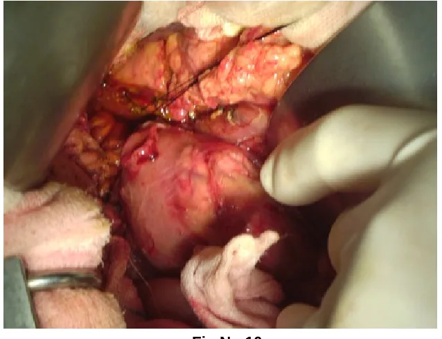
[image:77.612.114.483.159.697.2]
Fig No.10
[image:77.612.142.461.169.414.2]
Perforated Diverticulam Fig No.13
:
Small Intestinal Perforation
Fig No.14
ABBREVIATIONS
BS - Bowel Sounds
GIT - Gastro Intestinal Tract DUP - Duodenal Ulcer Perforation GUP - Gastric Ulcer Perforation IP - Ileal Perforation
NSAID - Nonsteroidal Anti Inflammatory Agents Pre OP Diag. - Pre Operative Diagnosis
Per Op Diag. - Per Operative Diagnosis Post Op Comp - Post Operative Complication Perf. Peritonitis - Perforation Peritonitis
Sc With Op - Simple Closure With Omental Patch
ABBREVIATIONS IN MASTER CHART
P - Pain VOM - Vomiting DIS - Distention Alc - Alcoholic F - Fever Rig - Rigidity BS - Bowel sound
GUD - Gas under the diaphragm POD - Pre op diagnosis
DIH - Delay in hours PRO - Procedure