Copyright © 1997, American Society for Microbiology
Sp1 Binds to the Precise Locus of End Processing within the
Terminal Repeats of Epstein-Barr Virus DNA
REN SUN,
1,2TAMMY A. SPAIN,
1SU-FANG LIN,
1ANDGEORGE MILLER
1,3*
Department of Molecular Biophysics and Biochemistry,
1Department of Genetics,
2and Departments of
Pediatrics and Epidemiology and Public Health,
3Yale University School of
Medicine, New Haven, Connecticut 06520-8064
Received 25 February 1997/Accepted 24 April 1997
Interconversion between the linear genome of Epstein-Barr virus (EBV) present in virions and intracellular
circular EBV DNA is a novel DNA recombination process. A previously characterized DNA binding activity
called terminal repeat or tandem repeat binding protein (TRBP) was found to recognize several G-rich
recombinogenic sequences in the EBV genome and in cellular DNA. TRBP was also found to be an autoantigen
recognized by sera from certain patients with undifferentiated connective-tissue disorders. Here the
transcrip-tion factor Sp1 has been identified as a component of TRBP and has been shown to be an autoantigen. Sp1
bound to recombination junctions of EBV DNA, such as those in the terminal repeats and in the large internal
repeats, as well as to recombinogenic regions of cellular DNA, such as variable-number tandem repeats and
switch regions of the immunoglobulin genes. We defined the ends of the linear EBV genome present in virions
and showed that Sp1 binds to the sequence (GGGGTGGGGCATGGG) within EBV terminal repeats at the
precise locus of interconversion of linear and circular viral DNA. Sp1 may be involved in DNA recombination.
Although the detailed mechanism of DNA recombination in
higher eukaryotes is unclear, certain sequence elements
pro-mote recombination. The human genome contains dispersed
minisatellite sequences or variable-number tandem repeat
(VNTR) sequences which are recombinogenic (15, 16, 22, 30,
52). Minisatellite and VNTR sequences often contain the
mo-tif GGNNGTGGGG. Immunoglobulin heavy-chain class
switch regions also consist of highly repetitive GC-rich
se-quences (8, 17, 18, 28, 31, 32, 34, 39, 55). The switch region of
gamma immunoglobulin genes (S
g
) contains repeats of
49-mers within which the sequence GGGGAGCTGGGG is often
found (38, 39). Chromosome telomere sequences, composed
of TTGGGG or TTAGGG repeats, are also recombinogenic
(3, 7, 35, 53). Furthermore, VNTR sequences, S
g
repeats, and
telomere sequences have been found to form stable
four-stranded structures in vitro, called G4 DNA, G quartet, or G
quadruplex (1, 23, 38). Such structures may be intermediates of
recombination or may promote stabilization of the ends of
DNA (24).
Similar to the cellular genome, the Epstein-Barr virus
(EBV) genome contains repeated GC-rich recombinogenic
se-quences (5). The number of these repeats varies remarkably in
different virus isolates, even after only one passage of the same
isolate (references 36 and 37 and unpublished results). The
double-stranded DNA viral genome is linear in virions but
circular in the nuclei of infected cells (2, 12, 19). The
circular-ization occurs 16 to 20 h postinfection and requires that the
infected cells be activated into the G
1phase of the cell cycle
(12). Furthermore, EBV infection activates the
immunoglob-ulin gene class switch recombination mechanism in B cells
(48). The EBV terminal repeats (TR), which are the sites of
linearization and circularization, are also loci of occasional
integration of EBV DNA into cellular DNA (27). TR of other
herpesviruses play a role in recombination events that are
involved in isomerization (29). The termini of some
herpesvi-ruses are similar to cellular telomeres (9, 21). Internal repeats
(IR) in EBV are also recombinogenic. During coinfection with
two different EBV strains, hybrid viruses containing crossovers
within the first IR were frequently isolated (unpublished
re-sults).
Our laboratory has previously identified a recombination
event between squirrel monkey retrovirus (SMRV) and EBV
(45). The recombination junction in EBV DNA was in the first
IR at the same position as one junction of the deletion in strain
P3J-HR-1 which removed the EBNA2 gene (14). The sequence
(GGGTGG) was present at the junctions of both
recombina-tion events (43). The sequences around the recombinarecombina-tion
sites in EBV are homologous to the common sequence of the
S
g
repeats and VNTRs. Similar sequences are found in EBV
TR.
A specific binding activity, called terminal repeat or tandem
repeat binding protein (TRBP), was identified (46). TRBP
bound to G-rich recombinogenic sequences in EBV and
cel-lular DNA, it formed a specific footprint on the EBV TR at the
site of terminal processing, it was present in the nuclear
ex-tracts of all mammalian cells tested, and it was a human
au-toantigen. Here we identify Sp1 as a component of this binding
activity and show that it binds precisely at the locus of EBV
terminal processing.
MATERIALS AND METHODS
Cell culture.Human and marmoset B lymphocytes [Raji, BJAB, BJAB-B1, P3HR-1/CL16(HH514-16), and B95-8] were grown in RPMI 1640 medium sup-plemented with 8% fetal calf serum. HeLa cells were cultured with RPMI 1640 medium supplemented with 6% calf serum. All media contained penicillin, amphotericin B, and streptomycin.
Nuclear extracts.Nuclei of cultured cells were prepared as described previ-ously (46); nuclei were extracted with 0.4 M KCl–0.2 mM EDTA (pH 8.0)–20 mM HEPES (pH 7.9)–20% glycerol–0.5 mM dithiothreitol–0.5 mM phenylmeth-ylsulfonyl fluoride. The protein concentrations of nuclear extracts, determined by the Bio-Rad protein assay, ranged from 3 to 10 mg/ml.
DNase I footprinting.The probe was a 282-bp BstEII/NarI restriction fragment which corresponds to bp 170538 to 170819 of the EBV sequence. The probe was radiolabelled at the BstEII end with [g-32P]ATP by using T4 polynucleotide kinase. DNA binding and DNase I digestion were performed under previously described conditions (4). Nuclear extract from P3HR-1/CL16(HH514-16) cells
* Corresponding author. Mailing address: 420 LSOG, 333 Cedar St.,
New Haven, CT 06520. Phone: (203) 785-4758. Fax: (203) 785-7194.
E-mail: [email protected].
6136
on November 9, 2019 by guest
http://jvi.asm.org/
or purified human Sp1 protein purchased from Promega (Madison, Wis.) was incubated with the labelled probe before DNase I digestion. A 24-mer oligonu-cleotide (59GTGACCCAGCCAAGCGTGACCAAG 39), corresponding to the 59end of the probe, was used as a primer in a DNA sequencing reaction. The sequencing reaction and the DNase I protection assay were run in adjacent lanes to determine the protected sequences.
Synthetic DNA oligonucleotides.The sequences of double-stranded oligonu-cleotides used in the electrophoretic mobility shift assay (EMSA) are shown in Table 1. Single-stranded oligonucleotides were annealed and radiolabelled by filling in recessed 39ends with Klenow fragment and [a-32P]dCTP (4).
EMSA.Binding reactions were carried out in 10ml of binding buffer [10 mM HEPES (pH 7.9), 100 mM KCl, 1 mM dithiothreitol, 0.5 mM EDTA (pH 8.0), 20% (vol/vol) glycerol, 1mg of poly(dI-dC)].32P-labelled oligonucleotides with 2.53104cpm at a final concentration of 5 nM and specific competitor DNAs were used as indicated below. The binding reactions were initiated by adding 5 to 10mg of nuclear extract or 0.1 footprint unit (FPU) of pure Sp1 protein to the initial mixture and incubating it for 10 min at room temperature. Bound and free probes in the EMSA were separated on a 4% polyacrylamide gel.
Antibodies and supershift assay.Rabbit polyclonal anti-Sp1 antibody was purchased from Santa Cruz Biotechnology (Santa Cruz, Calif.). Coded human sera from patients with autoimmune diseases were obtained from Yale New Haven Hospital. Antibody supershift assays were performed by adding 1ml of autoimmune serum to the EMSA reaction mixture and incubating it for an additional 10 min at room temperature. Bound and free probes in the EMSA and supershift assays were separated on a 4% polyacrylamide gel.
Western immunoblotting.After resolution by electrophoresis on sodium do-decyl sulfate (SDS)-polyacrylamide gels, proteins were electrotransferred to ni-trocellulose filters in a solution containing 0.25 M Tris, 0.15 M glycine, 0.1% SDS, and 25% methanol with a Bio-Rad Transblot apparatus for 2 h at 200 mA (4). Nitrocellulose filters were blocked in 5% nonfat dry milk for over 1 h before incubation with antibodies. Human sera were used at a 1:200 dilution, and rabbit sera were used at a 1:1,000 dilution. The bound primary antibodies were detected by125I-labelled protein A.
Primer extension with virion DNA.Primers were radiolabelled by T4 polynu-cleotide kinase at the 59end (4). EBV virion DNA was prepared as described previously (45). One hundred nanograms of EBV virion DNA was denatured and annealed with32P-labelled primer B (59ATCCCCGGAACGTCCGCCGC CATC) or primer C (59GTGACCCAGCCAAGCGTGACCAAGG) to map the 59nucleotides of the left and right ends, respectively, of the linear genome. Primer extension reactions were performed with Taq DNA polymerase. Ex-tended products were run next to a DNA sequencing reaction mixture which served as a molecular weight marker.
High-resolution Southern blotting.Virion DNA digested with a restriction endonuclease was run on a 6% acrylamide gel adjacent to a35S-labelled DNA sequencing reaction mixture which served as a molecular weight marker. ApaI was used to map the right end, and AvaII was used to map the left end. The DNA was transferred from the gel to nitrocellulose filters with a Bio-Rad gel dryer without heating. The DNA was cross-linked to the filters with UV light and hybridized with oligonucleotides radiolabelled with T4 polynucleotide kinase. Oligonucleotides H (TGACACAGGCAACCCTGACAAAGG) and I (GCCGT AGCGCCGCTCTGTGCG), respectively, were used to determine the 59ends of the termini. Then the filters were stripped and hybridized with kinase-labelled oligonucleotides J (CGGGGGTCTTTCCTGGGGGCCTTTGTCAGGGTTG) and K (CATTCCTGGAAAAAGTGGAGGGGGCGTGGCCTTCC), respec-tively, to determine the 39ends of the termini.
Ligation-mediated PCR.To generate adapters, oligonucleotide F (59GTAA ACGACGGCCAGTAAGAATTCCCC) was annealed to one of three oligonu-cleotides (59GGGAATTCTTAC, 59GGGGAATTCTTAC, or 59GGGGGAA TTCTTAC); oligonucleotide G (59GTAAAACGACGGCCAGTAAGAATTC
GGG) was also annealed to one of three oligonucleotides (59CCGAATTCTT AC, 59 CCCGAATTCTTAC, or 59 CCCCGAATTCTTAC). These adapters contained either one 39protruding nucleotide, a blunt end, or one 59protruding nucleotide. In each ligation reaction, 50 ng of EBV virion DNA was incubated overnight with 5 ng of annealed adapter. PCRs were performed with one of the two oligonucleotides, pB and pC, derived from EBV TR and with one of the two oligonucleotides, F and G, from the adapters. The PCR products were cloned into pBluescript KS(2) and sequenced.
RESULTS
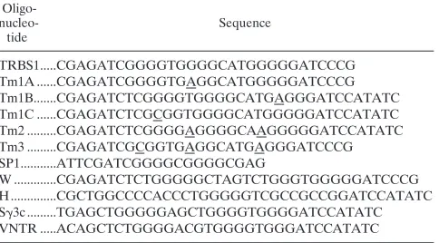
Sp1 binds specifically to the EBV TR sequences.
Preliminary
experiments that explored the biochemical properties of
TRBP, including its DNA binding specificity, its requirement
for Zn
21in DNA binding, its binding to wheat germ agglutinin,
and its behavior on gel filtration columns, suggested that
TRBP might be related to the Sp1 family of transcriptional
activators (43). The behaviors of TRBP and Sp1 were directly
compared in two DNA binding assays (Fig. 1). DNase
foot-printing showed that nuclear extract protected three regions on
the EBV TR designated TRBS1, TRBS2, and TRBS3 (Fig.
1A). Sp1 protein formed a footprint on the TRBS1 and TRBS2
sites but did not bind TRBS3. TRBS1 and TRBS2 bound the
same protein. They competed with each other for binding in
footprinting assays and in EMSAs (data not shown). TRBS3
seemed to bind a different protein. It did not compete with
TRBS1 or TRBS2 for the binding of TRBP.
Oligonucleotide competitions confirmed that TRBP and Sp1
bound related sequences (Fig. 1B). With nuclear extract as a
source of binding activity, three complexes, designated A, B,
and C, formed on oligonucleotide TRBS1 derived from the
EBV TR. Complexes of mobility identical to these formed by
nuclear extracts from Raji cells were formed with nuclear
ex-tracts from every mammalian cell tested, including P3HR-1/
CL16(HH514-16), Daudi, B95-8, BJAB, and a variety of
EBV-negative Burkitt lymphoma cell lines. Complexes A and B were
competed by an oligonucleotide containing a consensus Sp1
site from the simian virus 40 (SV40) promoter (Fig. 1B, lanes
3 to 7). Similarly, complexes A and B formed by nuclear extract
on the SV40 Sp1 site were competed by TRBS1 (Fig. 1B, lanes
19 to 23). Based on these competitions, the affinity of TRBP
for TRBS1 was about twofold greater than that for the Sp1 site
from SV40. In competition EMSA experiments with purified
Sp1 protein with labelled TRBS1 or Sp1 DNA probes, TRBS1
competed for the binding of Sp1 more efficiently than did the
Sp1 site (data not shown). Complexes C and D appear to
consist of factors that bind DNA nonspecifically (Fig. 1B and
2A). Complex C, which was not effectively competed by Sp1
oligonucleotide, was shown to contain the Ku autoantigen
(40a).
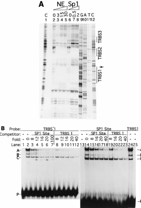
[image:2.612.59.301.82.216.2]To pinpoint the sequence within TRBS1 essential for the
binding of TRBP or Sp1, a series of mutant oligonucleotides
were used in EMSAs (Fig. 2). Mutations that decreased TRBP
binding decreased Sp1 binding concordantly. Mutants Tm1A,
Tm1B, Tm1C, and Tm3, containing interruptions in the G
tracts of TRBS1, all had reduced affinity for TRBP (Fig. 2A).
No complex A was formed with Tm1C or Tm3. Mutant Tm2,
in which two T nucleotides were replaced with A’s, retained
wild-type affinity for complexes A and B. Figure 2B shows that
Sp1 protein bound TRBS1 (T); however, only complex A was
formed. Mutations of TRBS1 caused similar effects on the
binding of TRBP and purified Sp1 (compare Fig. 2A and 2B).
Rabbit antibody against Sp1 supershifted complex A formed
on the wild type and the Tm2 mutant as well as the less
abundant complex A formed on Tm1A and Tm1B (Fig. 2C).
However, anti-Sp1 antibody did not recognize the unrelated
complexes that formed on Tm1C and Tm3.
TABLE 1. Sequences of double-stranded oligonucleotides
a Oligo- nucleo-tide Sequence TRBS1...CGAGATCGGGGTGGGGCATGGGGGATCCCG Tm1A ...CGAGATCGGGGTGAGGCATGGGGGATCCCG Tm1B...CGAGATCTCGGGGTGGGGCATGAGGGATCCATATC Tm1C ...CGAGATCTCGCGGTGGGGCATGGGGGATCCATATC Tm2 ...CGAGATCTCGGGGAGGGGCAAGGGGGATCCATATC Tm3 ...CGAGATCGCGGTGAGGCATGAGGGATCCCG SP1...ATTCGATCGGGGCGGGGCGAG W ...CGAGATCTCTGGGGGCTAGTCTGGGTGGGGGATCCCG H...CGCTGGCCCCACCCTGGGGGTCGCCGCCGGATCCATATC Sg3c ...TGAGCTGGGGGAGCTGGGGTGGGGATCCATATC VNTR ...ACAGCTCTGGGGACGTGGGGTGGGATCCATATCaMutated sequences are underlined.
on November 9, 2019 by guest
http://jvi.asm.org/
Sp1 binds to other EBV and cellular recombinogenic
se-quences.
The binding of TRBP and pure Sp1 was compared by
using oligonucleotides T (TRBS1), W, and H, which represent
recombinogenic sequences in the EBV genome, and
oligonu-cleotides S
g
3c and VNTR, which represent recombinogenic
sequences in the cellular genome (Table 1; Fig. 3). W and H
represent the sites of recombination that generate the deletion
in P3J-HR-1 DNA. Sp1 bound all the viral and cellular
se-quences that were bound by TRBP. In each instance, Sp1
protein formed a complex that comigrated with complex A
formed by TRBP. Anti-Sp1 antibody supershifted TRBP
com-plex A on all the viral and cellular sequences.
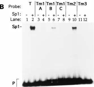
Sp1 is an autoantigenic component of TRBP.
Eight
autoim-mune sera from different patients recognized TRBP; all of
them also reacted with Sp1 in a supershift EMSA and in
immunoprecipitation experiments (data not shown). For
ex-ample, Fig. 4A shows that autoimmune serum 229, which
caused a supershift of TRBP in nuclear extract (lanes 7 and
17), also supershifted purified Sp1 (lanes 2 and 12). This
au-toantibody supershifted TRBP or Sp1 whether an Sp1 binding
site or TRBS1 was used as a probe in the EMSA. Figure 4B
shows that this autoantibody, which was reactive with TRBP
and Sp1 in EMSA, also recognized purified Sp1 protein on an
immunoblot (lane 8). In cell extracts (Fig. 4B, lanes 3, 4, and
7), this antibody recognized a 95-kDa protein corresponding in
size to Sp1 (lanes 6 and 8).
Mapping the ends of the EBV linear genome.
We reasoned
that if Sp1 played a role in EBV terminal processing, the
cleavage site should be close to, or at, the binding sites for Sp1.
At the time we initiated these experiments, the cleavage site
for EBV linearization had not been precisely mapped (43, 44,
46). Therefore, we mapped the ends of the EBV linear
ge-nome, initially using primer extension. Virion DNA was
ob-tained from viral particles purified on sucrose gradients. After
denaturation, viral DNA was annealed with primers located at
several positions on the EBV TR. Primer C, which was used to
map the right end of the genome, generated an unambiguous
pattern. The major extended product was 114 nucleotides (nt),
and a minor product was 113 nt (Fig. 5A). The longest
ex-tended product from primer B, which was used to map the left
end, was 171 nt (Fig. 5B). Based on the results of additional
experiments, described below, the shorter fragments were
likely the result of premature stops of polymerization.
The EBV ends were also mapped by Southern blotting (Fig.
5C and D). Virion DNA was digested with restriction
endo-nucleases which cut within the terminal repeat. ApaI was used
to map the right end, and AvaII was used to map the left end.
The digested products were separated on 6% denaturing
poly-acrylamide gels, transferred to nitrocellulose paper, and
probed with strand-specific oligonucleotides which were
lo-cated between the restriction enzyme cleavage site and the
putative site of terminal processing of EBV DNA. The right
termini were again found to be heterogeneous, since doublets
were detected on the Southern blots. The 3
9
end of the right
terminus was 86 or 85 nt away from the ApaI site (Fig. 5C). The
5
9
end was 89 or 88 nt away from the ApaI site. Considering
that ApaI digestion generates a 4-nt 3
9
overhang, the right end
of the EBV genome appears to have one extra nucleotide at
the 3
9
end. This estimate was based on the assumption that the
migration rate of the DNA fragments was dependent solely on
the number of nucleotides and was not affected by base
com-position. A single band of 117 nt represented the length
be-tween the 5
9
end of the left terminus and the AvaII site (Fig.
5D). The distance between the 3
9
end of the left terminus and
the AvaII site was 120 nt. Since AvaII digestion generates a
3-nt 5
9
overhang, the left terminus may be blunt.
[image:3.612.59.295.70.419.2]Unusual sequence at the EBV right terminus.
The results of
both primer extension and high-resolution Southern blotting
indicated that there was approximately 20 bp of extra sequence
present in the linear termini that was not present in the circular
genome. A ligation-mediated PCR method was used to
iden-tify these extra sequences in the EBV termini. Unlike other
FIG. 1. Binding of Sp1 to EBV TR sequences. (A) DNase I footprinting on EBV TR sequences by nuclear extract (NE) or purified Sp1 protein. Lanes 2 and 6, DNase I digestion of the probe in the absence of nuclear extract or Sp1 protein; lanes 3 to 5, incubation of the probe with 3, 15, and 30ml of nuclear extract before DNase digestion; lanes 7 and 8, incubation of the probe with 0.5 and 2 FPU of pure Sp1 protein before digestion. Nuclear extract for this exper-iment was prepared from P3HR-1/CL16(HH514-16) cells. The probe corre-sponds to EBV nt 170538 to 170819. An oligonucleotide corresponding to the 59 end of the probe was used as a primer in a DNA sequencing reaction. The sequencing reaction and the DNase I protection assay were run together to determine the protected sequences (lanes 1 and 9 to 12). The protected se-quences (TRBS1, TRBS2, and TRBS3) are shown in Fig. 7. In lanes 4 and 5, a hypersensitive site corresponding to the right end of the EBV genome is indi-cated by an arrow. (B) Competition for binding of TRBP by an Sp1 binding site or TR binding site 1 (TRBS1). Lanes 1 and 25,32P-labelled TRBS1 probe alone (EBV nt 170628 to 170644) (5); lane 2, labelled TRBS1 probe with no specific competitor added; lanes 3 to 7,32P-labelled TRBS1 competed with increasing amounts of unlabelled oligonucleotides containing the Sp1 binding site; lanes 8 to 12,32P-labelled TRBS1 probe competed with unlabelled TRBS1; lane 13, 32P-labelled Sp1 probe alone; lane 14, labelled Sp1 probe with no specific com-petitor added; lanes 15 to 18,32P-labelled Sp1 probe competed with unlabelled Sp1 oligonucleotide; lanes 19 to 23,32P-labelled Sp1 probe competed with unlabelled TRBS1 oligonucleotide; lane 24, labelled TRBS1 probe with no specific competitor added. “Fold” indicates the ratio of excess cold competitor to the labelled probe added into the binding reaction mixture. Shifted complexes from the top of the gel to the bottom are labelled A, B, and C. P, free probe. Nuclear extract for this experiment was prepared from Raji cells.
on November 9, 2019 by guest
http://jvi.asm.org/
methods of DNA cloning, this method does not require
mod-ification of the ends. Three types of adapters were ligated to
the left or right terminus of virion DNA: one contained a 1-nt
3
9
overhang, one was blunt, and one contained a 1-nt 5
9
over-hang. Separate PCRs were performed with each ligation, using
one primer in the TR and another primer in the adapter. Three
clones with lengths consistent with the results of primer
exten-sion and Southern blotting were obtained from PCR products
resulting from ligation of the right terminus, with the adapter
containing 1 nt (cytosine) protruding at the 3
9
end of the
bottom strand. These three independent clones contained
identical inserts, the sequence of one of which is shown in Fig.
6. The sequence revealed the identity of the extra 20 bp found
by primer extension and Southern blotting.
Linearization and circularization of the EBV genome are
novel DNA recombination processes.
The termini of the
en-capsidated linear EBV genome in the virion were recently
characterized by end labelling and by direct cloning and
se-quencing (56). The three different methods we used, primer
extension, high-resolution Southern blotting, and
ligation-me-diated PCR, confirmed these results (44). Linearization of the
viral genome is not due to simple cleavage of the circular or
concatemeric form; instead, the conversion of the linear to the
circular form and the reverse involved a novel form of DNA
processing (Fig. 7). Formation of the right terminus of linear
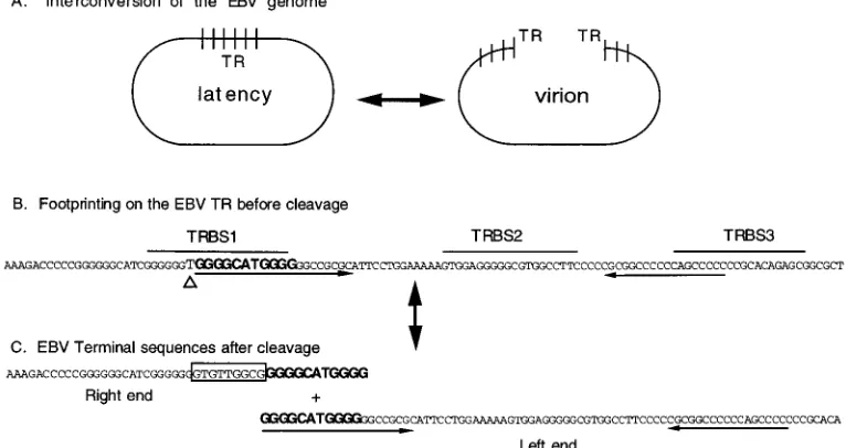
EBV DNA was associated with the insertion of 9 bp (GTGT
TGGCG) and the deletion of 1 bp (T) immediately adjacent to
the insertion. The right terminus of the EBV genome was
coterminal with the right end of TRBS1, one of the two major
binding sites for Sp1 (Fig. 1). The left terminus was in the
middle of TRBS1. According to previously published results,
the sequence of the left terminus of EBV DNA contains 11 bp
(GGGGCATGGGG) that are also present at the right end of
the genome (56).
DISCUSSION
[image:4.612.345.511.71.224.2]We show that Sp1, a well-characterized transcriptional
acti-vator, binds to recombination junction sequences in viral and
cellular DNA. Sp1 binds to TRBS1, the site of EBV terminal
processing. Sp1 binds to other recombinogenic sequences in
EBV and cellular DNA which contain the common motif
GGGTGG. The sequence TRBS1 contains three G
ntracts, all
FIG. 2. Electrophoretic mobility shifts with TRBS1 mutant oligonucleotides. (A) Shift of wild-type and mutant oligonucleotides with a nuclear extract from Raji cells. (B) Shift of mutant oligonucleotides with 0.1 FPU of purified Sp1 protein. (C) Supershift of complexes formed between Raji cell nuclear extract (3.25 mg of total protein) and mutant oligonucleotides with 1 ml of rabbit anti-Sp1. Probe T is an oligonucleotide containing the TRBS1 sequence. A, B, C, and D denote complexes from the top of the gel to the bottom. S, supershifted complex; P, free probe.FIG. 3. Complexes between TRBP or Sp1 and EBV or cellular recombina-tion juncrecombina-tions are supershifted by antibody to Sp1 (a-Sp1). Double-stranded oligonucleotides T (TRBS1) (lanes 1 to 5), W (2907 to 2927 in EBV BamHI W) (lanes 6 to 10), H (3586 to 3613 in EBV BamHI H) (lanes 11 to 15), Sg3c from the immunoglobulin heavy-chain g3 switch region, and VNTR common se-quences (lanes 21 to 25) were labelled with32P. Probes were shifted with 0.1 FPU of Sp1 protein (lanes 2, 7, 12, 17, and 22) or with 3.25mg of total protein of Raji cell nuclear extract (NE) (lanes 3 to 5, 8 to 10, 13 to 15, 18 to 20, and 23 to 25). One microliter of anti-Sp1 (lanes 4, 9, 14, 19, and 24) was added to binding reactions to supershift Sp1-containing complexes on the various probes. Rabbit antibody against the EBV immediate-early protein BRLF1 (a-R) was added as a negative control (lanes 5, 10, 15, 20, and 25).
on November 9, 2019 by guest
http://jvi.asm.org/
of which are required for optimal binding by Sp1. Additional
mutagenesis confirmed this conclusion (40a). Binding of Sp1 to
recombinogenic sequences suggests a novel function for the
protein in the process of DNA recombination.
The impetus for this study was the observation of a
recom-bination event between EBV and SMRV (43, 46). The
se-quence at the recombination crossover junction between EBV
and SMRV was TGGGTGG. A similar sequence, GGGTG
GG, has recently been found to promote recombination in a
functional assay with avian spleen necrosis retrovirus (33).
[image:5.612.129.488.70.221.2]FIG. 4. Sp1 is an autoantigenic component of TRBP. (A) Supershifts of TRBP-DNA complexes and Sp1-DNA complexes by autoimmune sera. Lanes 1 to 10 contain the32P-labelled Sp1 binding site probe. Lanes 11 to 20 contain the32P-labelled TRBS1 probe. Lanes 1 to 5 and 11 to 15 contain 0.1 FPU of Sp1 protein in each binding reaction mixture. Lanes 6 to 10 and 16 to 20 contain approximately 1.9mg of total protein from a HeLa cell nuclear extract. Lanes 1, 6, 11, and 16, no antibody; lanes 2, 7, 12, and 17, 1ml of human autoimmune serum 229 (anti-TRBP positive); lanes 3, 8, 13, and 18, 1ml of human autoimmune serum 6670 (anti-TRBP negative); lanes 4, 9, 14, and 19, 1ml of rabbit anti-Sp1 (a-Sp1); lanes 5, 10, 15, and 20, 1ml of rabbit anti-EBV immediate-early protein BZLF1 (a-Z). Sp1/A indicates the Sp1 shifted band and the top band in nuclear extract binding reactions. In lanes 6 to 10, complex B is visible on a longer exposure of the autoradiograph. (B) Recognition of Sp1 protein by an autoimmune serum on an immunoblot. Cellular extract from EBV-negative B-cell lymphoma line BJAB (lanes 1, 3, 5, and 7), the corresponding EBV (B95-8) converted cell line BJAB-B1 (lanes 2 and 4), and purified Sp1 protein (lanes 6 and 8) were electrophoresed on an 8% denaturing SDS-polyacrylamide gel. After transfer, the filters were probed with human serum RM (lanes 1 and 2), autoimmune serum 229 (lanes 3, 4, 7, and 8), and anti-Sp1 serum (lanes 5 and 6). The 80-kDa protein in the BJAB-B1 lanes is EBV nuclear antigen 1. The numbers at left are molecular masses, in kilodaltons.
FIG. 5. Mapping of EBV termini. (A) Determination of the 59end of the right terminus (59R) of the EBV linear genome by primer extension. Virion DNA was denatured and annealed with primer C (pC). Extended products were run next to DNA sequencing reaction mixtures which served as molecular weight markers (MW). (B) Determination of the 59end of the left end (59L) of the EBV linear genome by primer extension. Denatured EBV virion DNA was annealed with labelled primer B (pB). (C) Determination of the right end of the linear genome by high-resolution Southern blotting. ApaI-digested virion DNA was hybridized with kinase-labelled oligonucleotide H to determine the distance between the 59end of the right terminus and the ApaI site. Then the filter was stripped and hybridized with kinase-labelled oligonucleotide J to determine the 39end of the right terminus (39R). (D) Determination of the left end of the linear genome by high-resolution Southern blotting. AvaII-digested virion DNA was hybridized with labelled oligonucleotide I to determine the 59end of the left terminus. Then the filter was stripped and hybridized with labelled oligonucle-otide K to determine the 39end of the left terminus (39L). For panels A to D, numbers on each side are nucleotide lengths. (E) Diagrammatic summary of mapping of EBV termini. Restriction sites ApaI and AvaII and primers B and C are indicated. Dashed lines indicate the boundary of each TR. The distances between the dashed lines are indicated.
on November 9, 2019 by guest
http://jvi.asm.org/
Thus, recombination junctions of a primate and an avian
ret-rovirus contain related G-rich elements. Moreover, similar
se-quences are found at internal recombination junctions in EBV,
such as the recombination junction of the EBNA2 deletion in
EBV strain P3J-HR-1 and the EBV termini (Fig. 5). The
appearance of this motif in recombinogenic loci of unrelated
viruses supports the hypothesis of a functional role for such
sequences and suggests that they may interact with similar
cellular proteins.
Formation of the ends of EBV DNA is not a simple cleavage
and ligation reaction but an intricate process (Fig. 5 to 7). The
total number of TR may be increased or decreased in the
linear virion DNA relative to their number in the circular
latent DNA (19). A 9-bp sequence, GTGTTGGCG, also found
in unique sequences adjacent to TR, is newly added to the right
terminus. An 11-bp sequence, GGGGCATGGGG, found only
once in each fused TR of circular DNA, is duplicated at both
termini of linear DNA (56). Moreover, this 11-bp sequence is
embedded in an 18-bp sequence which is located 37 bp from a
nearly perfect inverted repeat (Fig. 7). A nucleotide, T, present
in circular DNA is absent from linear DNA. All these findings
point to the likely participation of DNA replication,
recombi-nation, and repair in the interconversion of the circular and
linear forms of EBV DNA. The recombination events
dis-cussed here are to be distinguished from the only
well-charac-terized eukaryotic recombination reaction in higher
eu-karyotes, namely, V(D)J recombination, which does not
involve DNA replication (40, 51). High rates of deletions,
insertions, and mutations, found close to the recombination
junctions in the spleen necrosis virus, likewise implicate DNA
replication and repair in this recombination process (33).
[image:6.612.73.282.69.267.2]Sp1 could play a role in either circularization or linearization
of the EBV genome. Both these events are likely to involve
DNA recombination processes. Several functions of Sp1 in
recombination can be envisioned. Sp1 has been shown to bring
two regions of double-stranded DNA together, either apposing
ends or forming loops (26, 41). This represents an essential
step in recombination and could play a role in circularization
of the input genome. Sp1 can bend DNA (13, 42). Distortion of
DNA near a point of cleavage may promote recombination.
For example, the binding of Sp1 protein to TRBS1 and TRBS2
together with another protein on TRBS3 may induce
confor-mational changes to direct the cleavage on the EBV TR.
Fi-nally, Sp1 can be phosphorylated by the complex of
autoanti-gen Ku and DNA-dependent protein kinase, which are
involved in DNA repair and V(D)J recombination (6, 10, 11,
FIG. 6. Sequence of the right terminus of the EBV genome. The sequence of the ligation-mediated-PCR product shows the unusual processing at the right end of the EBV genome. The adapter sequence was linked to the duplicated sequence, followed by the insertion of 9 bp. A base pair (T) is deleted before the sequence continues with the rest of the TR.
FIG. 7. Summary of EBV terminal processing. (A) Diagram of the interconversion of the EBV genome between the linear form in virions and the circular form in cells during latency. Short vertical bars represent the boundary between each direct TR. The linearization of EBV DNA results in molecules with variable numbers of TR at each end. (B) Partial sequence of EBV TR before cleavage. The footprinting areas, TRBS1, TRBS2, and TRBS3, are indicated (Fig. 1A). The arrows below the sequences indicate nearly perfect inverted repeats. One T, indicated by a triangle, is deleted during linearization. (C) EBV sequence at the termini after cleavage. The sequences in bold are present on both termini after cleavage, based on data shown in Fig. 5 and described in reference 56. The boxed sequences are introduced after cleavage and can be found at the junction between the short unique sequence and the TR (EBV nt 172232 to 172240) (5).
on November 9, 2019 by guest
http://jvi.asm.org/
[image:6.612.115.498.468.671.2]20, 40, 47, 49). Sp1 may attract components of the
recombina-tion machinery, such as Ku and DNA-dependent protein
ki-nase, to sites with the (GGGTGG) recombinogenic motif.
Other recent evidence implicates components of the
transcrip-tion apparatus in herpesvirus genome end processing.
TAF
II250, which is known to play a role in Sp1 transcriptional
activation, has recently been found to play a role in genome
end formation of herpes simplex virus (25, 50, 54). In summary,
Sp1 binds to the EBV termini at a locus intimately associated
with a novel form of DNA recombination.
ACKNOWLEDGMENTS
We are grateful to J. Craft for providing autoimmune sera and to P.
Olson and R. Dornburg for communicating results before publication.
J. Konner and S. Katz participated in this study as Yale University
undergraduate students.
This work was supported by a grant from the NIH (CA16038) to
G.M., a MARC predoctoral fellowship from the NIAID (GM14637) to
T.A.S., and a training grant from the NIH (T32CA09159) and a
Swe-bilius Cancer Research award to R.S.
REFERENCES
1. Aboul-ela, F., A. I. H. Murchie, and D. M. J. Lilley. 1992. NMR study of parallel-stranded tetraplex formation by the hexadeoxynucleotide d(TG4T). Nature 360:280–282.
2. Adams, A., and T. Lindahl. 1975. Epstein-Barr virus genomes with properties of circular DNA molecules in carrier cells. Proc. Natl. Acad. Sci. USA
72:1477–1481.
3. Ashley, T. 1994. Mammalian meiotic recombination: reexamination. Hum. Genet. 94:587–593.
4. Ausubel, F. M., R. Brent, R. E. Kingston, D. D. Moore, J. G. Seidman, J. A.
Smith, and K. Struhl (ed.).1995. Current protocols in molecular biology. John Wiley & Sons, New York, N.Y.
5. Baer, R., A. T. Bankier, M. D. Biggin, P. L. Deininger, P. J. Farrell, T. J.
Gibson, G. Hatfull, G. S. Hudson, S. C. Satchwell, C. Seguin, et al.1984. DNA sequence and expression of the B95-8 Epstein-Barr virus genome. Nature 310:207–211.
6. Blunt, T., N. J. Finnie, G. E. Taccioli, G. C. Smith, J. Demengeot, T. M.
Gottlieb, R. Mizuta, A. J. Varghese, F. W. Alt, P. A. Jeggo, et al.1995. Defective DNA-dependent protein kinase activity is linked to V(D)J recom-bination and DNA repair defects associated with the murine scid mutation. Cell 80:813–823.
7. de Bruin, D., M. Lanzer, and J. V. Ravetch. 1994. The polymorphic subte-lomeric regions of Plasmodium falciparum chromosomes contain arrays of repetitive sequence elements. Proc. Natl. Acad. Sci. USA 91:619–623. 8. Dunnick, W., G. Z. Hertz, L. Scappino, and C. Gritzmacher. 1993. DNA
sequences at immunoglobulin switch region recombination sites. Nucleic Acids Res. 21:365–372.
9. Gompels, U. A., J. Nicholas, G. Lawrence, M. Jones, B. J. Thomson, M. E.
Martin, S. Efstathiou, M. Craxton, and H. A. Macaulay.1995. The DNA sequence of human herpesvirus-6: structure, coding content, and genome evolution. Virology 209:29–51.
10. Gottlieb, T. M., and S. P. Jackson. 1993. The DNA-dependent protein kinase: requirement for DNA ends and association with Ku antigen. Cell
72:131.
11. Hartley, K. O., D. Gell, G. C. Smith, H. Zhang, N. Divecha, M. A. Connelly,
A. Admon, M. S. Lees, C. W. Anderson, and S. P. Jackson.1995. DNA-dependent protein kinase catalytic subunit: a relative of phosphatidylinositol 3-kinase and the ataxia telangiectasia gene product. Cell 82:849–856. 12. Hurley, E., and D. Thorley-Lawson. 1989. B cell activation and the
estab-lishment of EBV latency. J. Exp. Med. 168:2059–2075.
13. Ikeda, K., K. Nagano, and K. Kawakami. 1993. Possible implications of Sp1-induced bending of DNA on synergistic activation of transcription. Gene 136:341.
14. Jeang, K.-T., and S. D. Hayward. 1983. Organization of the Epstein-Barr virus DNA molecule. III. Location of the P3HR-1 deletion junction and characterization of the NotI repeat units that form part of the template for an abundant 12-O-tetradecanoylphorbol-13-acetate-induced mRNA tran-script. J. Virol. 48:135–148.
15. Jeffreys, A. J., A. MacLeod, K. Tamaki, D. L. Neil, and D. G. Monckton. 1991. Minisatellite repeat coding as a digital approach to DNA typing. Nature 354:204–209.
16. Jeffreys, A. J., V. Wilson, and S. L. Thein. 1985. Hypervariable ‘minisatellite’ regions in human DNA. Nature 314:67–73.
17. Kataoka, T., T. Kawakami, N. Takahashi, and T. Honjo. 1980. Rearrange-ment of immunoglobuling-1-chain gene and mechanism. Proc. Natl. Acad. Sci. USA 77:919–923.
18. Kataoka, T., T. Miyata, and T. Honjo. 1981. Repetitive sequences in class-switch recombination regions of immunoglobulin heavy chain genes. Cell
23:357–368.
19. Kintner, C. R., and B. Sugden. 1979. The structure of the termini of the DNA of Epstein-Barr virus. Cell 17:661–667.
20. Kirchgessner, C. U., C. K. Patil, J. W. Evans, C. A. Cuomo, L. M. Fried, T.
Carter, M. A. Oettinger, and J. M. Brown.1995. DNA-dependent kinase (p350) as a candidate gene for the murine SCID defect. Science 267:1178– 1183.
21. Kishi, M., G. Bradley, J. Jessip, A. Tanaka, and M. Nonoyama. 1991. In-verted repeat regions of Marek’s disease virus DNA possess a structure similar to that of the a sequence of herpes simplex virus DNA and contain host cell telomere sequences. J. Virol. 65:2791–2797.
22. Krowczynska, A. M., R. A. Rudders, and T. G. Krontiris. 1990. The human minisatellite consensus at breakpoints of oncogene translocations. Nucleic Acids Res. 18:1121–1127.
23. Laughlan, G., A. I. H. Murchie, D. G. Norman, M. H. Moore, D. M. J. Lilley,
and B. Luisi. 1994. The high-resolution crystal structure of a parallel-stranded guanine tetraplex. Science 265:520–524.
24. Liu, Z., and W. Gilbert. 1994. The yeast KEM1 gene encodes a nuclease specific for G4 tetraplex DNA: implication of in vivo functions for this novel DNA structure. Cell 77:1083–1092.
25. Maldonado, E., R. Shiekhattar, M. Sheldon, H. Cho, R. Drapkin, P. Rickert,
E. Lees, C. W. Anderson, S. Linn, and D. Reinberg.1996. A human RNA polymerase II complex associated with SRB and DNA-repair proteins. Na-ture 381:86–89.
26. Mastrangelo, I. A., A. J. Courey, J. S. Wall, S. P. Jackson, and P. V. Hough. 1991. DNA looping and Sp1 multimer links: a mechanism for transcriptional synergism and enhancement. Proc. Natl. Acad. Sci. USA 88:5670. 27. Matsuo, T., M. Heller, L. Petti, E. O’Shiro, and E. Kieff. 1984. Persistence of
the entire Epstein-Barr virus genome integrated into human lymphocyte DNA. Science 226:1322–1325.
28. Mills, F. C., J. S. Brooker, and R. D. Camerini-Otero. 1990. Sequences of human immunoglobulin switch regions: implications for recombination and transcription. Nucleic Acids Res. 18:7305–7316.
29. Mocarski, E. S., and B. Roizman. 1982. Structure and role of the herpes simplex virus DNA termini in inversion, circularization and generation of virion DNA. Cell 31:89–97.
30. Nakamura, Y., M. Leppert, P. O’Connell, R. Wolff, T. Holm, M. Culver, C.
Martin, E. Fujimoto, M. Hoff, E. Kumlin, and R. White.1987. Variable number of tandem repeat (VNTR) markers for human gene mapping. Sci-ence 235:1616–1622.
31. Nikaido, T., S. Nakai, and T. Honjo. 1981. Switch region of immunoglobulin C-mgene is composed of simple tandem repetitive sequences. Nature 292: 845–848.
32. Obata, M., T. Kataoka, S. Nakai, H. Yamagishi, N. Takahashi, Y.
Ya-makawi-Kataoka, T. Nikaido, A. Shimizu, and T. Honjo.1981. Structure of a rearrangedg-1-chain gene and its implication to immunoglobulin class-switch mechanism. Proc. Natl. Acad. Sci. USA 78:2437–2441.
33. Olson, P., and R. Dornburg. Capture of a recombination activating sequence from mammalian cell with homology to Chi. Submitted for publication. 34. Petrini, J., and W. A. Dunnick. 1989. Products and implied mechanism of H
chain switch recombination. J. Immunol. 142:2932–2935.
35. Pluta, A. F., and V. A. Zakian. 1989. Recombination occurs during telomere formation in yeast. Nature 337:429–433.
36. Raab-Traub, N., and K. Flynn. 1986. The structure of the termini of the Epstein-Barr virus as a marker of clonal cellular proliferation. Cell 47:883– 889.
37. Sato, H., T. Takimoto, S. Tanaka, J. Tanaka, and N. Raab-Traub. 1990. Concatameric [sic] replication of Epstein-Barr virus: structure of the termini in virus-producer and newly transformed cell lines. J. Virol. 64:5295–5300. 38. Sen, D., and W. Gilbert. 1988. Formation of parallel four-stranded com-plexes by guanine-rich motifs in DNA and its implications for meiosis. Nature 334:364–366.
39. Shimizu, A., and T. Honjo. 1984. Immunoglobulin class switch. Cell 36:801– 803.
40. Smider, V., W. K. Rathmell, M. R. Lieber, and G. Chu. 1994. Restoration of X-ray resistance and V(D)J recombination in mutant cells by Ku cDNA. Science 266:288.
40a.Spain, T. A., R. Sun, and G. Miller. Submitted for publication.
41. Su, W., S. Jackson, R. Tjian, and H. Echols. 1991. DNA looping between sites for transcriptional activation: self-association of DNA-bound Sp1. Genes Dev. 5:820.
42. Sun, D., and L. H. Hurley. 1994. Cooperative bending of the 21-base-pair repeats of the SV40 viral early promoter by human Sp1. Biochemistry 33: 9578–9587.
43. Sun, R. 1993. Ph.D. thesis. Yale University, New Haven, Conn.
44. Sun, R., et al. 1994. Presented at the 6th International Symposium of EBV and Associated Diseases, Cold Spring Harbor, N.Y.
45. Sun, R., E. Grogan, D. Shedd, A. F. Bykovsky, V. M. Kushnaryov, S. E.
Grossberg, and G. Miller.1995. Transmissible retrovirus in Epstein-Barr virus-producing B95-8 cells. Virology 209:374–383.
on November 9, 2019 by guest
http://jvi.asm.org/
46. Sun, R., T. A. Spain, S.-F. Lin, and G. Miller. 1994. Autoantigenic proteins that bind recombinogenic sequences in Epstein-Barr virus and cellular DNA. Proc. Natl. Acad. Sci. USA 91:8646–8650.
47. Taccioli, G. E., T. M. Gottlieb, T. Blunt, A. Priestley, J. Demengeot, R.
Mizuta, A. R. Lehmann, F. W. Alt, S. P. Jackson, and P. A. Jeggo.1994. Ku80: product of the XRCC5 gene and its role in DNA repair and V(D)J recombination. Science 265:1442–1445.
48. Thyphronitis, G., G. C. Tsokos, C. H. June, A. D. Levine, and F. D.
Finkel-man.1989. IgE secretion by Epstein-Barr virus-infected purified human B lymphocytes is stimulated by interleukin 4 and suppressed by interferong. Proc. Natl. Acad. Sci. USA 86:5580–5584.
49. Tuteja, N., R. Tuteja, A. Ochem, P. Taneja, N. W. Huang, A. Simoncsits, S.
Susic, K. Rahman, L. Marusic, J. Chen, et al.1994. Human DNA helicase II: a novel DNA unwinding enzyme identified as the Ku autoantigen. EMBO J. 13:4991–5001.
50. Umene, K., and T. Nishimoto. 1996. Inhibition of generation of authentic genomic termini of herpes simplex virus type 1 DNA in
temperature-sensi-tive mutant BHK-21 cells with a mutated CCG1/TAFII250 gene. J. Virol.
70:9008–9012.
51. van Gent, D. C., D. A. Ramsden, and M. Gellert. 1996. The Rag1 and Rag2 proteins establish the 12/23 rule in V(D)J recombination. Cell 85:107–113. 52. Wahls, W. P., L. J. Wallace, and P. D. Moore. 1990. Hypervariable minisat-ellite DNA is a hotspot for homologous recombination in human cells. Cell
60:95–103.
53. Wang, S., and V. A. Zakian. 1990. Telomere-telomere recombination pro-vides an express pathway for telomere acquisition. Nature 345:456–458. 54. Weinzierl, R. O. J., B. D. Dynlacht, and R. Tjian. 1993. Largest subunit of
Drosophila transcription factor IID directs assembly of a complex containing TBP and a coactivator. Nature 362:511–517.
55. Wu, T. T., M. Reid-Miller, H. M. Perry, and E. A. Kabat. 1984. Long identical repeats in the mouseg2b switch region and their implications for the mechanism of class switching. EMBO J. 3:2033–2040.
56. Zimmermann, J., and W. Hammerschmidt. 1995. Structure and role of the terminal repeats of Epstein-Barr virus in processing and packaging of virion DNA. J. Virol. 69:3147–3155.