JOURNAL OF VIROLOGY, June 1994, p. 3620-3630 Vol.68, No. 6 0022-538X/94/$04.00+0
Copyright © 1994, American Society for Microbiology
Changes
in
RNA
Expression Pattern
during the Malignant
Progression of Cottontail Rabbit
Papillomavirus-Induced
Tumors in
Rabbits
R.
ZELTNER,'
L. A.BORENSTEIN,2 F. 0. WETTSTEIN,2ANDT. IFTNERI*InstitutfOir Klinische und Molekulare Virologie der Universitat
Erlangen-Niimberg,
91054Erlangen,Germany,j
andDepartment
of
Microbiology
andImmunology,
UCLA Schoolof
Medicine,
LosAngeles, Califomia
900242Received 17 November 1993/Accepted 21February 1994
Cottontail rabbitpapillomavirus inducesstrictly epithelialtumorsinboth cottontail and domestic rabbits. Ahighproportion of the initial benignpapillomasprogresswithin8to 14months toinvasivecarcinomas. With the help ofmRNA-specific riboprobesforE6,E7,El,E2,LI,andL2,weinvestigated byin situhybridization
the RNA expression pattern of cottontail rabbit papillomavirus in tissue sections ofbiopsies from different stagesof tumordevelopment. Commonfeatures of alllesionswerehigh levels of E6 and E7 mRNAs and low levelsofEland E2 mRNAs. In agreement with earlier reports, there was no evidence for amajormRNA class equivalent to the prominent El^E4 RNA of human papillomavirus types 6/11 and 16. In cottontail rabbit
papillomas, high levels of E6 and E7 mRNAs were present in the upperdifferentiatedepithelial layers.These layers also contained mostoftheEl and E2 mRNAs and the viral DNA. In contrast,papillomasof domestic rabbits revealed the opposite differentiation-dependent expression pattern for the E6 and E7 mRNAs; there were strongsignals in the basallayers,andthesedeclinedwith increaseddifferentiation.Transcripts encoding
theLI mRNA were detectedonlyin afewisolatedcellsofthegranularlayer.Therewasnodifferencebetween the amounts ofE6,E7, El,and E2 mRNAs present inhighlydysplastic tissue and those present inadjacent
normal papillomatous epithelium within a progressing papilloma. However, latetranscripts and viral DNA detectableonly in the upperlayers of the papillomawere presentthroughoutthethickness of thedysplastic tissue, indicating a newlyacquired permissiveness of the dysplastic cells for viral DNAreplication and late
transcription. Carcinomasingeneralhad the sameexpressionpatternsforE6, E7,andElbut weredissimilar in the levels of expression of E2 and latetranscripts.
Papillomaviruses have attracted special attention because substantial experimental evidence suggests that certain types,
called high-cancer-risk papillomaviruses, play a role in the pathogenesis of human cancer (34). Cottontail rabbit papillo-mavirus (CRPV),whichwasthefirstpapillomavirus identified (25), induces strictly epithelial tumors inboth cottontail and domestic rabbits. The tumors are benign at first, but 8 to 14
months later carcinomasdevelopatthesamesite in25%of the
permissive hosts (the cottontail rabbits) and in 66 to80% of the nonpermissive hosts (the domestic rabbits) (17, 30). In some animals papillomas regress spontaneously or persist as
benign lesions for the entire life of the animal (30). These properties make CRPV an important model for high-cancer-risk humanpapillomaviruses(HPVs) in ordertostudythe role of viral gene expression during the early steps of cancer
development.
The viral transcripts in tumors of cottontail and domestic rabbits have been characterizedby Northern (RNA) blotting,
andthree early promoters and one late promoter were iden-tifiedby
Si
nucleaseandprimerextensionanalyses(21,23, 24,32). Transcripts initiated at the three early promoters (P1 to
P3)(Fig. 1)arepresent inbothpapillomasandcarcinomasof
domestic and cottontail rabbits as well as in the VX2
trans-plantable cancer line (6, 10, 22). The two major early
tran-scripts encode ashort E6protein, initiatedatthe second ATG of E6, and the E7 protein. Minor transcripts encode the
*Correspondingauthor. Mailing address: Institut
fur
Klinische und Molekulare Virologie, UniversitatErlangen-Nurnberg, Schlossgarten 4, 91054Erlangen, Germany. Phone: 9131-852103. Fax: 9131-852101. Electronic mail address: [email protected].full-length E6 protein, and two are polycistronic transcripts
serving as mRNAs for short E6 and E2 or for E7 and E2(1).
Threeof the early proteins, long E6, short E6, and E7, were shown tobe transformingwhen tested in the rabbit epithelial cell line sflEp, and the simultaneous expression of all three
proteins was required in order to induce papillomas (18). CRPVE5did not transformsflEp cells, and its expression was
dispensablefor the induction ofpapillomas in rabbits(4, 18). E2 is atrans-activating proteinwithpropertiessimilar tothose of otherpapillomaviruses (12).
Auniquefeature ofvirus-producingcottontail rabbit
papil-lomas was the presence of twomajortranscriptsof 2.6 and4.8
kb, encoding Li and L2, respectively, which are transcribed from the same promoter(Fig. 1,PL)located in thenoncoding
region,and of athird minortranscript of 1 kb which maps to the E4 region (21,24) and could represent an E4 mRNA.
Earlieranalysesof total mRNAfrom tumors(6, 10, 21, 22, 24, 31) did not provide anexplanation for thelong-established fact thatpapillomasof domesticrabbits progress to carcinoma more frequently than those ofcottontail rabbits(17, 30), nor
dothey explain therecentfinding ofaselective increase inthe
antibody response to viral structural proteins with tumor progression (17). The results of the in situ hybridization presented hereprovide apotential explanation for both phe-nomena.
MATERIALSAND METHODS
Specimens. Cottontail rabbits were inoculated with CRPV
by intradermalinjection,and NewZealand White rabbitswere
inoculatedby light scarificationor,inonecase(D.PAP-1), by
3620
on November 9, 2019 by guest
http://jvi.asm.org/
mRNA IN CRPV-INFECTED RABBIT TUMORS 3621
cmC') 0co 000
0)cmx0 T-U) N 0) T~~~- Me1. 0
COOD LC - (00 T-'
i I ii i iI 'I. I I
2
2a
2b
3
4
m,m -5
6
CRPV
RNA-probes
7
El
11
IE6
iri1
a1
E211
L2
7,0 7,5 7.8610 0.5 1.0 1.5 2.0 2.5 3.0 3.5 4.0 4.5 5.0 5.5 6.0 6.5 7.0 7.5 7.86 0.5
I
I
I I I II
II I
IPL
PI
P2
a
b
cP3
e
-_
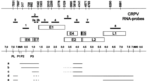
FIG. 1. Genetic and transcriptional maps of CRPV and localization ofmRNA-specific probes 1 to 9. The ORFs (open boxes) and the
nucleotide positions of the probeboundaries (top) correspond tothepublished sequence (11). Vertical lines within the ORFsrepresent the
translational startcodons. The general transcription mapwith the major transcripts (ato e) is depicted in the lowerpart(31). Dotted lines
representalternativesplicingpatterns,and dashed linesrepresentunknown5' ends. The earlypromotersareP1, P2, and P3, and the latepromoter
is PL.
infection with a pedo jet injector as previously described (3,
27). One DNA-induced papillomawas obtained by
intrader-mal inoculation with recombinant DNA (pLAII-CRPV
A&XbaI) asdescribedpreviously (20). All investigated carcino-masdevelopedafter 8to14monthswithout furthertreatment.
Biopsy specimensweresnapfrozenafter surgical excision and
storedat -70°Cuntil required.
Construction ofprobesfor in situhybridization analysis. To
construct expression vectors for the synthesis of mRNA-specific riboprobes, small subgenomic fragments of CRPV
were created by restriction enzyme digestions, purified by
agarose gel electrophoresis, and cloned, together with one
larger fragment(nucleotides [nt] 4571to6991), into thevector BluescribeM13+(Vector Cloning Systems, San Diego, Calif.).
The map positions (nucleotide numbers are according to
reference 11) ofthe 11 small subgenomic CRPV DNA
frag-mentsareillustrated inFig. 1. The identity of each clone and
the orientation of the insert were confirmed by restriction
enzyme digestions and additionally forprobe 7by sequence
analysis. All plasmids were linearized prior to transcription. Depending on the choice of the promoter, the in
vitro-synthesized RNAswere either in thesamepolarityasmRNA
(sense orientation) or in the opposite polarity (antisense
orientation). Invitro RNA synthesis in the presence of
3S-UTP, resulting in probeswitha specific activityof 3.5 x 10⁸
cpm/,ug, was followed by alkaline hydrolysis to reduce the
probe lengthto approximately 200 nt (5). Thequality of the transcribed RNAs was tested by polyacrylamide gel
electro-phoresis. The shortest probe (probe 2a,with acomplexity of
123 nt)wasapplied ata concentration of 3 x 10⁷ cpm/ml of
hybridization mixture, and the concentrations of the others
werenormalized inrelationtothe probe lengths.
In situhybridization. ForRNAinsituhybridization, serial
cryostatsections ofbiopsymaterial mountedon
aminopropyl-silan-coated slides were fixed in 4% paraformaldehyde in
phosphate-buffered saline and then dehydrated through graded ethanols. The dry sections thenwereacetylated in 0.1
Mtriethanolamine-0.25%aceticanhydride for 10min, washed
in 0.2x SSC (lx SSC is 0.15 MNaCl plus 0.015 M sodium citrate),andpreincubatedfor 2 h at42°C covered by solution
I,which contained45% formamide, 0.6 M NaCl, 2.5x
Den-hardt's solution, 10 mM Tris-HCl (pH 7.5), 1 mM EDTA,
0.1% sodium dodecyl sulfate, and 0.15 mg of tRNAper ml. After removal of the prehybridization solution, the tissue
sections were incubated for 16 h at 42°C with 35S-labelled antisense riboprobes in solution I containing 10% dextran sulfate. The incubated slides were washed in 50% form-amide-lx SSC at 55°C, treated with RNase A (10 ,ug/ml),
washedtwice for 2 h each in 0.1x SSCat 60°C, dehydrated,
andfinallycoated with Kodak NTB-2 emulsion. Theconditions for the DNA-hybridization with asense strandriboprobefor
CRPV (nt4571 to6991) have beendescribedpreviously (33).
Allslidesfromoneexperimentweredevelopedafter thesame
exposuretime(10to12days)toallowthe directcomparisonof
signals obtained with different probes.
Hematoxylin-and-eosine (H&E)-counterstained sections were evaluated and
photographedwithaZeissAxiophot microscopewitha
dark-orbright-fieldcondenser. The level ofbackground signalsfor RNAhybridizationwasdeterminedby usingsenseriboprobes
CO ) 1~
co
cO
-e r CO
l
9
I
0 T-co
11
E4
_I
8
II
LI
VOL.68, 1994
on November 9, 2019 by guest
http://jvi.asm.org/
[image:2.612.66.564.76.352.2]3622 ZELTNER ET AL.
5
7
E4
7
X
CRPV
RNA-probes
8
11
L
IE
1
[E
I11
E2
JF-
L2
1
probe
# 1 2 3 4 5 6 7 8 9 DNAup
4-.4-4-
4-44-l4-4
+44-4-4-4-C.
PAP-1
Ilo
++*
+ nd ++up
+l
+4-l
++ +4- +4 + 4-4-4-4-4-4-4-4-C.
PAP-2
lo
+++ --
+-
_ +up + 44- _
D. PAP-I lo 4-4-4- ++++ nd
++
++P - - nd ndup +-- +-_
+bb
* b4b
-+-/_a
D. PAP-2
lo
++|.*.
+ nd ++up +
4b4-
4-b b -4 +4+ +a+
+D.
PAP-3/4
io 4-4-i4 ++++4-4-4-4-
-
-D. DYSPL +++ ++
+
+++
upl-+l ++
D. PAP-X
lo
Ho i4 + nd + + ++++ - ++ nd+-I-D. CA-I
14
++++ + + ++ +++++1-D. CA-2 +4-4-
4+-44*4+
+
*-4
+4-
--
-D. CA-4 ++++
nd
nd ++ --
nd0 a
D. CA-5 4--4I-4 nd ++++ - +1- ++
D.
MET-112
+++ 4-+++4+ nd 4- ++4++
- - nd ++VX2 +++ ++++ + + +
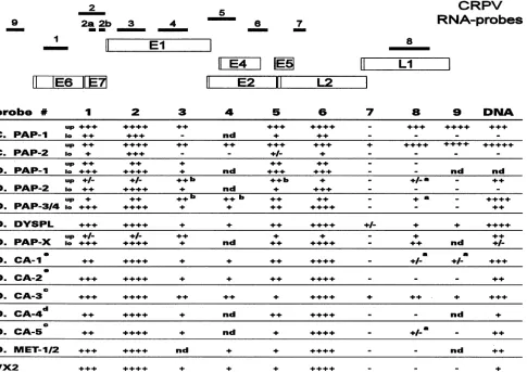
FIG. 2. The upperpartshows the
genetic
mapand thelocalization ofmRNA-specific riboprobes1
to9 used in thisstudy. The lowerpartgivestheintensitiesof the in situ
hybridization signals
detected with theCRPV-specificriboprobes for eachindividualcase. +,weak; + +,moderate;+++,strong; +
±+
+
,verystrong;nd,notdetermined;up, upperlayersof theepithelium; lo,lower layers of the epithelium;C., cottontailrabbit;D., domesticrabbit; PAP,
papilloma;
DYSPL,dysplastic
epithelium;CA, carcinoma;MET, metastasis. Signalsjust abovethe level ofbackgroundhybridization
areindicatedby
+/-.
a,signals
onlyinafewsinglecells; b, signalsonlyin some areas ofthe tissue section; c, well-differentiated carcinoma; d,moderately
differentiated carcinoma; e,poorlydifferentiated carcinoma.and by pretreatment of the tissue with DNase-free RNase before hybridization.
RESULTS
The followingspecimens werechosen for in situ
hybridiza-tionanalysis: twopapillomas from cottontail rabbits(Fig.2,C.
PAP-1 andC.PAP-2),oneearlypapilloma ofadomesticrabbit
(Fig.2, D. PAP-1) excised 3 weeks after infection with virus, and three other papillomas of domestic rabbits (Fig. 2, D.
PAP-2, -3, and-4) takenatlater times. One of thepapillomas
(Fig.2, D.PAP-4)contained asmallregion of highlydysplastic epithelium(Fig. 2, D.DYSPL) exhibiting aloss oftheregular
arrangement of the epithelial layersandmicroinvasion intothe
connective tissue. Another papilloma (Fig. 2, D. PAP-X)was
inducedbyinfection withamutantDNAof CRPV withalarge
deletion within the L2 ORF (pLAII-CRPV AXbaI). Two of
five malignant tumors could be classified histologically as
well-differentiatedkeratinizingsquamouscell carcinomas
con-tainingnestsof neoplastic cells with smooth borders (Fig. 2, D.
CA-3 and D. CA-5), one of the carcinomas was moderately
differentiated (Fig. 2, D. CA-4), and two other carcinomas
consistedmainly of poorly differentiatedcells(Fig. 2, D.
CA-1
and D.
CA-2).
We further included in our analysis two lungmetastases
(Fig.
2,
D.MET-1/2)
and the VX2 transplantablecancerline
(6,
15).
Biopsy specimens
D.PAP-3/4 and D. CA-I as well as D. PAP-2 and D.MET-1
were from the sameanimals.
Toassessthe presence ofdifferentmRNAspecies intumor
tissues,
appropriate
CRPVfragments
were cloned into the BluescribeM13+
RNAexpression
vector.The orientations of the inserts weredetermined,
and35S-labelled
antisense andnegative
control senseriboprobes
were synthesized in vitro.The
hybridization
specificities
of the different probes for themapped
RNAspecies
areasfollows. Probe 1 isspecific for thelong
E6and short E6mRNAsstarting
atpromotersP1 andP2(Fig. 1), respectively.
Probe 2 additionally recognizes the E7mRNAinitiatedat
promoter
P3aswellasthe mRNAsforLIand L2
(Fig.
1).
Since latetranscripts
are not detectable onNorthern blots of domestic rabbit tumors (21, 24), a higher
level of
hybridization
withprobe
2 than with probe1
indomestic rabbit tumors would indicate the presence of E7
mRNA. Probes3 and4
identify
potential
transcripts coveringthe open
reading
frame(ORF)
El but do not hybridize tomRNAs thatwere
processed
by
cleavage
at the splice donorsite 1371
(6).
The lack ofan El mRNA inthe transcription9
2
2a 2b
I
3 4
11
El
E
J.VIROL.
on November 9, 2019 by guest
http://jvi.asm.org/
[image:3.612.63.548.78.421.2]mRNA IN
CRPV-INFECTED
RABBIT TUMORS 3623map of CRPV in Fig. 1 is due to the fact that the original studies (31)could not distinguish whether the weak hybridiza-tion ofprobes colinear to ORFEl with polyadenylated RNA isolated from rabbit tumors represented nuclear precursors of the major transcripts or specific El mRNA (31a). Probe S
hybridizes to mRNA species with the coding capacity for a full-length E2 protein. Since El mRNA has not been mapped and the structure of an E4 mRNA is still unclear, it is possible thatprobe5 might also hybridize to El mRNA and E4 mRNA in addition to a full-length E2 mRNA. Probe 6 hybridizes to themajorearlytranscripts and to the minor E2 mRNA as well
asto the L2 mRNA and a potential E4 mRNA. Evidence for
aputativeE4 mRNA (21), however, could be obtained only if thatmRNAwaspresent in epithelial layers that contain small
amountsof major early transcripts and L2 mRNA. To evaluate further the existence of an E1'E4 RNA and potentially
provideinformation about the location of its cap site, two small
probes,2aand 2b, wereprepared (Fig. 1). Stronger signals with probe 2b than with probe 2a could indicate a start site within E7,inanalogy to the
El^E4
RNA ofHPV6/11 and HPV16 (2, 7, 13, 19,26). Probe 7 is unique for the L2 mRNA, and probes 8 and 9 hybridize to both major late transcripts.In allspecimens investigated we observed high levels of E6 andE7mRNAs andlow levels ofEl and E2 mRNAs. In none of the tumors was there evidence for a major mRNA class homologous to the prominent El^E4 RNA of HPV6/11 and HPV16(2, 7, 13, 19, 26). No difference was found between the intensities of thehybridization signals of probes 2a and 2b (Fig.
1),which were designed to detect prominent transcripts initi-ating within ORF E7, or between those of a probe located within ORF E7 and one located within ORF E4. This clearly indicated the absence of a prominent El E4 mRNA.
With papillomas of two cottontail rabbits (Fig. 2, C. PAP-1 and -2), we observed for all riboprobes an increase in the intensity of thehybridization signal in the more differentiated
layers of the epithelium. The high content of pigmented
granules, which were present only in cottontail rabbit warts, resulted inartificial signals in dark-field observation (Fig. 3H). These granules, however, could be clearly distinguished from
specific silver grains under higher magnification with bright-field optics (Fig. 3H, inset). Hybridization with probe 1 (spe-cific for E6 mRNAs) and with probe 5 (identifying mRNAs with a coding capacity for afull-length E2 protein) showed in addition weak signals in the lower epithelium (Fig. 3B and E,
respectively). The comparison of the hybridization signals of
probes 1, 2, and 8 revealed a fair amount of E7-specific
transcripts in the less-differentiated keratinocytes and basal cells(Fig. 3C).Strong signals were observed after hybridization with late-mRNA-specific probes 8 (Fig. 3G) and 9 exclusively in the granular cell layer, whereas weak L2-specific signals
(probe 7; data not shown) could be detected in only one
papilloma (Fig. 2, C. PAP-2).
Anearlypapillomaofa domestic rabbit (Fig. 2, D. PAP-1), which developed 3 weeks after infection with virus particles, showed ahistology reminiscent of that of an inverted
condy-loma; it projectedinto the corium and grew laterally to come
to underlie the normal epithelium. The granular layer was thickened, and beginning keratinization could be observed.
Hybridizationwithriboprobes 1and 2 revealed high levels of
transcriptsencoding E6 and E7 (data not shown); however, the differentiation-dependent expression pattern was opposite to thatin cottontailrabbitpapillomas (Fig. 3B and C). This was
even more pronounced in an older papilloma with deeper
papillae(Fig. 2,D.PAP-2), in which E6 and E7 mRNA levels
wererather highwithin thelowest layers of the epithelium and
graduallydeclined along with the differentiation of the
kerati-nocytes (Fig. 4). In all papillomas of the domestic rabbits, a weak signal was observed throughout the epithelium with riboprobes 3 (Fig.
5D)
and 4 (data not shown), which cover the El region, and a moderate signal was observed with an E2-specific probe (Fig.SE).
In addition, theEl and E2 probes showed, in some areas, clustered, mostly nuclear signals in the upper third. None of the domestic rabbit papillomas revealed transcripts encoding the minor capsid protein L2. Signalsspecific for Li were detected only in a few isolated spots of more highly differentiated cells in the granular layer (Fig. 5G, inset), with the exception of one papilloma (Fig. 2, D. PAP-X), which was induced by infection with a mutant DNA of CRPV with a large deletion within the L2 ORF
(pLAII-CRPV
AXbaI).
In this mutant DNA, however, the Li ORF is separated from the early transcription unit by the insertion ofpLAII
vector DNA sequences, which may be responsible forthe unusual expression of the Li gene. The transcription
patterns
of all other mRNAs and the replication of the viral DNA for this wart and for virus-induced papillomas showed no further difference. Overall, the patterns of viral gene expres-sion related to epithelial differentiation were clearly different for papillomas of cottontail and domestic rabbits.One papilloma of a domestic rabbit (Fig. 2, D. PAP-4) contained a small region of highly dysplastic epithelium (Fig. 2, D. DYSPL; Fig. 6A) exhibiting a complete lack of epithelial differentiation, the loss of a sharp border against the connec-tive tissue (Fig. 6B), and microinvasion into the connecconnec-tive tissue of single cells that were positive by in situ hybridization (Fig. 6C). Interestingly, the in situ hybridization with all early riboprobes showed no difference in the amounts of E6, E7,El, and E2 mRNAs for the highly dysplastic and adjacent papillo-mateous tissue (Fig.
SB,
C, D, E, and F). However, a clear difference could be observed in the expression patterns for late transcripts and in the permissiveness of the cells for the replication of the viral DNA (Fig.5H).
Whereas signals specific for Li were detectable only in a few isolated spots of more highly differentiated cells in the papilloma (Fig.5G,
inset), a weak signal was observed after hybridization with probe 8 (Fig.5G)
and probes 7 and 9 throughout the whole dysplastic epithelium. The same change was true for the distribution of viral DNA signals, which were strongest in the granular layer of the papilloma but were present in all cells of dysplastic origin (Fig.5H).
The investigation of five invasive carcinomas, two metasta-ses, and the transplantable VX2 cancer line revealed in general the same expression pattern for E6, E7, and El but dissimi-larities in the expression levels of E2 and late transcripts. In two well-differentiated carcinomas (D. CA-3 and -5), two lung metastases (D.
MET-1
and -2), and the VX2 tumor, we observed low levels of E2 transcripts (D. CA-3 is shown in Fig. 7D), whereas two poorly differentiated carcinomas (D.CA-1
and -2; Fig. 8E) and one moderately differentiated carcinoma (Fig. 2, D. CA-4) revealed higher levels of transcripts with a coding capacity for a full-length E2 protein. One of the well-differentiated carcinomas (D. CA-3) exhibited moderate levels of Li (Fig. 7E) and low levels of L2 (Fig. 7F), whereas most of the other tumors contained barely detectable late transcripts. The predominant transcripts in the VX2 line and the lung metastases encoded the E6 and E7 ORFs; all other early genes were expressed only at a low level.Hybridization with probes 7 to 9, which are specific for late transcripts, resulted in no signal above the background level. In the VX2 cancer line, viral DNA was present at a low copy number, in contrast to the case for most other tumors investigated (Fig.2).VOL. 68,1994
on November 9, 2019 by guest
http://jvi.asm.org/
'\
-lkl* " z'- - ;
[image:5.612.80.527.46.664.2]G
HJ
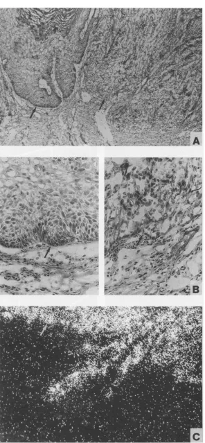
FIG. 3. Insitu hybridization of adjacent tissue sections of a cottontail rabbit papilloma (Fig. 2, C. PAP-2). (A) Histopathology observed by bright-fieldmicroscopy after staining with H&E stain. Individual adjacent tissue sections after hybridization with riboprobes 1 (B), 2 (C), 3 (D), 5(E),6(F),and 8(G)andwith probe 7(H) in the sense orientation as a negative control are shown as observed by dark-field microscopy. The silvergrainsgenerated in the film emulsion after exposure to the35S-labelledprobes are seen aswhite grains under dark-field illumination.The highcontentofpigmented granules in the lower part of the epithelium of cottontail rabbit warts (see bright-field inset in panel H) can be mistaken forahybridization signalwhen evaluated by dark-field observation. Panel H demonstrates the level of artificial signalcaused by thosepigmented granules(irregularlyshaped largegrains) togetherwith the level of background signal afterhybridizationwith a sense probe(smallgrains)as seen in dark-field observation.
3624
on November 9, 2019 by guest
http://jvi.asm.org/
mRNA IN CRPV-INFECTID RABBIT TUMORS 3625
A
B
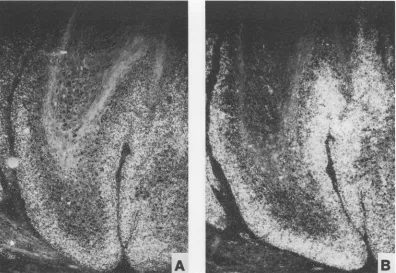
FIG. 4. Insitu hybridizationof adjacent tissue sections of a domestic rabbit papilloma (Fig. 2, D. PAP-2) after hybridization with riboprobes
1
(A) and2(B).
DISCUSSION
Grossanalysis of transcriptsencoding early genes by North-ernblot,
Si
nuclease, and primer extension analyses revealednomajor quantitativeor qualitativedifferences between
pap-illomas of cottontailand domestic rabbits or between benign
tumorsandcarcinomas(21,24,31).Togain more insight into the rolesofindividual genes in the viral lifecycle and in the
progression of virus-inducedlesions, it was necessary to study theirexpressionin the immediatecontextof tissuemorphology with the help of in situ hybridization.
There was amajor difference in the expressionpatterns of the E6 and E7 genes between papillomas of cottontail and
domestic rabbits.Wartsof the cottontail rabbit had the highest levels of E6 and E7 mRNAs in the upper third of the
epithelium,and thiswasalsotruefor all other viral transcripts. This transcription pattern has some similarities to that ob-served withHPV16-infected low-grade intraepithelial lesions (2, 9, 13, 28),whichwereshowntocontain themajorityof the viral transcripts in the more highly differentiated epithelial layers. In contrast, inpapillomas of domesticrabbits, expres-sion of E6 and E7 was restricted to the lower third of the
epithelium,an
expression
patternmorereminiscent of HPV6-induced condylomata accuminata (14). An additionaldiffer-enceintheexpressionof the E6 and E7 geneswastherelative
amountsofE6andE7transcriptsin cottontailversusdomestic rabbits. In cottontail rabbitpapillomas, E6 expression in the less-differentiated epitheliallayerswas lower than E7
expres-sion,whiletheywereabout equalin domestic rabbit papillo-mas.Thehigh level of expression of both viral oncogenesinthe undifferentiatedepitheliumof domestic rabbitpapillomasmay
provide an explanation for the high frequency of tumor progression.Ithas been shown for HPV16 that increased levels
of E6 and E7 inundifferentiatedcells may beanessential step
to evoke full transformation of human keratinocytes(8). El and E2 mRNAs were in general much less abundant than
mRNAs encoding E6and E7. In productive cottontail rabbit
papillomas and in nonproductive domestic rabbit papillomas,
El-specific transcripts were observed in the same epithelial
layerswhere viralDNAalso accumulated, and this would be in line with the functiondescribedfor theElprotein of
papillo-maviruses (reviewedin reference 16).Inaddition to detecting cytoplasmicsignals withEl and E2 probes, we observed asan
unusual phenomenon the presence of strong nuclear signals. Similar observations have been made previously for
HPV-infectedcondylomas, for intraepithelial lesions, and for
carci-nomas(14,28, 29).The nuclearsignalsmay representintrons of theunspliced major transcriptsassuggested previously(13, 28), or they could also indicate anaccumulation of El- and
E2-specificmRNAs in the nucleus.
In contrast to the results obtained with HPVs, in situ
hybridizationwith aprobe containing sequences of ORF E4 (Fig. 1, probe 6) did not result in a signal higher than that
expectedfor the hybridization to the major early transcripts. Thisresult,of course, doesnotexclude the presenceofanE4
mRNA as aminorspecies,assuggested bythe Northern blot
analysisofcottontail rabbitpapillomaRNA(21, 24).
Therewas no fundamental difference inthe expression of
earlygene transcripts betweenpapillomas and carcinomas of domestic rabbits. High levels of E6 and E7 mRNAs were
present inthe whole spectrum oflesions, from a 3-week-old papillomatotheVX2 cancerline,which hasbeen
continuously
maintainedin rabbitsformorethan 50years
(15).
Incontrast, therewas astrikingdifferenceinpermissiveness
for late geneexpression andviral DNAreplication betweendomestic
pap-VOL. 68, 1994
I
on November 9, 2019 by guest
http://jvi.asm.org/
[image:6.612.119.515.78.351.2]*;rj.:r |g w n ij
*.;'.' VLi..jf St ,. S
A .-r
[image:7.612.78.531.49.679.2]_~
G
H
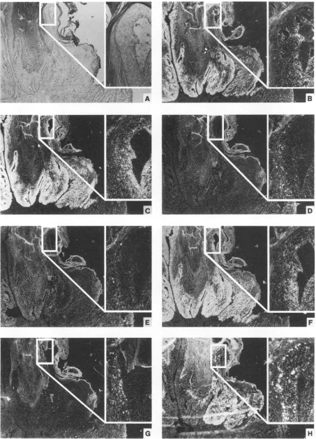
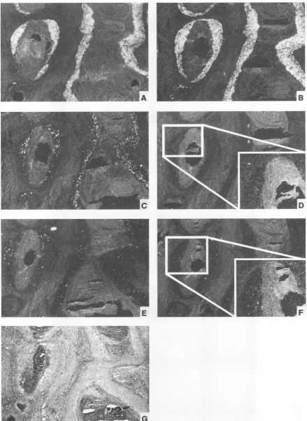
FIG. 5. In situ hybridization of adjacent tissue sections of a progressing domestic rabbitpapilloma (Fig. 2, D. PAP-4) containinga highly dysplastic region (Fig. 2, D. DYSPL). Histopathology is shown by light microscopy after staining with H&E stain (A). An enlargement of papillomateous epithelium is given as an inset in the upper right of each photography, displaying individual adjacent tissue sections after hybridization with riboprobes 1 (B), 2 (C),3 (D),5 (E),6 (F),and 8 (G)andwith a senseprobe (nt4571 to6991)for DNA detection(H)in dark-field illumination.
3626
on November 9, 2019 by guest
http://jvi.asm.org/
.,~~~~~~~~~~~~~~~~5'VX ' X X
c
FIG. 6. (A) Histopathology of a progressing domestic rabbit papilloma (Fig. 2, D. PAP-4). (B) Higher magnification of the papillomateous epithelium (left) and the highly dysplastic area (right). (C) In situ hybridization of epithelial cells invading into the connective tissue after hybridizing with probe 1, shown in dark-field illumination. Arrows denote the epithelial basement membrane.
3627
AL
40r4
on November 9, 2019 by guest
http://jvi.asm.org/
[image:8.612.168.464.44.683.2]3628 ZELTNER ET AL.
FIG. 7. In situ hybridization ofawell-differentiated domestic rabbit carcinoma (Fig. 2, D. CA-3). Individual adjacent tissue sections after hybridization with riboprobes 1 (A), 2 (B), 3 (C), 5 (D), 8 (E), and 7 (F) are shown in dark-field illumination. Histopathology is shown bylight microscopyafterstaining with H&E stain (G).
J. VIROL.
on November 9, 2019 by guest
http://jvi.asm.org/
[image:9.612.85.524.77.679.2]mRNA IN CRPV-INFECTED RABBIT TUMORS 3629
[image:10.612.98.533.74.528.2]E
_FF
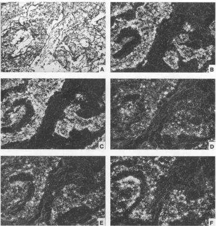
FIG. 8. Insituhybridizationofapoorly differentiated domestic rabbit carcinoma (Fig. 2, D. CA-1). Histopathology is shown by light microscopy afterstainingwith H&E stain(A).Individualadjacenttissuesections after hybridization with riboprobes 1 (B),2(C), 3 (D), and 5 (E) and with
asense-orientedprobe forDNAdetection (F)areshown in dark-fieldillumination.
illoma cellsanddysplastic cells. Inpapillomas,transcriptionof structural geneswasconfined tothe most-differentiated cells, while carcinomas, containing mainly less-differentiated cells,
werepermissive for latetranscription. The detection of signals specific for the late exon (probe 9) clearly indicated that the late promoter wasactiveinsomecarcinomas.The level oflate transcription in carcinomaswasquite variable andisprobably related to the highly variable viral DNA content of domestic rabbit tumors,which varies from 10 to afew hundredcopies
perdiploid-cellDNAequivalent(27).Sincealarge majorityof carcinoma-bearing rabbits (90%) have a strong antibody re-sponse toLI (17), the detection ofLI transcriptsin less than
half ofthe carcinomas analyzed may reflecta sensitivity limit for in situ hybridization. A likely reason for the increased
antibodyresponse to LI incarcinoma- compared with papil-loma-bearing rabbits is the change in the location of the Li transcript. Inpapillomas, LI is expressed only in the upper-most layers of the epithelium, which does not allow efficient recognition by the immune system, while in carcinomas,
ex-pression is observed throughout the thickness of the whole tumor,which is amuch more favorable situationforimmune stimulation.
Overall, the results of the in situhybridization experiments revealed a basic difference in the differentiation-specific
ex-pression of the viral transforminggenes and have provided a
possible explanation for the increased frequency of tumor progression in domestic compared with cottontail rabbits. Furthermore,the loss ofdifferentiation-dependent expression
VOL.68, 1994
on November 9, 2019 by guest
http://jvi.asm.org/
3630 ZELTNER ET AL.
of structural genes with tumor progression can explain the
selectively increased immune responsetostructuralproteinsin
carcinoma-bearing rabbits.
ACKNOWLEDGMENTS
We thankA.Schmitt for excellenttechnicalassistance.
This work was supported by grant 89.042.2 from the Wilhelm Sander-Stiftungto T.I. andby PublicHealth Service grant CA18151 awardedby theNationalCancer InstitutetoF.O.W.
REFERENCES
1. Barbosa, M. S., and F. 0. Wettstein.1988.E2 of cottontailrabbit papillomavirus is a nuclear phosphoprotein translated from an
mRNAencodingmultipleopenreadingframes. J. Virol. 62:3242-3249.
2. Bohm, S., S. P. Wilczynski, H. Pfister, and T. Iftner. 1993. The predominant mRNAclass in HPV16-infected genital neoplasias does not encode the E6 or the E7 protein. Int. J. Cancer 55:791-798.
3. Brandsma, J. L., Z. Yang, S. W. Barthold, and E. A. Johnson. 1991. Use of a rapid, efficient inoculation method to induce papillomas by cottontail rabbit papillomavirus DNAshows that the E7 geneisrequired.Proc.Natl.Acad.Sci.USA88:4816-4820. 4. Brandsma, J. L., Z. Yang, D. DiMaio, S. W. Barthold, E. A.
Johnson, and W. Xiao.1992. Theputative E5openreadingframe ofcottontail rabbit papillomavirus is dispensable for papilloma
formationindomesticrabbits. J. Virol.66:6204-6207.
5. Cox,K.H.,D.V.DeLeon,L. M.Angerer,and R. C.Angerer.1984. Detection ofm-RNAsinseaurchinembryos byin situ hybridiza-tionusingasymmetricRNAprobes.Dev.Biol. 101:485-502. 6. Danos, O., E. Georges, G. Orth, and M. Yaniv. 1985. Fine
structure of the cottontail rabbit papillomavirus mRNAs
ex-pressed in the transplantable VX2 carcinoma. J. Virol. 53:735-741.
7. Doorbar,J.,A.Parton, K. Hartley, L.Banks,T.Crook,M.Stanley, and L.Crawford. 1990.Detectionof novel splicingpatterns in a
HPV16-containing keratinocytecell line. Virology 178:254-262. 8. Durst,M., F. X. Bosch, D.Glitz,A.Schneider,and H.zurHausen.
1991. Inverserelationship between humanpapillomavirus(HPV)
type 16 early gene expression and cell differentiation in nude
mouseepithelial cysts and tumors inducedby HPV-positive
hu-mancelllines. J.Virol. 65:796-804.
9. Durst, M., D. Glitz, A. Schneider, and H. zur Hausen. 1992. Human papillomavirus type 16 (HPV16) gene expression and DNAreplicationin cervicalneoplasia: analysis byinsitu
hybrid-ization.Virology189:132-140.
10. Georges, E., F. Breitburd, N. Jibard, and G. Orth. 1985. Two Shope papillomavirus-associated VX2 carcinoma cell lines with different levels ofkeratinocyte differentiation and
transplantabil-ity.J.Virol. 55:246-250.
11. Giri, I.,0.Danos,andM. Yaniv.1982. Genomicstructureof the cottontail rabbit (Shope) papillomavirus. Proc. Natl. Acad. Sci. USA 82:1580-1584.
12. Giri,I., andM. Yaniv. 1988. Structural and mutationalanalysisof E2transactivating proteinsofpapillomaviruses reveals three dis-tinct functional domains. EMBO J. 7:2823-2829.
13. Higgins, G. D.,D. M.Uzelin, G.E.Phillips,P.McEvoy,R.Marin,
andC.J.Burell. 1992. Transcriptionpatterns of human papillo-mavirustype 16 ingenital intraepithelial neoplasia: evidence for promoter usagewithin the E7 openreadingframeduring epithe-lial differentiation. J. Gen. Virol. 73:2047-2057.
14. Iftner,T.,M.Oft, S.Bohm,S. P.Wilezynski,andH. Pfister.1992. Transcription of the E6 and E7 genes of humanpapillomavirus type6 inanogenital condylomataisrestrictedtoundifferentiated celllayersof theepithelium.J.Virol. 66:4639-4646.
15. Kidd, J.G.,andP. Rous.1940. Atransplantablerabbit carcinoma
originatinginavirus inducedpapillomaandcontainingthe virus in maskedoralteredform. J.Exp.Med.71:813-838.
16. Lambert, P.F. 1991. Papillomavirus DNA replication. J.Virol. 65:3417-3420.
17. Lin, Y.-L.,L. A.Borenstein,R.Selvakumar,R.Ahmed,and F.0. Wettstein. 1993. Progression from papilloma to carcinoma is
accompanied by changes inantibodyresponsetopapillomavirus
proteins.J. Virol. 67:382-389.
18. Meyers, C., J. Harry, Y.-L. Lin, and F. 0. Wettstein. 1992. Identificationof threetransforming proteinsencodedbycottontail rabbitpapillomavirus.J. Virol. 66:1655-1664.
19. Nasseri, M.,R.Hirochika,T. R.Broker,and T. L.Chow. 1987. A human papillomavirus type 11 transcript encoding an E1IE4 protein. Virology159:433-439.
20. Nasseri, M., C. Meyers, and F. 0. Wettstein. 1989. Genetic
analysis of CRPV pathogenesis: the Li open reading frame is
dispensablefor cellular transformation but isrequiredfor
tumor-igenicity. Virology170:321-325.
21. Nasseri, M.,and F.0. Wettstein. 1984.Differencesexist between viraltranscriptsin cottontailrabbitpapillomavirus-induced benign and malignanttumors as well asnon-virus-producingand
virus-producingtumors.J.Virol.51:706-712.
22. Nasseri, M.,and F.0.Wettstein. 1984. Cottontailrabbit
papillo-mavirus-specific transcripts in transplantable tumors with
inte-gratedDNA.Virology138:362-367.
23. Nasseri, M.,F.0.Wettstein,andJ.G. Stevens. 1982. Twocolinear and spliced viral transcripts are present in non-virus-producing
benign and malignant neoplasms inducedby the Shope (rabbit)
papillomavirus. J.Virol. 44:263-268.
24. Phelps,W. C.,S. L. Leary,and A. J.Faras. 1985.Shope papillo-mavirus transcription in benign and malignant rabbit tumors.
Virology 146:120-129.
25. Shope,R. E.,andE. W. Hurst.1933. Infectiouspapillomatosisof rabbits. J.Exp.Med. 68:607-623.
26. Smotkin, D.,H. Prokoph, and F.0.Wettstein. 1989. Oncogenic
and nononcogenichuman papillomavirusesgenerate the E6and E7 mRNAsbydifferentmechanisms.J.Virol. 63:1441-1447. 27. Stevens, J.G.,andF.0.Wettstein.1979.Multiple copiesofShope
virus DNAarepresentin cells ofbenignandmalignant
non-virus-producing neoplasms.J.Virol. 30:891-898.
28. Stoler,M.H., C. R.Rhodes,A.Whitbeck,S. M.Wolinsky, L. T.
Chow,and T. R. Broker.1992. Humanpapillomavirustype16and 18 gene expressionin cervicalneoplasias. Hum. Pathol. 23:117-128.
29. Stoler,M.H.,S. M.Wolinsky,A.Whitbeck,T. R.Broker,and L. T. Chow. 1989.Differentiation-linked humanpapillomavirustypes 6 and 11 transcription in genital condylomata revealed byin situ
hybridization with message-specific RNA probes. Virology 172: 331-340.
30. Syverton,J.T.1952. Thepathogenesisof the rabbit
papilloma-to-carcinoma sequence.Ann.N.Y.Acad.Sci. 54:1126-1140. 31. Wettstein,F.0.1987.Papillomavirusesandcarcinogenic
progres-sion. I. Cottontail rabbit(Shope)papillomavirus,p. 167-186. In N. Salzman and P. M. Howley (ed.), The Papovaviridae, vol. 2. Plenum PublishingCorp.,NewYork.
31a.Wettstein,F.0.Unpublisheddata.
32. Wettstein,F.O.,M.Barbosa,and M. Nasseri.1989. Identification of the major cottontail rabbit papillomavirus late cap site and
mappingandquantitation ofanE2and minor E6codingmRNA inpapillomasand carcinomas. Virology159:321-328.
33. Wilczynski,S.P.,M.Oft,N.Cook,S.Y.Liao,and T. Iftner.1993. Humanpapillomavirustype 6 in squamouscell carcinoma of the bladder and cervix. Hum. Pathol.24:96-102.
34. zurHausen,H.1991. Humanpapillomavirusesinthepathogenesis
ofanogenital cancer.Virology184:9-13.
J. VIROL.