0022-538X/88/072380-06$02.00/0
CopyrightC 1988, American Society for Microbiology
A Sequence-Specific
Single-Strand-Binding Protein for the
Late-Coding
Strand of the Simian
Virus 40
Control Region
CLAIRE GAILLARD, MICHELE WEBER, AND
FRANCJOIS
STRAUSS* Institut Jacques Monod, 2 PlaceJussieu, 75251 Paris05, FranceReceived 28 January 1988/Accepted5 April 1988
We have purified a protein from uninfected monkey CV1 cells that binds specifically in vitro to the late-coding simian virus 40DNAstrandin the region of transcription control withoutanydetectable binding to the complementary single strand. Nuclease protection experiments detected two binding sites in the
21-base-pairrepeatregion. Theprotein
did
notbindtothis regionin the double-stranded form,nordid itbind toRNA synthesized invitroby using either DNA strandasatenplate. This protein, and perhaps other DNAsjngle-strand-seqpence-specjfic proteins,mayplayarole in the control ofgeneexpression in higher organisms.
Dissection of eucaryotic geneshas allowed the
identifica-tion of DNA sequence elements that play a role in the
control ofgene expression, and mnany of themarethoughtto act at the level of transcription control by interacting with sequence-specific DNA-binding proteins. Purification of the factors that modulate gene expression by interacting with
regulatory DNA sequences has thus become an important
step towards a better understanding ofgene regulation in
higherorganisms.
The controlregion of simian virus 40 (SV40) is a
particu-larly goodsystemfor this kind of study, since it comprises, in a few hundred base pairs (bp), several DNA sequence
elements thatareimportant for viralgenetranscription (fora
review, see reference 23). Upstream from the gene for T antigen and the origin of replication, for example, is an
AT-rich stretch similartoaTATAbox; there are'three direct
repeatsofaGC-rich, 21-bp sequencethat also belongtothe early promoter and to which the transcription factor Spl binds; and preceding the late genes is the enhancer
se-quence, which includes two 72-bp repeats and has the characteristic propertyofstimulating transcription in either orientationfroma distance.
Tantigen
is'the
only virus-coded protein which interactswith'the control region, and all other factors involved in
transcription must necessarily arise from the host itself. Proteins from uninfected cells that bind to specific portions of this region include the well-characterized transcription factor Spi, which binds to the sequence'GGGCGG in the 21-bp repeats (5, 8-10, 14, 15, 19). A second transcription factor which also binds to the 21-bp repeat region has recently been identified (20). In
addition,
several researchgroups have shown, in different ways, the existence of
factors thatinteract with the enhancer(1, 2, 4, 7, 18, 21, 22, 24, 27, 29, 30, 32-36, 40-42).
Herewereporttheidentification ofaprotein that bindsto the late-coding
sipgle
strandat twospecific sites in the 21-bp repeatregion.MATERIALSANDMETHODS
Cell culture. African green monkey cells (CV1 line) were
maintained as monolayers in Eagle minimal essential
me-dium supplemented with penicillin and streptomycin and 10% newborn calfserum(Boehringer Mannheim
Biochemi-cals). For nuclei preparations, cells were grown to
conflu-* Corresponding author.
ence jn 20 25-cm-square plates (Nunc), yielding approxi-mately 109 cells.
DNAsubstrates. Aplasmid (pMW1) containing the control region of
SV40
with a single 72-bp repeat was constructedwith SV40 DNA (strain 776): the HindIII fragment that
encompassesthe controlregion (map position [m.p.] 5171to
1046) was inserted at the HindIII site of pBR322 and the 72-bp NsiI fragment (SV40 m.p. 126 to 198) was deleted.
Fragments obtained from this plasmid were used in most experiments; experiments performed with uncloned SV40 DNAgaveidentical results (datanot shown).
DNA labeling was performed with either' [y-32PIATP
(5,000 Ci/mmol; Amersham Corp.) and polynucleotide
ki-nase or [a-32P]dCTP (3,000 Ci/mmol) and the Klenow
frag-ment of'DNA polymerase. Following labeling, DNA frag-mentsorsinglestrandswerepurified by chloroform-isoamyl alcohol extraction, gel electrophoresis, electroelution, and ethanolprecipitation.
For strand separation, DNA wasdenatured at90°Cfor2 min in 10 mM Tris hydrochloride (pH 7.5)-l mM EDTA, chilled in ice-cold water,adjustedtoaconcentration of 2%in glycerol, and immediately loaded on a 4% polyacrylamide gelsimilartothegelsusedfor fractionation ofDNA-protein complexes (37;seebelow).Inmany cases,particularlyin the
caseof the SV40controlregion, strands of DNAfragments
up to 250 bp could be separated on this type ofgel. After electroelution of the-single strands, their identities were
determined by chemical seqiencing. The slow-migrating strand of
the
SV40controlregionwasthus foundtobe the late strand(see Fig. 1 and3).RNAsubstrates. For in vitro synthesisofRNA substrates forprotein binding,a plasmid containingapromoterfor T7 RNApolymerasewasused(pTZ19R; Pharmacia).The
BglI-HpaIIfragment(SV40m.p.5235to346)ofpMW1wasmade
bluptended with T4 DNA polymeraseand cloned in either orientation atthe HindIIl site ofpTZ19R by using Hindlll linkers (New England BioLabs, Inc.). Both plasmids were
linearized and transcribed with T7 RNApolymerase (Boehr-inger kit) and [a-32P]CTP (800Ci/mmol; Amersham).
Nonradioactive CTP was addedto the incubation mix in quantitiessuch that thespecificactivities of bothRNAswere
approximately equaltothespecific activityofthe DNA used in DNA-protein-binding experiments.
The identities of the RNAs were confirmed by checking that. their sizes depended in the expected way on the restriction site used for linearizing the plasmids. For the
2380
on November 10, 2019 by guest
http://jvi.asm.org/
experiment shown in Fig. 5, the PstI site in the plasmid polylinker was used, yielding RNA molecules 304
nucleo-tideslong.
Following synthesis, the RNAs were purified by electro-phoresis on a4% polyacrylamide gel,followed by electroe-lution.
Purification of protein H16. Frozen nuclei from
approxi-mately 109cells, purified as described elsewhere (37), were quickly thawed, pelleted, and suspended by gentle vortexing
in 7 ml of cold 0.6 M NaCi-50 mM sodium HEPES
(N-2-hydroxyethylpiperazine-N'-2-ethanesulfonicacid; pH
7.5)-i
mM EDTA-1 mM dithiothreitol (DTT) containing the five proteinase inhibitors used during nuclei purification (phen-ylmethylsulfonyl fluoride, antipain, leupeptin, chymostatin,and pepstatin A). After 30 min at 0°C with occasional stirring,thesuspensionwascentrifugedat 10,000 x gfor 30 min at 4°C, and the pellet was discarded.
The 0.6 MNaCInuclear extract was diluted sixfold with 50 mM sodium HEPES (pH
7.5)-i
mM EDTA-1 mM DTT. Alight precipitate that formedupondilutionwas discarded by centrifugation. Theextract wasapplied to a20-ml phospho-cellulose column (P11;Whatman, Inc.)preequilibrated with 50 mMsodium HEPES(pH7.5)-i mMEDTA-1 mM DTT-75 mMNaCI. Thecolumnwas washed with the samebuffer andelutedwithalineargradient of75 mM to 2 MNaClin 50 mMsodium HEPES (pH
7.5)-i
mM EDTA-1 mM DTT.Without the DNA-binding activities being assayed, four separate pools were made with the fractions eluting under 100 mM, between 100 and 250 mM, between 250 and 500 mM, andabove500mM. Each pool was dialyzed against 25 mMTris hydrochloride (pH
7.5)-i
mM DTT-30 mM NaCIand applied to a 1-ml fast-protein liquid chromatography mono Q column(Pharmacia) equilibrated in the same buffer.
Elutionwas done witha20-mllineargradient of30mM to 1 MNaCIin 25 mMTris hydrochloride(pH
7.5)-i
mMDTT.Fraction H16, which contained the protein described here,
originated fromthesecond phosphocellulose pool(100 to 250 mMNaCI) and elutedfromthe mono Q column at 250 mM
NaCI.
Both columns were run at room temperature, with
frac-tions immediately transferred to 0°C after collection. For long-term storage, fractions were adjusted to 15% glycerol and 1 mgbovineserumalbumin per ml and kept at -70°C. Forroutineuse, smallaliquotswere stored at -20°C in 50%
glycerol and 1 mgofbovine serum albumin per ml. Electrophoresis of DNA-protein complexes. For
denatur-SV40
Late Strand Early Strand
ation, DNA (or RNA) was heated at 90°C for 2 min in 10 mM Tris hydrochloride (pH
7.5)-i
mM EDTA and cooled to room temperaturejust before use. NonradioactiveEsche-richia coli DNA sonicated to an average length of 1 kilobase pair was used as competitor DNA.
DNA (5,000 cpm, about 0.1 ng) was incubated with proteins (1 ,ul) at room temperature (about 22°C) for 30min
in 25
,ll
of50 mMNaCI-10mMTrishydrochloride (pH7.5)-1 mM EDTA-7.5)-1 mM DTT-7.5)-1mg of bovine serum albumin per ml.Electrophoresison a4% polyacrylamidegel was done as
describedelsewhere(37). The gelwasdried and autoradio-graphed.
Digestion ofDNA-protein complexes withT4DNA polymer-ase. Thecomplex ofprotein H16 with 5'-end-labeled DNA wasformed as described above butwithoutthe addition of
competitorDNA. MgCl2was then addedto2 mM, and the mixture wasdigestedat37°C for2minwith0.3UofT4DNA
polymerase (New England BioLabs). The reaction was stopped by the addition of EDTA.
RESULTS
Protein thatbindsspecifically to one of the single strands of SV40 control region. Nuclear extracts were prepared from
uninfected Africangreenmonkey CV1 cells, permissivefor
SV40, to search for proteins that bind specifically to the
SV40 StyI restrictionfragmentthat extendsfromm.p. 37to 333(numberingasin reference 38) and encompasses both the enhancer and the 21-bp repeat region. The actual DNA
fragment used was acloned, 224-bp-long segment obtained bydeletingoneofthe72-bprepeats.Afterendlabeling, this
fragmentwasincubated withproteinextractsin the presence
of various amounts of nonradioactive competitor E. coli DNA, and the DNA-protein complexes formed were de-tected by polyacrylamide gel electrophoresis and autoradi-ography. Since thistechnique detects abundantnonspecific
proteins as well as thespecificones, thesequencespecificity oftheproteinswasassessedby comparingtheirbindingtoa labeled fragmentfrom pBR322 (37).
Initial assaysperformedwith crude nuclear extractsfailed
toreveal anyproteinbutSpl. However, sincerareproteins
may be masked in the assay by abundant specificproteins, such as Spl, or even by nonspecific proteins, we fraction-ated thenuclear extracts. We first loaded acrudeextract on a phosphocellulose column and arbitrarilymade fourpools
with the eluted fractions without assaying their
DNA-pBR322
Strand 1 Strand 2
SV40
Double Strand-
I-...
123456789C1 23456789 C 1 23 4 5 678 9 C1 2 3 4 5678 9 C
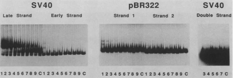
FIG. 1. Interaction ofproteins from fraction H16 withDNA. Polyacrylamide gel electrophoresiswasusedtoassaythe bindingof proteins
contained infraction H16tothe following five 5'-end-labeledDNAfragments:bothsinglestrandsofthe 224-bp StyI fragment from theSV40
control region (the late strand is slightly contaminated with the early strand; see late strand, lane C),both single strands of the 185-bp
EcoRI-EcoRVfragment from pBR322, and thesame StyIfragment from SV40 asdescribedabove but double stranded. Incubation of the
labeledDNA(about 0.1ng)with fraction H16wasdoneinthepresenceof increasingamountsof denatured competitor DNA (2,4,8, 15. 30,
60, 125, 250, and 500ngin lanes 1 through 9, respectively). LanesC, Control, freeDNA, noproteinadded.
34567C
on November 10, 2019 by guest
http://jvi.asm.org/
[image:2.612.118.493.539.665.2]a
<-
4 mA/T 21 21 21
Sty! FokI
37 80
A B C D E F G
H
72
H
Nsil BstNI
126 160
3,
*
* |,I .. ... .
*...
5,
* *
A
B
CD
E
F
G
H
12 34 Cl 2 34 Cl 234C1 234 C 1 234 C1 2 34Cl 23 4C1 234 C
Mi
[image:3.612.141.490.71.408.2]. ~ ~ _
FIG. 2. Binding of protein H16tosingle-stranded subfragments of theSV40controlregion.(a)Schematicmapofthe controlregionof
SV40 with adeletion ofoneof the 72-bprepeats. Arrowsrepresent the sites ofinitiation of transcription (from left toright: early-early, late-early, and late transcripts). The restriction sites and the DNA fragments(Athrough H) usedfortheexperiment shown in panel bare
indicated, with the position of the 32P end label either 5'or3'(*). Numbers indicate base pairs. (b) The latestrandsoftheeight end-labeled fragments (A through H),schematically shown in panela, werepurified, mixed with proteinH16in thepresenceofincreasingamountsof denaturedcompetitor DNA (2, 8,30,and 125ngin lanes1through4,respectively), and electrophoresedon apolyacrylamidegel.Equalmolar
amountsoflabeled DNAwereused. Lanes C, Control, free DNA,noprotein added.
binding activities (see Materials and Methods). Subsequent fractionation of eachpoolonthefast-protein liquid
chroma-tography anionexchange monoQ columngave us some 40
fractionsthatwere individually assayed. Inaddition toSpl,
we wereableto identifytwoother specificprotein activities that were capable of binding to the SV40 control region (unpublished data). We used a labeled DNAfragment that
was slightly denatured, and we observed in one of the
fractions (fraction H16) a third binding activity that was
specific foronlyone of the single strands.
Subsequently, both strands of the SV40 DNA fragment andthoseof thepBR322 controlfragmentwerepurified from
their complementary counterparts and used individually in bindingassayswithfraction H16(Fig. 1).The labeled single strandswereincubated with agiven amountof fraction H16 in the presence of increasing amounts of nonradioactive, sonicated, heat-denaturedE.coli DNA (lanes1to9).A band ofslower mobility was observed only with the SV40 late
strand. In contrast, few band migration differences were
observed with the SV40 early strand and the pBR322 strands. Note that the small amount of early strand that contaminated the late strand in Fig. 1 remained
uncom-plexed even at the lowest amount of competitor used,
whereas the latestrand wascompletely shifted tothe
com-plex. We interpret the retarded band as a protein-DNA
complex, since it was proteinase K sensitive and RNaseA resistant; itsformation was indifferent tothe presence of2 mMmagnesiumand 1 mM calcium in the incubation buffer (datanot shown). The decrease in intensityof the complex
upon increasing the amount of competitor DNA is not surprising, since any DNA-binding protein is expected to findsomecognate sequencesin thegenomeof E. coli. Such sites are probably rare or weak, however, since we have
tested theprotein with several single-stranded fragmentsof pBR322,coveringabout80% of theplasmid,without observ-inganyinteraction (datanotshown).
Incubation of fractionH16 with the double-strandedSV40 DNAfragmentdidnotshowanybands with retarded migra-tionevenatlowcompetitoramountsanduponoverexposure of theautoradiogram (Fig. 1).
Localization of binding sites with smaller restriction frag-ments. To determine more precisely where protein H16
binds, we prepared eight subfragments of the initial StyI
fragment (Fig. 2a, fragmentslabeled Athrough H).The late strands were individually purified and used for binding
assays. FractionH16 stronglyboundtofragments A, B, C,
PvuIl 270
Styl
333
b
_ o
on November 10, 2019 by guest
http://jvi.asm.org/
G
- + +
urn
-30 3m.40
50
60
80
90
100
.110
1
120
em 3
Or
A/T
2 1
=
21
21
72
. 130
FIG. 3. T4 DNA polymerase digestion of protein H16-DNA complexes. The late strand of SV40 (fragment BstNI, m.p. 5092 through 160)was5' end labeled atposition160 and digested by the 3'exonuclease activity ofT4 DNApolymerasein the absence (-) or presence (+)of protein H16. DNA was extracted and electropho-resedon an 8% sequencing gel with a G+Achemical sequencing reaction. The originof replication, 21-bp repeats, and 72-bp repeat areshown onthe right.Numbers to the right of the gel indicate SV40 mappositions.
21
andDbut didnotbindtofragments E, F, G,and H(Fig. 2b).
For fragments E through H, retarded bands of different
mobilities and much lower intensities than those observed for A, B, C, and D may indicate the presence of a contam-inating bindingprotein, perhaps not sequence specific. We conclude that protein H16 binds specifically to the late strand of SV40 in the 21-bp repeat region (see Fig. 2a). The appearanceoftworetarded bands may represent fragments with either one orboth ofthe twobinding sites (seebelow) occupied bythe protein.
Binding sites localization by nuclease digestion. For foot-printing experiments with a single-strand-binding protein, most enzymes are unsuitable: DNase I does notefficiently digest single-stranded DNA; micrococcal nuclease has a strong sequence specificity; DNase II, Si nuclease, and mungbeannuclease donotworkatthe neutralpH required
for formation of a stable protein H16-DNA complex. In
addition, dimethyl sulfate interference experiments gave
negativeresults.Therefore,weused thesingle-strand3'-*5' exonucleaseactivity ofT4DNApolymerase. Figure3 shows the result ofT4 polymerase digestion of a 5'-end-labeled
late-strand fragment that encompasses the 21-bp repeatsin theabsenceorthe presenceof fractionH16. Therewere two regions in which theprogression ofthe enzymewasstrongly slowed only when H16 was present: the first one, just precedingthe firstofthe21-bp repeats,isaroundnucleotide 38; the second one, in the second 21-bp repeat, is around nucleotide70(Fig.4).Immediately5'totheregionsin which the enzyme pauses, i.e. at the putative binding sites of protein H16, the sequence 5'-CCGCCCC-3' was found.
However,athirdCCGCCCC motifpresentin thethird21-bp
repeat did notseemtobindprotein H16. This suggests that the sequencerecognizedisnotstrictlylimitedtoCCGCCCC
and that neighboringbases on its 5' side arealso important
determinants ofthe specificity. In any case, precise knowl-edgeofthe nucleotidesequencesandofthe DNAstructure recognized by the protein will require an analysis of its
binding siteson alarge set ofdifferent DNAfragments.
Failure of protein H16 to bind to RNA. To exclude the
possibility that H16 is a sequence-specific RNA-binding protein that also binds DNA in vitro when presented with
homologous single-stranded DNA sequences, we
investi-gated its RNA-binding properties. The SV40 controlregion
was clonedineither orientation in plasmid pTZ19R, next to a promoterfor T7 RNApolymerase, and RNAwas
synthe-sized in vitro with either theearlystrandorthe late strandas thetemplate. Figure5 shows theinteraction ofproteinH16 with both RNAs and with the late strand of DNA in the presenceof variousamountsofdenaturedcompetitorDNA. Nocomplex wasobserved with RNA, while the same band
21
21
I III
*- early
I
"IV
V
zI
5ATTAGTCAGCCATGGGGCGGAGAATGGGCGGAACTGGGCGGAGTTAGGGGCGGGATGGGCGGAGTTAGGGGCGGGACTATGG
TAATCAGTCGGT,ACCCCGCCTCTTACCCGCCTTGACCCGCCTCAATCCCCGCCCTACCCGCCTCAATCCCCGC,CCTGATACC5e
30 40 s0 G0 70 so 90 100
late
FIG. 4. DNAsequenceof the 21-bprepeatregion. Theupperstrand is the early strand; the lower strand is the late strand. The six Spl-binding sitesareindicatedby rectangles above the sequence,withshading intensity increasing with Spl affinity. Thetworegions inwhich
T4 DNApolymerase digestionpausesinthepresenceof proteinH16arerepresented by dark rectangles below thesequence.Numbers below thesequenceindicatenucleotide positions.
VI
on November 10, 2019 by guest
http://jvi.asm.org/
[image:4.612.124.229.70.496.2] [image:4.612.96.518.597.683.2]pattern as described above (Fig. 1) was observed with the late strand of DNA.
DISCUSSION
We have purified asingle-strand-binding proteinthatbinds in vitro to specific regions in the 21-bprepeatsequence of the late strand of the control region ofSV40. Itdoes not bind to the complementary singlestrand, thedoublestrand,or RNA synthesized with either DNA strand as a template.
A conservative estimate of the affinity ofthe protein for the late strand relative to that for the early strand can be obtained from the data presented in Fig. 1 by applying the equations of equilibria (12) tohomologouslanes. With regard to lanes 4, for example, the ratioofboundDNA to free DNA is about 5 for the latestrandandlessthan 0.5% for the early strand, as determined by quantitation of the bands on the autoradiogram. This leads to an estimate of 3 orders of magnitude for the ratio of specific to nonspecific affinities.
However, it should be stressed that the protein is not pure and that the faint bands observedwith theearly strand might be due to acontaminatingprotein. This value is therefore a minimum estimate, and the actual value might be much higher.Purifying the protein tohomogeneitywillallowus to determine precisely thisratio, aswellastheabsolute affinity constants.
The transcription factor Spl also binds to SV40 DNA in this region. However, these two proteins are distinct, as judged by theirdifferent chromatographic behaviors
(unpub-lished data) and,mostconvincingly, by thefact thatprotein
H16 does not bind to double-stranded DNA whereas Spl
RNA
RNADNA
Early Strand Late Strand Late Strand
Transcript Transcript
1 23 45C 1 2 345C 1 2 345C
FIG. 5. Interactionof protein H16 with RNA. 32P-labeledRNA,
synthesizedin vitro fromaT7polymerase promoterby usingeither
thelate strandortheearly strand of theSV40 controlregion asthe
template (see Materials and Methods), wasmixedwithprotein H16
in thepresenceof increasingamountsof denaturedcompetitor DNA
(0, 1, 2, 4, and 8 ng, lanes 1 through 5, respectively) and
electro-phoresed on a 4% polyacrylamide gel. The same experiment was
donewith late-strand DNA.Lanes C, Noprotein added.
does.Although we havetriedseveraldifferent conditionsfor binding, including the presence ofZn2+ or ATP, we have been unabletoobserve theformationof a complexofprotein
H16 with double-stranded DNA. We therefore think that protein H16 itself is unable to open the DNA double helix
andmake itssites on the late strand accessible. An
interest-ing possibilitywouldbe thatSplparticipates in this process. Inthis respect, it isinteresting to note that both H16 sites are located atthe levelof the two weakest Spl sites on the DNA sequence (15) (Fig. 4). In addition, the first H16 site near
position 45 was found by mutagenesis to be the most
important of the six GC boxes for transcription initiationat the early-early sites(3).
Several single-stranded DNA-binding proteins have been isolated in higher eucaryotes. Many of those that bind reasonably well to single-stranded DNA-cellulose appar-ently have norelation with DNA in vivo (e.g., dehydroge-nases, a protocollagen precursor, serum proteinsinvolved in
complement activation, a1-antichymotrypsin; seereferences
in 43). Proteins UP1from calf thymus and HDP-1 from mice are related proteins that bind to single-stranded DNA, de-stabilize duplex DNA, and stimulate DNA polymerase,
similar to proteins encoded bygene32 of bacteriophage T4 and ssb ofE. coli(17, 28, 43). It hasbeensuggested thatthey
derive from heterogeneous nuclear ribonucleoproteins by
proteolysis (25), and they have no known sequence speci-ficity. Recently, two proteins believed to be involved in replication have been detected and shown to possess some sequence specificity to single-stranded DNA (13, 39). The transcription factor of the 5S RNA genes, TFIIIA, binds to 5S RNA, to 5S DNA at specific sites (11, 26, 31), and to single-stranded DNA but apparently not in a sequence-specific manner (16). Its binding properties are thus very
different from those ofprotein H16.
With respect to the role of this protein, we have no direct evidence to suggest that it binds to the late strand in vivo. However, short stretches of single-stranded DNA are sup-posed tobe transiently present in the genome, for example, during replication and transcription, and the possible role of protein H16 could be related to either of these processes. Given the importance of the 21-bp repeat region in the control of transcription, we are currently investigating the
effect ofthe protein in in vitro transcription experiments. In 1971, F. Crick published a model forthe chromosomes
of higher organisms in which "the recognition sites needed for control purposes are mainly unpaired single-stranded stretches of double-stranded DNA" (6). Indeed, single-stranded DNA presents more structuralflexibility, as well as a greaternumberof contact points for specific protein-DNA interactions. Achieving the high sequence specificity neces-sary to properly control gene activity in higher organisms may thus be made easierby the use of such interactions.
ACKNOWLEDGMENTS
We are grateful to Peter Brooks andMichael Ellison for critical readingof themanuscript.
This work was supportedby theCentre National de la Recherche Scientifique, the University of Paris 7, the Association pour la Recherche surle Cancer, andthe Ligue contre le Cancer.
LITERATURECITED
1. Angel, P., M. Imagawa, R. Chiu, B. Stein, R. J. Imbra, H. J. Rahmsdorf, C.Jonat, P. Herrlich, and M. Karin. 1987. Phorbol ester-inducible genescontain acommon cis elementrecognized by a TPA-modulated trans-actingfactor. Cell 49:729-739. 2. Azorin, F., and A. Rich. 1985. Isolation of Z-DNA binding
io
low,wvmw
:: :.sf,
;,Oao.w
a
.:M:
on November 10, 2019 by guest
http://jvi.asm.org/
[image:5.612.87.287.395.649.2]proteinsfromSV40minichromosomes: evidence for binding to
the viral controlregion. Cell 41:365-374.
3. Barrera-Saldana, H., K. Takahashi, M. Vigneron, A. Wildeman, I. Davidson, and P. Chambon. 1985. All six GC-motifs of the SV40earlyupstreamelement contributetopromoter activity in vivoandin vitro. EMBOJ. 4:3839-3849.
4. Bohmann, D.,W. Keller, T. Dale, H. R. Scholer, G. Tebb, and I. W. Mattaj. 1987. A transcription factor which binds to the enhancers of SV40, immunoglobulin heavy chain and U2 snRNA genes. Nature (London)325:268-272.
5. Briggs,M. R., J. T. Kadonaga, S. P. Bell, and R. Tjian. 1986. Purification andbiochemical characterization of the promoter-specifictranscription factor, Spl. Science 234:47-52.
6. Crick,F. 1971. General model forthe chromosomes of higher organisms. Nature(London)234:25-27.
7. Davidson, I., C. Fromental, P. Augereau, A. Wildeman, M. Zenke,and P.Chambon.1986.Cell-type specificprotein binding
totheenhancer of simian virus40 in nuclear extracts. Nature (London)323:544-548.
8. Dynan, W. S., and R. Tjian. 1983. Isolation of transcription factors that discriminate between different promoters recog-nizedby RNApolymerase II. Cell32:669-680.
9. Dynan, W. S., and R. Tjian. 1983. The promoter-specific tran-scription factorSpl binds to upstream sequences inthe SV40 earlypromoter. Cell 35:79-87.
10. Dynan,W.S.,and R.Tjian.1985.Control of eukaryoticmRNA synthesis by sequence-specific DNA binding proteins. Nature (London) 316:774-778.
11. Engelke,D.R., S.-Y.Ng,B.S. Shastry, and R. G. Roeder. 1980. Specific interaction ofa purified transcription factor with an internalcontrolregion of SS RNA genes. Cell 19:717-728. 12. Fried, M.,and D. M. Crothers. 1981. Equilibria andkinetics of
lacrepressor-operator interactions bypolyacrylamidegel elec-trophoresis. Nucleic Acids Res. 9:6505-6525.
13. Fry, M., F.W. Perrino, A. Levy,and L. A. Loeb. 1988. Factor D is a selective single-stranded oligodeoxythymidine binding protein. NucleicAcids Res. 16:199-211.
14. Gidoni, D.,W. S. Dynan, and R. Tjian. 1984. Multiple specific
contacts between a mammalian transcription factor and its cognatepromoters. Nature(London) 312:409-413.
15. Gidoni, D., J.T.Kadonaga, H.Barrera-Saldana, K.Takahashi, P.Chambon, and R. Tjian. 1985. Bidirectional SV40 transcrip-tion mediatedby tandem Splbindinginteractions. Science 230: 511-517.
16. Hanas,J.S.,D. F.Bogenhagen, and C.-W. Wu. 1984.Bindingof Xenopus transcription factor A to 5S RNA and to single strandedDNA. Nucleic AcidsRes. 12:2745-2758.
17. Herrick, G., and B. Alberts. 1976. Purification and physical characterization of nucleic acid helix-unwindingproteins from calfthymus.J. Biol. Chem. 251:2124-2132.
18. Johnson, P. F., W. H. Landschulz, B. J. Graves, and S. L. McKnight. 1987.Identification ofa ratliver nuclear protein that binds to the enhancer core element of three animal viruses. GenesDev. 1:133-146.
19. Kadonaga, J. T., K. R. Carner, F. R. Masiarz, and R. Tjian. 1987.Isolation ofcDNAencodingtranscription factorSpland functional analysis ofthe DNA binding domain. Cell 51:1079-1090.
20. Kim,C.H.,C. Heath, A.Bertuch, and U. Hansen. 1987. Specific stimulation of simian virus 40 late transcription in vitro by a cellular factor binding the simian virus 40 21-base-pairrepeat promoter element.Proc. Natl. Acad. Sci. USA84:6025-6029. 21. Lee,W.,A.Haslinger, M.Karin, and R. Tjian. 1987. Activation
oftranscription bytwofactors that bindpromoter and enhancer sequencesofthe human metallothionein geneandSV40.Nature (London)325:368-372.
22. Lee, W.,P. Mitchell, and R. Tjian. 1987. Purifiedtranscription factor AP-1 interacts with TPA-inducible enhancer elements. Cell49:741-752.
23. McKnight, S.,andR.Tjian.1986.Transcriptional selectivityof viralgenesin mammalian cells. Cell 46:795-805.
24. Mitchell, P. J., C. Wang, and R. Tjian. 1987. Positive and negative regulation of transcription in vitro: enhancer-binding protein AP-2 is inhibited bySV40Tantigen. Cell 50:847-861. 25. Pandolfo, M., 0. Valentini, G. Biamonti, C. Morandi, and S.
Riva. 1985. Single stranded DNA binding proteins derive from hnRNP proteins by proteolysis in mammalian cells. Nucleic Acids Res. 13:6577-6590.
26. Pelham, H. R. B., and D. D. Brown. 1980. A specific transcrip-tion factor that can bind either the SS RNA gene orSS RNA. Proc. Nati. Acad. Sci. USA 77:4170-4174.
27. Piette, J., and M. Yaniv.1987. Twodifferent factors bindtothe a-domain of the polyoma virus enhancer, one of which also interacts with theSV40and c-fosenhancers. EMBOJ. 6:1331-1337.
28. Planck, S. R., and S. H. Wilson. 1980. Studieson thestructure
of mouse helix-destabilizing protein-1. DNA binding and controlled proteolysis with trypsin. J. Biol. Chem. 255:11547-11566.
29. Robbins, P. D., D. C. Rio, and M. R. Botchan. 1986.
trans-Activation of the simian virus 40 enhancer. Mol. Cell. Biol. 6:1283-1295.
30. Rosales, R., M. Vigneron, M. Macchi, I. Davidson, J. H.Xiao, and P. Chambon. 1987. In vitro binding of cell-specific and ubiquitous nuclear proteins to the octamer motif ofthe SV40 enhancer and related motifs present in other promoters and enhancers. EMBO J. 6:3015-3025.
31. Sakonju, S., D. D. Brown, D. Engelke, S.-Y. Ng, B. S.Shastry, and R. G. Roeder. 1981. The binding of a transcriptionfactorto deletion mutants of aSS ribosomal RNA gene. Cell 23:665-669. 32. Sassone-Corsi, P., A. Wildeman, and P. Chambon. 1985. A trans-acting factor is responsible for the simian virus 40 en-hancer activity in vitro. Nature (London) 313:458-463. 33. Scholer, H., A. Haslinger, A. Heguy, H. Holtgreve, and M.
Karin. 1986. In vivo competition between a metallothionein regulatory element and theSV40 enhancer. Science 232:76-80. 34. Scholer, H. R., and P. Gruss. 1984. Specific interaction between enhancer-containing molecules and cellular components. Cell 36:403-411.
35. Scholer, H. R., and P. Gruss. 1985. Celltype-specific transcrip-tionalenhancement invitro r'equires thepresenceof trans-acting factors. EMBO J. 4:3005-3013.
36. Sergeant, A., D. Bohmann, H. Zentgraf, H. Weiher, and W. Keller. 1984. A transcription enhancer acts in vitro over dis-tances of hundreds of base-pairs on both circular and linear templates but not on chromatin-reconstituted DNA. J. Mol. Biol. 180:577-600.
37. Strauss, F., and A. Varshavsky. 1984. A- protein binds to a satellite DNA repeat at three specific sites that would be brought into mutual proximity by DNA folding in the nucleo-some. Cell37:889-901.
38. Tooze, J.1982. DNA tumor viruses, 2nd ed. Cold Spring Harbor Laboratory, Cold Spring Harbor, N.Y.
39. Traut,W., and E. Fanning. 1988. Sequence-specific interactions betweena cellular DNA-binding protein and the simian virus 40 origin of DNA replication. Mol. Cell. Biol. 8:903-911. 40. Wang, X.-F., and K. Calame. 1986. SV40 enhancer-binding
factors are required at the establishment but not the mainte-nance step of enhancer-dependent transcriptional activation. Cell 47:241-247.
41. Wildeman, A. G., P. Sassone-Corsi, T.Grundstrom,M. Zenke, and P. Chambon. 1984. Stimulation of in vitro transcription from the SV40 early promoter by the enhancer involves a specific trans-actingfactor. EMBO J. 3:3129-3133.
42. Wildeman, A. G., M. Zenke, C. Schatz, M. Wintzerith, T. Grundstrom, H. Matthes, K. Takahashi, and P. Chambon. 1986. Specific protein binding to the simian virus 40 enhancer in vitro. Mol. Cell. Biol. 6:2098-2105.
43. Williams, K. R., K. L. Stone, M. B. LoPresti, B. M. Merril, and S. R.Planck. 1985. Amino acid sequence of the UP1 calf thymus helix-destabilizing protein and its homology to an analogous protein from mouse myeloma. Proc. Natl. Acad. Sci. USA 82: 5666-5670.