Int. J. Electrochem. Sci., 6 (2011) 819 - 829
International Journal of
ELECTROCHEMICAL
SCIENCE
www.electrochemsci.org
Electrocatalytic Oxidation of NADH on Graphene Oxide and
Reduced Graphene Oxide Modified Screen-Printed Electrode
Lei Zhang1, Yang Li1, Li Zhang1, Da-Wei Li1, Dimitre Karpuzov2, Yi-Tao Long1,*
1
Shanghai Key Lab of Functional Materials Chemistry, East China University of Science and Technology, Shanghai, 200237, P.R. China
2
Alberta Centre for Surface Engineering and Science, University of Alberta, Edmonton, AB, T6G 2G6, Canada
*
E-mail: [email protected]
Received: 11 January 2011 / Accepted: 9 February 2011 / Published: 1 March 2011
A facile method, Graphene Oxide (GO) modified electrochemically pre-anodized screen-printed carbon electrodes (SPCEs), was developed for the determination of nicotinamide adenine dinucleotide (NADH) in neutral aqueous solution. Compared with the bare SPCEs and the reduced Graphene Oxide (rGO) modified SPCEs (rGO/SPCEs), the GO/SPCEs exhibit greatly enhanced electrocatalytic oxidation toward NADH. The scanning electron microscopy (SEM) and X-ray photoelectron spectroscopy (XPS) provide the substantial evidence for the formation of edge-plane sites on the surface of SPCEs, rGO/SPCEs and GO/SPCEs. The GO/SPCEs show the excellent electrochemical character for the determination of NADH in the range from 0.8 to 500 μM with a detection limit of 0.10 μM. The formation of oxidation product NAD+ was confirmed by in-situ UV-Vis spectroelectrochemistry. It shows a great promise for the design of the disposable amperometric biosensor for NADH.
Keywords: Graphene oxide, nicotinamide adenine dinucleotide, screen-printed electrode, biosensors, XPS
1. INTRODUCTION
reactions. Particularly, NAD+ and NADH are involved in cellular energy metabolism, including glycolysis, tricarboxylic acid cycle and oxidative phosphorylation [2]. Consequently, there is a great need to develop a robust detection for NADH. However, direct electrochemical detection by oxidation of NADH to its corresponding oxidized form NAD+ at bare electrodes is accompanied by very high overpotential at platinum or carbon [3, 4]. Moreover, the strong adsorption of not only NADH itself but also its oxidation NAD+ leads to electrode surface fouling and, as a consequence, producing a system which lacks sensitivity, reproducibility and stability [5]. Numerous efforts have been devoted to identifying mediators that will accelerate the interfacial electron transfer between NADH and a solid electrode, such as quinone derivatives [6, 7], thionine derivatives [8], diamines [9], ferrocene [10], ruthenium complexes [11], catechols [12], phenazine [13] and L-glutamate [14] derivatives adsorbed onto graphite or as electrodeposited films. Despite the extensive work carried out, these redox mediators still need improvements, mainly in terms of stability and reproducibility because of the water soluble mediator loss from the electrode surface. In this direction, some works employing nanostructured carbon materials such as pyrolytic graphite [15], carbon nanofibers [16, 17], carbon microparticles [18], especially carbon nanotubes [19-21] have been used widely to reduce the high-overpotential for NADH oxidation, to minimize the surface fouling and to improve electron-transfer kinetics without the help of redox mediators due to their edge-plane sites/defects present in the carbon materials. Despite advantages of these carbon nanomaterials, there still has been significant interest in the development of new carbon nanomaterials to accelerate the development of electroanalytical chemistry.
The screen-printing technology has widely been preformed for the fabrication of the mass, disposable, low-cost electrochemical sensors [22]. Normally carbon materials used as the screen-printed mediators are widely applied in electrochemical research for the determination of biological analytes such as glucose [23], NADH [24] and dopamine [25, 26]. Disposable pre-anodized SPCEs have shown highly improved electrocatalytic activity toward NADH [27]. The pre-anodized SPCEs with the introduction of edge plane carbonyl groups has been demonstrated to act similar to an edge plane graphite or carbon nanotubes (CNTs) modified electrode. Graphene, as a two-dimensional macromolecular sheet of carbon atoms with only one atom thickness, has been extensively used in electrochemistry applications in the recent years because of its unique structure and electrical and mechanical characteristics [28-32]. On the other hand, the graphene oxide (GO) and reduced GO (rGO) have the good electrochemical characters due to the oxygen-containing groups in their edge plane sites. Similar with carbon nanotubes, the higher proportion of edge plane defects may lead to more rapid electron transfer [33]. As a novel carbon nanomaterial, using graphene to develop high-performance sensors for biomolecular is highly expected.
magnitude by ranging of 0.8~500 μM for NADH with a detection limit of 0.10 μM on the GO/SPCEs, which is of great interest to minimize interferences and for the design of the disposable amperometric biosensor for NADH.
2. EXPERIMENTAL SECTION 2.1. Reagents and apparatus
Carbon ink, silver ink, and silver-silver chloride ink were obtained from Camnano Technology Co., Ltd. Xuzhou, China. Reduced form of β-nicotinamide adenine dinucleotide was purchased from Sigma. All solutions were prepared with Nanopure water (18 MΩ cm–1) from a Millipore MilliQ system. All chemicals used were of analytical grade and used as received without any further purification.
All electrochemical experiments were conducted with a CHI Instruments (CHI 660C) with an integrated screen-printed three electrode device: a carbon working electrode, a carbon counter electrode, and an Ag/AgCl reference electrode. And all experiments were typically conducted at room temperature (25 ± 2ºC). A 0.1 M phosphate buffer (0.05 M NaH2PO4 and 0.05 M Na2HPO4 adjusted to
pH 7.4 containing 0.1 M KCl) solution (PBS) was used throughout since enzyme-catalysed reactions of NADH and NADPH-dependent dehydrogenases are pH dependent with pH 7 and above. Consequently, the pH of 7.4 used in the experiments was selected based on the previous report [34].
The scanning electron micrographs were taken out by using a JEOL JSM6360 LV scanning electron microscope (JEOL Ltd., Tokyo, Japan). X-ray photoelectron spectroscopy (XPS) analysis was carried out on an AXIS HIS 165 spectrometer (Kratos Analytical) using a monochromatized Al Kr X-ray source (1486.71 eV). Survey spectra (0-1100 eV) were taken at constant analyzer pass energy of 160 eV, and all high-resolution spectra for C1s and O1s were acquired with a pass energy of 20 eV, a step of 0.1 eV and a dwell time of 200 ms. The takeoff angles between the film surface and the photoelectron energy analyzer were of 90 degree. The typical operating pressure was around 5×10⁻¹⁰ Torr in the analysis chamber. The peaks were fit using the CasaXPS application software. The samples for XPS measurements were prepared from dropping 2 μL sample solution onto SPCE working electrode surface, drying in N2 stream.
2.2. Preparation of graphene oxide
GO was prepared from graphite powder according to the modified Hummers method [35, 36]. Briefly, graphite powder (5 g) was oxidized by adding to a hot solution (80°C) of concentrated H2SO4
(30 mL) containing K2S2O8 (10 g), and P2O5 (10 g). The resultant dark blue mixture was thermally
resulted mixture was further stirred at 35ºC for 2 h and distilled water (200 mL) was added. The reaction was stopped with the addition of a mixture of 560 mL distilled water and 10 mL 30% H2O2.
For purification, the mixture was washed with 1:10 HCl and then with water. The GO product was re-suspended in water to a brown dispersion, which was subjected to dialysis to remove residual metal ions and acids. The purified GO dispersion was sonicated for about 1.5 h to exfoliate GO, and un-exfoliated GO was removed by centrifugation (3000 rpm, 5 min). In order to obtain rGO, hydrazine hydrate was added to the GO dispersion and stirred at 80°C for about 1 h.
2.3. Preparation and modification of the electrode
The integrated three-electrode devices were mass manufactured by a screen-printing process using an AT-25P machine (ATMA CHAMP ENT. Corp.). The fabrication of SPCEs has been reported in our previous work [37]. On 0.5 mm thick glass fiber plate, as the substrate material, the circular working electrode (WE) (planar area: 3.1 mm2, thickness: 15 μm) and the ring-shaped counter electrode were firstly fabricated from carbon ink and silver ink acting as conductive medium was printed as the second layer. The reference electrode was prepared with the silver-silver chloride ink. The insulating ink (Jelcon AC-3G, JUJO Chemical Co., Ltd., Japan) was used to provide the insulation layer which defines the thin-layer electrochemical micro-cell. The integrated three-electrode devices needed vacuum drying at 100ºC for 30 min after each step of the printing process. Finally, the integrated devices were vacuum dried at 140ºC for 90 min, then stored in vacuum container before use.
In order to modify the working electrode at the integrated SPCEs, the SPCEs was first washed with distilled water and dried by N2 stream. Then the SPCEs was pre-anodized in a 0.1 M (pH = 7.4)
PBS containing 0.1 M KCl by applying an anodic potential of +1.9 V (vs. Ag/AgCl) for 120 s [38]. The rGO/SPCEs (or GO/SPCEs) was prepared by coating 5 µL 0.3 mg/mL of the rGO (or GO) homogeneous suspension onto the SPCEs and then dried at room temperature overnight. All modified electrodes were cleaned by cyclic voltammetric technique between –0.5 and +0.5 V at a scan rate of 50 mV/s in PBS (pH 7.4) until a stable cyclic voltammetric response was obtained, and then rinsed with water and dried under a nitrogen stream.
3. RESULTS AND DISCUSSION
3.1. Characterization of GO (or rGO) modified SPCEs
[image:5.596.65.532.136.251.2]
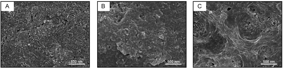
and the carboxylate carbon. Thus, GO could be spread uniformly on the surface of SPCEs, which provides the large amount of edge plane sites for the electrocatalytic oxidation toward NADH.
Figure 1. SEM images of SPCEs (A), GO/SPCEs (B) and rGO/SPCEs (C).
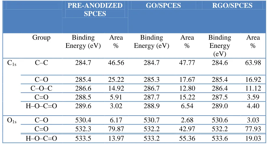
X-ray photoelectron spectroscopy (XPS) is a quantitative surface analysis technique that measures the elemental composition, empirical formula, chemical state and electronic state of the elements. The chemical composition of the bare SPCEs, GO/SPCEs and rGO/SPCEs were monitored by XPS. The high-resolution spectra for C1s and O1s are shown in Fig. 2A, B, C and E, F, D,
respectively. The peak fitting of C1s and O1s spectra was applied the mixed Gaussian/Lorentzian
function to fit peak shape after a Shirley background correction. The C1s XPS spectra could be
deconvoluted into five components corresponding to carbon atoms in different oxygen-containing functional groups[39]: (a) the non-oxygenated C at ~284.5 eV, (b) the carbon in C–O at ~285.5 eV, (c) the epoxy/ether group (C–O–C, ~286.5 eV), (d) the carbonyl carbon (C=O, ~288 eV), and (e) the carboxylate carbon (H–O–C=O, ~289 eV). The assignment of peak in O1s spectra can further confirm
the analysis information of C1s components. The O1s chemical shift at ~ 530.5 eV, ~532 eV and ~533.5
Figure 2. Deconvoluted the C1s and O1s XPS spectra of the pre-anodized SPCEs (A, D), GO/SPCEs
[image:6.596.113.482.69.310.2](B, E) and rGO/SPCEs (C, F). Solid square symbol: experimental data; solid line: fitting curve.
Table 1. XPS spectra results for C1s and O1s in the samples of SPCE, GO/SPCEs and rGO/SPCEs.
PRE-ANODIZED SPCES
GO/SPCES RGO/SPCES
Group Binding Energy (eV)
Area %
Binding Energy (eV)
Area %
Binding Energy
(eV)
Area %
C1s C–C 284.7 46.56 284.7 47.77 284.6 63.98
C–O 285.4 25.22 285.3 17.67 285.4 16.92
C–O–C 286.6 14.92 286.7 12.80 286.4 11.12
C=O 288.5 5.91 287.7 15.22 287.5 3.59
H–O–C=O 289.6 3.02 288.9 6.54 289.0 4.40
O1s C–O 530.4 6.17 530.7 2.68 530.6 3.03
C=O 532.3 79.87 532.2 42.97 532.2 77.93
H–O–C=O 533.5 13.97 533.2 55.36 533.6 19.03
3.2. Electrocatalytic oxidation of NADH
[image:6.596.72.522.407.651.2]
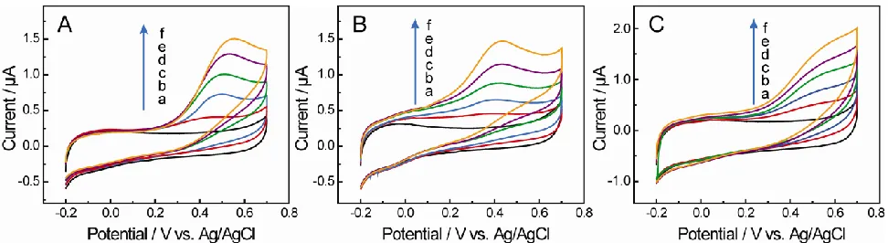
GO/SPCEs and rGO/SPCEs at a formal potential (E1/2) of –0.07 V and –0.04 V were observed in
background solution. These may be caused by the electrochemical redox of the oxygen-containing groups at the edge plane sites of GO and rGO, which could be supported by the components of C=O in the XPS C1s spectrum (Table 1).
[image:7.596.56.542.418.551.2]For the GO/SPCEs, as shown in Fig. 3B, a remarkable oxidation peak is observed starting around -0.1V with a peak at +0.41 V in the presence of 0.5 mM NADH in pH 7.4 PBS. The peak current of electrocatalytic oxidation of NADH at GO/SPCEs increases more significantly at +0.41V compared with SPCEs and rGO/SPCEs after the successive addition of NADH into the solution. However, the oxidation activity is observed at the SPCEs (Fig. 3A) and rGO/SPCEs (Fig. 3C) starting ca. 0.1 V and 0.05 V with peaks at +0.49 V and +0.51 V, respectively. Obviously, the oxidation peak potential at the GO/SPCEs is more negative than that at the bare SPCEs (Fig. 3A) and rGO/SPCEs (Fig. 3C), suggesting that the electron transfer kinetics for NADH oxidation on GO/SPCEs is essentially faster than the bare SPCEs and rGO/SPCEs. The effect of scan rates on electron transfer between NADH and the surface of electrode was evaluated (not shown). With an increasing scan rates, the anodic peak potential of NADH shifted to a more positive value, and the oxidation current were proportional to the square root of scan rates, indicating a diffusion-controlled behavior. From these demonstrations, we may conclude that the fast electron transfer kinetics for the NADH oxidation at the GO/SPCEs could be mainly associated with the presence of the oxygen-containing groups at this kind of carbon materials with edge plane sites.
Figure 3. Cyclic voltammograms curves of SPCEs (A), GO/SPCEs (B) and rGO/SPCEs (C) in 0.1 M PBS containing various concentration of NADH, a), 0; b),0.1; c), 0.2; d), 0.3; e), 0.4; f), 0.5 mM, pH = 7.4, scan rates: 10 mV/s.
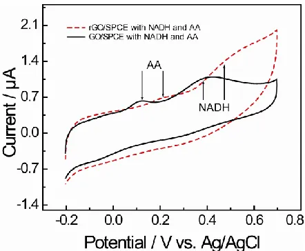
separation of over than 270 mV. In contrast, for the rGO/SPCEs, there are two small peaks at +0.22 V and +0.50 V respectively, which are not stable in the subsequent scan cycles. This suggests that the GO/SPCEs could be used to detect NADH and ascorbic acid simultaneously.
Figure 4. Cyclic voltammograms of the GO/SPCEs (solid line) and rGO/SPCEs (dot line) in 0.1 M PBS in the presence of 0.5 mM NADH and 0.5 mM AA. pH = 7.4, scan rate: 10 mV/s.
3.3. Amperometric detection of NADH
[image:8.596.188.409.146.328.2]
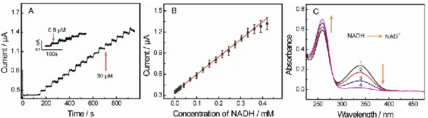
absorbance peak at 259 nm was increased slightly, which is present of the formation of NAD+. The results provided the evidence of the oxidation process of NADH to NAD+.
Figure 5. (A) Current–time curves of GO/SPCEs in PBS with various concentration of NADH, (B) The liner relationship of electrocatalytic oxidation current and concentration. (C) Real-time UV-vis spectrum of 0.2 mM NADH at different time, curve 1: 0s; curve 2: 10s; curve 3: 30s; curve 4: 90s. PBS: pH 7.4, applied potential: 0.4 V.
3.4 Stability of the biosensor
Long-term stability is one of the most important properties for biosensor application. The stability of GO/SPCEs was investigated by cyclic voltammetry. The response currents can retain almost constant upon continuous 20 cyclic sweeps over the applied potential ranging from −0.2 to +0.7 V with the scan rate of 10 mV/s. After stored in refrigerator at 4 °C for 90 days, the potentials of oxidation peak in CVs remained at the same positions and the peak currents decreased by only about 3.3% of its initial current response.
4. CONCLUSIONS
The present investigation reveals that the GO/SPCEs shows excellent electrocatalytic oxidation activity towards NADH with a remarkable decrease in overpotential and enhanced stability compared to the performance observed at a bare SPCEs and rGO/SPCEs. This could be attributed to the oxygen-containing functional groups on the surface of SPCEs, rGO/SPCEs and GO/SPCEs. The results of XPS provide a solid support to the oxygen-containing groups of edge plane sites on the surface of SPCEs, rGO/SPCEs and GO/SPCEs. The disposable SPCEs could act as the edge plane graphite electrode in the electrocatalytic oxidation of NADH. The detection range of GO/SPCEs is 0.8 to 500 μM with a lower detection limit of 0.10 μM. These facts offer a boarder application in the disposable amperometric NADH biosensors with immobilizing GO on the SPCEs surface.
ACKNOWLEDGEMENTS
[image:9.596.91.509.129.244.2]
Program for Professor of Special Appointment (Eastern Scholar) at Shanghai Institutions of Higher Learning.
References
1. A.L. Lehninger, D. L. Nelson, M. M. Cox, Lehninger principles of biochemistry 5th edn: W.H. Freeman; (2008)
2. A.Radoi, D. Compagnone, Bioelectrochem. 76 (2009) 126. 3. H. Jaegfeldt, J. Electroanal. Chem. 110 (1980) 295
4. J. Moiroux, P. J. Elving, Anal. Chem. 50 (1978) 1056. 5. M. A. Hayes, W. G. Kuhr, Anal. Chem. 71 (1999) 1720.
6. F. Pariente, F. Tobalina, M. Darder, E. Lorenzo, H. D. Abruña, Anal. Chem. 68 (1996) 3135. 7. D. C. -S Tse, T. Kuwana, Anal. Chem. 50 (1978) 1315.
8. K. Hajizadeh, H. T. Tang, H. B. Halsall, W. R. Heineman, Anal. Lett. 24 (1991) 1453. 9. A.Kitani, Y. H. So, L. L. Miller, J. Am. Chem. Soc. 103 (1981) 7636.
10.T. Matsue, M. Suda, I. Uchida, T. Kato, U. Akiba, T. Osa, J. Electroanal. Chem. 234 (1987) 163. 11.M. Somasundrum, J. Hall, J. V. Bannister, Anal. Chim. Acta 295 (1994) 47.
12.C. Ueda, D. C.-S Tse, T. Kuwana, Anal. Chem. 54 (1982) 850. 13.A.Torstensson, L. Gorton, J. Electroanal. Chem. 130 (1981) 199.
14.P. Norouzi, F. Faridbod, H. Rashedi, M. R. Ganjali, Int. J. Electrochem. Sci. 5 (2010) 1713. 15.R. R. Moore, C. E. Banks, R. G. Compton, Anal. Chem. 76 (2004) 2677.
16.L. N. Wu, X. J. Zhang, H. X. Ju, Anal. Chem. 79 (2007) 453.
17.A. Arvinte, F. Valentini, A. Radoi, F. Arduini, E. Tamburri, L. Rotariu, G. Palleschi, C. Bala, Electroanalysis 19 (2007) 1455.
18.B. Pérez, M. del Valle, S. Alegret, A. Merkoçi, Talanta 74 (2007) 398. 19.J. Wang, M. Musameh, Anal. Chem. 75 (2003) 2075.
20.Z. Liu, A. Fan, S. Sherlock, A. Goodwin, X. Chen, Q. Yang, D. Felsher, H. Dai, Angew. Chem. Int. Engl. 48 (2009) 7668.
21.M. Musameh, J. Wang, A. Merkoci, Y. H. Lin, Electrochem. Commun. 4 (2002) 743. 22.O. D. Renedo, M. A. Alonso-Lomillo, M. J. A. Martinez, Talanta 73 (2007) 202.
23.P. Norouzi, F. Faridbod, B. Larijani, M. R. Ganjali, Int. J. Electrochem. Sci. 5 (2010) 1213. 24.Q. Gao, X. Q. Cui, F. Yang, Y. Ma, X. R. Yang, Biosens. Bioelectron. 19 (2003) 277. 25.H. R. Zarea, N. Nasirizadeh, Int. J. Electrochem. Sci., 4 (2009) 1691.
26.A. S. Adekunle1, K. I. Ozoemena, Int. J. Electrochem. Sci., 5 (2010) 1726.
27.K. S. Prasad, J. C. Chen, C. Ay, J. M. Zen, Sensors and Actuators B: Chem. 123 (2007) 715. 28.C. Liu, S. Alwarappan, Z. Chen, X. Kong, C. Z. Li, Biosens. Bioelectron. 25 (2010) 1829. 29.S. He, B. Song, D. Li, C. Zhu, W. Qi, Y. Wen, L. Wang, S. Song, H. Fang, C. Fan, Adv. Funct.
Mater. 20 (2010) 453.
30.S. Alwarappan, C. Liu, A. Kumar, C. Z. Li, J. Phys. Chem. C, 114 (2010) 12920. 31.K. E. Toghill, R. G. Compton, Int. J. Electrochem. Sci. 5 (2010) 1246.
32.X. Zuo, S. He, D. Li, C. Peng, Q. Huang, S. Song, C. Fan, Langmuir, 26 (2010) 1936. 33.C. E. Banks, R. G. Compton, Analyst 130 (2005) 1232.
34.P. N. Bartlett, E. N. K. Wallace, J. Electroanal. Chem. 486 (2000) 23. 35.W. S. Hummers, R. E. Offeman, J. Am. Chem. Soc. 80 (1958) 1339.
36.N. I. Kovtyukhova, P. J. Ollivier, B. R. Martin, T. E. Mallouk, S. A. Chizhik, E. V. Buzaneva, A. D. Gorchinskiy, Chem. Mater. 11 (1999) 771.
37.L. Zhang, D. W. Li, W. Song, L. Shi, Y. Li, Y. T. Long, Sensors Journal, IEEE 10 (2010) 1583. 38.J. Wang, M. Pedrero, H. Sakslund, O. Hammerich, J. Pingarron, Analyst 121 (1996) 345.
40.O. C Compton, S. T. Nguyen, Small 6 (2010) 711.
41.J. F. Wang, S. L. Yang, D. Y. Guo, P. Yu, D. Li, J. S. Ye, L. Q. Mao, Electrochem. Commun. 11 (2009) 1892.
42.L. D. Zhu, J. G. Zhai, R. L. Yang, C. Y. Tian, L. P. Guo, Biosens. Bioelectron. 22 (2007) 2768. 43.D. W. Yang, H. H. Liu, Biosens. Bioelectron. 25 (2009) 733.
44.F. Pariente, E. Lorenzo, F. Tobalina, H. D. Abruna, Anal. Chem. 67 (1995) 3936.
45.F. Pariente, F. Tobalina, G. Moreno, L. Hernández, E. Lorenzo, H. D. Abruña, Anal. Chem. 69 (1997) 4065.
46.J. Chen, J. C. Bao, C. X. Cai, T. H. Lu, Anal. Chim. Acta 516 (2004) 29.