R E S E A R C H A R T I C L E
Open Access
Comparative genome and transcriptome analyses
of the social amoeba
Acytostelium subglobosum
that accomplishes multicellular development
without germ-soma differentiation
Hideko Urushihara
1*, Hidekazu Kuwayama
1, Kensuke Fukuhara
1, Takehiko Itoh
2, Hiroshi Kagoshima
3, Tadasu Shin-I
3,
Atsushi Toyoda
3, Kazuyo Ohishi
3, Tateaki Taniguchi
4, Hideki Noguchi
2, Yoko Kuroki
5, Takashi Hata
1, Kyoko Uchi
1,
Kurato Mohri
1, Jason S King
6, Robert H Insall
6, Yuji Kohara
3and Asao Fujiyama
3,7Abstract
Background:Social amoebae are lower eukaryotes that inhabit the soil. They are characterized by the construction of a starvation-induced multicellular fruiting body with a spore ball and supportive stalk. In most species, the stalk is filled with motile stalk cells, as represented by the model organismDictyostelium discoideum, whose developmental mechanisms have been well characterized. However, in the genusAcytostelium, the stalk is acellular and all
aggregated cells become spores. Phylogenetic analyses have shown that it is not an ancestral genus but has lost the ability to undergo cell differentiation.
Results:We performed genome and transcriptome analyses ofAcytostelium subglobosumand compared our findings to other available dictyostelid genome data. AlthoughA. subglobosumadopts a qualitatively different developmental program from other dictyostelids, its gene repertoire was largely conserved. Yet, families of
polyketide synthase and extracellular matrix proteins have not expanded and a serine protease and ABC transporter B family gene,tagA, and a few other developmental genes are missing in theA. subglobosumlineage. Temporal gene expression patterns are astonishingly dissimilar from those ofD. discoideum, and only a limited fraction of the ortholog pairs shared the same expression patterns, so that some signaling cascades for development seem to be disabled inA. subglobosum.
Conclusions:The absence of the ability to undergo cell differentiation inAcytosteliumis accompanied by a small change in coding potential and extensive alterations in gene expression patterns.
Keywords:Multicellular development, Cell differentiation, Signaling cascade, Gene expression, Evolution
Background
Morphogenesis and cell differentiation are the major components of multicellular development. In multicel-lular organisms, somatic cells, which are free from re-generative obligations, accomplish a variety of tasks to support complex body structures and functional integ-rity. The differentiation of mortal or sacrificial somatic cells from reproductive germ cells was the key event for
the establishment and diversification of multicellular sys-tems. How this was achieved in the history of life is an interesting and complex issue [1,2].
The social amoebae are unique organisms that exhibit conditional multicellularity and serve as an excellent model system to address this issue; they grow as solitary amoeba in the presence of sufficient food, but when starved, they gather together and form a multicellular fruiting body composed of a spore ball(s) and a sup-portive stalk(s). In many species, the stalk is filled with vacuolated stalk cells to stiffen it using osmotic pressure and cellulose walls that are deposited and polymerized * Correspondence:[email protected]
1
Faculty of Life and Environmental Sciences, University of Tsukuba, 1-1-1 Tennodai, Tsukuba, Ibaraki 305-8572, Japan
Full list of author information is available at the end of the article
on the extracellular matrix (ECM). While spores trans-mit their genetic information to their offspring, the stalk cells are no longer regenerative and represent one of the simplest forms of terminally differentiated somatic cells. In Dictyostelium discoideum, the most widely analyzed social amoeba species, cells in the migratory slug are committed to either the spore (prespore cells) or stalk lineage (prestalk cells) [3]. The latter further diversifies to generate the prestalk subpopulations PstB, PstO, and PstU, which end up in the basal disk and upper and lower cup structures, in addition to PstA constituting the main stalk body. These developmental processes are mainly controlled by the external levels of chemical cues such as cyclic nucleotides, ammonia, polyketides, pep-tides, and steroids to activate the corresponding intracel-lular signaling cascades [4].
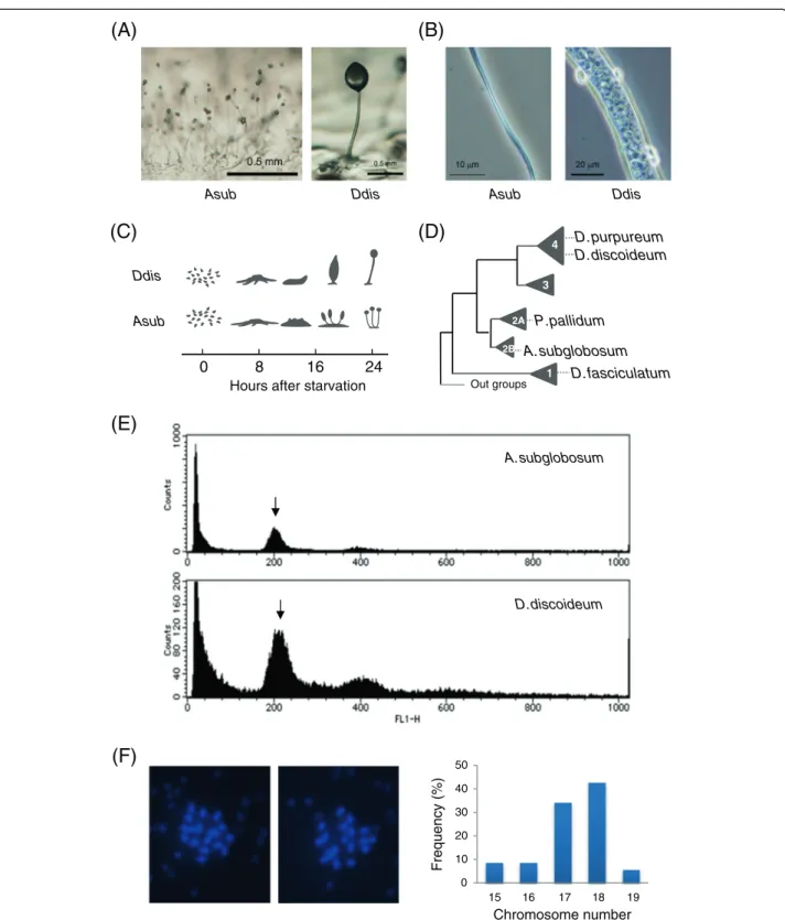
In spite of the common nature of starvation-induced fruiting body formation, species in the genus Acytoste-liumform an acellular stalk [5] and all aggregated amoe-bae become spores [6] (Figure 1A, B). They skip the migratory stage and multiple tips arise directly from the cell aggregates to generate crown-type fruiting bodies (Figure 1C). Recently, we studied the development of
Acytostelium subglobosumand found that the aggregated cells first became prespore-like cells producing spore coat proteins, and then function as if they were prestalk cells to synthesize and secrete the ECM and cellulose, and finally achieve terminal differentiation of the spores at the top of culminants [6]. Thus, temporal but not per-manent division of labour is observed in this species.
According to molecular phylogenetic analyses, D.
dis-coideumbelongs to the newest evolutionary clade (group
4), while the genus Acytostelium is in an older clade
(group 2) [7,8] (Figure 1D). There are two possibilities for the lack of the ability to undergo cell differentiation inAcytostelium: this ability was either acquired in a later species or it had been acquired in a common ancestor and lost in theAcytosteliumlineage. Since the species in the oldest clade, group 1, form fruiting bodies with cellular stalks, the latter possibility seems more likely, although it is still possible that cellular stalks arose inde-pendently multiple times, as pointed out by Swanson
et al. [9]. In either case, it is intriguing to determine what genetic information correlates with the ability to undergo germ-soma differentiation.
In the present study, we analyzed the genome and tran-scriptome ofA. subglobosumin comparison with other
so-cial amoebae making cellular stalks. The D. discoideum
genome was reported initially in 2005 [10]. Since then, the
genomes of Dictyostelium purpureum (group 4) [11],
Polysphondylium pallidum (group 2), and Dictyostelium fasciculatum (group 1) [12] have become available. The
developmental transcriptome of D. purpureumwas
com-pared with that ofD. discoideumto reveal their remarkable
conservation [13]. Our comparative analyses showed that dissimilarities in the gene repertoire between differentiating and non-differentiating species were limited, but that their transcriptomes had diverged. We suppose that the critical loss of early developmental genes relevant to cell-type specification affected the gene networks and led to the in-vention of a new developmental program where the entry of the entire amoeba to germ-line spores was traded off against the low efficiency of their dispersal due to short and fragile acellular stalks that were unable to support siz-able spore balls.
Results and discussion
Structure and general features of theA. subglobosum genome
The genome ofA. subglobosumLB-1/A1 was sequenced
using a whole genome shotgun sequencing approach and assembled into 371 contigs (DDBJ:BAUZ01000001-BAUZ01000371) arranged into 106 supercontigs (Table 1, Additional file 1: Figure S1). The total extension of the nucleotide sequence was 30.9 Mbp and close to the size ofD. fasciculatum, the smallest dictyostelid genome ana-lyzed so far. Since there was no available information on the genome size ofA. subglobosum, we used comparative flow cytometry analysis of nuclear DNA content to estimate its size as approximately 29 Mbp (Figure 1E). Although there is a small discrepancy between the two numbers, we assume that the present data adequately
represent theA. subglobosumgenome. The chromosome
number is 18 (Figure 1F). This is much larger than in the other dictyostelid species, but the possibility that it is diploid was shown to be unlikely from the results of flow cytometry. The (A + T) content of the A.
subglobo-sumgenome is 55%, which is remarkably lower than the
other dictyostelid species and naturally results in diffe-rential codon usage. It is much less biased compared to that of D. discoideum (Additional file 2: Table S1). The rRNA genes were found clustered in one of the super-contigs (SC 64). The number of tRNA genes was the smallest among the analyzed species. Simple sequence repeats and transposons were also not abundant. As a whole, our sequencing data revealed the neutral base composition, compact nature, and rather static features
of theA. subglobosumgenome.
Protein coding potential ofA. subglobosum
We performed expressed sequence tag (EST) analysis of vegetative and developmental cDNA libraries to
deter-mine the protein coding potential of A. subglobosum.
Altogether, 32000 clones were read from both ends (DDBJ:HY448297-HY508708), and the obtained sequences were clustered into 7439 non-redundant groups derived from 5749 genes, 98.4% of which were successfully mapped
(A)
D.discoideum
(E)
D. purpureum D. discoideum
P.pallidum
A. subglobosum D. fasciculatum Out groups
4
3
2A
2B
1 Ddis
Asub
24 16
8 0
Hours after starvation
(D)
(C)
(F)
0 10 20 30 40 50
16
15 17 18 19
Frequency (%)
Chromosome number
Asub Ddis
Asub Ddis
(B)
A. subglobosum
Representative cDNA clones were chosen for each of these groups and re-sequenced. The transcript information thus obtained (Additional file 1: Figure S2) was incorporated into a gene prediction program based on dicodon analysis [15] (Additional file 1: Figure S3). We also constructed
homology-based gene models usingD. discoideumprotein
sequences. The three sets of gene models, actual transcript sequences,ab initiopredictions, and homology based pre-dictions were combined to generate 12722 non-redundant protein coding genes (Additional file 2: Table S2; Additional file 3). This gene number is smaller than D. discoideum, but larger than 3 other species (Table 1). Lack of stalk cells inA. subglobosummay therefore not be explained simply by the absence of gene sets required for cell differentiation and stalk-cell function. This supports the previous argu-ment that the ability to undergo cell differentiation was lost inA. subglobosumrather than its acquisition in later species.
Orthology and gene family analyses
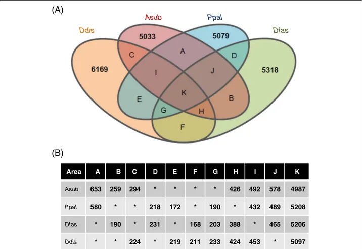
The gene orthology relationships were analyzed by the standard bidirectional best hit approach. When the e-value
threshold was set to 1E-10, 4987 A. subglobosum genes
were shared as orthologs with 3 other species (Figure 2, area K), but 190P. pallidum, 203D. fasciculatum, and 233
D. discoideumgenes had no orthologs in theA. subglobo-sum lineage (Figure 2, area G). Heidel et al. [12] took a more conscientious approach by employing identity and
coverage factors and described that 6569 D. discoideum
genes were shared with P. pallidumand D. fasciculatum. The corresponding gene number was 5330 in our analysis (sum of K and G in Figure 2). Since changing the
threshold value did not result in an increased number of orthologs, we did not change the above number and exam-ined the genes of interest individually in the later analyses.
To analyze the lineage-specific expansion of gene fam-ilies inD. discoideum, we combined the homology search
results and OrthoMCL [16] data of D. discoideum.
Namely, homologous genes for eachD. discoideum gene
were assigned to the same gene family. Clustering of
D. discoideum proteins by OrthoMCL resulted in similar but slightly bigger families than those reported by Eichinger
et al. [10] (Additional file 1: Figure S4). For the 5414
OrthoMCL OG-ids of D. discoideum[17], which did not
exclude the unique genes in this species, 4582A.
subglobo-sum, 4468 P. pallidum, and 4445 D. fasciculatum gene
families (and unique genes) were associated.
Besides common expansions, a significant number of lineage-dependent family expansions were observed (Additional file 1: Figure S5). Notable examples of them are marked and detailed in Table 2. The polyketide syn-thase (PKS) family (OG5_id 126633), whose members function in the biosynthesis of diverse classes of natural products such as those involved in intercellular and ecological interactions [10,18,19], is very small inA. sub-globosum. This suggests that these activities are less extensive in this species. Yet, A. subglobosum does have homologs of stlB, involved in the biosynthesis of diffe-rentiation inducing factor-1 (DIF-1) [20], which is ne-cessary for the induction of stalk-cell subpopulations [21,22], and stlA [23,24], which is involved in the
sig-naling cascade for spore maturation in D. discoideum
(Figure 3). Likewise, the ECM family (OG5_133822), which is expressed predominantly in prestalk cells and is Table 1 General features ofA. subglobosumand other dictyostelid genomes
Asub Ddis Dpur Ppal Dfas
Phylogeny group 2 4 4 2 1
Genome size (Mbp)a 31 34 33 33 31
Chromosome number 18 6 7 6
Supercontigs 106 6 799 41 25
Contigs 371 226 1,213 52 33
Genome (A + T) content (%) 55.0 77.6 68.0 66.2
Protein coding genes 12722b 13213 12410 12373 12173
Gene density (CDS/Mbp) 410 396 376 375 392
tRNAs 167c 390 273 198
Simple sequence repeat (%) 3.8 11.0 4.4
DNA transposons 1 215
LTR transposons 13 315 1.1 Mb
Non-LTR transposons 57 235
Reference This work [10] [11] [12] [12]
a
Total extension.
b
Only proteins predicted to be >50 amino acids were counted.
c
Asub Ppal
Ddis Dfas
Area A B C D E F G H I J K
Asub 653 259 294 * * * * 426 492 578 4987
Ppal 580 * * 218 172 * 190 * 432 489 5208
Dfas * 190 * 231 * 168 203 388 * 465 5206
Ddis * * 224 * 219 211 233 424 453 * 5097
(A)
(B)
Figure 2Gene orthology analysis of 4 social amoeba species. A: Results of the bidirectional best hit approach are shown schematically. Numbers represent species-specific genes. Colors orange, red, blue, and green representD. discoideum,A. subglobosum,P. pallidum, andD. fasciculatum, respectively. Genes in area G are absent in theA. subglobosumlineage but present inD. discoideum,P. pallidum, andD. fasciculatum.B: Gene numbers of each species contained in the areas A–K of the Venn diagram.
Table 2 Lineage-dependent expansion of gene families
OG5-id Description Family size in Mark#
Ddis Asub Ppal Dfas
Expansion in non-Dfaslineage
126643 Zinc finger, B-box domain and FNIP repeat-containing protein 241 381 206 55 a
Expansion in non-Ddislineage
153020 IPT/TIG domain-containing protein, EGF-like domain-containing protein, C-type lectin domain-containing protein
9 43 35 50 b
Expansion in Group 2
138577 Colossin D, Cna B-type domain-containing protein 5 33 22 7 c
Expansion inA. subglobosumlineage
181792 N-terminal delta endotoxin domain-containing protein 4 27 6 4 d
Lack of expansion inA. subglobosum
133822 Cellulose-binding domain-containing protein, putative extracellular matrix protein 30 4 25 18 e
126633 Putative polyketide synthase, beta-ketoacyl synthase family protein 41 6 21 25 f
#
required for cellulose deposition and polymerization [25], has not expanded in A. subglobosum, reflecting the small and less complex structure of its fruiting bodies.
Search for genes associated with the ability to undergo cell differentiation
The main purpose of this study was to mine out genetic information that was correlated with the ability to undergo cell differentiation. It was noted, at the early
stage of the study, that the major D. discoideum genes
related to stalk-cell differentiation were present in A. subglobosum despite the absence of stalk cells in this species. Although this is seemingly contradictory, it is not irrational considering the fact that A. subglobosum
does make stalks. Apparently, a simple loss of “stalk
genes”cannot explain the unique developmental process
ofA. subglobosum.
As already mentioned, 233 D. discoideum genes with
orthologs inP. pallidumandD. fasciculatumdid not have specific orthologs in theA. subglobosumlineage. Only 5 of them, 3 unique genes and 2 family-constituting genes, are currently known to be related to culmination and cell dif-ferentiation (Table 3). Of special interest is thetagAgene, which encodes a putative serine protease and ABC trans-porter B family protein and has been reported to play a crucial role in cell-fate determination and maintenance of the spore lineage [26]. Although there are a large number
of ABC transporters in A. subglobosum, as in other
dictyostelids, there is only 1 member (gene_6301) of the B subgroup with a serine protease domain (OG5_134947). Gene_6301 encodes a protein orthologous to TagC (Additional file 1: Figure S6) and is expressed similarly to it at the later stages of development (Additional file 2: Table S2).
Other genes in Table 3 are expressed at the later stages ofD. discoideumdevelopment and their relevance to
cell-fate determination is less likely. Two genes, rtaA and
warA, are concerned with prestalk-cell diversification. The
rtaAgene encodes a putative GPCR [28,29] expressed in
PstU cells [30], which finally populate the upper cup of a fruiting body whose function is to lift up the spore mass [31]. The latter gene,warA, is a homeodomain-containing putative transcription factor expressed in prestalk cells and determines the proportion of PstO cells in the slug, which occupy the zone between the prestalk and prespore regions [32]. Since the corresponding cell populations do
not exist even temporarily in A. subglobosum, the
me-chanistic aspect of culmination seems different in this
species, as pointed out by Bonner [2]. The expl7 gene
encodes a member of the expansin-like protein family and is homologous to the plant cellulose-binding protein expansin [33]. Although the over-expression of expl7 re-sulted in an anomaly of stalk morphology, its disruption did not affect the fruiting body morphology inD. discoi-deum[34], suggesting complementation by its paralog(s). The disruption mutant of rsc12 resulted in aberrant cul-minant at the final stage of development [35], but detailed analysis has not been carried out.
Loss of function can also be caused by the acquisition of new genes with suppressive effects. Although it is possible that some such genes may act to suppress the
A. subglobosum counterparts of developmental genes in stalk-cell-making species, their functions in relation to fruiting body formation are elusive without molecular biological analyses.
Comparative transcriptome analysis
The above mentioned analysis on the gene repertoires demonstrated that the majority of genes involved in fruiting body formation inD. discoideumhave orthologs in A. subglobosum and only few of them are without counterparts. To clarify whether the conserved genes are
actually expressed, the developmental transcriptome of
A. subglobosum was analyzed. Approximately 4.5 Gb of cDNA reads, generated at each of 0, 8, 16, and 24 h of development, were combined, clustered, and mapped to the A. subglobosum genome contigs (Additional file 1: Figure S7), resulting in the association of at least 9067
gene models. For the 5961 A. subglobosum orthologs to
D. discoideum genes with a significant level of ex-pression, expression data for 5062 (85%) genes were obtained. As for the developmental gene orthologs, 70 had no clear evidence of expression during growth and asexual development.
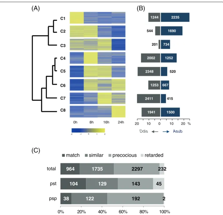
In an attempt to compare the temporal expression patterns of orthologous pairs, the expression data of A. subglobosum obtained here and those of D. discoideum
[13] were clustered collectively by K-means clustering, after normalization and standardization (Additional file 2: Table S3), using the cluster number 8 (Figure 4A). The resulting clusters fell into 2 major groups according to their mutual distances: clusters 1–3 and 4–8. The average expression pattern of the former group is down-regulation and the latter, up-regulation during development. There was a striking difference between the 2 species in gene distribution among the clusters (Figure 4B). Clusters 4 and 8 contain comparable fractions ofA. subglobosumand
D. discoideum genes, while the other clusters are ex-tremely biased to either species.More than 50% of the an-alyzed genes were in the down-regulated clusters (C1–C3) inA. subglobosum, while less than 20% were so inD. dis-coideum. These differences are in contrast to a report on
D. purpureumtranscriptome analysis that demonstrated a remarkable conservation in gene expression patterns with
D. discoideum [13,36], indicating the distant anatomical nature of the A. subglobosum fruiting body. (Additional file 1: Figure S8).
In accordance with the overall dissimilarity, only 18% of the orthologous gene pairs were assigned to the same clusters and nearly half of the ortholog pairs belonged to Table 3D. discoideumdevelopmental genes lacking orthologs in theA. subglobosumlineagea
Geneb Product Mutant informationc
Involved in cell fate-determination
tagA(DDB_G0293002) ABC transporter B family protein, serine protease Multiple tips in mound (partial KO); Aberrant fruiting body morphology (KO)
Involved in stalk-cell diversification
rtaA*(DDB_G0271852) Lipid-translocating exporter family protein Expressed in PstU cells (lacZfusion)
warA*(DDB_G0291075) Putative homeobox transcription factor Development arrests at slug stage, decreased prespore cell differentiation, and increased PstO cell differentiation (KO)
Involved in terminal differentiation
expl7(DDB_G0288331) Expansin-like protein Aberrant culminant morphology (OE)
rsc12*(DDB_G0277871) Unknown Aberrant culmination (KO) a
Only those related to morphogenesis and cell differentiation are listed.
b
Unique genes are asterisked. Parentheses indicate DDB_G ID.
cPhenotypes of disruptants (KO) and overexpressors (OE), and product localization (lacZ
different groups (C1–C3 vs.C4–C8) (Figure 4C). As can
be seen in Figure 4C, the A. subglobosum orthologs for
prespore-specific genes, showed the stronger propensity for precocious expression, while prestalk-specific gene orthologs displayed the opposite pattern. This tendency for differential expression coincides with our previous
finding that A. subglobosum produces prespore vesicles
soon after aggregation and then makes stalk materials
[6]. In consideration of development under starvation stress, it should be a safe strategy to transcribe the germ-line (spore lineage) genes first and to use the rest of the available energy for stalk formation, unless the population is divided into germ (spore) and soma (stalk cells) lineages; the stalk volume can be variable, but the amount of spore coat material is definite. The D.
discoi-deum developmental genes whose orthologs belong to
(B)
(A)
(C)
0h 8h 16h 24h
2 -2 -1 0 1 C1
C2
C3
C4
C5
C6
C7
C8 1500
415 667
520 1252 734
1690 2235
1941 2411
1253 2348
2002 201 544
1244
20 10 0 10 20 %
Asub Ddis
38
104
964
122
129
1735
192
143
2297
2
45
232
0% 20% 40% 60% 80% 100%
psp pst total
match
similar
precocious
retarded
distant clusters are listed in Table 4. Interestingly, genes important for the terminal differentiation of spores and
stalk cells in D. discoideum such as acbA, dgcA, and
gtaCwere expressed very early and down-regulated
rap-idly in A. subglobosumdespite their presumed functions at the latest stage (Table 4). They may be translationally or post-translationally regulated or might have different functions inA. subglobosum.
In addition to the differential expression time course, al-tered mRNA levels, both increased and decreased, were no-ticed in a substantial number of ortholog pairs (Additional file 1: Figure S9). It caught our attention that counting factor and related components involved in determination of aggregate size (cfn50-1, ctnA, and cf60) were greatly re-pressed. Only one of the related genes,cfaD, is expressed at a comparable level and in the same pattern, but this gene is presumed to control the growth-development transition.
Since inactivation of these genes inD. discoideumresults in larger aggregates, the implications of the above finding on the small fruiting bodies of A. subglobosumare unclear. It may be related to the fact thatA. subglobosumdevelopment is possible only at lower cell densities than for D. dis-coideum development [6]. It is also interesting that the expression of genes for G proteinαsubunits 6 (gpaF) and 9 (gpaI) were altered in mutually opposite directions. These transcriptome differences should exert significant influen-ces on the signal-response cascades and gene networks during development.
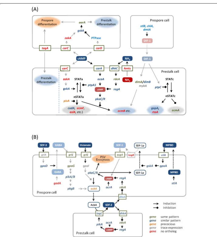
Signaling cascades for cell differentiation and morphogenesis
The cell differentiation process inD. discoideumproceeds in 3 major steps: cell-fate determination, diversification of the prestalk cell lineage, and terminal differentiation of
Table 4D. discoideumdevelopmental genes with altered expression inA. subglobosum Cluster # Cell-type specificity inD. discoideum
Ddis Asub Prespore Prestalk Non-specific
C1 0 (nxnA) (4)
C4 gabT, proB aprA*, cf50-1*, cmfA, ctnA*
C6 scrA
C8 gpaF
C2 0 (2)
C4 icmA cnxA
C5 psmC1
C8 gpaI*
C4 0 (krsA), (paxB) (14)
C1 mgp2, mppA1, sglA, tmem184C, DG1060
C2 DG1112, gsr, spkA* alg9,midA, ppp4C, sgcA, DG1104, DDB_G0287723
C3 crtA, psmA1 clc, phlp1, DG1040
C5 0 (11)
C1 plbG hdaB, sr
C2 adrm1, DG1122 exoc2 cshA*, ctr9, Dd5P4, nfaA, snfA, DDB_G0278945
C3 kif12, lvsD, DDB_G0269680, DDB_G0285083, DDB_G0288007
C6 0 (12)
C1 amtA*, dymA*, srfA
C2 DG1124 fpaA atg5, dgkA, DDB_G0270344
C3 ahhA*
C7 0 adprt3, tipC (elmoA) (5)
C2 cf60* captC, ifkA,DG1003
C3 torA,dnmA
C8 0 (mhcA) (6)
C1 dgcA alrA, dokA, phyA, vmp1
C2 acbA gtaC
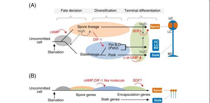
the spores and stalk cells (Figure 5A). To examine how genomic and transcriptomics properties ofA. subglobosum
related to these steps, the results of gene orthology and
transcriptome analyses of A. subglobosum were overlaid
on the known signaling cascades controlling D.
discoi-deumdevelopment (Figure 6).
The spore lineage cells induced by a high concentra-tion of extracellular cAMP in turn induce uncommitted cells to the prestalk lineage in D. discoideum. The im-portant gene at this step, tagA, is missing, as mentioned above, and orthologs of two cAMP receptors, cAR3 and cAR4 (encoded bycarCand carD), are also absent inA. subglobosum, corresponding to the lack of cell-type dif-ferentiation in this species. The specific substrate of TagA is suspected to be the acyl coenzyme A binding protein (AcbA) from the genetic evidence [37]. The facts
that lack of the transporter domain of tagA caused the
multi-tip phenotype inD. discoideum but that complete
disruption of this gene resulted in a single gnarled stalk suggest the dual function of TagA during development.
Stalk-cell diversification is triggered by DIF-1, which is secreted from prespore cells in D. discoideum. Despite
the fact that A. subglobosum does not make the basal
disk or upper and lower cup structures of the fruiting body, genes for the components of the DIF-1 signaling cascade do exist and are expressed at more or less simi-lar timings. On the other hand, we were unable to detect
DIF-1 production in A. subglobosum either
bioche-mically or biologically [38]. Therefore, it is possible that
a similar but different substance is produced in A. sub-globosumand induces a modified version of the polyke-tide signaling cascade to activate ECM genes and the cellulose synthase required for stalk formation.
The molecular mechanisms of synchronized and rapid spore encapsulation caused by exocytosis of spore-coat materials in the prespore vesicles are relatively well understood. They employ peptides called spore differen-tiation factor 1 (SDF-1) and SDF-2, which are secreted as precursors from prespore cells and processed by the serine protease-ABC transporters TagB and TagC, re-spectively [39,40]. The whole process is accelerated by GABA- and MPBD-mediated cascades, the former being triggered by a steroid type SDF (SDF-3) [41]. The accu-mulation of SDF precursors in the prespore cells is con-trolled by intracellular cAMP levelsviaprotein kinase A
[42]. Overlaying A. subglobosum orthology and
expres-sion data suggested that only the core cascade involving SDF-2 is intact in A. subglobosum(Figure 6B); enhance-ment by the SDF-3-GABA route is probably disabled by the lack of its essential gene, gadA. The SDF-1 cascade
seems to be hampered by the absence of tagB.
Consi-dering the small size of the A. subglobosum sorus, the
synchronous encapsulation of spore precursor cells may not require multiple cascades, although the possibility of functional complementation by paralogous genes still exists.
Overall developmental signalling is compared between the 2 species in Figure 5. We already showed that a
(B)
(A)
Spore genes
Spore
Stalk
Stalk genes
Encapsulation genes cAMP,DIF-1 like molecule SDF?
Uncommitted cell
Starvation
Spore lineage
Pst B,O (PstU)
Spore
Stalk
Uncommitted cell
tagA
Stalklineage tagC
UC LC BD
cAMP
DIF-1
SDFs
Terminal differentiation Diversification
UC
LC
BD Fate decision
Starvation
PstA
c-di-GMP
tagB
sequential gene expression of “prespore” and “prestalk” genes was observed in individual cells [6]. The results presented here suggest that this finding can be extended:
In contrast to the developmental process of D.
discoi-deum achieved by 2 types of cells in parallel, the
deve-lopmental program of A. subglobosum seems to depend
(B)
(A)
dhkC
rdeA
regA
cAMP
Prespore cell
carC
cAMP
zakA
gskA
carA
acaA STATa
nSTATa ptpA1
cudA,ecmF,
aslA, etc.) ecmBetc.
DIF-1
ptpC
nSTATc
gapA,
rtoA ecmA
DIF-1
stlB, chlA, dmtA
carC zakA
aarA
carD
Prestalk differenaon
PTPase
tagA
Prestalk differenaon
Amts
STATc
pisA gskA
NH3
NH3
Prespore differenaon
pkaC/R
Prestalk cell
dimA/dimB
mybA
largely on sequential gene expression, which is regulated cell-autonomously, and on a few cell-cell interactions.
Conclusions
Our genome analysis ofA. subglobosumrevealed the unex-pected conservation of D. discoideum stalk-specific genes. However, alterations in developmental transcriptomes were extensive. This suggests that non-differentiating species utilize fundamentally different developmental programs, even though their final morphologies appear similar. Since gene losses at the early stages of cell-fate determination must disturb the later developmental processes enor-mously, they are likely to have been compensated by diffe-rential gene regulations.
Methods
Cell culture and asexual development
The clonal line of A. subglobosum strain LB-1/A1 was
described previously (6). A1 cells were grown in sha-king HL5 medium at 22°C. For asexual development, the cells were harvested at their early growth phase (1.0–3.0 × 10⁶
cells/mL), washed twice with KK2 buffer (20 mM K2HPO4/KH2PO4, pH 6.8), and spread on a
cellu-lose ester membrane (48 mm in diameter) (Advantech) at a density of 2.5 × 10⁵cells/cm2. This was the upper limit for efficient fruiting body formation in A. subglobosum. Ten membranes were put on a 20 cm × 20 cm plate of plain agar containing charcoal to enhance development, and incubated at 22°C.
Genome size determination
Approximately 1 × 10⁸ cells were harvested from the
HL5 culture, washed in phosphate-buffered saline (PBS) and pelleted by centrifugation. Their nuclei were pre-pared using a nuclei extraction kit NE-PER (Pierce), resuspended in 2 mL PBS containing 1 mM EDTA,
200 μg/mL RNase A, and 50 μg/mL propidium iodide,
and then analyzed on a FACS Calibur platform (Becton Dickinson) using an excitation wavelength of 488 nm. To ensure single-nucleus measurement, the gate was set using the FLS-A and FL2-W parameters of the doublet discrimination module.
Chromosome number determination
Approximately 5 × 10⁶cells were seeded in a 5 cm dish containing acid-washed coverslips and incubated for 2 h in 5 ml HL5 medium to allow cells to adhere. The culture
medium was replaced with fresh HL5 containing 33μM
nocotazole. After incubation for 4 h, the coverslips were placed in chilled distilled water for 10 min and fixed for 1 h in ice-cold 3:1 ethanol/glacial acetic acid, followed by 10 min re-fixation in the fresh fixative. The coverslips were air dried and mounted on glass slides in 3μL DAPI/
Vectashield and observed under a wide-field fluorescent microscope using a 100 × 1.4 NA objective.
Genome sequencing and assembly ofA. subglobosum Genomic DNA was extracted from the nuclei of growth phase A1 cells and processed for nucleotide sequencing. We constructed a hybridde novoassembly based on Sanger pair-end whole genome shotgun (WGS) sequences from plasmid clones with a ~3 Kb insert and supplemented with Illumina WGS sequences. The Sanger sequence data were assembled into sequence contigs using PCAP [43] and the subsequently independently assembled Illumina contigs using Platanus [44] were used to extend them and to close gaps between Sanger-based contigs. The transcriptome data (see below) were also used to fill the contig gaps where pos-sible. A fosmid library was also constructed and its 6912 clones were end-sequenced to aid scaffold construction. Some contig gaps were filled by manual walking-in.
Transcriptome analysis
To construct the growth phase cDNA libraries, mRNA was extracted using Oligotex-dT30 < super > (TAKARA), reverse transcribed, and ligated with (asgl library) or with-out (asgs library) size fractionation (>1 Kb) to pSPORT1 using the SuperScript Plasmid System with Gateway Technology (Invitrogen) and transformed intoEscherichia coliDH10B ElectroMax (Invitrogen). For the preparation of developmental RNA, the developing cells were de-tached from the membranes by incubation in cold PBS containing 5 mM EDTA for 5 min followed by vigorous shaking, and washed twice with cold PBS. A full-length developmental cDNA library (asdv) was constructed from the 3 combined preparations of 20 h cells by the SMART method (TAKARA) using pDNR-LIB as a vector. The inserts of randomly chosen clones from these 3 cDNA libraries were sequenced from both ends using an ABI 3730 DNA Analyzer (Applied Biosystems). The obtained EST data were assembled by the CAP3 program to obtain non-redundant sequences. We selected and re-sequenced 700 clones with an unfilled internal sequence to generate high quality cDNA sequences.
For mRNA massive sequencing, we combined 6 inde-pendent preparations of total RNA from 0, 8, 16, and 24 h of development. The cDNA templates for Solexa sequencing were synthesized using an mRNA-Seq RNA Sample Prep Kit (Illumina) according to the manufac-turer’s instructions. The sequence data were assembled using ABySS [45] and mapped to the genome contigs using the exonerate assembly program [46].
Gene model construction
genome contigs and those with an identity≥95% and coverage≥80% were selected. Where it was appropriate, the forward and reverse sequences of each singlet were joined. The longest open reading frames starting with the initiation codon were adopted. 2) Dicty_Pept: the
trans-lated A. subglobosum genome sequences homologous to
D. discoideumprotein sequences at a similarity≥30% and coverage≥50% were extracted and joined where appropri-ate. 3) Ab initio prediction: a gene prediction program based on dicodon analysis [15] was used employing the real transcript information obtained here. The final gene models were constructed by unifying the above 3 models and, in part, by manual curation.
Genome information and gene models of other dictyostelid species
The genome sequences and gene models ofD. discoideum,
D. purpureum, P. pallidum, and D. fasciculatum were
downloaded from dictyBase [27]. For D. discoideum,
the genes located on the duplicated region of chromosome
2 [10] were eliminated. D. discoideum “developmental
genes”were extracted from published reports summarized in the Dicty Stock Center website [47].
Gene orthology and family assignment
Orthologous gene pairs were determined between 2 spe-cies by the bidirectional best hit approach setting the blastp threshold to an e-value of 1E-10. Genes of non-orthologous hits were regarded as paralogs in each spe-cies. We used OrthoMCL [48] to cluster the proteins of
D. discoideum and manually supplemented the results using information from the dictyBase gene list and re-ports by Sucgang et al. [11] and Heidel et al. [12]. Ortho-logous genes in other species and their paralogs were assigned to the same gene family.
Transcriptome comparison
The mRNAseq data of A. subglobosum obtained as the
mean of 6 biological replicates, excluding contaminating rRNA sequences, were converted to reads per kilobase
per million as in the case of D. discoideum. For the
downloaded D. discoideum data of Parikh et al. [13],
those from 0, 8, 16, and 24 h were extracted and the mean of 2 biological replicates was obtained. To nor-malize the data of the 2 species, each value was multi-plied by [107/the sum of the relative expression levels for each time point of each species]. Genes that were not expressed throughout development were eliminated, and all remaining data of the 2 species were combined. K-means clustering was performed using Orange soft-ware [49] with distance measure, Pearson correlation, initialization, random, and restart 100 times. Cluster number 8 was employed after trials using larger and smaller numbers.
Availability of supporting data
The data sets supporting the results of this article were deposited to DDBJ under project ID PRJDG1513. Their accessions are HY448297-HY508708 for 60412 EST, BAUZ01000001-BAUZ01000371 for 371 WGS and DF837573-DF83768 for 106 CON (Contiguous sequence) entries. CON entries include 11687 CDS loci (locus_tag: SAMD00019534_000010- SAMD00019534_126860; pro-tein_id: GAM116827-GAM29510). Protein sequences of gene models are also supplied in Additional file 3.
Additional files
Additional file 1: Figure S1.Size distribution of analyzedA. subglobosumsupercontigs.Figure S2.Properties ofA. subglobosum coding genes deduced from cDNA and EST analyses.Figure S3.Results ofA. subglobosumgene prediction.Figure S4.Gene family distributions inD. discoideum.Figure S5.Lineage-specific family expansions. Figure S6.A phylogenetic tree of the ABC transporter B family, serine protease family proteins.Figure S7.Statistics of mRNAseq results. Figure S8.K-means clustering of developmental transcriptomes. Figure S9.Differential expression of orthologous gene pairs between A. subglobosumandD. discoideum.
Additional file 2: Table S1.Comparison of codon frequencies. Table S2.Homology and Expression data ofA. subglobosumgenes. Table S3.Downloaded and processedD. discoideumdata.Table S4.D. discoideumdevelopmental genes with altered expression inA. subglobosum (shown by DDB_G ID).
Additional file 3:Protein sequences of gene models used in the present study.
Competing interests
The authors declare that they have no competing interests.
Authors’contributions
HU conceived of the study, and participated in its design and coordination and drafted the manuscript. HKuwayama, KU, TH participated in the library construction. TI, HKagoshima, TS, AT, KO, HKuwayama, KM, YKohara, AF participated in the sequencing and alignment. TT, HN performed gene model construction. YKuroki, AF carried out flow karyometry. JSK, RHI determined the chromosome number. FK performed the statistical analyses. All authors read and approved the final manuscript.
Acknowledgements
This work was supported by a Grant-in-aid for Scientific Research on Priority Areas (#20017004) and a Grant-in-Aid for Scientific Research (B) (#17310112) to H. Urushihara from the Ministry of Education, Culture, Sports, Science, and Technology of Japan. We acknowledge Mr. Ryuji Yoshino for his assistance in setting upA. subglobosumculture and experiments.
Author details
1Faculty of Life and Environmental Sciences, University of Tsukuba, 1-1-1
Tennodai, Tsukuba, Ibaraki 305-8572, Japan.2Tokyo Institute of Technology,
Yokohama, Japan.3National Institute of Genetics, Mishima, Japan.4Mitsubishi
Research Institute, Tokyo, Japan.5RIKEN Advanced Science Institute,
Yokohama, Japan.6Beatson Institute for Cancer Research, Glasgow, UK. 7National Institute of Informatics, Tokyo, Japan.
Received: 12 August 2014 Accepted: 23 January 2015
References
1. Wolpert L, Szathmary E. Multicellularity: evolution and the egg. Nature. 2002;420:745.
3. Kessin RH. Dictyostelium - Evolution, Cell Biology, and the Development of Multicellularity. Cambridge, UK: Cambridge Univ. Press; 2001.
4. Schaap P. Evolutionary crossroads in developmental biology:Dictyostelium discoideum. Development. 2011;138:387–96.
5. Cavender JC, Vadell EM. The genusAcytostelium. Mycologia. 2000;92:992–1008. 6. Mohri K, Kiyota Y, Kuwayama H, Urushihara H. Temporal and non-permanent
division of labor during sorocarp formation in the social amoebaAcytostelium subglobosum. Dev Biol. 2013;375:202–9.
7. Schaap P, Winckler T, Nelson M, Alvarez-Curto E, Elgie B, Hagiwara H, et al. Molecular phylogeny and evolution of morphology in the social amoebas. Science. 2006;314:661–3.
8. Romeralo M, Skiba A, Gonzalez-Voyer A, Schilde C, Lawal H, Kedziora S, et al. Analysis of phenotypic evolution in Dictyostelia highlights developmental plasticity as a likely consequence of colonial multicellularity. Proc Biol Sci. 2013;280:20130976.
9. Swanson AR, Spiegel FW, Cavender JC. Taxonomy, slime molds, and the questions we ask. Mycologia. 2002;94:968–79.
10. Eichinger L, Pachebat JA, Glockner G, Rajandream MA, Sucgang R, Berriman M, et al. The genome of the social amoebaDictyostelium discoideum. Nature. 2005;435:43–57.
11. Sucgang R, Kuo A, Tian X, Salerno W, Parikh A, Feasley CL, et al.
Comparative genomics of the social amoebaeDictyostelium discoideumand Dictyostelium purpureum. Genome Biol. 2011;12:R20.
12. Heidel AJ, Lawal HM, Felder M, Schilde C, Helps NR, Tunggal B, et al. Phylogeny-wide analysis of social amoeba genomes highlights ancient origins for complex intercellular communication. Genome Res. 2011;21:1882–91. 13. Parikh A, Miranda ER, Katoh-Kurasawa M, Fuller D, Rot G, Zagar L, et al. Conserved developmental transcriptomes in evolutionarily divergent species. Genome Biol. 2010;11:R35.
14. Stand-alone tRNAscan-SE. [http://selab.janelia.org/software/] 15. Long M, deSouza SJ, Rosenberg C, Gilbert W. Relationship between
“proto-splice sites”and intron phases: Evidence from dicodon analysis. Proc Natl Acad Sci U S A. 1998;95:219223.
16. Chen F, Mackey AJ, Stoeckert CJ, Roos DS. OrthoMCL-DB querying a comprehensive multi-species collection of ortholog groups. Nucl Acids Res. 2006;34:D363–8.
17. OrthoMCL. [http://orthomcl.org/orthomcl/]
18. Zucko J, Skunca N, Curk T, Zupan B, Long PF, Cullum J, et al. Polyketide synthase genes and the natural products potential ofDictyostelium discoideum. Bioinformatics. 2007;23:2543–9.
19. Gokhale RS, Sankaranarayanan R, Mohanty D. Versatility of polyketide synthases in generating metabolic diversity. Curr Opin Struct Biol. 2007;17:736–43.
20. Austin MB, Saito T, Bowman ME, Haydock S, Kato A, Moore BS, et al. Biosynthesis ofDictyostelium discoideumdifferentiation-inducing factor by a hybrid type I fatty acid-type III polyketide synthase. Nat Chem Biol. 2006;2:494–502.
21. Thompson CRL, Kay RR. The role of DIF-1 signaling in Dictyostelium development. Mol Cell. 2000;6:1509–14.
22. Saito T, Kato A, Kay RR. DIF-1 induces the basal disc of theDictyostelium fruiting body. Dev Biol. 2008;317:444–53.
23. Anjard C, Su Y, Loomis WF. The polyketide MPBD initiates the SDF-1 signaling cascade that coordinates terminal differentiation inDictyostelium. Eukaryot Cell. 2011;10:956–63.
24. Narita TB, Koide K, Morita N, Saito T.Dictyosteliumhybrid polyketide synthase, SteelyA, produces 4-methyl-5-pentylbenzene-1,3-diol and induces spore maturation. FEMS Microbiol Lett. 2011;319:82–7.
25. Morrison A, Blanton RL, Grimson M, Fuchs M, Williams K, Williams J. Disruption of the gene encoding the EcmA, extracellular matrix protein of Dictyosteliumalters slug morphology. Dev Biol. 1994;163:457–66.
26. Good JR, Cabral M, Sharma S, Yang J, Van Driessche N, Shaw CA, et al. TagA, a putative serine protease/ABC transporter ofDictyosteliumthat is required for cell fate determination at the onset of development. Development. 2003;130:2953–65.
27. Basu S, Fey P, Pandit Y, Dodson R, Kibbe WA, Chisholm RL. DictyBase 2013: integrating multiple Dictyostelid species. Nucleic Acids Res. 2013;41:D676–683. 28. Soustre I, Letourneux Y, Karst F. Characterization of theSaccharomyces
cerevisiaeRTA1 gene involved in 7-aminocholesterol resistance. Curr Genet. 1996;30:121–5.
29. Sillo A, Bloomfield G, Balest A, Balbo A, Pergolizzi B, Peracino B, et al. Genome-wide transcriptional changes induced by phagocytosis or growth on bacteria inDictyostelium. BMC Genomics. 2008;9:291.
30. Yamada Y, Kay RR, Bloomfield G, Ross S, Ivens A, Williams JG. A new Dictyosteliumprestalk cell sub-type. Dev Biol. 2010;339:390–7.
31. Fukuzawa M. Control of prestalk-cell differentiation by transcription factors. Dev Growth Differ. 2011;53:538–47.
32. Han Z, Firtel RA. The homeobox-containing gene Wariai regulates anterior-posterior patterning and cell-type homeostasis inDictyostelium. Development. 1998;125:313–25.
33. Yi L, Louise J, Simon M. Expansins and cell growth. Curr Opin Plant Biol. 2003;6:603–10.
34. Ogasawara S, Shimada N, Kawata T. Role of an expansin-like molecule in Dictyosteliummorphogenesis and regulation of its gene expression by the signal transducer and activator of transcription protein Dd-STATa. Dev Growth Differ. 2009;51:109–22.
35. Sawai S, Guan XJ, Kuspa A, Cox EC. High-throughput analysis of spatio-temporal dynamics inDictyostelium. Genome Biol. 2008;8:R144. 36. Kessin RH. Two different genomes that produce the same result. Genome
Biol. 2010;11:114.
37. Cabral M, Anjard C, Loomis WF, Kuspa A. Genetic evidence that the acyl coenzyme A binding protein AcbA and the serine protease/ABC transporter TagA function together in Dictyostelium discoideum cell differentiation. Eukaryot Cell. 2006;5:2024–32.
38. Mohri K, Hata T, Kikuchi H, Oshima Y, Urushihara H. Defects in the synthetic pathway prevent DIF-1 mediated stalk lineage specification cascade in the non-differentiating social amoeba, Acytostelium subglobosum. Biology Open. 2014;3:553–60.
39. Anjard C, Chang WT, Gross J, Nellen W. Production and activity of spore differentiation factors (SDFs) inDictyostelium. Development. 1998;125:4067–75. 40. Cabral M, Anjard C, Malhotra V, Loomis WF, Kuspa A. Unconventional
secretion of AcbA in Dictyostelium discoideum through a vesicular intermediate. Eukaryot Cell. 2010;9:1009–17.
41. Anjard C, Su Y, Loomis WF. Steroids initiate a signaling cascade that triggers rapid sporulation inDictyostelium. Development. 2009;136:803–12. 42. Wang N, Soderbom F, Anjard C, Shaulsky G, Loomis WF. SDF-2 induction of
terminal differentiation inDictyostelium discoideumis mediated by the membrane-spanning sensor kinase DhkA. Mol Cell Biol. 1999;19:4750–6. 43. Huang X, Wang J, Aluru S, Yang SP, Hillier L. PCAP: a whole-genome
assembly program. Genome Res. 2003;13:2164–70.
44. Kajitani R, Toshimoto K, Noguchi H, Toyoda A, Ogura Y, Okuno M, et al. Efficient de novo assembly of highly heterozygous genomes from whole-genome shotgun short reads. Genome Res. 2014;24:1384–95. 45. Birol I, Jackman SD, Nielsen CB, Qian JQ, Varhol R, Stazyk G, et al. De novo
transcriptome assembly with ABySS. Bioinformatics. 2009;25:2872–7. 46. Slater GS, Birney E. Automated generation of heuristics for biological
sequence comparison. BMC Bioinformatics. 2005;6:31.
47. Fey P, Dodson RJ, Basu S, Chisholm RL. One stop shop for everything Dictyostelium: dictyBase and the Dicty Stock Center in 2012. Methods Mol Biol. 2013;983:59–92.
48. Li L, Stoeckert Jr CJ, Roos DS. OrthoMCL: identification of ortholog groups for eukaryotic genomes. Genome Res. 2003;13:2178–89.
49. Curk T, Demsar J, Xu Q, Leban G, Petrovic U, Bratko I, et al. Microarray data mining with visual programming. Bioinformatics. 2005;21:396–8.
Submit your next manuscript to BioMed Central and take full advantage of:
• Convenient online submission
• Thorough peer review
• No space constraints or color figure charges
• Immediate publication on acceptance
• Inclusion in PubMed, CAS, Scopus and Google Scholar
• Research which is freely available for redistribution