Capsule endoscopy of the future: What’s on the horizon?
Piotr R Slawinski, Keith L Obstein, Pietro Valdastri
Piotr R Slawinski, Keith L Obstein, Pietro Valdastri, STORM Lab, Department of Mechanical Engineering, Vanderbilt University, Nashville, TN 37212, United States
Keith L Obstein, Pietro Valdastri, Division of Gastroenterology, Hepatology, and Nutrition, Vanderbilt University Medical Center, Nashville, TN 37235-1592, United States
Author contributions: All authors contributed to this review.
Supported by (in part) The National Institute of Biomedical Imaging and Bioengineering of the National Institutes of Health, United States, under Award No. R01EB018992; in part by the National Science Foundation, United States, under grant No. CNS-1239355 and No. IIS-1453129, as well as the National Science Foundation Graduate Research Fellowship Program under Grant No. 1445197.
Conflict-of-interest statement: The authors declare no conflict of interest.
Open-Access: This article is an open-access article which was selected by an in-house editor and fully peer-reviewed by external reviewers. It is distributed in accordance with the Creative Commons Attribution Non Commercial (CC BY-NC 4.0) license, which permits others to distribute, remix, adapt, build upon this work non-commercially, and license their derivative works on different terms, provided the original work is properly cited and the use is non-commercial. See: http://creativecommons.org/ licenses/by-nc/4.0/
Correspondence to: Piotr R Slawinski, BS, STORM Lab, Department of Mechanical Engineering, Vanderbilt University, Olin Hall Room 406, 2400 Highland Avenue, Nashville, TN 37212, United States. [email protected]
Telephone: +1-402-5702864
Received: April 27, 2015
Peer-review started: April 27, 2015 First decision: June 2, 2015 Revised: June 22, 2015 Accepted: August 31, 2015 Article in press: August 31, 2015 Published online: October 7, 2015
TOPIC HIGHLIGHT
DOI: 10.3748/wjg.v21.i37.10528 © 2015 Baishideng Publishing Group Inc. All rights reserved.2015 Advances in Gastrointestinal Endoscopy
Abstract
Capsule endoscopes have evolved from passively moving diagnostic devices to actively moving systems with potential therapeutic capability. In this review, we will discuss the state of the art, define the current shortcomings of capsule endoscopy, and address research areas that aim to overcome said shortcomings. Developments in capsule mobility schemes are emphasized in this text, with magnetic actuation being the most promising endeavor. Research groups are working to integrate sensor data and fuse it with robotic control to outperform today’s standard invasive procedures, but in a less intrusive manner. With recent advances in areas such as mobility, drug delivery, and therapeutics, we foresee a translation of interventional capsule technology from the bench-top to the clinical setting within the next 10 years.
Key words: Capsule endoscopy; Capsule robot mobility; Diagnostic capsule; Magnetic capsule endoscopy; Therapeutic capsule
© The Author(s) 2015. Published by Baishideng Publishing Group Inc. All rights reserved.
Slawinski PR, Obstein KL, Valdastri P. Capsule endoscopy of the future: What’s on the horizon? World J Gastroenterol 2015; 21(37): 10528-10541 Available from: URL: http://www.wjgnet. com/1007-9327/full/v21/i37/10528.htm DOI: http://dx.doi. org/10.3748/wjg.v21.i37.10528
EXISTING TECHNOLOGIES
In recent history, the standard tool for visualizing the gastrointestinal (GI) tract has been the flexible endoscope. Unfortunately, endoscopy is invasive, cannot visualize the entire GI tract in an easy manner, and requires reprocessing. Due to invasiveness, the procedure usually requires sedation. Capsule endoscopy was introduced in 2000 and has been utilized as a less-invasive mode for screening the gastrointestinal tract[1]. The most prevalent clinically
used capsules today include the PillCam® SB 3 (small
bowel), ESO 3 (esophageal), and COLON 2 (Medtronic Plc., Minneapolis, MN, United States - formerly Given Imaging Ltd), CapsoCam® SV-2 (CapsoVision Inc.,
CA, United States), MiroCam® v2 (IntroMedic Co.
Ltd., Seoul, South Korea), OMOM® (Jinshan Science
and Technology Co. Ltd., Chongqing, China), Hitron® (Jinan Nefisa Medical Trade Co., Ltd, Jinan, Shandong,
China), and the EndoCapsule® (Olympus Corporation,
Tokyo, Japan). Each of these capsules, with exception of the CapsoCam, contains cameras at either one or both longitudinal ends. The CapsoCam has a novel approach of a transparent shell and four radially oriented cameras around the middle of the capsule that provide the examiner with a view of the lumen at any given moment (www.capsovision.com). Typical capsule endoscopy systems include the capsule, a wearable data receiver, an image processor, and video viewing software.
Clinical capsules have also been developed for purposes besides visualization. Given Imaging Ltd.’s Bravo® and SmartPill® Capsule collect non-visual
data from the GI tract. The Bravo® adheres to the
esophagus by pinning suctioned tissue and remains fixed in esophagus and transmits pH data wirelessly to an external receiver. Such procedure has a duration of 48 h, but can be extended to 96 h[2]. Similarly, the
SmartPill® wirelessly transfers motility and pH data
from throughout the digestive tract. Medimetrics (a Philips company, Netherlands) introduced IntelliCap®
which has been developed to study drug absorption in the GI tract. The IntelliCap® can control quantity, rate,
and location of drug release and has received the CE mark
as well as DEKRA certification, but to our knowledge is not
yet FDA approved[3]. The Enterion™ Capsule (Quotient
Clinical, England) is used in assessment of drug absorption in the GI tract (http://www.quotientclinical. com/enterion/). The CorTemp® (HQ Inc., Palmetto, FL)
telemetry capsule, originally developed by the Johns Hopkins Applied Physics Laboratory in collaboration
with the Goddard Space Flight Center, is an FDA cleared device that monitors internal body temperature while passing through the digestive system (“CorTemp® Core
Body Temperature Monitoring Systems” Brochure, www.spinoff.nasa.gov, www.hqinc.net). The capsule wirelessly transmits data to a recorder worn by the patient and has been shown to be a reliable source for intestinal temperature measurement[4].
These clinically accepted technologies are less invasive than traditional flexible endoscopes, but are still subject to several limitations of which the main can be categorized as follows: (1) Capsules are restricted to passive movement through the GI tract; (2) Capsule endoscopy systems lack position and orientation tracking systems that localize a capsule with respect to both body landmarks and a tridimensional world frame; (3) Effective drug delivery capsules for localized therapy have not yet been developed that enable the transition from research to clinical use; and (4) A
unified model of capsule-gut interaction has not been
developed.
This review aims to present recent work to address the aforementioned limitation areas. We have selected what we believe to be the most relevant manuscripts
and scientific publications from a larger pool of works.
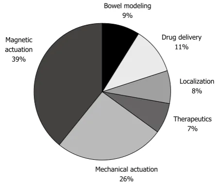
The topic distribution of the 142 selected papers is shown in Figure 1. The themes shown are recurring topics of review papers in the field and we believe
these to be the most influential subfields[5-7]. This list
does not include information from company websites utilized, though some are specified throughout the text.
OVERCOMING LIMITATIONS
Addressing challenges: Then and now
Several solutions to the aforementioned limitations have been developed in the last ten years, though many of these are limited by system complexity or little possibility of gaining regulatory approval for
Bowel modeling 9%
Drug delivery 11%
Localization 8%
Therapeutics 7%
Mechanical actuation 26% Magnetic
[image:2.595.307.529.77.268.2]actuation 39%
translating and commercializing the research. A representation of a futuristic capsule robot from before 2010 is shown in Figure2. This was the vision of Paolo Dario’s research team at Scuola Superiore Sant’Anna and served as the ground for the Versatile Endoscopic Capsule for Gastrointestinal Tumor Recognition and Therapy (VECTOR) project. VECTOR was an integrated project funded by the European Union Commission from 2006-2011 that generated relevant momentum in robotic capsule endoscopy in the last decade. Project outcomes such as[8-12] have resulted
in successful, though mechanically complex, systems. Issues in reliability and repeatability accompany mechanical complexity that inhibits the translation of technology from research to clinical use. Magnetic driving of capsules, a more recent focus of research, does not depend on on-board mechanical components for actuation and thus requires less power storage. The extra space and power can be used for integrating interventional tools and sensing systems, or the capsules can be further miniaturized.
In the following sections, we address what we believe are the pertinent developments in capsule technology that either show great promise, or have been relevant steps in bringing technology to where it is today.
Mobilization of capsule robots
Soon after the introduction of capsule endoscopy into the marketplace, researchers began publishing work on methods for enabling active locomotion (as opposed to traditional passive locomotion enabled by physiological peristalsis). The following section discusses mechanical as well as magnetic research approaches for enabling capsule locomotion.
Mechanical actuation
The most explored methods for inducing active locomotion in capsules are crawling through lumens, inchworm-like actuation, and swimming through the stomach cavity. The study of legged capsule locomotion through the GI tract began in 2004 with Menciassi et al[13] who had a goal of achieving both
tissue contact for force transmission and the ability
to displace these contact points and eventually utilize this combination for both diagnostic and therapeutic
purposes. Soon after the development of this field of
mechanical capsule actuation, Kim et al[14] (also 2004)
developed a shape memory alloy (SMA) actuated capsule robot that biomimiced micro hooks of an insect and traversed via an anchor-and-pull effect.
As part of the aforementioned VECTOR project, Quirini et al[15] and Valdastri et al[10] developed
prototypes of 4, 8, and 12 legged capsules before 2010. The 4-leg device had a single degree of freedom (DoF) where all legs moved simultaneously and a balloon in the front was used to distend collapsed tissue. The 8-leg capsule had ability to open and close 4 legs at a time (one set in front and one set in back) which allowed for synchronizing motion and preventing back slippage while opening a single leg set[8,16].
The 12-leg capsule developed in 2009 operated in a similar manner and was designed for generating large propulsive force while maintaining an ingestible body size[10].
In 2010, Kim et al[17] introduced a paddling based
capsule that traverses a lumen using anchored propulsion. With only one DoF of actuating legs, a linear actuator inside the capsule allows for avoidance of the back slippage problem experienced by Quirini et al[16] The capsule was able to traverse at a satisfactory
rate during in vivo porcine trials, as seen in Table 1, though erythematous mucosal injuries were noted. In 2010 and 2011 Woo et al[18,19] conducted studies
[image:3.595.57.289.80.205.2]on utilizing an electrical stimulus to contract the small intestine. An electrical stimulus applied to the bowel causes contraction and inhibits capsule motion until the electrical potential is released. A 2010 study utilized this phenomenon for resisting peristalsis while the 2011 study targeted active locomotion. During the locomotion study, unexpected contractions were
Figure 2 Artistic representation of a robotic capsule from before 2010 (Credit: Virgilio Mattoli).
Table 1 Select mechanically actuated endoscopic capsules
Ref. Year Actuation mode Highest
testing level (cm/min)Speed Standard
colonoscopy[28] - - - 7
Menciassi et al[13] 2004 Legged Ex vivo NA
Kim et al[14] 2004 Inchworm Ex vivo 1.47
Kim et al[26] 2005 Inchworm Ex vivo 13.4
Wang et al[22] 2006 Inchworm In vitro NA
Quirini et al[8] 2008 Legged Ex vivo 3
Quirini et al[16] 2008 Legged Ex vivo 6
Valdastri et al[10] 2009 Legged Ex vivo 5
Tortora et al[11] 2009 Swimming In vivo 90
Kim et al[17] 2010 Paddling-based In vivo 17
Woo et al[18,19] 2010 Electrical
stimulus
In vitro 17.46
Morita et al[32] 2010 Swimming In vivo 300
Sliker et al[20] 2012 Treads In vivo 18
Lin et al[23] 2012 Inchworm Ex vivo 30
Chen et al[24] 2013 Inchworm In vitro NA
Chen et al[25] 2014 Inchworm Ex vivo 2.3
[image:3.595.307.540.103.309.2]generated sufficient propulsive force.
In 2009, Tortora et al[11] developed a swimming capsule robot for exploration of a fluid filled stomach.
The device includes four propellers to be controlled
via joystick. This system consists of the wirelessly controlled capsule, and triaxial joystick and was tested
in vitro, ex vivo, and in vivo using a porcine model. The same approach for locomotion was also tested with a tridimensional inductive link to transmit a 400 mW power supply to operate the four propellers[12].
Further work on swimming robots was continued by the group into 2014, this time adding a 3.5 mm diameter wireless camera in a capsule that was 22 mm in diameter[31].
In 2010, Morita et al[32] developed a self-propelling
swimming system. The system consisted of a PillCam (Given Imaging Ltd.) with a fin and a mounted magnetic vibratory propeller at the aft of the capsule. A user’s interaction with a joystick changed magnetic field that in turn changed the capsule’s velocity and orientation. This disposable device was successfully tested in a dog, clear images were recorded, and a velocity of 300 cm/min was achieved.
Mechanical actuation summary: The majority of legged locomotion work was done before 2011 and
this field of research halted owing to on-board power
limitations and mechanical complexity. Capsules are generally subject to a size limitation in order to be swallowable. Each mode of locomotion discussed (crawling, inch-worm, and swimming) requires mechanical actuators that take up a large portion of on-board volume that leaves limited space for energy storage, that is needed to drive the actuators. Developments in energy storage technology may resurrect this area of research. Reliability, cost of development, and design hazards such as the need for apertures in the capsule needed for legs or the interaction of legs with mucosa, are all limiting factors in translating legged locomotion technology to clinical use. Other mechanical locomotion schemes that have been developed include rolling on treads that surround a capsule robot, inchworm like capsule motion induced by anchoring mechanisms coupled with on-board linear actuation, and submarine-like motion induced by propellers for gastric diagnostics. None of these technologies has translated to clinical trials so far, and show little promise.
Resisting peristalsis
Peristalsis resistance methods for capsules are generally developed for enabling detailed diagnostic examination of a target site or for enabling therapeutic intervention. This is a subsidiary field to active capsule locomotion - if an effective locomotion method is developed, then that method may be used to inhibit capsule movement. Studied methods that are suitable for lumens include: the use of miniature legs, applying an electric stimulus
to artificially induce tissue collapsing, and mucoadhesive
observed after stimulus application. The authors warned that electric stimulation may cause damage to electrically sensitive organs, such as the liver, and may disturb natural peristalsis[18,19].
In 2012, Sliker et al[20] developed a tethered robotic
capsule endoscope to enable active locomotion within
a collapsed lumen that was able to generate sufficient
biopsy forces and reduce chance of capsule retention. The device was composed of a mid-section housing motors, LEDs, and a camera, that was surrounded on four sides by micro-patterned polydimethylsiloxane threads. During in vivo testing, the capsule traversed through bowel as well as other tissue surfaces inclu-ding mesentery, abdominal wall, and liver. This group has continued work to develop a capsule traction force measurement platform for application in developing robotic capsule colonoscopies[21].
In 2006, Wang et al[22] developed one of the first
capsules to be propelled via both internal mechanical system and electromagnetism in the capsule body. In this preliminary study, a capsule that biomimics the motion of an inchworm was presented. In 2012, Lin
et al[23] developed a 3-leg micro robot for actuation
and anchoring in the intestinal lumen. This robot utilizes this anchoring to generate an inchworm like motion and is thus able to propel itself. Chen et al[24]
developed a wireless inchworm type colon-traversing robot in 2013 with interventional capability that utilizes a power-transmitting coil for power supply. The robot contains air-balloon anchors that may be safer than previously developed capsule legs, and an extending mechanism to produce forward motion. The following year, this same group developed a robot on a similar principle but utilizing extending spiral legs[25]. Other
inchworm concepts include a piezo-actuated concept from 2005[26] on which further optimization work was
done in[27].
As seen in Table 1, the works that monitored traversing speed in vivo include Kim et al[14] (1.47
cm/min), Quirini et al[8] (6 cm/min), Valdastri et al[10] (5
cm/min), Kim et al[17] (17 cm/min), Woo et al[19] (17.46
cm/min), and Lin et al[23] (30 cm/min). A standard
colonoscopy has duration of 21.1 min (SD, 10.4 min) where a mean total distance of approximately 140 cm must be traversed resulting in a desired velocity of approximately 7 cm/min[28]. Not all of these devices
are targeted for colonoscopy; however, this number is useful as a point of reference for comparison.
In 2008, Kósa et al[29,30] proposed a miniature
patches. Given Imaging’s Bravo pH Monitoring System is widely used but is limited to the proximal part of the GI tract and requires manual insertion with a company-provided tool. The only aforementioned device to undergo human trials is a magnetic sheath that is worn over the abdomen to hold a gastric capsule stationary.
The ability to resist peristalsis is critical for enabling therapeutic intervention in capsule endoscopy. Though most actuation techniques may allow for anchoring, research has also been done on capsules passing through the bowel passively and anchoring only once a target area is reached. Multiple methods for anchoring have been studied, some of which are intermittent to enabling active locomotion (e.g., anchoring legs), while others are developed with the sole purpose of anchoring. In 2006, Karagozler et al[33] developed
a legged capsule with biomimetic micro-patterned adhesives for resisting peristalsis in the small bowel. Also in 2006, Dodou et al[34] conducted experiments on
developing mucoadhesive polymers whose frictional properties may be altered reversibly via external stimuli for use in alternative colonoscopic devices. Water and air may be used to detach polymers from the mucosa, but environmentally sensitive polymers are needed to enable repeatability.
In 2008, Glass et al[35] developed, and tested in
vitro, an anchoring mechanism that utilizes pulleys for the distension of 4 legs that contain high friction adhesive pads. In 2009, Tognarelli et al[36] proposed a
force controlled stopping mechanism for esophageal capsule endoscopy. The device contains 3 SMA
elastic flaps that distend upon wireless triggering. An
aforementioned study was done by Woo et al[18] in
2010 in utilizing electrical stimulus to resist peristalsis. In 2011, this group proposed the use of mucoadhesive films for locking surgical assistive tools inside the gastric cavity. They suggest that this technique overcomes the challenges associated with magnetic coupling: exponential magnetic force relationship with distance, inability to use other magnetic devices in the area, and limitations of use in obese patients owing to larger minimum magnet-tool distance. The authors found that as with most adhesives, a greater application area results in higher detachment forces and thus an ability to hold a larger tool[37].
A promising concept was presented in 2014 by Kim et al[38] who introduced a magnetic belt to be
worn over the abdomen to restrict movement of a magnetic capsule in the stomach and allow monitoring of gastric motility. This device was tested on a human volunteer using a MiroCam (IntroMedic) capsule that was embedded with permanent magnetic disks. Once swallowed, the capsule was maneuvered to a desired location after which the magnet was fastened by a belt. Without the need for insufflation during this procedure, and owing to the small size of the
capsule that does not have significant effect on gastric
behavior, motility could be monitored. The magnetic
capsule motility observation was conducted along with cutaneous electroencephalography with a hope of establishing a relation between the two monitoring methods.
Resisting peristalsis summary: Peristalsis resi-stance methods for capsules are generally developed for enabling detailed diagnostic examination of a target site or for enabling therapeutic intervention.
This is a subsidiary field to active capsule locomotion
- if an effective locomotion method is developed, then that method may be used to inhibit capsule movement. Studied methods that are suitable for lumens include: the use of miniature legs, applying an
electric stimulus to artificially induce tissue collapsing,
and mucoadhesive patches. Given Imaging’s Bravo pH Monitoring System is widely used but is limited to the proximal part of the GI tract and requires manual insertion with a company-provided tool. The only aforementioned device to undergo human trials is a magnetic sheath that is worn over the abdomen to hold a gastric capsule stationary.
Magnetic actuation
Though the mechanical actuation method of capsules in the bowel has been made possible in research settings, it is accompanied by complexity of mechanical design as well as having large space and power requirements. Implementing magnetic actuation may allow for further miniaturization of capsules by decreasing dependence on internal mechanical hardware for locomotion and minimizing on-board power needs. Researchers are faced with the challenge of developing reliable magnetic actuation techniques that are not trivial owing to the exponential decrease of magnetic coupling force with distance[39].
Magnetic capsule actuation consists of inducing motion on a capsule with an embedded magnet. Orientation of such capsule may be governed by a uniform magnetic field generated from outside of the patient, while a magnetic field gradient in space
induces relative motion. This field may be generated by
permanent magnets or electromagnets. In comparing the two, electromagnets provide an additional DoF in varying the magnitude of magnetic field, though the
volumetric magnetic flux density generated is lower than
that of permanent magnets. Control of magnetic capsule endoscopes has evolved from mobilizing an ingested capsule via hand-held permanent magnet, to robotic
control in both a static and rotary magnetic field manner.
Hand-held magnet actuation
Carpi et al[40] published one of the first uses of magnetics
in capsule endoscopy in 2007. The group conducted bench trials, using porcine tissue, on M2A® capsules
This preliminary study was a starting point for years of magnetic actuation research to come.
The first human trial on actuating magnetic capsules via hand-held external magnet occurred in 2010 during a study conducted by Swain et al[41] The
compartment for one of the two cameras of a PillCam COLON was replaced by permanent magnets. During this trial, a capsule was moved in the esophagus and stomach and was reported by a volunteer to not cause discomfort. In a similar study by the same group, 10 healthy volunteers swallowed magnetic capsules that were manipulated externally by a hand held magnetic paddle (Given Imaging Ltd.). The authors reported that the esophageal transit time was highly variable and that magnetic forces were not strong enough to hold the capsule against peristalsis near the gastroesophageal junction[42].
In 2010, Valdastri et al[43] developed a method of
steering an endoluminal camera mounted inside a capsule that could be manipulated to achieve viewing in a specific direction by use of both an external magnet and an internal motor coupled with a magnet. The external field was generated by a permanent magnet mounted on a passive hydraulic arm that could be manipulated by hand. This device achieved viewing steps of 1.8° and was shown to be feasible during in vivo porcine trials. In 2012 Lien et al[44] developed the magnetic field navigator system that enables
button-controlled camera view adjustment as a capsule is actuated via a hand-held device. The hand-held navigator contains an embedded motor coupled with a permanent magnet for inducing capsule rotation. Early efforts of magnetic manipulation are starting to be seen in clinic: Jinshan Science and Technology Co. Ltd. (Chongquing, China) has developed the OMOM®
Controllable Capsule System (http://jinshangroup. gmc.globalmarket.com/products/details/omom-controllable-capsule-system-4544573.html). Besides the capsule, image recorder, and image workstation, the system includes a hand held magnetic controller. This system is not available for use in the US.
The SupCam European project is one of the latest developments in hand assisted magnetic capsule control. For use in the colon, Tozzi et al[45] developed
a spherical capsule that contains an internal core and transparent shell that rotate independent of each other so the camera view does not roll. The spherical capsule is actuated via a low cost external magnet that is hung
from a fixture and contains a handle for easy grip. The
group is anticipating testing this in ex vivo and in vivo
settings.
Robotic actuation
Magnetic capsule control consists of actuating a capsule via magnetic gradient manipulation, while responding to sensory data. The evolution of magnetic capsule control will be described by four paradigms with progressive computer assistance: [A] - [D] where
[D] is autonomous.
[A] Hand-held magnetic capsule actuation: as described in the previous subsection, consists of a human closing the control loop by receiving sensory feedback by means of vision, and in response, generating actuating movements. This implementation involves qualitative sensory feedback and inaccuracies in actuation i.e., the user has no feedback on magnetic field strength and must move the actuating magnet iteratively to achieve desired motion; [B] An evolution of such control is the use of computer-assisted actuation where magnetic field may be manipulated achieve desired capsule actuation, or a robot is utilized in moving a permanent magnet. A human is a key part of this control loop and has the responsibility of handling sensory (vision) errors and sending commands to an actuator; [C] Introducing a method of closing the control loop without direct human error handling, but rather human assistance, may further
improve procedural precision. A final evolution of this
control, that we will refer to as closed loop control, would consist of a robot controlled permanent magnet or
computer generated magnetic field that is respondent
to both sensory feedback in human vision as well as proprioceptive capsule feedback, i.e., magnetic field strengths and localization. A human now directs a capsule in response to visual information, while a range of assistive control schemes such as teleoperation, shared control, or autonomous control may direct a capsule to achieve the user defined motion; and [D] A platform with image processing and aforementioned sensing may someday perform procedures in full autonomy, without a human in the control loop.
In 2009, Ciuti et al[46] published the first study on in
vivo actuation of magnetic capsules via external robot. The study compared the effectiveness of an industrial robot for holding an external magnet as opposed to the magnet being held by hand. Ten total in vivo
trials were performed (5 hand-held, 5 robotic). The authors reported an ability to locate a target during hand-held trials, though were unable to approach the target without losing it in view. Using robotic control, more targets were reached (87% ± 13% vs 37% ± 14%) and precision of movement was improved, but mean trial completion time more than doubled (201 ± 24 s vs 423 ± 48 s). The movement precision stems from the ability to adjust the magnet robotically around a particular DoF to tilt or nudge the capsule, while unstable and jerky movements may occur if holding the magnet by hand. Implementing the control scheme as described by [C] above may eliminate such procedural delays. A similar study was conducted using the Niobe magnetic navigation system (Stereotaxis, St. Louis, MO, United States). Carpi et al[47] were
localization was implemented via real time fluoroscopic
imaging and thus requires exposing the patient to ionizing radiation.
In 2012, Arezzo et al[48] conducted a study to
compare the performance of a robotically-driven magnetic capsule for colonoscopy system to standard colonoscopy. The study included 22 subjects (11 experts, 11 trainees) who were to complete a full colonoscopy on an ex vivo swine bowel. Of 672 target pins, 80.9% were detected by capsule procedure as compared to 85.8% by traditional colonoscopy. Detection rate was promising, but the procedure time was nearly three times longer for the capsule procedure (556 ± 188 s vs 194 ± 158 s). The authors also observed that though experts performed better in the traditional procedure, trainees using the robotic platform were able to outperform experts using traditional procedure. Implementing a control scheme as described by [C] may alleviate these procedural delays and potentially eliminate discomfort and need of sedation from the procedure, while maintaining detection rates of standard procedures.
In 2012, Keller et al[49] presented a magnetic capsule
mobility human study on 53 patients and volunteers using a magnetic driving system developed by Olympus Medical Systems Corp. and Siemens Healthcare. This system operates under a control scheme as described in [B], where a human dictates desired motion via
vision feedback and computer assisted actuation occurs by magnetic field specification. This driving system resembles an MRI machine. The group was
able to implement “functions” that caused pre-specified
capsule motions, such as “rotation” or “parking” that, in example, resets the capsule in the middle of the stomach to restart the examination. The authors examined which functions were most pertinent for completing a screening. Yim et al[50] (2012) developed
magnet-driven capsules that are made of soft elastomer
structures. Specific applications of these capsules will be
discussed in the “Therapeutic Capsules” section of this manuscript.
In 2014, Sun et al[51] presented a novel capsule
driving technique that utilizes two actuating magnets mounted on either lateral side of a patient with the magnetic capsule held coplanar. This set-up allows for the use of simpler, and thus less expensive, robotic arms having less DoF. In 2015, Mahoney et al[52]
developed a 5 DoF capsule manipulation method, subject to aforementioned control schema [C], for the screening of a fluid-distended stomach. The group utilized an industrial 6 DoF serial manipulator with a permanent magnet mounted at the end effector for manipulating a capsule that was submerged in a water-filled translucent tank. A vision system was used to obtain 3 DoF position feedback, though this localization method is not applicable in vivo. Capsule control was analyzed while the robot was in singular
configurations and a control scheme was developed to
maintain capsule position while momentarily sacrificing
orientation control.
Spiral capsules and actuation via rotating external
magnet
Magnetic capsule actuation by direct magnetic link with an external magnet may be dangerous if the distance between magnets is abruptly decreased and the coupling could potentially cause tissue perforation. The use of a rotating external magnet to induce screw-like capsule motion has been a subject of research for 13 years. In 2002, Sendoh et al[53] proposed the first
work on actuating a magnetic device with embedded spiral threads for converting rotation to linear actuation and soon afterwards, applied the technique to actuate a capsule endoscope. The device consists of a capsule with an embedded permanent magnet and a spiral thread-like structure wrapped around the capsule’s
exterior. Applying an external rotational magnetic field
causes rotation of the capsule that is converted to linear motion via capsule threads[54]. This method showed
potential and has been a subject of research since then[55-60].
In 2011, Mahoney et al[60-62] proposed a
mathe-matical model to optimize (not necessarily threaded) capsule driving by rotating magnetic field. The motivation behind this work was elimination of potentially hazardous direct magnetic pull that may be experienced by an in vivo magnetic device. The group demonstrated that an external magnet rotating at non constant speed according to a specific open-loop rotation trajectory may relinquish direct attractive force while directly applying a nearly constant lateral force[61]. Later work by this group has investigated
the inverse problem of determining necessary axis of rotation of an external permanent magnet to apply a
force in a specified direction on the internal magnet.
Magnetic actuation summary: The evolution of magnetic capsule control may be characterized as follows: [A]: Human controlled actuation in response to human sensing; [B]: Human controlled actuation with computer assistance in response to human sensing; [C]: Human controlled actuation with computer assistance in response to computer sensing; and [D]: Computer controlled actuation in response to computer sensing: no human in the loop. The field has progressed to
groups having developed control scheme [B]. The first
human studies on magnetic capsule actuation were conducted using an external hand-held permanent magnet that is manipulated in response to live video feedback from the capsule. Though hand-held control
is simpler to implement, studies have shown difficulty
non-manual magnetic driving device to undergo human trials. Several groups are developing robotic driving systems that involve either a rigidly mounted magnet at the robot’s end effector or a rotating magnet. To achieve control schemes [C] and eventually [D], proprioceptive capsule sensing and integration with actuation modes is critical. The field may then progress to
intelligent-assistive actuation. This field shows potential to transfer technology from research field to clinical use in coming
years.
LOCALIZATION OF
IN VIVO
CAPSULES
Knowledge of capsule endoscope position can be considered with either a diagnostic or global respect, both of which are crucial for implementing closed-loop magnetic control. Diagnostic localization refers to monitoring capsule position with respect to anatomical landmarks, while global localization (proprioceptive sensing) refers to monitoring position and orientation in a tridimensional Cartesian frame. Fischer et al[63], in 2004, were one of the first groups to develop a capsule
endoscopy localization system. This algorithm was developed to be used with Given Imaging Ltd.’s M2A®
capsule and is based on measuring the received signal strength (RSS) of a capsule’s wireless transmission data
via 8 external antennas. No extra on board hardware is needed and all implementation is incorporated into Given Imaging Ltd.’s existing video processing software (RAPID®).
In 2006, Hu et al[64] proposed the first magnet
based localization algorithm. This group used a capsule with an internal magnet and 3-axis magnetic field sensors placed outside the body to obtain the capsule’s global position and, unlike Fischer’s work, implementation required additional hardware.
A localization method based on microwave imaging was presented in 2013 by Chandra et al[65] The group
utilized electric properties of tissue as well as tissue positions to aid in resolving the device’s position. Preliminary testing resulted in errors of 1 cm or less and the algorithm is currently limited to 2D application. In 2012, Salerno et al[66] proposed a novel concept
of using magnetic field sensors inside a capsule for localization. Using a pre-computed magnetic field model along with the sensor readings, the group reported position errors of 14 mm, 11 mm, and 19 mm in the X, Y, Z coordinate directions.
In 2013, Yim and Sitti[67] proposed a magnetic
localization method that provides 2.0-3.7 mm in 3D position error. The authors developed the magnetically actuated soft capsule endoscope (MASCE) that uses magnet-induced rolling to move through the stomach and is the device on which the localization study is based. The localization system is based on capsule deformation as the external magnet nears the body.
Miller et al[68] (2012) developed a method for measuring the magnetic field produced by an external
magnet at the center-point of an internal magnet
embedded in a capsule, without interference of the internal magnet. Knowledge of such magnetic field state allows for manipulating the external field in a controlled manner to achieve a control schema as described by [C] above. Popek et al[69] (2013)
proposed a non-iterative localization method that utilizes a rotating magnetic dipole and was shown to produce sufficiently small errors when generating 6 DoF localization data. This method is limited by need of a 30 s post-processing period.
In need of a position and orientation detection
method of capsule in presence of a magnetic field, Di
Natali et al[70] developed a localization algorithm that
employs sensor readings and pre-defined magnetic
field maps. This algorithm provides 6 DoF localization
data where errors are below 5 mm in position and below 19° in orientation, and allows for real time application during capsule actuation via external permanent magnet. To account for magnetic dipole assumption inaccuracies, this group improved this algorithm the following year by utilizing the Jacobian of the magnetic field in relation to capsule pose. This iterative algorithm, operating at faster than 100 Hz, resulted in errors below 7 mm. The authors reported that experimental results suggest that the methodology was effective and reliable at realistic clinical capsule movement speeds[71].
Capsule localization summary: Capsule position and orientation may be described with respect to landmarks in the GI tract (diagnostic) or by a coordinate position in space (global). Diagnostic and global localization systems that provide both the position and orientation of a capsule endoscopy are vital for magnetic actuation. Current capsule endoscopy systems used clinically rely on RSS measurements on video data that is transmitted, which does not communicate the capsule’ s orientation. Several research groups are actively investigation global localization methods, though no in vivo human trials have been performed.
THERAPEUTICS
Currently available capsule endoscopes are still inferior to traditional endoscopes owing to both passive actuation and inability to conduct biopsies or therapeutic intervention[5]. Implementing modes of
tissue intervention is necessary for moving capsule technology forward. The first biopsy capsule was developed in 1957 by William H. Crosby and Heinz W. Krugler. The Intestinal Biopsy Capsule operated by sucking in mucosa and then releasing a spring-actuated rotary knife[72]. This work has provided
motivation for many of the following devices.
A biopsy module to be used in a capsule endoscope was developed in 2005 by Kong et al[73]. This device
operates similarly to Crosby and Krugler’s capsule, via
a cow and rabbit. In 2008, Valdastri et al[74] developed
an interventional surgical clip capsule for use in both capsule endoscopy and natural orifice transluminal endoscopic surgery (NOTES). The capsule was tested
in vivo via porcine model and steered to the target lesion site via external magnetic arm and successful surgical clipping was observed. Again inspired by the design of the Crosby and Krugler capsule, Simi et al[75] (2010) developed a magneto-mechanical elastic
torsion spring biopsy mechanism that was driven via
external magnetic field. Ex vivo trials were performed using excised porcine intestine where the capsule was actuated via hand held magnet from the outside of the intestine. Authors reported that external permanent magnet driving provided stabilization, anchoring, and
sufficient torque to acquire biopsies and further in vivo
testing is needed. Ryou et al[76] (2011) introduced the
self-assembling magnets for endoscopy (SAMSEN) platform for creating a gastric anastomosis to be used in gastrojejunostomy. This device that relies on assembly via laparoscopic graspers provides a simple method for mating tissue walls and has been tested in porcine and human cadaver trials. The same group (Kong et al[77]) that developed the biopsy module in
2005 went on to develop a robotic biopsy device for capsule endoscopes in 2012. This device consists of three modules: a tissue monitoring module, an anchor module, and a biopsy module in vitro testing of this device was successful.
Therapeutics summary: Interventional technology in capsule endoscopy is heavily dependent on development of active locomotion systems for capsules. Nearly all research areas involving therapeutic capsules also involves active locomotion. The rapid development of actuation techniques suggests that applications for therapeutic systems in the capsule will be necessary. This is relevant research, though it has not yet been applied in human trials.
DRUG DELIVERY
Drug delivery capsules have been a prevalent field of study in recent years owing to applications in both treating GI tract diseases and drug absorption studies[78]. Targeting drug delivery sites in the GI tract
may maximize local drug concentration while avoiding drug effects in unwanted areas[79].
In 2008, Hongying et al[80] developed a site-specific delivery capsule that utilizes a heating array,
elastomeric bellows, and piston to release a drug. The main limitation of this device was a small drug reservoir volume that was limited by large size of internal electrical and mechanical components. Animal trials as well as a study on 12 healthy volunteers suggested this device to be reliable. That same year, Cui et al[81] developed a microelectromechanical
systems (MEMS) microcapsule for real-time drug
release and for GI fluid sampling. The hermetic,
non-digestible, and wirelessly controlled capsule was deemed reliable following in vivo trials.
In 2009, Groening et al[82] developed a wirelessly
controlled capsule that utilized hydrogen gas pro-duction induced by current to activate a piston that dispensed a drug. Preliminary testing of this device was successful and authors stated hopes of making such device biodegradable. Later that year, Pi et al[83,84]
introduced a remote controlled capsule that is actuated wirelessly and utilizes combustion for drug release. The telemetry signal ignites microthrusters that actuate a piston and release the drug. Though the device was tested in vivo on beagles, serious safety concerns exist and authors recommend precise calculation of propellant dosage.
Yim and Sitti[50] (2011) developed a compliant
magnetic capsule to be used for drug delivery in the stomach. The device is actuated via magnet induced rolling. The drug release feature is actuated by an external magnet being moved closer to the capsule. Series of devices like this have been developed by this group with focus on the device’s shape manipulation, capsule localization, multimodal drug release, and carrying mirogrippers for biopsy[67,85-87]. In that same
year, Antipina et al[88] described possibilities of utilizing physical influences such as magnetic field, ultrasound
or light for drug delivery. Laser light illumination on microcapsules can be utilized to open nanomembrane
channels for releasing capsule content. Magnetic field
can be utilized to both locomote capsules as well as trigger a drug release mechanism. Pirmoradi et al[89]
(2011) introduced a magnetically controlled MEMS device that utilizes an external magnetic field to deform an internal membrane that increases reservoir pressure that triggers drug release.
Dietzel et al[90] (2012) developed the Magnetic
Active Agent Release System (MAARS) for drug delivery that is triggered via magnetic field rather than
potentially harmful media such as heating elements
or radiation. Magnetic flux forces metallic components
of the capsule together and once demagnetized, a compartment is opened and the drug is released. Human trials on 13 healthy volunteers to release a solid drug (acetylsalicylic acid) showed that the technology is safe and the device is well tolerated. In 2013, Woods et al[91] developed a capsule that housed
a rotatable drug injection needle while having capability to anchor in the intestine via mechanical extensions. With video feedback, the operator can approximate a target site for injecting the drug. Preliminary testing has shown this device to be feasible.
To prevent drugs from passing through and being inadvertently affected by bacteria and pH variance throughout the GI tract, Traverso et al[92] (2014)
peristalsis, which, in turn, releases the drug. In vivo
studies were conducted successfully and the group intends to investigate the possibility of fabricating the microneedles from biocompatible polymers that can become lodged in the mucosa and slowly release a drug.
Medimetrics Personalized Drug Delivery Inc., a Phillips associated company, has introduced a drug delivery capsule for clinical use. Proclaiming itself as the pioneer and global leader in electronic oral drug deli-very, Medimetrics developed the IntelliCap® telemetry
capsule. The IntelliCap® (CE Mark, Medimetrics) is
an intelligent oral drug delivery system that contains a microprocessor, employs real-time wireless com-munication, and contains pH and temperature sensors (www.medimetrics.com). This device has the ability to communicate its approximate location to a physician wirelessly via diagnostic localization by tracking variability of pH values[93]. The Enterion™ capsule
(Quotient Clinical, England) is targeted towards assessing drug absorption in the GI tract and can deliver both liquids and solids. This device underwent 120 clinical studies (4000 capsules) in the United kingdom (http://www.dddmag.com/news/2012/05/quotient-receives-enterion-approval).
Drug delivery summary: A breadth of drug delivery concepts has been developed and human trials were conducted on a piston-based drug release system as well as the MAARS system that utilizes a simple-magnetically driven design and was developed for conducting new drug absorption studies. The IntelliCap (Medimetrics) and Enterion capsules have undergone clinical trials and the former has received the CE mark. Drug delivery via capsules continues to be a thriving area of development and we expect further clinical applications in coming years.
QUANTIFYING DESIGN PARAMETERS
FOR ROBOTIC CAPSULE ENDOSCOPES
Robotic capsules for active locomotion or resisting peristalsis may be designed without awareness of the bowel’s tribological properties, though this may result in inefficient or inadequate systems. Quantification of mechanical response of the bowel to assist with developing endoscopic devices is pertinent to effective capsule design and has been an area of research since 2000[94]. Though groups have studied forces
exerted by traditional endoscopes[95], we focus on
the mucosal forces relevant to the design of capsule robots. In 2007, Kim et al[96] developed an analytical
frictional resistance model for the development of capsule endoscopes to be used in the small intestine, where the main forces applied to the capsule are frictional force owing to capsule weight, stress due to tissue deformation, and peristaltic contractions of the mucosa and the capsule was modelled as a pressure
vessel with induced hoop stress. Results suggest that the frontal shape of the robot was a major contributor in the frictional resistance. Resistive properties of the small bowel were studied in 2010 by Wang et al[97]
and the group concluded that the capsule’s size and moving speed affect the amount of resistive (traction) force experienced. This traction force may be as high as 8 times the magnitude of a capsule’s weight[98]. To
minimize the friction between GI tissue and the surface of a capsule that causes this traction, Ciuti et al[98]
(2011) developed a vibratory magnetic capsule with a wireless on-board inertial sensor and reported traction force reduction of up to 30% during implementation.
In 2011, Terry et al[99] quantified the radial (contact)
component of peristaltic forces generated by the contractions (migrating motor complex) of the mutually orthogonal circular and longitudinal bowel lumen muscle layers. Previous groups have focused on robot-specific modeling and force quantification and thus the purpose of this work was to develop a unanimous characterization of the bowel to assist in general capsule robot development. The group developed a migrating motor complex force sensor (MFS), a biaxial stress/strain apparatus, an in vitro mucous adhesivity protocol, as well as an in vivo tribometer. This work experimentally confirmed the previously developed theoretical force values by Miftahof et al[100]. Further in
vivo studies using the MFS were conducted in[101].
In 2013, Ze Wang et al[102] developed a frictional
resistance determination model for a capsule under radial compression and, as opposed to Kim et al[96] (2007), considered the friction force owing to
peristalsis, modeled as a sine wave. Authors report that modeling peristaltic force allows for better observation of influence of capsule radius, length, speed, and contact angle with the mucosa. Zhang
et al[103] (2014) developed a capsule resistance
force quantification model that where the static and kinetic friction coefficients are analyzed as a stick-slip phenomenon. The group conducted in vitro trials and concluded that the model sufficiently predicted experimental results. A further work quantifying force resistance in magnetically rotating capsules can be seen in[104].
In 2014, Natali et al[105] developed a system to
enable tracking of the resistive force experienced by an untethered capsule. The system consists of a wireless capsule with embedded permanent magnet that is
manipulated by an external magnetic field.
resistive force, though none of these systems have yet been applied clinically. Work has been done to mechanically characterize theoretical models of capsule-tissue interaction, though no unified model exists that may be applied for any lumen traversing capsule.
THE HORIZON: FIVE-YEAR OUTLOOK
We have now seen the evolution of capsule technology from a primitive biopsy capsule in the 1950s, to wireless video capsule endoscopes (2000), to robotic capsule endoscopes (mid-to-late 2000’s). Currently, the majority of work has been in developing active mobility schemes. In this review, we discussed relevant capsule methods for resisting peristalsis, actuation, drug delivery, therapeutics, and bowel modeling. One of the main ubiquitous challenges has been energy storage on board these devices. Owing to the ability to transfer mechanical force through a physical barrier, magnetics have applications in mobility, therapeutics, study of motility, and bowel force quantification. Development of electronic ingestible devices has also expanded from academic laboratories to corporations. The market for “smart pills” is expected to grow to $1 billion by 2017[106]. Studies are being conducted
on observing autofluorescence emission from tissue by use of a capsule. This method may be used for detecting diseased tissue without the use of on-board cameras[107].
A crucial evolution in capsule mobility has been the shift from mechanical actuation techniques, such as legs, to magnetic manipulation which does not consume internal capsule power and does not require internal components for mechanical actuation. Robotic magnetic manipulation seems to have improved precision and reliability when compared to hand-held magnet actuation; however, currently at a cost of longer procedure time[46]. Capsule actuation schemes
range from user dependency in both sensing and direct actuation to fully autonomous robotic control. The near-future direction of the field is to utilize proprioceptive capsule data to assist the user in driving capsules in an intuitive manner. Clinical applications of such technologies seem feasible within the next 10 years.
ACKNOWLEDGEMENTS
Any opinions, findings, and conclusions, or recom-mendations expressed in this material are those of the
authors and do not necessarily reflect the views of the
National Institutes of Health or the National Science Foundation.
REFERENCES
1 Iddan G, Meron G, Glukhovsky A, Swain P. Wireless capsule endoscopy. Nature 2000; 405: 417 [PMID: 10839527 DOI:
10.1038/35013140]
2 Roman S, Mion F, Zerbib F, Benamouzig R, Letard JC, Bruley des Varannes S. Wireless pH capsule--yield in clinical practice.
Endoscopy 2012; 44: 270-276 [PMID: 22275050 DOI: 10.1055/ s-0031-1291541]
3 Better Drug Development for Oral Products. IntelliCap®, Electronic
Capsule for Personalized Oral Controlled Release. MediMetrics-a Philips Company; 2013 (cited Sep 21, 2014). Available from: URL: http://medimetrics.com/upload/file/medimetrics_intellicap.pdf 4 Ruddock AD, Tew GA, Purvis AJ. Reliability of intestinal
temperature using an ingestible telemetry pill system during exercise in a hot environment. J Strength Cond Res 2014; 28: 861-869 [PMID: 24561595 DOI: 10.1519/JSC.0B013E3182AA5DD0]
5 Sliker LJ, Ciuti G. Flexible and capsule endoscopy for screening, diagnosis and treatment. Expert Rev Med Devices 2014; 11: 649-666 [PMID: 25148269 DOI: 10.1586/17434440.2014.941809] 6 Toennies JL, Tortora G, Simi M, Valdastri P, Webster RJ.
Swallowable medical devices for diagnosis and surgery: the state of the art. Proc Inst Mech Eng Part C J Mech Eng Sci 2010; 224: 1397-1414 [DOI: 10.1243/09544062JMES1879]
7 Valdastri P, Simi M, Webster RJ. Advanced technologies for gastrointestinal endoscopy. Annu Rev Biomed Eng 2012; 14: 397-429 [PMID: 22655598 DOI: 10.1146/annurev-bioeng-071811-150006] 8 Quirini M, Scapellato S, Valdastri P, Menciassi A, Dario P. An
approach to capsular endoscopy with active motion. Conf Proc IEEE Eng Med Biol Soc 2007; 2007: 2827-2830 [PMID: 18002583 DOI: 10.1109/IEMBS.2007.4352917]
9 Quirini M, Webster RJ, Menciassi A, Dario P. Design of a Pill-Sized 12-legged Endoscopic Capsule Robot. IEEE ICRA 2007; 1856-1862 [DOI: 10.1109/ROBOT.2007.363592]
10 Valdastri P, Webster RJ, Quaglia C, Quirini M, Menciassi A, Dario P. A New Mechanism for Mesoscale Legged Locomotion in Compliant Tubular Environments. IEEE Trans Robot 2009; 25: 1047-1057 [DOI: 10.1109/TRO.2009.2014127]
11 Tortora G, Valdastri P, Susilo E, Menciassi A, Dario P, Rieber F, Schurr MO. Propeller-based wireless device for active capsular endoscopy in the gastric district. Minim Invasive Ther Allied Technol 2009; 18: 280-290 [PMID: 19707936 DOI: 10.1080/1364 5700903201167]
12 Carta R, Tortora G, Thoné J, Lenaerts B, Valdastri P, Menciassi A, Dario P, Puers R. Wireless powering for a self-propelled and steerable endoscopic capsule for stomach inspection. Biosens Bioelectron 2009; 25: 845-851 [PMID: 19775883 DOI: 10.1016/ J.BIOS.2009.08.049]
13 Menciassi A, Stefanini C, Gorini S, Pernorio G, Kim B, Park JO, Dario P. Locomotion of a legged capsule in the gastrointestinal tract: theoretical study and preliminary technological results.
Conf Proc IEEE Eng Med Biol Soc 2004; 4: 2767-2770 [PMID: 17270850 DOI: 10.1109/IEMBS.2004.1403791]
14 Kim B, Lee S, Park JH, Park JO. Design and fabrication of a locomotive mechanism for capsule-type endoscopes using shape memory alloys (SMAs). IEEE Trans Mechatron 2005; 10: 77-86 [DOI: 10.1109/TMECH.2004.842222]
15 Quirini M, Menciassi A, Scapellato S, Stefanini C, Dario P. Design and Fabrication of a Motor Legged Capsule for the Active Exploration of the Gastrointestinal Tract. IEEE Trans Mechatron
2008; 13: 169-179 [DOI: 10.1109/TMECH.2008.918491] 16 Quirini M, Menciassi A, Scapellato S, Dario P, Rieber F, Ho CN,
Schostek S, Schurr MO. Feasibility proof of a legged locomotion capsule for the GI tract. Gastrointest Endosc 2008; 67: 1153-1158 [PMID: 18513557]
17 Kim HM, Yang S, Kim J, Park S, Cho JH, Park JY, Kim TS, Yoon ES, Song SY, Bang S. Active locomotion of a paddling-based capsule endoscope in an in vitro and in vivo experiment (with videos). Gastrointest Endosc 2010; 72: 381-387 [PMID: 20497903]
18 Woo SH, Kim TW, Cho JH. Stopping mechanism for capsule endoscope using electrical stimulus. Med Biol Eng Comput 2010;
intestinal model for electrically propelled capsule endoscopy.
Biomed Eng Online 2011; 10: 108 [PMID: 22177218 DOI: 10.118 6/1475-925X-10-108]
20 Sliker LJ, Kern MD, Schoen JA, Rentschler ME. Surgical evaluation of a novel tethered robotic capsule endoscope using micro-patterned treads. Surg Endosc 2012; 26: 2862-2869 [PMID: 22538681 DOI: 10.1007/s00464-012-2271-y]
21 Sliker LJ, Kern MD, Rentschler ME. An Automated Traction Measurement Platform and Empirical Model for Evaluation of Rolling Micropatterned Wheels. IEEE Trans Mechatron 2014; 1-9 [DOI: 10.1109/TMECH.2014.2357037]
22 Wang XN, Meng MQH. An Inchworm-like Locomotion Mechanism Based on Magnetic Actuator for Active Capsule Endoscope. In: Proceedings of IEEE/RSJ International Conference on Intelligent Robots and Systems. Beijing: IEEE, 2006: 1267-1272 [DOI: 10.1109/IROS.2006.281887]
23 Lin W, Yan G. A study on anchoring ability of three-leg micro intestinal robot. Engineering 2010; 4: 477-483 [DOI: 10.4236/ eng.2012.48062]
24 Chen W, Yan G, He S, Ke Q, Wang Z, Liu H, Jiang P. Wireless powered capsule endoscopy for colon diagnosis and treatment.
Physiol Meas 2013; 34: 1545-1561 [PMID: 24149981 DOI: 10.10 88/0967-3334/34/11/1545]
25 Chen W, Yan G, Wang Z, Jiang P, Liu H. A wireless capsule robot with spiral legs for human intestine. Int J Med Robot 2014; 10: 147-161 [PMID: 23843276 DOI: 10.1002/rcs.1520]
26 Kim B, Park S, Jee CY, Yoon SJ. An earthworm-like locomotive mechanism for capsule endoscopes. IROS 2005; 2997-3002 [DOI: 10.1109/IROS.2005.1545608]
27 Kwon J, Park S, Park J, Kim B. Evaluation of the critical stroke of an earthworm-like robot for capsule endoscopes. Proc Inst Mech Eng H 2007; 221: 397-405 [PMID: 17605397 DOI: 10.1243/09544119JEIM134]
28 Childers RE, Williams JL, Sonnenberg A. Practice patterns of sedation for colonoscopy. Gastrointest Endosc (cited Apr 19, 2015). Available from: URL: http://www.sciencedirect.com/ science/article/pii/S0016510715000589
29 Kósa G, Jakab P, Jolesz F, Hata N. Swimming capsule endoscope using static and RF magnetic field of MRI for propulsion. ICRA
2008; 2922-2927 [DOI: 10.1109/ROBOT.2008.4543653] 30 Kósa G, Jakab P, Székely G, Hata N. MRI driven magnetic
microswimmers. Biomed Microdevices 2012; 14: 165-178 [PMID: 22037673 DOI: 10.1007/s10544-011-9594-7]
31 De Falco I, Tortora G, Dario P, Menciassi A. An integrated system for wireless capsule endoscopy in a liquid-distended stomach.
IEEE Trans Biomed Eng 2014; 61: 794-804 [PMID: 24216631 DOI: 10.1109/TBME.2013.2290018]
32 Morita E, Ohtsuka N, Shindo Y, Nouda S, Kuramoto T, Inoue T, Murano M, Umegaki E, Higuchi K. In vivo trial of a driving system for a self-propelling capsule endoscope using a magnetic field (with video). Gastrointest Endosc 2010; 72: 836-840 [PMID: 20883863 DOI: 10.1016/J.GIE.2010.06.016]
33 Karagozler ME, Cheung E, Kwon J, Sitti M. Miniature Endoscopic Capsule Robot using Biomimetic Micro-Patterned Adhesives. IEEE/RAS-EMBS BioRob 2006; 105-111 [DOI: 10.1109/BIOROB.2006.1639068]
34 Dodou D, Breedveld P, Wieringa PA. Stick, unstick, restick sticky films in the colon. Minim Invasive Ther Allied Technol 2006; 15: 286-295 [PMID: 17062403 DOI: 10.1080/13645700600929144] 35 Glass P, Cheung E, Sitti M. A legged anchoring mechanism
for capsule endoscopes using micropatterned adhesives. IEEE Trans Biomed Eng 2008; 55: 2759-2767 [PMID: 19126455 DOI: 10.1109/TBME.2008.2002111]
36 Tognarelli S, Quaglia C, Valdastri P, Susilo E, Menciassi A, Dario P. Innovative Stopping Mechanism for Esophageal Wireless Capsular Endoscopy. Procedia Chem 2009; 1: 485-488 [DOI: 10.1016/ J.PROCHE.2009.07.121]
37 Tognarelli S, Pensabene V, Condino S, Valdastri P, Menciassi A, Arezzo A, Dario P. A pilot study on a new anchoring mechanism for surgical applications based on mucoadhesives. Minim Invasive
Ther Allied Technol 2011; 20: 3-13 [PMID: 20704525 DOI: 10.310 9/13645706.2010.496955]
38 Kim HM, Choi JS, Cho JH. A pilot trial of ambulatory monitoring of gastric motility using a modified magnetic capsule endoscope.
J Neurogastroenterol Motil 2014; 20: 261-264 [PMID: 24840379 DOI: 10.5056/jnm.2014.20.2.261]
39 Furlani EP. Chapter 3 - Field Analysis. In: Furlani EP, editor. Permanent Magnet and Electromechanical Devices [Internet]. San Diego: Academic Press, 2001: 97-205 [cited Mar 26, 2015]. Available from: URL: http://www.sciencedirect.com/science/ article/pii/B9780122699511500048
40 Carpi F, Galbiati S, Carpi A. Controlled navigation of endoscopic capsules: concept and preliminary experimental investigations.
IEEE Trans Biomed Eng 2007; 54: 2028-2036 [PMID: 18018698 DOI: 10.1109/TBME.2007.894729]
41 Swain P, Toor A, Volke F, Keller J, Gerber J, Rabinovitz E, Rothsteain RI. Remote magnetic manipulation of a wireless capsule endoscope in the esophagus and stomach of humans (with videos).
Gastrointest Endosc 2010; 71: 1290-1293 [PMID: 20417507 DOI: 10.1016/j.gie.2010.01.064]
42 Keller J, Fibbe C, Volke F, Gerber J, Mosse AC, Reimann-Zawadzki M, Rabinovitz E, Layer P, Swain P. Remote magnetic control of a wireless capsule endoscope in the esophagus is safe and feasible: results of a randomized, clinical trial in healthy volunteers. Gastrointest Endosc 2010; 72: 941-946 [PMID: 20855064 DOI: 10.1016/J.GIE.2010.06.053]
43 Valdastri P, Quaglia C, Buselli E, Arezzo A, Di Lorenzo N, Morino M, Menciassi A, Dario P. A magnetic internal mechanism for precise orientation of the camera in wireless endoluminal applications. Endoscopy 2010; 42: 481-486 [PMID: 20506065 DOI: 10.1055/S-0029-1244170]
44 Lien GS, Liu CW, Jiang JA, Chuang CL, Teng MT. Magnetic control system targeted for capsule endoscopic operations in the stomach--design, fabrication, and in vitro and ex vivo evaluations.
IEEE Trans Biomed Eng 2012; 59: 2068-2079 [PMID: 22581127 DOI: 10.1109/TBME.2012.2198061]
45 Tozzi A, Ciuti G, Lucarini G, Mura M, Quaglia C, Menciassi A, Battaglia G. Supcam European project: preliminary prototyping and test of a new generation active endooscopic colon capsule. Vienna, Australia: United Eur Gastroenterol Week, 2014. Available from: URL: http://endoiov.it/wp-content/uploads/2014/11/Tozzi-A-SUPCAM-P1148-UEGW-Vienna-2014.pdf
46 Ciuti G, Donlin R, Valdastri P, Arezzo A, Menciassi A, Morino M, Dario P. Robotic versus manual control in magnetic steering of an endoscopic capsule. Endoscopy 2010; 42: 148-152 [PMID: 20017088 DOI: 10.1055/S-0029-1243808]
47 Carpi F, Kastelein N, Talcott M, Pappone C. Magnetically controllable gastrointestinal steering of video capsules. IEEE Trans Biomed Eng 2011; 58: 231-234 [PMID: 20952324 DOI: 10.1109/ TBME.2010.2087332]
48 Arezzo A, Menciassi A, Valdastri P, Ciuti G, Lucarini G, Salerno M, Di Natali C, Verra M, Dario P, Morino M. Experimental assessment of a novel robotically-driven endoscopic capsule compared to traditional colonoscopy. Dig Liver Dis 2013; 45: 657-662 [PMID: 23453360 DOI: 10.1016/j.dld.2013.01.025]
49 Keller H, Juloski A, Kawano H, Bechtold M, Kimura A, Takizawa H, Kuth R. Method for navigation and control of a magnetically guided capsule endoscope in the human stomach. BioRob 2012; 859-865 [DOI: 10.1109/BioRob.2012.6290795]
50 Yim S, Sitti M. Design and Rolling Locomotion of a Magnetically Actuated Soft Capsule Endoscope. IEEE Trans Robot 2012; 28: 183-194 [DOI: 10.1109/TRO.2011.2163861]
51 Sun ZJ, Cheng XG, Cao S, Ye B, Zhang HH, Liu S. Multi-applications of a magnet configuration in actuating capsule endoscope. AIM 2014; 106-111 [DOI: 10.1109/AIM.2014.6878063] 52 Mahoney AW, Abbott JJ. 5-DOF Manipulation of an Untethered
Magnetic Device in Fluid using a Single Permanent Magnet. Proc Robot Sci Syst 2014; 1-19 [DOI: 10.1177/0278364914558006] 53 Sendoh M, Ishiyama K, Arai KI. Direction and individual