0022-538X/82/091123-06$02.00/0 Vol.
Detailed Characterization
of
anApparently
Unspliced
,
Herpes Simplex
Virus
Type 1
Gene Mapping
in the Interior
of
Another
KENNETH G. DRAPER, RAYMONDJ. FRINK, AND EDWARDK. WAGNER*
DepartmentofMolecular Biology andBiochemistry, UniiersityofCalifornia, Inrine, Irvline, Cailifornia 92717
Received 19 February 1982/Accepted20 May 1982
We precisely localized the coding region and determined the nucleotide sequenceofa 1.2-kilobase herpes simplex virus type1 mRNA which underlies
the3' region of the 5.2-kilobase mRNA mapping in HindlIl fragment K. This
mRNA, which lacks readily detectable splices, has its own promoter by the
criteriaofidentificationof putative herpes simplexvirus type1control sequences and invitro transcription bya Manley polymerase system.
Previously, we characterized the major mRNA species of herpes simplex virus type 1 (HSV-1) mapping inHindlllfragments K (0.527 to 0.592) and L (0.592 to 0.647) and EcoRI fragment 1 (0.633 to 0.721) (1, 9, lla). Many of these mRNAs overlap each other. As a good example, consider the 6.9-, 5.2-, 1.7-, and 1.2-kilobase (kb) mRNAs mapping between 0.555 and 0.600 in HindIll fragments K and L. The 5.2-kb(3) mRNA has its 5' end in the interior of the6.9-kb(y)mRNAjuxtaposed to the 3' end of the 1.7-kb(y) mRNA(see Fig. 1A). The 6.9-kb mRNA, then, is a result of an inefficient tran-scription termination at the end of the "gene" for the 1.7-kb mRNA. We previously showed that the promoter for the 5.2-kb mRNA maps just upstream ofthe 5' end of this mRNA and shares certain features with the promoter of another( HSV-1 mRNA,thymidinekinase(tk). The 5'end ofthe 1.2-kb ( mRNAunderlyingthe 3' end ofthe 6.9- and 5.2-kb mRNAs could be generated in the same way orcould result from usingthe 5.2-kb mRNA promoter and splicing. Asshown inthis note, the regionjustupstream ofthe 5' end ofthe 1.2-kb mRNA has sequence similarities tothe other HSV-1 promoters previ-ously characterized. Further,
transcription
of this mRNA can be initiatedby using a Manley uninfected cell lysate transcription system (10, 13), indicating that this mRNA is under itsownpromotercontrol and lacksdetectable
splicing.
Welocatedthe 5'end ofthe1.2-kb
(1)
mRNAto be
approximately
300 bases upstream of the HindIll siteat 0.592 and the common 3' endof allthree mRNAstobe 900 bases downstream of thissite(1).Ahigh-resolution
restriction map of theregion inquestion
is shown inFig.
1B.Here,
Hinfl,
Sall,AvaI,HindIll,
PvuII, and DdeIsitesareindicated. WeusedtwoHSV-1 DNAclones
forthesestudies,BamHI-HindIII fragmentO-K (0.576 to 0.592) and HindIII-BamHI fragment
L-0
(0.592 to 0.602). Details of our cloning procedures weredescribed elsewhere(1, 8). We located the 5' end of the 1.2-kb mRNA bySI
nuclease digestion ofhybrids between the Sall-AvaI fragment5'end labeledattheAvaIsiteand infected-cell
polyadenylated
mRNA(viral
mRNA). The basic details of isolation of viral mRNAviatheuseof the PalmiterMg't precipi-tationofpolyribosomes has beendescribed pre-viously (1, 18). We also have described our methods for end labeling viral DNA fragments (9, 10, 15). In this case, BamHI-HindIII frag-ment O-K was digested with AvalI, 5' end la-beled,and redigestedwith Sall,and the labeled fragmentwasstrandseparatedon a nondenatur-ing acrylamide gel as described by Maxam and Gilbert (15). S1 nuclease
digestion
ofhybrids
gave two distinctbands (Fig. 2A); onewas 370 baseslong,
owing
toprotection
of the fulllength
ofthe DNAbythe 6.9-and5.2-kb
mRNAs,
andone was 250 bases
long,
owing
toprotection by
the1.2-kb mRNA. The relative
intensity
ofthetwobands(Fig. 2A)
provides
agood
measureof the abundance ofthe 1.2-kb mRNA relative tothetwolarger
species.
Weprecisely
located the 5' end ofthe mRNA to be 251 bases upstream fromtheAvaI site (data not shown)by
running
the
Si
nuclease-protected fragment
against aMaxam-Gilbertsequenceladder. The 5' endwas
located in the sequence TGTACT at the A
residue(see sequencedata
below).
Wesimilarly
locatedthe 3' end ofthe three colinear mRNAs
tobe
approximately
100 bases downstream(3')
of the
rightmost
DdeI siteby using
strand-separated
DNA fromHindIII-BamHI
fragment
L-0 (0.592
to0.602)
3'-labeled at theDdeI
site. Details of 3' endlabeling
have been described1123
on November 10, 2019 by guest
http://jvi.asm.org/
1124 NOTES
0.576 0.592 0.602 Kpn PVuI Pvut[
Pvull Kpn X Ba BaSa XBgl So H Ba ¢lOOOn, p3-1700
5-6900 58,000
1-1700
F---* /3~-5200
54~~~00d
/3~~~1200
140,000d40,000d
B (5')
(On
Hen Hint
IHinf So Hinf Hinf H PvuIE DDE DDE HinfHinf Hinf 11
H
4-TATA" Ava 1
(ATG)
(tGA
(TGA)FIG. 1. (A) Schematic localization of mRNA spe-cies mapping between 0.567 and 0.620 on the P ar-rangement of the HSV-1 genome. Valuesarebasedon
S1 nucleaseanalysisand in vitro translational studies (1). Arrows indicate direction of transcription and approximate 5' and 3' termini of mRNA species.The totallength of the mRNAs and their relative times of appearance are shown above the arrows, and the size of the polypeptide encoded by each species is shown below the bracket. (B) High-resolution map of the HSV-1region (0.587to0.602)encodingthe1,200-base
P mRNA, with flanking regions. Specific sites are based on results described in the text. Note (i) the location of the 5' and 3' termini of the 1,200-base mRNA, (ii)the presence of the canonical TATAbox,
and (iii)the putative site of the initiation(ATG) and
termination(TGA) codons of the40,000-dalton
poly-peptide encodedby this mRNA. The restriction sites
usedtoderivethe sequencefound inFig.3areshown;
enzymes used were Hinfl (Hinf), Sall (Sa), AilaIl
HindllI (H), PFill, and Ddel (DDE).
FIG. 2. (A)S1 nuclease analysis of mRNA species mapping proximal to the 5' terminus of the
1,200-base
3 mRNA ofHSV-1. Five micrograms of the HSV-1 clone BamHI-HindlIl fragment O-K (0.576 to 0.592) wasdigested withAvI, 5'labeled with [32P]ATP, redigested withSall,andelectrophoresedon a 6% acrylamide gel
bythe methodof Maxam and Gilbert (15). The 370-base piece representing the HSV-1 region of 0.588 to 0.591 wasfurtherpurified, afterdenaturation, byelectrophoresison anondenaturing5%acrylamide (1:50cross-link) gel.Samples containingtheresultant 370-base single strand of DNA and 10 ,ug of viralpolyadenylatedRNA were
hybridized in50p.1of hybridization buffer (80% formamide, 0.4 M Na+, 0.1 M HEPES [N-2-hydroxyethylpipera-zine-N'-2-ethanesulfonic acid] [pH 8.0], 0.005 M EDTA) for 16 h. Si nuclease analysis was done essentially by
the method of Berk and Sharp as described previously (4-6. 9). After nuclease digestion, samples were J. VIROL.
A 0.554
i
AM I..
AMN.
on November 10, 2019 by guest
http://jvi.asm.org/
[image:2.501.53.391.94.582.2] [image:2.501.130.373.261.589.2]NOTES 1125
previously (9; data not shown). The DNA frag-ment ranging between the second DdeI site and the BwlnHI site at 0.602 was separated from a digest by using a nondenaturing 6% acrylamide fragment separation gel as described by Maxam and Gilbert (15).
The precise localization of the 5' end of the 1.2-kb (a) mRNA allowed us to examine the nucleotide sequence upstream of it (see below) and suggested that this mRNA is unspliced, since a TATA box and a CAT box sequence could be identified. Uninfected-cell RNA poly-merase II recognizes the promoter for the 1 tk gene, since this gene is expressed in biochemi-cally transformed cells and in micro-injected amphibian oocytes (17, 24). The early (1) alka-line exonuclease of HSV-1 is similarly express-ible in amphibian oocytes (19), and we suggested that the ability ofuninfected-cell polymerase to recognize 3 promoters is general, since we found that the Manley polymerase recognized major 1 promoters mapping in Hindlll frag-ments K and L (10). The situation with the Manley system is not absolutely clear, since several workers have reported that multiple transcription initiation sitesareseenwhen HSV-1 BaonHI fragment P (0.298 to 0.318). which encodes tk, is used as a template. Our rather moreclear-cut results suggested that we can use
the uninfected system to identify early HSV-1 promoters.
We used three templates in a transcription runoff experiment (10, 21) to determine whether accurate initiation at the 5' end of the 1.2-kb mRNA occurred. We used a commercial sys-tem, in a 50-,ul total volume, with
Qx-[32P]UTP
(400 Ci/mmol; Amersham Corp.) and 4 ,ug of template made by appropriately digesting the BamnHI-HindIIIfragmentO-K (0.576 to 0.592) in the pBR322 vector. Use of template formed by digestion with
BarnHI
andHinidIII
gave a radio-active band 370 bases long (Fig. 2B1 upper panel), a size expected from the position of the 5' endofthe 1.2-kb mRNA (371 bases upstream of theHindIII site; see below). This same band was seen when the template was cut with both Sall andBarnHI-HindIII
suggesting that any recognition sites for polymerase are between the Sallsite (120 bases upstream of the 5' end of the mRNA; see below) and the 5' end of the mRNA. When the template was formed by digestion of the template withAalI,BaitHI,
andHinidlll,
a product 250 bases long was seen. Several other size bands were also seen, notably one about 150 bases long and one 600 bases long; these sizes were unaffected by the enzymes used to cut the template, and we suggest they were due to the presenceof promoters on the pBR portion of thesuspended in 30 ,ul of 98% formamide-1( mM HEPES (pH 8.0). heat denatured at 95°C for 2 min. and
electrophoresedina5%acrylamide (1:40 cross-link). 8 M urea gel. The results of such an analysis areshownin the column markedSal-Ava*. Size standard (S.S.) was prepared by digesting 5 p.g ofpBR322 with Hinfl and
EcoRI restrictionendonucleases and subsequent 5' labeling with 132P]ATP. Marker DNA waxs also denatured
before electrophoresis. Numbers represent sizes of DNA fragments in nucleotides. Exposure was overnight without intensifying screens. (B)In vitro transcription, using the HSV-1 cloneBa,n1HI-HindlIl fragmentO-Kas a template. Fourmicrogramsof the HSV-1 cloneBainHI-Hindlllfragment O-K was suitably digested and used as
atemplate in transcriptional runoffexperiments.using a Manley HeLa cell lysate system (13). Incubations in a
50-,ul total volume were performed as described previously (10). RNA was isolated and analyzed by
electrophoresis at 600 Vfor2.5 h on denaturing5% acrylamide (40:1). 8 M ureagels (1.8 by 40 by 200 mm).
Column 1, Template DNAdigestedwithBatinHI andHindlll: column2. template DNAdigestedwithBamnHI. Sall,andHindlll;column3,templateDNAdigestedwithBamfHll Ailail.andHindlll. Size markersarebasedon comigration with5' end-labeled fragmentsproduced by Hinfl digestionofpBR322.The 250- and 370-base RNA
transcription products are indicated. Exposurewas for 5 days with intensifyingscreens (22). (C) Analysis of
HSV-1 DNA sequence proximal to the 5' end ofthe 1.2-kb l mRNA. Ten micrograms of the HSV-1 clone
BatiHI-HindIll fragment O-K was digested with Sall and Hindlll. The 490-base DNA fragment was 5' end
labeled, strand separated. and sequenced from both ends as descried by Maxam and Gilbert (15). Chains
interruptedatG, G+A,C+T, and C residues werefractionated on an8% acrylamide gel (80by 30)cmby0.5 mm). The sequence shown represents a portion of the strand which was labeled at the Still site (t).588) and
proceeds throughthe 5' end ofthe 1.2-kb mRNA toward theHindlll site. Note thepresence of the ATATAA sequence 29 bases 5' tothe transcription initiation site of the mRNA. (D) Hybridization of unlabeled invitro transcription producttothecodingstrandof HSV-1 DNA. Invitrotranscriptions wereperformedasdescribed above, except equimolar concentrations (0.5 mM) of all four base triphosphates were used, and no labeled
nucleotidewaspresent.Templatewas4FLg of cloned HSV-1 DNA fromBamiiHl-HindlllfragmentO-Kdigested withBainHIandHindlll.Thirtymicroliters ofdilutionbuffer(10)wassubstituted for the HeLa celllysatein the ''noenzyme" reactions. Transcription productswerepurifiedfromtemplateDNAasdescribedinthe text. After
extensive dialysis, each set of transcription products was hybridized with 5-pLg equivalents of the
single-stranded,5'-labeled, 370-base HSV-1 (Avial-Sall)DNAfragmentdescribedinthe legendtoFig. 2A. Hybridiza-tionswerecarriedoutin101.d ofaqueous buffer(0.2 MNa+-0.1 M HEPESIpH 8.01-0.005M EDTA) in sealed
capillarytubesat65°Cfor16h. Reactions werequenched bydilution into400[LIofSI buffer. andSI nuclease
digestion was performed (1, 9). Products of thedigestion weredenatured and analyzedon denaturing gels as
shown in panel B. 5' End-labeled fragments of Hinfl-digested pBR322 were used as size standards.
Exposure wasfor4days with intensifyingscreens (22).
VOL. 43, 19821
on November 10, 2019 by guest
http://jvi.asm.org/
1126 NOTES
template, such as those which have been de-scribed previously (10).
Weconfirmed the synthesisofanRNA prod-uctwith a 5' end very near the location of the 1.2-kb mRNA by using Si nuclease analysis of in vitro transcription products (Fig. 2D). The Manley polymerase lysate was incubated with BamHI-HindIII-digested template and unla-beled base triphosphates (0.5 mM each); a
secondincubationwith noenzymeswascarried out as a control. Radioactive carrier RNA (100,00032p cpm;10
[ig)
wasaddedtotheprod-ucts of reaction, and the RNA was separated from thetemplate by36 h of
centrifugation
ina1-mlCaCl gradientofastartingdensityof 1.6 g/ cm3. Centrifugation wascarried out inanSW60 Ti rotor at 50,000 rpmat17°C. These conditions werefoundadequate to removeessentiallyall of the template DNA from the RNA pellet in separate experiments. The pelleted RNA was
redissolved in water, dialyzed versus 0.1 M NaCl-0.01 MTris (pH 7.4)-0.001 M
EDTA,
and then hybridized under aqueous conditions with strand-separated HSV-1 DNA 5' end labeledatthe AvaI site as described above and in the legend toFig.2D. Hybridization for18 hyielded several
Si-resistant
bands, causedbythe struc-ture of the radiolabeled DNAprobe, as well assome undigested material, as shown by the bands of 220, 240, and 370 bases in both the control and experimental tracks. However, a
band of 250 bases was consistently seenin the enzyme incubation butwas missing in the
con-trol. We concluded that this was owing to syn-thesis of RNA initiating 250 bases upstream of theAvaI site (1).
The in vitro transcription data indicated that the 1.2-kb mRNA has a functional promoter within 120 bases upstream of its 5' end. We carried out Maxam-Gilbert DNA sequence anal-ysis of the DNA encoding this mRNA. We sequenced both strands ofDNA end labeled at the restriction sites indicated in Fig.
lB.
The methodology in which strand-separated DNA was used was exactly as described by Maxam and Gilbert and previously by us (10, 15). An example of the sequence data proceedingdown-stream from the Sall site at 0.588 is shown in Fig. 2C. The sequence is of the same sense as the mRNA, and the sequence ATATAA starting 29bases upstream of the 5' end of the mRNA is indicated.The full mRNA sense sequence of the DNAbeginning 313 bases upstream of the 5' end of the 1.2-kb mRNA and going 1,244 bases downstream is shown in Fig. 3.
The 5' endof the mRNA is, as noted, 29 bases downstream from a putative TATA box se-quence. The sequence TCAC is seen 90 bases upstream. In the 5.2-kb I mRNA, thesequence ACATCis seen at -90 bases (10), and in tk, the
sequence TCATT is seen at -88 bases(16, 23). We suggest that this is aCAT box sequence (2, 3, 14). More significantly, the region between -110 and -97 is mainlyA's andC's, whichwas
also seen inthe twoother 3 mRNAs character-ized, but not with 13y or y mRNAs. Further comparative studies in progress will indicate how significant andgeneral this finding is.
Theonlytranslation start signal in the mRNA sequence is seen atposition 151. The sequence around this translation start (GCCATGG) is a
favored one for eucaryotic translation starts
(12). The reading frame defined by this start
signal is open for 1,017 bases, defining a 339 amino acidpolypeptide whose predicted compo-sition wouldgiveamolecularweight of 37,970,a
value ingood agreement with ourin vitro
trans-lation value of40,000.
The 3' end of the 6.9-, 5.2-, and 1.2-kb mRNAs is verynearthe sequenceATAATAAA. The sequence ATAAAA is found 69 bases downstream of this sequence (data not shown), and the sequence TAATTTTATT is
down-stream another 60 bases from that (data not
shown). Since this region encodes the 3' end of mRNAsonbothstrands, we must regard it asan
efficient polyadenylation region. The sequence AATAAAA has been implicated as the 3' stop signal of both HSV-1 tk (16, 23) and other eucaryotic mRNAs (20). Whether the departure from this nominal sequence seen in the present case is significant is as yet unknown, but AT-rich regions are seen in the areaencoding the 3' ends of several HSV-1 mRNAs around the HindlIl site at 0.647 (unpublished data), so the general character of the polyadenylation signal seems well established.
In light of the fact thatasimilar size polypep-tide immunologically cross-reacting with a 140,000-dalton HSV-2 polypeptide is encoded by the analogous region of HSV-2 and can be detected in some HSV-2-transformed cells (11), it is interesting to ask what the relationship is between the HSV-2 polypeptides and the one encoded by HSV-1. The following facts are clear: inHSV-1, there is no open reading frame upstreamof the 5' end of the 1.2-kb mRNA that would reasonably allow an in-phase fusion pro-tein to be synthesized from the 5.2-kb mRNA. The frame that is used as the phase for the reading of the 1.2-kb mRNA is terminated many times upstream of the translation initiator in the mRNA. Similar results have been reported by Clements and McLauchlan (7). Another area of differencebetween HSV-1 and HSV-2 is that the 1.2-kb mRNA in HSV-2 appears to be a major mRNA (11), whereas the protein analogous to the 140,000-dalton product of the major 5.2-kb a HSV-1 mRNA is not readily seen, and then only late. Therefore, some interesting differences in
J. VIROL.
on November 10, 2019 by guest
http://jvi.asm.org/
VOL. NOTES
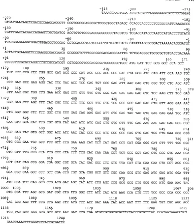
-313 -300 -271
TAAAGGAACTGGA ACGCACGTTTAGGGGAAGCGCCTCCTGGAG
-270 -240 -210 -181
GTGATGAACAGCTCGACGCCAAGCAGGGTT CCGTGGCGCAGGCGCTCCCGTGCCTAGAGC CCACCCACCCCCTCCGGCGATTCAAGACCG
-180 -150 -120 -91
CGTTTGACTACGACCAGAAGTTGCTGATCG ACCTGTGTGCGGACCGCGCCCCCTACGTCG TCGACCATAGCCAATCCATGACCCTGTATG
-90 -60 -30 -1
TCACGGAGAAGGCGGACGGGACCCTCCCAG CCTCCACCCTGGTCCGCCTTCTGGTCCACG CATATAAGCGCGGACTAAAAACAGGGATGT
+1 30 60 90
ACTACTGCAAGGTTCCGAAGGCGACCAACA GCGGGGTCTTTGGCGGCGACGACCCAATTG TCTGCACGGCTGCGCGCTGTGACCGACAAA
+91 120 150 165 171
CCCCCTCCGCGCCAGGCCCGCCGCCATCGT CGTCGCCGTCCCACGCGCTCCCCCGCTGCC ATG GAT TCC GCG GCC CCA GCC
+172 180
TCT CCC CCG
+241
CCC
+310
CTT
+379
GGC
+448
CTG
+517
GAA
+586 CGC +655
GTG
+724
CTC
+793
CCT GAC CCC 315 AAC CGC GAG CTC
GGC GGC 525 GTC GCA
GAG TAC 660 CGG GAA GTT TTG
CAT CAG
CTC TGG GCC CAT 255 GAG AGG TAC TTC
330 TGG CTG GAA ACC
390
AGC TTT TAC CGC 465 CTC TCC GGC CTG
CAC TCG CGC GTG 600 GTG GCC GGC ACC
675 TGC GCC TCC GTT 735
CCG CCA TCG CCC 810 CCG GGA CGA GGC
195
ACG GGC
210
CAT AGC GCG ACG GCG GAC CTA GCG
225 240
ATC CAG ATT CCA AAG TGC
270 285 300 309
TAC ACC TCC CAG TGT CCC GAC ATT AAC CAC CTG CGC TCC CTC AGC ATC
345 360 375 378
GAG CTT GTT TTC GTG GGG GAC GAG GAG GAC GTC TCC AAG CTT TCC GAG
405 420 435 447
TTC CTC TTC GCT TTC CTG TCG GCC GCC GAC GAC CTG GTT ACG GAA AAC
480 495 510 516
TTT GAG CAG AAG GAC ATT CTC CAC TAC TAC GTG GAG CAG GAA TGC ATC
540 555 570 585
TAC AAC ATC ATC CAG CTG GTG CTT TTC CAC AAC AAC GAC CAG GCG CGC
615 630 645 654
ATC AAC CAC CCG GCC ATC CGC GCC CAG GTG GAC TGG CTG GAA GCG CGG
690 705 720 723
CCG GAA AAG CAT TCT CAT GAT CCT CAT CGA GGG CAT CTT TTT TGC CGC
750 765 780 792
CTA CCT TCG CAC CAA CAA CCT TCT GCG GGT CAC CTG CCG GTC AAA CGA
825 840 855 861
CGT GCA CAC GAC GGC CTC GTG TTA CAT CTA CAA CAA CTA CCT AGG CGG
+862 870 885 900 915 930
OCA CGC CAA GCC CCC GCC CGA CCG CGT GTA CGG GCT GTC CGC CAA GCG GTC GAG ATC GAG ATC GGA TTT
+931 945 960 975 990 999
ATC CGA TCC CAG GCa CCG ACG GAC AGC CAT ATC CTG AGC CCG GCa GCG CTG GCG GCC ATC GAA AAC TAC
1000 1005 1020 1035 1050 106' 1068
GTG CGA TTC AGC GCG GAT CGC CTG TTG GGC CTT ATC CAC ATG AAG CCA CTG TTT TCC GCC CCA CCC CCC
1069 1080 1095 1110 1125 1137
GAC GCC AGC TTT CCG CTG AGC CTC ATa TCC ACC GAC AAA CAC ACC AAT TTT TTC GAG TGT CGC AGC ACC
1138 1155 1170 1200 1215
TCC TAC GCC GGG GCG GTC GTC AAC GAT CTG TGA GTGTCGCGGCGCGCTTCTACCCGTGTTTGC CCATAATAAACCTCT
1216 1244
GAACCAAACTTTGGGTCTCATTGTGATTC
FIG. 3. Nucleotide sequence of thenoncoding strand of HSV-1 DNAencodingthe1,200-base 3 mRNA and
its5' and 3' flanking regions. DNAsequenceanalysiswas performed by the procedureof Maxam and Gilbert
(15). ClonedDNAwasend labeledwith[32P]ATP,andisolatedstrands of DNAwerethen
sequenced, using
gels (30by 80cmby0.5mm).All sequencesweredoneatleast induplicate,and both DNA strandsweresequenced. The sequence from 313 bases upstream of the 5' end of the mRNAto1,244bases downstream is shown for the mRNAsensestrand. As discussed in the text, theputativeCAT boxsignalisatposition-90, the AC-rich regionisat-112through -104,and the TATA box is between -29 and -24. The translation termination codon TAA is
seen at-313 anddefinesreadingframe 1. Otherterminator sequences in this frameare seenatpositions201, 285,
312, 471,693,and 855.Translationterminationcodons in frame 2areseenatpositions -290, -216, -156,-102, and -15. The frame isopenedwith theAUG (ATG)codon atposition151 and closedagain atpositions1168,
1204, and1207. This is thereadingframefor the encodedpolypeptide whose molecularweightis 37,970, based
on its calculated amino acid composition. The third potential reading frame has translation terminators at
positions -269, -266,-176,-113, -26, +80, +218, +965,+1040, and +1085. The restriction sites indicated in
Fig.1Bare asfollows: Hinfl sites(GANTC)areatpositions-192, +154, +228, +1004, and +1240; theSallsite
(GTCGAC)isat-121,the AvaI site(CPyCGPuG)isatposition +247;theHindlllsite(AAGCTT) is at position +367;thePvuIIsite(CAGCTG)isatposition+550;andDdelsites(CTNAG)areatpositions+964 and + 1084.
on November 10, 2019 by guest
http://jvi.asm.org/
[image:5.501.56.450.68.524.2]1128 NOTES
temporal control may be
operating
in the two infectious cycles.This work was supported by Public Health Service grant CA11861fromtheNational Institutesof Health.
We thank R. Costa and L. Hallfor assistanceanddiscussion and J.Wagnerforeditingandpreparingthismanuscript.
LITERATURE CITED
1. Anderson, K. P., R. J. Frink, G. B. Devi, B. H.Gaylord, R. H.Costa, and E. K. Wagner. 1981.Detailed charaicter-ization of the mRNAmappingintheHioidlIl fragment K regionoftheherpessimplexvirustype genome. J.Virol. 37:1011-1027.
2. Benoist, C., and P. Chambon. 1981. In vivo sequence requirements oftheSV40 earlypromoterregion. NatuLre (London)290:304-310.
3. Benoist, C., K.O'Hare, R. Breathnach, and P. Chambon. 1980.The ovalbumin gene-sequence ofputative control regions. NucleicAcidsRes. 8:127-142.
4. Berk, A.,and P. Sharp. 1977. Sizing andmatppingof early adenovirusmRNAsbygelelectrophoresisofS1 endonu-clease-digested hybrids.Cell 12:721-732.
5. Berk, A., and P. Sharp. 1978. Splicedearly mRNAs of simianvirus 40. Proc. Natl. Acad. Sci. U.S.A. 75:1274-1278.
6. Berk, A., and P.Sharp. 1978.StructureottheadenoviruLs 2earlymRNAs. Cell 14:695-71 1.
7. Clements,J., and J. McLauchlan. 1981. Control regions involved in the expression of two 3' co-terminal eairly
mRNAs. p. 57.IniA. S. Kaplan, M.Lat Placa. F. Raipp, and B.Roizman(ed.).International workshopon herpes-viruses. Esculapio Publishing Co.,Bologna.Italy. 8. Costa, R. H., B. G. Devi, K. P. Anderson, B. H. Gaylord,
and E. K.Wagner. 1981.Characterizationof amaijorlate herpessimplexvirus type 1 mRNA. J. Virol.38:483-496. 9. Frink, R. J., K. P. Anderson, and E. K. Wagner. 1981. Herpessimplexvirus type I Hio1dIlI fragmentLencodes spliced and complementary mRNA species. J. Virol. 39:559-572.
10. Frink, R., K. Draper, andE.Wagner. 1981. Uninfected cellpolymerase efficiently transcribesearly but not late herpes simplex virus type I mRNA. Proc. Natl. Acad.
Sci.U.S.A. 78:6139-6143.
11. Galloway, D. A., L. C. Goldstein, and J. B. Lewis. 1982. Identification ofproteins encoded by a fragment of herpes simplexvirustype 2 DNA that hastransformingactivity. J.Virol. 42:530-537.
1la.Hall,L.M.,K. G.Draper,R.J. Frink,R. H.Costa,and E. K.Wagner. 1982.Herpessimplexvirus mRNAspecies mappingin EcoRIfragment I. J. Virol.43:594-607. 12. Kozak, M. 1981. Possible role offlankingnucleotides in
recognition of the AUG initiator codon by eukar-yotic ribosomes. Nucleic Acids Res.9:5233-5252.
13. Manley,J.,A. Fire, A.Cano, P. Sharp,andM. Gefter. 1980. DNA-dependent transcription ofadenovirus genes in a soluble whole cell extract. Proc. Nattl. Acad. Sci. U.S.A.77:3855-3859.
14. Mathis, D.,andP.Chambon. 1981. TheSV40 earlyregion TATA boxis required for accurate in vitro initiationof transcription.Nature(London)290:310-315.
15. Maxam,A.,andW. Gilbert.1980.Sequencingend-labeled DNA with base-specific chemical cleavages. Methods
Enzymol.65:499-559.
16. McKnight, S. 1980. The nucleotide sequence and
train-scriptmap of theherpessimplex viruLs thymidine kinase gene. Nucleic Acids Res. 8:5949-5964.
17. McKnight, S., and E. Gavis. 1980. Expression of the herpes thymidinekinase gene inXenopus aiievisoocytes: anassay for thestudyof deletion mutants constructed in vitro. Nucleic AcidsRes.8:5931-5948.
18. Palmiter,R. D. 1974. Mg'- precipitationof ribonucleo-protein complexes. Expedient techniques ftrtheisolation ofundegraded polysomesand messengerribonucleicaicid.
Biochemistry 13:3606-3614.
19. Preston,C.1981.SynthesisofHSV exonuclease in Xeno-puslaevis oocytes, p. 58. In A. S. Kaplain.M. LaPlaicat, F. Rapp, and B.Roizman (ed.), International workshop
on herpesviruses. Esculapio Publishing Co., Bologna. Italy.
20. Proudfoot, N., and G. Brownlee. 1976. 3' Non-coding regionsequences in eucaryotic mRNA.Natture (London) 263:211-214.
21. Rio, D., A. Robbins, R. Myers, and R. Tjian. 1980(. Regulation of SV40) early transcription in vitro bv al purified tumor antigen. Proc. Natl. Acad. Sci. U.S.A. 77:5706-5710.
22. Swanstrom, R., and P. Shank. 1978. X-ray intensifying screensgreatly enhance the detection byautoradiography of the radioactive isotopes 32Pand 1251 Anal. Biochem. 86:184-192.
23. Wagner, M.,J. Sharp, and W. Summers. 1981. Nucleotide sequence of the thymidine kinase gene of herpes simplex virus type 1. Proc.NatI. Acad. Sci.U.S.A.78:1441-1445. 24. Wigler, M., S. Silverstein, L.-S. Lee, A. Pellicer, Y.-C. Cheng, and R. Axel. 1977. Transfer of purified herpes virus thymidine kinase gene to culturedmotusecells. Cell 11:223-232.
J.VIROL.