T E C H N I C A L N O T E
Open Access
Evaluation of six different DNA extraction
methods for detection of
Mycobacterium
tuberculosis
by means of PCR-IS6110: preliminary
study
Isabela Neves de Almeida
1, Wânia da Silva Carvalho
1, Maria Lúcia Rossetti
2, Elis Regina Dalla Costa
2and Silvana Spindola de Miranda
3*Abstract
Background:Developments in molecular detection and strain differentiation of members ofMycobacterium tuberculosiscomplex have proved to be useful. The DNA extraction method influences the amplification efficiency, causing interference on the sensitivity and respective inhibitors. The aim of this study was to standardize a simple and fast DNA extraction method, providing DNA amplification by IS6110-PCR effectively free from undue interferences. Findings:The efficiency of the six different protocols tested inM. tuberculosiscultures has varied from 75% to 92.5%. This preliminary study evaluating the IS6110 PCR sensitivity and specificity was developed in DNA extracted from microscope slides, and achieved 100% of efficiency.
Conclusions:DNA extraction byChelex + NP-40method from both, cultures ofM. tuberculosisand smear slides, resulted in good quantity of interference free DNA, especially in samples with low concentrations of genetic material; therefore, such technique may be used for the molecular diagnosis of tuberculosis.
Keywords:M. tuberculosis, IS6110, DNA extraction, PCR
Background
Tuberculosis (TB) is one of the leading chronic bacterial infections, with mortality of almost 3 million and more than 8 million new cases every year [1]. Early diagnosis, effective treatment, and successful termination of trans-mission are major strategies for TB control [2].
Polymerase Chain Reaction (PCR) is a fast and sensi-tive diagnostic method for detection and identification of M. tuberculosis, especially in samples with poor load of bacilli [3]; and the most used molecular marker for DNA amplification is the IS6110 insertion element; which is specific for the genome ofM. tuberculosis com-plex species [4].
Some studies report different sensitivity and specificity results, when using PCR techniques as diagnostic
investigation tool; such results vary from 11 to 81% [5]. There are many reasons for this frequent variability of PCR results; however, earlier studies have suggested that PCR outcomes largely depend on the DNA extraction method [6].
DNA extraction methods should be effective, simple, and rapid, eliminating also the presence of PCR inhibi-tors during extraction [6,7].
Thus, the aim of this study was to standardize a sim-ple, fast, and less complex method of DNA extraction, providing also DNA amplification by IS6110-PCR free from any undue interference.
Methods Clinical data
The samples included in this study came from patients assisted in the Hospital of Clinics of the Federal University of Minas Gerais (UFMG), in the years of 2010 and 2011. Patients care comprised only individuals older * Correspondence:[email protected]
3Research Group Coordinator FM/UFMG_REDE-TB, Belo Horizonte, Minas Gerais, Brazil
Full list of author information is available at the end of the article
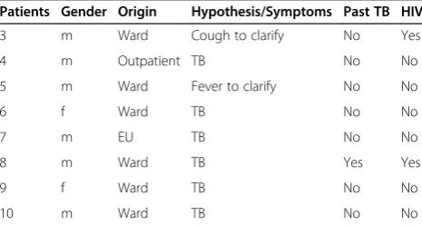
than 18 years, who are presenting TB suspicion and have not started the treatment yet, as demonstrated in Tables 1 and 2. All patients that received TB diagnosis treated with anti-TB drugs.
Samples
Ten bacterial dilutions of M. tuberculosis in Löwenstein-Jensen culture medium have been tested, using serial dilu-tions of 1/10, 1/100 and 1/1000; a total of 230 diludilu-tions were tested with different DNA extraction methods.
Two bacterial suspensions of H37Rv strain of M. tuberculosiswere used as positive control.
Five slides with positive bacilloscopy sputum (one + and two ++) and one with negative bacilloscopy were selected, coming from the Mycobacterial Laboratory of the Hospital of Clinics, at the Federal University of Minas Gerais. The slides were stained by fluorescence method (Auramine O), to be submitted to extraction procedure.
Samples preparation M. tuberculosis inactivation
For DNA extraction from culture, a bacterial suspension containing 1.5 mL of sterile water and around 3 to 5 col-onies of H37Rv strain ofM. tuberculosiswas prepared in Eppendorf tubes. This suspension was inactivated at 100°C for 30 minutes in thermo block, and then centri-fuged at 14,000 rpm for 10 minutes, at 4°C. The super-natant was discarded, and the sediment was used in Extraction Protocols 1, 2, 3, 4, and 5. For Extraction Protocol 6, the initial stage was different and will be de-scribed further.
Preparation and staining of smears slides
The smears were done before the samples’ deconta-mination step with 0.5%N-acetyl-L-cysteine/2% NaOH (NaLC/NaOH) [8].M. tuberculosisand Non-tuberculous Mycobacteria (NTM) species were confirmed after growth in Löwenstein-Jensen culture medium, and identified by
basic biochemical methods, in the Ezequiel Dias Founda-tion Reference Center [9].
Extraction
M. tuberculosis culture DNA extraction
Extraction protocol 1–extraction using phenol-chloroform
DNA fromM. tuberculosisstrains was prepared as follows, after neutralization: a pellet of 400μL of lysozyme solution (10 mg/mL) was added to the suspension, and incubated for 1 hour at 37°C. Afterwards, 20μL EDTA (50 mM) + 400μL of proteinase K solution (10 mg/mL) were added, and the mixture was incubated at 60°C for 1 hour. Then, the solution was stored at -20°C overnight, and the DNA was extracted with phenol-chloroform and ethanol. After freezing, the solution was divided into two parts, and 400 μL of phenol-chloroform were added in each vortex tube and centrifuged at 12,000 rpm for 15 minutes. The supernatant was then transferred to another tube, and 0.6 volume of isopropanol and 1/10 volume of sodium acetate were added; homogenizing then the solution until white color disappearance. The solution was then stored again at -20°C, for 30 minutes. After that, it was centrifuged at 12,000 rpm for 10 minutes, and the supernatant was dis-carded. The pellet was washed twice with 500μL of 70% ethanol, and after complete evaporation 20μL of TE (Tris 100μM + EDTA 50μM) were added to it. The DNA was conserved at 2-8°C, until PCR development. This method was considered the standard method [10].
Extraction protocol 2–extraction using 70% alcohol
500μL of 70% Ethanol were added to a pellet in a tube, and incubated for 2 hours. Then, mycobacterial cells were centrifuged at 13,000 rpm for 10 minutes, the supernatant was discarded, and the pellet was washed twice with sterile distilled water. After washing, the pel-let was resuspended in 500 μL of sterile distilled water within an Eppendorf tube of 1.5 mL, being then used for PCR [11].
Extraction protocol 3–extraction using Chelex 100 + Nonidet P-40 (NP-40)
200 μl of solution were added to a pellet from Chelex suspension containing 5% Chelex-100, 1% Nonidet P-40, 1% Tween 20, and distilled water. After mixing thor-oughly, the samples were maintained for 30 minutes at 100°C. The samples were then centrifuged for 10 mi-nutes at 13,000 g, and the solution was transferred to a fresh microcentrifuge tube and used for PCR [8].
Extraction protocol 4–extraction using Chelex 100
[image:2.595.57.290.591.715.2]200 μL of a solution containing 40 mg Chelex 100 + 100 mL of water were added to a pellet, and the resulting material was maintained at 95°C for 20 minutes. Then, it Table 1 Clinical data from patients included in the
protocols of extraction from cultures
Patients Gender Origin Hypothesis/Symptoms Past TB HIV
3 m Ward Cough to clarify No Yes
4 m Outpatient TB No No
5 m Ward Fever to clarify No No
6 f Ward TB No No
7 m EU TB No No
8 m Ward TB Yes Yes
9 f Ward TB No No
10 m Ward TB No No
was centrifuged at 12,000 g for 15 minutes. The super-natant was used as a DNA source for PCR [12].
Extraction protocol 5–extraction using Chelex 100 + 70% Alcohol
A total of 150μL of ice-cold 70% ethanol was added to a tube, mixed thoroughly, and maintained in ice (−20°C) for 20 minutes. The suspension was then centrifuged at 12,000 rpm for 5 minutes, and 200μl of 20% Chelex solu-tion were added to the pellet. The mixture was then vigor-ously stirred with a shaker, and incubated at 55°C for 1 hour. It was then placed in the vortex again, at high speed, for 10–20 seconds. The tubes were maintained at 100°C for 15 minutes. Then, the tubes were centrifuged at 12,000 rpm for 5 minutes, at 4°C. Afterwards, the super-natant was transferred to a new tube and used for PCR [6].
Extraction protocol 6–extraction using chloroform + CTAB(N-cetyl-N,N,Ntrimethyl ammonium bromide)
Several bacteria loopfuls were resuspended in 400μL of TE 1X buffer, and then inactivated at 80°C for 20 mi-nutes. 50 μL of lysozyme solution were added to the vortex and incubated at 37°C for at least 1 hour, under stirring. 70 μL of SDS 10% and 5 μL of proteinase K were added to the vortex, and incubated at 65°C for 10 minutes. 100μL of NaCl 5 M and 100μL of CTAB/NaCl solution were added to the vortex until the liquid con-tent becoming white; then, it was incubated for 10 mi-nutes at 65°C. 750 μL of chloroform/isoamyl alcohol (24:1) were added to the vortex for 10 seconds, and cen-trifuged at room temperature for 5 minutes, at 14,000 g. The supernatant was transferred to a clean tube and 0.6 volume of isopropanol was added. It was incubated at -20°C for 30 minutes, and centrifuged for 15 minutes at 14,000 g. The supernatant was discarded and the pel-let washed with 1 mL of 70% ethanol and centrifuged for 5 minutes at 14,000 g. To precipitate the DNA, 20– 30μL of TE were added [13].
M. tuberculosisDNA extraction from smear slides of sputum Chelex 100 + Nonidet P-40 (NP-40) on slides
The sputum smears on slides were submitted to Chelex 100+Nonidet P-40 (NP-40) extraction, as it was the most effective method among the six tested ones.
A volume of 25 μL of a suspension containing 5% Chelex-100, 1% Nonidet P-40, 1% Tween 20, and dis-tilled water was spread on the smear slides with a pipette tip. The liquid was transferred to an Eppendorf tube and 75μL of the same suspension were added. After mixing thoroughly, the samples were incubated for 30 minutes at 100°C. The samples were then centrifuged for 10 minutes at 13,000 g, and the solution was transferred to a fresh microcentrifuge tube; then, 5μL were used for PCR [8].
PCR and electrophoresis
PCR was performed in a final volume of 50μL contain-ing 7.0 μL of Buffer (10x), 3.0 μL of MgCl2 (50 mM),
0.2μL of DNTP (25 mM), 25 pmol of each oligonucleo-tide (IS1- 5′ CCT GCG AGC GTA GGC GTC GG 3′ and IS2- 5′ CTC GTC CAG CGC CGC TTC GG 3′) and 0.5μL of Taq DNA Polymerase (500 U) Invitrogen®. Amplification was carried out for 40 cycles, each consist-ing of initial denaturation at 94°C for 2 min, denatur-ation at 94°C for 30 seconds, annealing at 64°C for 2 minutes, extension at 71°C for 1 minute, followed by a final extension at 72°C for 10 minutes. PCR products were analyzed by gel electrophoresis in 2% agarose gel. Target DNA fragments of one hundred twenty-three base pairs (bp) were viewed under UV light.
Quantitative assessment of extracted DNA
The DNA dosing was executed by spectrophotometry (SpectraMax Plus®). The absorbance and DNA concen-tration (ideal 5–100 ng/μL) were evaluated as indicative of nucleic acids purity (ideal ratios: A260/A280≥1. 8 and
A260/A230= 2) [14,15].
A volume of 5μL of concentrated DNA, and the respect-ive dilutions (1/10, 1/100, and 1/1000), were submitted to PCR reaction, and dosed in the spectrophotometer Spectra-Max Plus® (using 2μL as per manufacturer’s instruction).
Findings
M. tuberculosis DNA dosing exhibited concentrations
ranging from 1.048 ng/μL; 4.33 ng/μL; 47.78 ng/μL; 182.5 ng/μL; 112.3 ng/μL; 7.0 ng/μL in protocols 1, 2, 3, 4, 5, and 6, respectively (Figure 1).
The average value of A260/A280and A260/A230ratios is
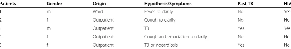
[image:3.595.58.538.101.183.2]demonstrated in Table 3. Table 2 Clinical data of patients included in protocols of extraction from microscope slides
Patients Gender Origin Hypothesis/Symptoms Past TB HIV
1 m Ward Fever to clarify No Yes
2 f Outpatient Cough to clarify No No
3 m Outpatient TB Yes Yes
4 f Outpatient Cough and emaciation to clarify No No
5 f Outpatient TB or nocardiosis Yes No
The efficiency of Extraction Protocols 1, 2, 3, 4, 5 and 6, developed inM. tuberculosis.
Cultures was respectively of: 75% (30/40), 75% (30/40), 90% (36/40), 75% (30/40) 77.5% (31/40), and 92.5% (37/40).
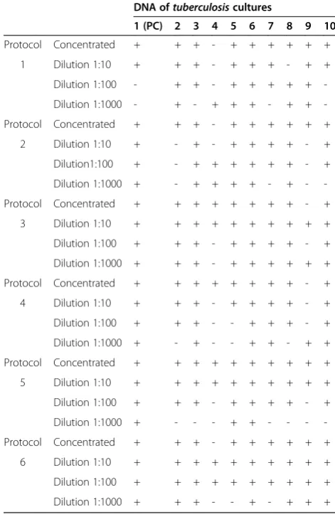
The methods presenting better performance were Extrac-tion Protocols 3 and 6 (90% and 92.5%); however, protocol 3 did not show negative results even in dilution 1/1000.
The amplification results after DNA extraction from tenM. tuberculosiscultures are shown in Table 4.
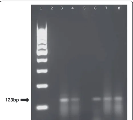
Figure 2 shows electrophoresis of amplifications per-formed with Extraction Protocol 3.
There was amplification in 4 slides withM. tuberculosis
positive cultures, and the slide with M. kansasii culture was not amplified by IS6110-PCR. The DNA extracted from the smear slides using Extraction Protocol 3 are shown in Table 5. Electrophoresis from smear slides amp-lification is shown in Figure 3.
Discussion
DNA dosing from cultures after extraction is an important parameter to quantify the extracted amount and evaluate its quality (purity); however, as evidenced in our results, it is not an absolute parameter [16,17], as large variation was verified either in ‘DNA concentration’ or ‘absorbance’ values, with no relationship with PCR-IS6110 amplification. This study demonstrated the application of several DNA extraction methods, and found an inter-methods efficiency variation of 75% to 92.5% of PCR-IS6110,
which proves that the extraction method does not exert direct influence on the IS6110-PCR effectiveness.
The number of steps and reagents, and the nature of chemical reagents (proteolytic enzymes, organic sol-vents, alcohols, and resins) used show the difference be-tween the six extraction methods. Extraction Protocols 1 and 6 (Phenol-chloroform and Chloroform + CTAB) used lysozyme and proteinase K in the first extraction phase, which led to cellular membranes rupture and release of cytoplasmic components, due to digestion by proteolytic enzymes. Organic solvents as phenol and chloroform were also used, separating DNA from lipids and other biochemical compounds; the addition of ethanol oc-curred as well, to recover and purify the DNA. The use of CTAB allowed to improve the sensitivity, as it has better action on DNA purification [14].
Extraction Protocols 2 and 5 (70% AlcoholandChelex 100 + 70% Alcohol) used 70% alcohol aiming DNA puri-fication. 70% alcohol helps in the removal of organic waste that could act as PCR inhibitors [10]. Within these
[image:4.595.55.291.89.219.2]ng/µL
[image:4.595.302.538.113.475.2]Figure 1Dosages of the extracted DNA using six different methods.Concentrated DNA.“blue square”.
Table 3 Average absorbance with extraction protocols A260/A280 A260/A230
(≥1.8) (<2.0)
Protocol 1 1.96 1.69
Protocol 2 1.24 −0.74
Protocol 3 1.28 0.62
Protocol 4 0.46 0.13
Protocol 5 1.26 0.46
Protocol 6 −1.13 0.21
Table 4 Amplification after extraction of tenM. tuberculosis cultures
DNA oftuberculosiscultures
1 (PC) 2 3 4 5 6 7 8 9 10
Protocol Concentrated + + + - + + + + + +
1 Dilution 1:10 + + + - + + + - + +
Dilution 1:100 - + + - + + + + +
-Dilution 1:1000 - + - + + + - + +
-Protocol Concentrated + + + - + + + + + +
2 Dilution 1:10 + - + - + + + + - +
Dilution1:100 + - + + + + + + - +
Dilution 1:1000 + - + + + + - + -
-Protocol Concentrated + + + + + + + + - +
3 Dilution 1:10 + + + + + + + + + +
Dilution 1:100 + + + - + + + + - +
Dilution 1:1000 + + + - + + + + + +
Protocol Concentrated + + + + + + + + - +
4 Dilution 1:10 + + + - + + + + - +
Dilution 1:100 + + + - - + + + - +
Dilution 1:1000 + - + - - + + - + +
Protocol Concentrated + + + + + + + + + +
5 Dilution 1:10 + + + + + + + + + +
Dilution 1:100 + + + - + + + + - +
Dilution 1:1000 + - - - + + - - -
-Protocol Concentrated + + + - + + + + + +
6 Dilution 1:10 + + + + + + + + + +
Dilution 1:100 + + + + + + + + + +
Dilution 1:1000 + + + - - + - + + +
[image:4.595.57.291.623.732.2]two protocols, elimination of large amounts of organic waste in the concentrated samples is evidenced through the action of 70% alcohol, which is best shown in Ex-traction Protocol 5, as all concentrated cultures were amplified, and resin (Chelex 100) was used to help the DNA removal from cell inside [6].
Extraction Protocols 3 and 4 (Chelex + NP-40 and
Chelex 100) used Chelex 100 resin as principal reagent for DNA extraction, which is associated to thermal shock and NP-40 in order to purify the DNA. These ex-traction protocols are fast; however, Exex-traction Protocol 3, which uses NP-40, had a higher efficiency when DNA was amplified (90%), demonstrating its possible use for saving time as compared to Extraction Protocol 1 (Phenol Chloroform), which has been described by some authors as the gold standard for DNA extraction [18].
Extraction Protocol 3 presented the best culture results in extraction; so, using this protocol in smear slides en-ables the recovery of smaller amounts (dilution 1/1000) of
M. tuberculosisDNA, consuming less reagents, with less stages, and without inhibitors; so, this method is effective, practical, and fast.
In this work, we have noticed that a simpler protocol (Extraction Protocol 3), presenting fewer stages and
consuming less reagents, presented results similar to those from more complex protocols (Extraction Protocol 6), pre-senting also higher efficiency with lower DNA concentra-tions (1/1000). Protocol 3 has been employed by some authors, since it does not use any organic solvent, elimi-nates multiple stages of purification, and uses only two Eppendorf tubes per sample, decreasing so the costs and time spent [7].
The authors used Extraction Protocol 3 (Chelex + NP-40) for DNA extraction in smear slides, due to its higher effi-ciency between protocols tested in culture. The good PCR performance exhibited in samples extracted by Chelex +
NP-40 may be associated to the fact that NP-40 is a
[image:5.595.61.540.89.243.2]123bp
[image:5.595.305.539.474.684.2]Figure 2Agarose gel electrophoresis of amplifications performed with protocol 3.Lane 1 = Molecular marker 100 bp, Lane 2 = Negative Control, Lane 3 = Positive Control, Lane 4 = Sample 8, Lane 5 = Sample 6, Lane 6 = Sample 7, Lane 7 = Sample9, Lane 8 = Sample 5, Lane 9 = Sample 10, Lane 10 = Sample 4, Lane 11 = Sample 3, Lane 12 = Sample 1, Lane 13 = Sample 2.
Table 5 Results of amplifications after extraction of DNA from five smears in sputum slides
AFB AU PCR Culture
Sample 1 ++ pos M. tuberculosis
Sample 2 + pos M. tuberculosis
Sample 3 - pos M. tuberculosis
Sample 4 ++ pos M. tuberculosis
Sample 5 + neg M. kansasii
+ and ++ positives; pos = positive; neg = negative; PCR = Polymerase Chain Reaction; AFB AU = Acid-fast bacilli by Auramine.
123bp
[image:5.595.57.292.633.716.2]detergent with high capacity for breaking lipid-protein interactions, performing so the cells lysis and facilitating DNA release from them; which results in good extraction and reduces the presence of PCR inhibitors. In addition, it is a single stage extraction method, eliminating so the DNA loss that occurs when using multiple stages.
This is a preliminary study with low number of sam-ples; however, as molecular tests are very expensive in developing countries and, therefore, not routinely re-quired as diagnostic tools by physicians, the implant-ation of these protocols in such countries could be a valid alternative to investigate TB; in addition, the cases included in this study are suspected of having TB and, effectively, they proceed from a high complexity hospital of the public health system.
The limitation of smear slides requires the use of a large panel in the study, to confirm sensitivity and speci-ficity values; so, Extraction Protocol 3 would be ideal for laboratories where only bacilloscopy is performed, as is the case of several locations in Brazil. Additionally, it would eliminate the ‘biosafety issue’ during transporta-tion of slides to molecular biology labs. The relevance of amplifying DNA extracted directly from smear slides is because bacilloscopy does not differentiate bacterial spe-cies, while IS6110 sequence amplification allows a rapid identification of these mycobacteria, as it is a specific se-quence of MTC, distinguishing them from MNT. There-fore, new “in house” extraction methods should be standardized and tested, as shown in this study.
Conclusion
The results of this work lead to conclusion thatChelex + NP-40method (Extraction Protocol 3) forM. tuberculosis
DNA extraction from cultures and slides, is able to pro-vide a good quantity of interference free DNA, mainly in samples with low concentrations of genetic material; which justifies its use in the molecular diagnosis of TB.
Abbreviations
M. tuberculosis:Mycobacterium tuberculosis; TB: Tuberculosis;
MTC:Mycobacterium tuberculosiscomplex; PCR: Polimerase chain reaction.
Competing interests
The authors declare no competing interests.
Authors’contributions
SSM, WSC and INA planned and designed the experiments. INA performed the experiments. SSM, WSC and INA analyzed the data. SSM, WSC, INA, MLR and ERDC wrote the paper. All authors read and approved the final manuscript.
Acknowledgements
The Minas Gerais Research Foundation (FAPEMIG) and the Graduate department of the Medicine Faculty, Federal University of Minas Gerais for the financial support. To the collaborators: Lúcia Tavares Paradizi, Ana Lethícia Figueiredo and Lida Jouca, and all integrated laboratories employees.
Author details 1
Federal University of Minas Gerais, Belo Horizonte, Brazil.2State Foundation for Production and Research in Health (FEPPS), Porto Alegre, Brazil.3Research Group Coordinator FM/UFMG_REDE-TB, Belo Horizonte, Minas Gerais, Brazil.
Received: 30 May 2013 Accepted: 16 December 2013 Published: 28 December 2013
References
1. Amim I, Idrees M, Awan Z, Shashid M, Afzal S, Hussain A:PCR could be a method of choice for identification of both pulmonary and extra-pulmonary tuberculosis.BMC Research Notes2011,4:332.
2. Van Der Zandem AGM, Te Koppele Vije EM, Vijay Bhanu N, Van Soolingen D, Schouls LM:Use of DNA Extracts from Ziehl Neelsen Stained Slides for Molecular Detection of Rifampin Resistence and Spoligotyping of Mycobacterium tuberculosis.J Clin Microbiol2003,41(3):1101–1108. 3. Haldar S, Chakravorty S, Bhalla M, Majumdar DS, Tyagi SJ:Simplified detection of
Mycobacterium tuberculosis in sputum using smear microscopy and PCR with molecular beacons.J Med Microbiol2007,56(10):1356–1362. 4. Kolk AHJ, Noordhoek GT, De Leeuw O, Van Emden JDA:Mycobacterium
smegmatis strain for detection of Mycobacterium tuberculosis by PCR used as internal control for inhibition of amplification and for quantification of bacteria.J Clin Microbiol1994,32(5):1354–1356. 5. Barani R, Saranga G, Antony T, Periyasami S, Kindo AJ, Srikanth P:Improved
detection ofMycobacterium tuberculosisusing two independent PCR targets in a tertiary care centre in South India.J Infect Dev Ctries2012,6(1):46–52. 6. Nagdev KJ, Kashyap RS, Deshpande PS, Purohit HJ, Taori GM, Daginawala
HF:Determination of polymerase chain reaction efficiency for diagnosis of tuberculous meningitis in Chelex-100® extracted DNA samples.
Int J Tuberc Lung Dis2010,14(8):1032–1038.
7. Suresh N, Arora J, Pant H, Rana T, Singh UB:Spoligotyping of
Mycobacterium tuberculosis DNA from Archival Ziehl–Neelsen-stained sputum smears.J Microbiol Methods2007,68(2):291–295.
8. Van Der Zanden AGM, Hoeilmann FGC, Weltevred EF, Schouls LM, Van Embden JDA:Simultaneous detection and strains differentiation of Mycobacterium tuberculosis complex in paraffin wax embedded tissues and stained microscopic preparation.J Clin Pathol1998,51(4):209–214. 9. da Saúde M:Manual Nacional de Vigilância Laboratorial da tuberculose e
outras Micobactérias.Brasília: Ministério da Saúde; 2009.
10. Otal I, Martin C, Frebault LVV, Thierry D, Gicquel B:Restriction fragment length polymorphism analysis using IS6110 as an epidemiological marker in tuberculosis.J Clin Microbiol1991,29(6):1252–1254. 11. Elbir H, Mushin A, Babiker A:Short report: a one-step DNA PCR-based
method for the detection ofMycobacterium tuberculosiscomplex grown on Lowenstein-Jensen media.Am J Trop Med Hyg2008,78(2):316–317. 12. Al-Mutairi NM, Ahmad S, Mokkadas E:Performance comparison of four methods for detecting multidrug-resistantMycobacterium tuberculosis
strains.Int J Tuberc Lung Dis2011,15(1):110–115.
13. Leao SC, Martin A, Mejia GI, Palomino JC, Robledo J, Telles MAS, Portaels F: Pratical handbook for the phenotypic and genotypic identification of mycobacteria.Section II - Methodological Procedures2004, 115–116. Printed by Vanden B. Brugues.
14. Sambroock J, Fritsch EF, Maniats T:Molecular Cloning–a laboratory manual on the web. Chapter 8. InVitro Amplification of DNA by the Polymerase Chain Reaction.Edited by Cold Spring Harbor Laboratory Press. USA; 2001:696. 15. Sambroock J, Russell DW:Spectrophotometry of DNA or RNA.Molecular
Cloning2001,A8:20–21. Third edition.
16. Mesquita RA, Anzai EK, Oliveira RN, Nunes FD:Avaliação de três métodos de extraction de DNA de material parafinado para amplificação de DNA genômico pela técnica da PCR.Pesqui Odonto Bras2001,15(4):314–319. 17. Barea JA, Pardini MIMC, Gushiken T:Methodos of DNA extraction from
archived materials and rare sources for utilization in polymer reaction.
Rev bras Hematol Hemoter2004,26(4):274–281.
18. Telenti A, Marchesi F, Balz M, Bally F, Bottger EC, Bodmer T:Fast identification of mycobacteria to species level by polymerase chain reaction and restriction enzyme analysis.J Clin Microbiol1993,31(2):175–178.
doi:10.1186/1756-0500-6-561