BOSNIAN JOURNAL OF BASIC MEDICAL SCIENCES 2010; 10 (1): 89-97
&
Abstract
Pregnancy represents a risk factor in the occurrence of vaginal candidosis. Th e objectives of our study were: to make determination of the microscopic fi ndings of vaginal swab, frequency of Candida species in the culture of pregnant women and patients who are not pregnant, determine the Candida species in all cultures, and to determine the frequency and diff erences in the frequency of C. albicans and other non-albicans species. In one year study performed during year, we tested patients of Gynaecology and Obstetrics clinic of the Clinical Centre in Sarajevo and Gynaecology department of the General hos-pital in Sarajevo. woman included in the study were separated in two groups: pregnant (in the last trimester of pregnancy), and non-pregnant woman in period of fertility. Each vaginal swab was examined microscopically. Th e yeast, number of colonies, and the species of Candida were determined on Sabouraud dextrose agar with presence of antibiotics. For determination of Candida species, we used germ tube test for detection of C. albicans, and cultivation on the selective medium and assimilation tests for detection of non-albicans species. Th e results indicated positive microscopic fi ndings in the test group (,), as well as greater number of positive cultures (,). Th e most commonly detected spe-cies for both groups was C. albicans ( test group . and control group ,). Th e most commonly detected non-albicans species for the test group were C. glabrata (, ) and C. krusei (,), and for the control group were C. glabrata (,) and C. parapsilosis (,). Th e microscopic fi ndings correlated with the number of colonies in positive cultures. In the test group, we found an increased number of yeasts (,), and the pseudopyphae and blastopores by microscopic examination as an indication of infec-tion. In the control group, we found a small number of yeasts (,) , in the form of blastopores, as an indication of the candida colonisation. Our results indicate that gravidity, as the risc factor for incidence of infection, has the signifi cant role in the incidence of vaginal candidosis.
KEY WORDS: vaginal candidosis, pregnancy- risk factor, Candida albicans, non-albicans species
CANDIDA ALBICANS
AND NON-ALBICANS
SPECIES AS ETIOLOGICAL
AGENT OF VAGINITIS IN
PREGNANT AND
NON-PREGNANT WOMEN
Mirela Babić *, Mirsada Hukić
Institute for Clinical Microbiology, University of Sarajevo Clinics Centre, Bolnička , Sarajevo, Bosnia and Herzegovina
BOSNIAN JOURNAL OF BASIC MEDICAL SCIENCES 2010; 10 (1): 90-97frequent attacks of recurrent vulvovaginal candidosis. Point-prevalence studies indicate that Candida species may be isolated from the genital tract of approximately (range -) of asymptomatic, healthy women in the child-bearing age (). Twenty fi ve to of women who are culture positive for Candida species in the vagi-nal area are asymptomatic carriers. Th e natural history of asymptomatic colonization is unknown, although limited human studies suggest that vaginal carriage may continue for several months and perhaps years. In the United States, Candida species is now the second most common cause of vaginal infections, while in Eu-rope it is listed as the primary cause (). Until recently, the problem of vaginal candidosis was often ignored by medics, or treated as an insignificant problem for the female population. It received more attention only after Herman Gardner said: “Vaginitis can cause more inconvenience than any other gynaecological disease. In addition, many mental and emotional problems are associated with vaginitis “(). Documented risk fac-tors of vaginal candidosis are pregnancy (-), use of high estrogen content oral contraceptives, antibiot-ics, steroids, chemotherapeutantibiot-ics, attendence at sexually transmitted diseases clinics and age (). Th e increased secretion of reproductive hormones during pregnancy favors the formation of infection (). High levels of es-trogen provide an increased amount of glycogen in the vagina, furthermore providing a good source of carbon needed for candida growth and their germina-tion. Th ese hormones accelerate the formation of yeast pseudopyphae. Vaginal candidosis is rare in postmeno-pausal women and girls, due to hormonal dependence of vaginal candidosis (). There is a balance between candida, normal bacterial fl ora, and immune defence mechanisms. When this balance is disturbed, coloniza-tion is replaced by infeccoloniza-tion. Vaginal candidosis is one of the mucous opportunistic infections, and also one of the most common infections of the genital system. It is still not defi nitively determined what exactly leads to
disrup-is responsible for the largest number of symptomatic episodes of vaginal candidosis. Percentage of infection that causes C. albicans was high in the past decades, and varied from to (, ). Non-albicans species are most commonly represented by C. glabrata, and C. tropicalis (, ). During the s, the incidence of non-albicans species in the United States amounted to -. Th e objectives of our study were: to analyze the microscopic fi ndings of vaginal swabs, determine the frequency of Candida species occurrence in cultures of pregnant and non-pregnant women; determine Can-dida species in all cultures; determine the frequency of C. albicans occurrence, as well as diff erences in the frequency of C. albicans and other non-albicans species.
Materials and Method
Respondents
Th is one year study performed during year. Th e trial included female patients with Gynaecology-Ob-stetrical Clinics at the University of Sarajevo Clinics Centre, and female patients of the Gynaecology De-partment at the General Hospital in Sarajevo. Study included total of women, divided into two groups. Test group included pregnant women in the last trimester of pregnancy, while control group includ-ed non-pregnant women in the fertile period. In both groups, female patients were handled with clear clinical picture of colpitis, and showed no symptoms that would be indicators of any diseases. Age of fe-male patients in both groups was within the range of to . Criteria for inclusion of subjects into the test group was eight or ninth lunar month of pregnancy.
Research Methodology
Sampling
BOSNIAN JOURNAL OF BASIC MEDICAL SCIENCES 2010; 10 (1): 91-97
under the gynaecological review. A number of women in both groups exhibited symptoms that indicated a possible existence of vaginal candidosis, while others had no symptoms or indications of other problems. Microscopic examination
Examination was done at the Institute for Clinical Microbiology, University of Sarajevo Clinics Centre. Each sample of vaginal swab was stained by methy-lene blue and examined microscopically (x). Pres-ence of regular blastopores, as well as pseudopyphae and blastopores, was determined by this method. Yeast isolation
Mycological vaginal cultures were prepared at Insti-tute for Clinical Microbiology, University of Sarajevo Clinics Centre. Each sample of vaginal swab was fur-ther cultivated on Sabouraud’s agar, with the addi-tion of antibiotics to prevent growth of bacterial fl ora. After hours of incubation at ºC, cultures were separated into positive (growth of yeast) or negative cultures (no yeast growth). Using this approach, we isolated and determined the number of yeast colonies. Yeast identifi cation
a) Germ tube test
Test proves yeast germination, and it is
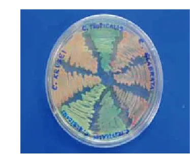
charac-teristic for the detection Candida albicans (un-less when yeast germination is not characteristic). b) Cultivation on the selective medium (Chrom agar) Chrom agar is selective medium, which indicates an increase in the number of non-albicans species based on a colour change. It can be used for identifi cation of individual non-albicans species, as well as C. albicans, if germ tube test was not characteristic. All positive cul-tures from both experimental groups were cultivated on a Chrom agar. After incubation in the thermostat ( hours at ˚C), identification of yeast was pre-formed based on a colony colour. Using this method, we were able to identify the following individual non-albicans species: C. glabrata (dark pink colonies, wet), C. tropicalis (blue colonies, wet), C. krusei (light pink
colonies, dry), and C. albicans (green colonies, wet). c) Yeast assimilation test (API test)
non-albi-
BOSNIAN JOURNAL OF BASIC MEDICAL SCIENCES 2010; 10 (1): 92-97cans species using this test, confi rming the results ob-tained by cultivation on Chrom agar. We also tested for other non-albicans species (egg, Candida kefyr, Candida guilliermondii, Candida famata and others).
Results
Results of microscopic examination
From the test group, of the total number samples of test group, , (/) showed negative using the microscopic examination, while , (/) were observed as positive. From the control group, of the to-tal number samples of control group, , (/) of microscopic fi ndings were determined as negative, while , (/) correlated to positive. Positive microscopic fi ndings (Figure . and .) were more fre-quent in the test group, as opposed to the control group (Graph .). Th e diff erences between the two groups is statistically signifi cant (p=,; p<,).
Results of isolation
Results growth of Candida species on Sabouraud’s agar Test group showed , (/) positive cultures and , (/) negative cultures. From the control group showed , (/) positive cul-tures while , (/) correlated to negative cultures. Positive cultures (Figure .) were more fre-quent in the test group, as opposed to the control group (Graph. ). The difference between the two groups is statistically significant (p=,; p<,). Th e results are separated in four groups: the growth of up to yeast colonies, an increase from to colonies, an increase from to colonies, and yeast colo-nies and more, for both test and control groups. Th e larg-est number of colonies was observed in the tlarg-est group (, , /), while the lowest number of colonies was observed in the control group (, , /) (Table ).
Determination and the frequency of C. albicans; germ tube test
The positive germ tube test (Figure ) determined a greater number of C. albicans , (/) in the test group, compared to the control group , (/). Germination test for the test group was negative , (/) of sampled subjects, while , (/) turned negative in the control group (Graph ). The difference between the two groups is statistically significant (p=,; p<,).
Determination and frequency of the non-albicans species on selective medium
Experiment shows the number of positive cultures of non-albicans species in test and control groups,
Colony
number Test group Control group
Positive culture (total no.) <50 16 (16,8 %) 25 (40,4 %) 41 (26,1 %) 50-100 18 (18,9 %) 15 (24,2 %) 33 (21,0 %) 100-200 38 (40,1 %) 13 (20,9 %) 51 (32,5 %) >200 23 (24,2 %) 9 (14,5 %) 32 (20,4 %) Positive culture
(total no.) 95 (100 %) 62 (100 %) 157 (100 %)
BOSNIAN JOURNAL OF BASIC MEDICAL SCIENCES 2010; 10 (1): 93-97
elaborated on a selective medium (Chrom agar) (Fig-ure .). Cultivation on Chrom agar could not be used for identifi ed of all non-albicans species, therefore, we tested all positive cultures using an API test. In the test group, the most commonly identified species were C. glabrata (, , /) and C. krusei (, , /). In the control group, the most commonly detected spe-cies were C. glabrata (,, /) and undetermined
non-albicans species (,, /) (Table ). Th ere is no signifi cant diff erence between the results of identifi ca-tion and frequency to individual non-albicans speciesin relation to the test and control group (p=,; p<,).
Determination and the frequency non-albicans species using assimilation test (API test)
Analysis of all positive culturesfrom the test group (n=) yielded detection of C. glabrata in samples (,), C. krusei in samples (,), C. tropicalis in samples (,), C. parapsilosis in samples (, ), and C. guilliermondii in sample (,).
Analy-Non-albicans
Candida species test group control group C. glabrata 4,2 % (4) 3,2 % (2)
C. tropicalis 2,1 % (2) 1,6 % (1)
C. krusei 3,2 % (3) 1,6 % (1) undetermined
non-albicans species 3,2 % (3) 3,2 % (2)
C. albicans 87,4 % (83) 90,3% (56) Positive culture Candida
species (total no.) 100,0 % (95) 100,0 % (62)
BOSNIAN JOURNAL OF BASIC MEDICAL SCIENCES 2010; 10 (1): 94-97sis of all positive cultures from the control group (n=) yielded detection of C. glabrata in samples (,), C. parapsilosis in samples (,), C. krusei in sample (, ), and C. tropicalis in sample (, ), while C. guilliermondii was not detected, as was the case with the test group (Graph ). Th ere is no signifi -cant difference between the results of identification and frequency to individual non-albicans species in relation to test and control groups (p=,; p>,).
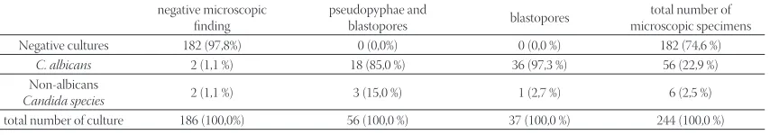
Correlation between the results of germ tube test and the microscopic findings (only blastospores or blastospores with pseudomycelium) of vaginal swab
In the test group, the maximum frequency of pseudo-pyphae and blastopores, established at C. albicans in , (/), and blastopores, at non-albicans species in , (/) of the samples. (Table , Graph. ). In the control group, the maximum frequency of pseu-dopyphae and blastopores, established at non-albicans species in , (/), and blastopores, at C. albicans in , (/) of the samples (Table , Graph. ). There is a significant difference between deter-mined Candida species and microscopic ex-amination for both groups (p=,; p<,).
Discussion
Candida species in the vaginal mucosa was found in of healthy women (). Numerous studies world-wide show that Candida albicans are responsible for the greatest number of symptoms associated with the vagi-nal candidosis. It is important to emphasize that in the past three decades there has been an increasing percent-age of infections caused by non-albicans species. Th ese non-albicans species are often resistant to conventional therapy (). In the he microscopic specimens, simple blastopores are usually detected. Th ere is a delicate bal-ance between Candida species, normal bacterial fl ora, and immune defence mechanisms. When this balance is disturbed, colonization usually results in the occurrence of vaginal candidosis. It is one of the mucous oppor-tunistic infections, and also one of the most common infections of the genital system. Mechanisms that lead to the disruption of this balance are still unclear, and origins of the infection remain uncertain (). Disorders of immune systems and host defence mechanisms con-tribute to the colonization of the genital system by yeast, which is then transformed into the manifest form of
vag-negative microscopic fi nding
pseudopyphae and
blastopores blastopores
total number of microscopic specimens Negative cultures 182 (97,8%) 0 (0,0%) 0 (0,0 %) 182 (74,6 %)
C. albicans 2 (1,1 %) 18 (85,0 %) 36 (97,3 %) 56 (22,9 %) Non-albicans
Candida species 2 (1,1 %) 3 (15,0 %) 1 (2,7 %) 6 (2,5 %)
total number of culture 186 (100,0%) 56 (100,0 %) 37 (100,0 %) 244 (100,0 %) negative microscopic
fi nding
pseudopyphae and
blastopores blastopores
total number of microscopic specimens Negative cultures 108 (90,0%) 0 (0,0%) 0 (0,0 %) 108 (53,2 %)
C. albicans 11 (9,2 %) 53 (94,6 %) 19 (70,3 %) 83 (40,9 %) Non-albicans species 1 (0,8 %) 3 (5,4 %) 8 (30,7 %) 12 (5,9 %) Total number of culture 120 (100,0 %) 56 (100,0%) 27 (100,0%) 203(100,0 %)
BOSNIAN JOURNAL OF BASIC MEDICAL SCIENCES 2010; 10 (1): 95-97
inal candidosis. Th is condition is accompanied by appro-priate symptoms, clinical features and mycological fi nd-ings. Blastopores stretch, and hyfe become formed and carried out the invasion of tissue. Vaginal candidosis is the infection of women in the reproductive age, and it is very rare in postmenopausal women and girls, which indicates the hormone dependence of infection (). Hamad at al. () examined the ability of estro-gens to induce of vaginal candidosis in the case when there is no infection, or if it already exists. The obtained results clearly indicate that estro-gens are able to disrupt the relationship between Candida species and host and lead to infection. During pregnancy, which is listed as a risk factor, vagina is more sensitive, and the infections occur signifi cantly more often. Th is is especially true in the last trimester of pregnancy, due to the increased amount of glycogen in the vagina and high levels of hormones (with estrogens being the most important). It provides a good source of carbon, which favours the growth of Candida species Fidel at al. () demonstrated the effect of reproduc-tive hormones on experimental vaginal candidosis, and put forward the hypothesis that despite high levels of progesterone during pregnancy, the high incidence of vaginitis pregnant women is related to levels of estro-gens, which is in turn considered the primary factor for the occurrence of infection. Vaginal candidosis usu-ally has pronounced symptoms in women in the last trimester of pregnancy, and infection is often repeated. Our results clearly demonstrate signifi cantly increased the number of positive microscopic fi ndings in pregnant women (,, /), compared to , (/) in non-pregnant women. Newmann at al. () investi-gated two groups of respondents, pregnant (n=) and non-pregnant (n=) women, in a similar way, using the microscopic preparation. Their results indicated a greater representation of the positive microscopic findings in the test group (,), compared to the
contro-
BOSNIAN JOURNAL OF BASIC MEDICAL SCIENCES 2010; 10 (1): 96-97detected in , (/) sampled individuals from the test group, and (/) sampled individuals in the control group. Using cultivation on a selective basis, and identifying colonies based on different co-lour and consistency, we were able to detect three non-albicans species: C. glabrata, C. tropicalis and C. krusei. Assimilation test of Candida species (API test) con-firmed these findings, and also determined the other non-albicans spp. (C. parapsilosis and C. guilliermondii). Based on these results, we can conclude that the most common non-albicans species, in tested samples, were C. glabrata, . (/) and C. krusei , (/), while most common species in the control samples were C. glabrata , (/) and C. parapsilosis , (/). Th ere are diff erences in results regarding the representa-tion of C. albicans compared to non-albicans species be-tween the individual studies. In most studies, - of vaginal isolates were C. albicans. Incidence of candidal vulvovaginitis caused by non-albicans species increased during the last decade (). In a study done by Polish re-searchers (), wich was examined genital candidiasis; C. albicans was detected in of positive cultures. Th e most commonly detected non-albicans species were C. glabrata and C. tropicalis. Th ese results are very similar to the results that we obtained in our study. Holland at al. () showed was slightly lower percentage of C. albi-cans detected in the positive cultures of vaginal swabs in pregnant women ( instead of ) but the diff erence has no statistical signifi cance. Our results correlate to results of mentioned studies. Most commonly detected non-albicans species in both experimental groups were C. glabrata (,), C. parapsilosis, C. krusei, and C. tropi-calis. Rihter at al. () showed in their study that the C. albicans was the most commonly detected vagi-nal yeast (,). It should be emphasized that the num-ber of non-albicans species has been growing,
BOSNIAN JOURNAL OF BASIC MEDICAL SCIENCES 2010; 10 (1): 97-97
Conclusion
Th ere is a clear correlation between the frequency of positive microscopic fi ndings and positive cultures in vaginal swabs, in both pregnant and non-pregnant women. Greater frequency of positive cultures and microscopic fi ndings was estab-lished in pregnant compared to non-pregnant women. Th e number of colonies and pseudopyphae and blastopores in microscopical fi ndings, were found in the vaginal swabs of pregnant women. It is characteristic for infection. Identifi ca-tion of species showed that the most common isolated candida, of the pregnant and non-pregnant women was Candida albicans. Frequency of detection C. albicans was greater in pregnant, compared to non-pregnant women. Th e most com-monly identifi ed non-alibcans species, in both groups, were C. glabrata, C. parapsilosis, and C. tropicalis. Correlation between C. albicans and microscopic specimens showed that the C. albicans was mostly commensal for the control group, but for the test group mostly caused a vaginal candidosis.Correlation between non-albicans spp. and microscopic specimens showed, non-albicans spp. were mostly caused a vaginal candidosis for the control group, but for the test group, were commensally.
List of Abbreviations
API C AUX - Yeast identifi cation kit VVC - Vulvovaginal candidosis
References
() Sobel J.D. Candidal vulvovaginitis. Seminars in Dermatology ; (): -.
() Sobel J.D. Genital candidiasis. In: Brodey G.P. Candidiasis. nd ed. New York: Raven Press, ; .
() Kaufman R.N., Freidrich E.G., Garden H.L. Benign diseases of the vulva and vagina, rd ed. Chicago, Year Book Medical Publishers, : -.
() Sobel J.D., Faro S. Force R.W., Foxman B., Ledger W.J., Nyersey P.R., Reed B.D., Summers R. Vulvovaginal candidiasis: Epidemio-logic, diagnostic and therapeutic considerations. Am. J. Obstet. Gynecol., ; (): -.
() Odds F.C. Candidosis of genitalia. In: Odds F.C. (ed): Candida and candidosis, nded. London: Balliere Tindall, ; -. () Sobel J.D. Epidemiology and pathogenesis of recurrent
vulvovagi-nal candidiasis. Am. J. Obstet.Gynecol., ; : . () Numanović F., Hukić M., Nurkić M., Gegić M., Delibegović Z.,
Imamović A., Pasić S. Importance of isolation and biotypization of Gardnarella vaginalis in diagnosis of bacterial vaginosis. Bosn. J. Basic. Med. Sci. ; (): -.
() Babic-Cemalovic M., Ozegovic L., Subasic Đ., Zvizdic A., Seremet M. Morphotypization and genotypization of candida albicans during two attacs of reccurent vaginitis at pregnant woman. Iber-noam. Micologia, ;
() Horowitz B.J., Edelstein S.W., Lippman L. Candida tropicalis vul-vovginitis, Obstet. Gynecol., ; : -.
() Goldacre M.J., Watt B., Loudon N., Milne LJ., Loudon J.D., Vessey M.P. Vaginal microbial fl ora in normal young women. Br. Med. J., ; (): -.
() Babic - Cemalovic M., Babić M., Hukić M. Ožegović L, Arapčić S. Examing of yeasts from Candida species on infl uence of fl ucona-sole. Micologia, ;
() Howard L., Kent M.D. Epidemiology of vaginitis, Am. J. Obstet. Gynecol., ; (): -.
() Hamad M. Estrogen–dependent induction of persistent vaginal candidosis in naive mice. Mycoses, ; (): -. () Fidel P.L., Cutright J., Steel C. Eff ect of reproductive hormones on
experimental vaginal candidiasis, Infection and Immun., ; , ():-.
() Newman G., Kaben U. Blastomycoid fl ora of the urogenital tract in non-pregnant and pregnant patients. Zentralbl. Gynakol. ; (): -.
() Holland J., Young M.L. Vulvovaginal carriage of yeasts other than Candida albicans. Sex. Transm. Infect. ; : -. () Eckert L.O., Hawes S.E. Vulvovaginal candidiasis: clinical
mani-festations, risk factors, management algorithm. Obstet. Gynecol., , ():
() Enweani I.B., Gugnani H.C., Okobia R., Ojo S.B. Eff ect of contra-ceptives on the prevalence of vaginal colonization with Candida species in Endo State, Nigeria. Rev. Iberoam. Micol., ; :
() Nyirsey P. Chronic focal vaginitis: Th e value of cultures. Am. J. Obstet. Gynecol., ; ():
() Sobel J.D., Sabastian F. et.al. Vulvovaginal candidiasis: Epidemio-logic, diagnostic and therapeutic considerations. Am. J. Obstet. Gynecol., ; ():-.
() Evans EG. Diagnostic laboratory techniques in vaginal candido-sis. Br. J. Clin. Pract. Suppl., ; :-.
() Kent H.L. Epidemiology of vaginitis. Am. J. Obstet. Gynecol., ; (): -.
() Kwasniewska J., Jaskolowska A., Choczaj-Kukula A. Candido-sis of genitalorgans in sexual partners. Mikologia lekarska , (): -.
() Rihter S., et al. Antifungal susceptibilities of Candida species caus-ing vulvovaginitis and epidemiology of reccurent cases. J. Clin. Microbiol. ; (): -.
() Odds F.C. Candida and Candidosis, nded, Bailliè Tindall, Lon-don, : .
() Horowitz B.J. Mycotic vulvovaginitis. Am. J. Obstet. Gynecol., ; , (): -.
() Osmanaglaoglu O., et al. Identifi cation of diff erent Candida spe-cies isolated in various various hospitals in Ankara by fungichrom test kit and their diff erentiation by SDS-Page. Turk. J. Med. Sci., , : -.
() Evans E.G. Diagnostic laboratory techniques in vaginal candido-sis. Br. J. Clin. Pract. Suppl., ; : -.