COMPARATIVE EVALUATION OF BONE REGENERATION AFTER SURGICAL REMOVAL OF IMPACTED
MANDIBULAR THIRD MOLARS USING HYDROXYAPATITE WITH AND WITHOUT PLATELET RICH FIBRIN
A Dissertation submitted in
partial fulfillment of the requirem ents for the degree of
MASTER OF DENTAL SURGERY
BRANCH – III
ORAL AND MAXILLOFACIAL SURGERY
THE TAMIL NADU DR. M.G.R. MEDICAL UNIVERSITY
CHENNAI – 600 032
ENDORSEMENT BY HEAD OF THE DEPARTMENT /
HEAD OF THE INSTITUTION
This is to certify that the Dissertation entitled “COMPARATIVE EVALUATION OF
BONE REGENERATION AFTER SURGICAL REMOVAL OF IMPACTED
MANDIBULAR THIRD MOLARS USING HYDROXYAPATITE WITH AND
WITHOUT PLATELET RICH FIBRIN” is a bonafide work done by Dr. ISHA
KOCHAR, Post Graduate student (2013-2016) in the Department of Oral and Maxillofacial
Surgery, under the guidance of Dr. B. SARAVANAN, M.D.S., Ph.D., Professor, Department
of Oral and Maxillofacial Surgery, Tamil Nadu Government Dental College and Hospital,
Chennai – 600 003.
Dr. P. SRIMATHI, M.D.S., DR. B. SARAVANAN, MDS., Ph.D.,
PROFESSOR & H.O.D., PRINCIPAL DEPARTMENT OF ORAL AND
MAXILLOFACIAL SURGERY
CERTIFICATE BY THE GUIDE
This is to certify that Dr. ISHA KOCHAR, Post Graduate student (2013-2016) in the
Department of Oral and Maxillofacial Surgery, Tamil Nadu Government Dental College
and Hospital, Chennai – 600 003 has done this dissertation titled “COMPARATIVE
EVALUATION OF BONE REGENERATION AFTER SURGICAL REMOVAL
OF IMPACTED MANDIBULAR THIRD MOLARS USING HYDROXYAPATITE
WITH AND WITHOUT PLATELET RICH FIBRIN” under my direct guidance and
supervision in partial fulfillment of the regulations laid down by The Tamil Nadu Dr.
M.G.R. Medical University, Chennai – 600 032 for Master of Dental Surgery, Oral
and Maxillofacial Surgery (Branch III) Degree Examination.
PROF. DR. B. SARAVANAN, M.D.S., Ph.D.,
PROFESSOR AND GUIDE
DEPT. OF ORAL AND MAXILLOFACIAL SURGERY
DECLARATION BY THE CANDIDATE
I hereby declare that this dissertation titled “COMPARATIVE EVALUATION OF
BONE REGENERATION AFTER SURGICAL REMOVAL OF IMPACTED
MANDIBULAR THIRD MOLARS USING HYDROXYAPATITE WITH AND
WITHOUT PLATELET RICH FIBRIN” is a bonafide and genuine research work
carried out by me under the guidance of Prof. Dr. B. SARAVANAN, M.D.S., Ph.D.,
Professor, Department of Oral and Maxillofacial Surgery, Tamil Nadu Government
Dental College and Hospital, Chennai - 600003.
Dr. ISHA KOCHAR
Signature of the candidate
TRIPARTITE AGREEMENT
This agreement herein after the “Agreement” is entered into on this day ……….
between the Tamil Nadu Government Dental College and Hospital represented by its
Principal having address at Tamil Nadu Government Dental College and Hospital, Chennai- 600 003 , (hereafter referred to as, ‘the college’)
And
Dr. B. SARAVANAN, aged 55 years working as Professor in the Department of Oral and Maxillofacial Surgery at the college, having residence address at Triplicane,
Chennai (hereafter referred to as the 'Principal Investigator')
And
Dr. ISHA KOCHAR, aged 28 years currently studying as final year Post Graduate student in the Department of Oral and Maxillofacial Surgery, Tamil Nadu Government Dental College and Hospital, Chennai -3 residing at Kellys, Chennai.
(hereafter referred to as the ‘PG and co- investigator’)
Whereas the ‘PG student as part of her curriculum undertakes to research on
“COMPARATIVE EVALUATION OF BONE REGENERATION AFTER SURGICAL REMOVAL OF IMPACTED MANDIBULAR THIRD MOLARS USING HYDROXYAPATITE WITH AND WITHOUT PLATELET RICH FIBRIN” for which purpose the Principal investigator shall act as Principal investigator and the College shall provide the requisite infrastructure based on
availability and also provide facility to the PG student as to the extent possible as a
Co-investigator.
Whereas the parties, by this agreement have mutually agreed to the various issues
Now this agreement witnessed as follows:
1. The parties agree that all the Research material and ownership therein shall
become the vested right of the college, including in particular all the copyright
in the literature including the study, research and all other related papers.
2. To the extent that the college has legal right to do go, shall grant to license or
assign the copyright so vested with it for medical and/or commercial usage of
interested persons/entities subject to a reasonable terms/conditions including
royalty as deemed by the college.
3. The Royalty so received by the college shall be shared equally by all the three
parties.
4. The PG/Research student and PG/Principal Investigator shall under no
circumstances deal with the copyright, Confidential information and
know-how-generated during the course of research/study in any manner whatsoever,
while shall sole west with the college.
5. The PG student and Principal Investigator undertake not to divulge (or) cause
to be divulged any of the confidential information or, know-how to anyone in
any manner whatsoever and for any purpose without the express written
consent of the college.
6. All expenses pertaining to the research shall be decided upon by the principal
investigator/Co-investigator or borne sole by the PG student.(co-investigator)
7. The college shall provide all infrastructure and access facilities within and in
other institutes to the extent possible. This includes patient interactions,
introductory letters, recommendation letters and such other acts required in
this regard.
8. The Principal Investigator shall suitably guide the Student Research right from
selection of the Research Topic and Area till its completion. However the
selection and conduct of research, topic and area research by the Student
Researcher under guidance from the Principal Investigator shall be subject to
the prior approval, recommendations and comments of the Ethical Committee
9. It is agreed that as regards other aspects not covered under this agreement, but
which pertain to the research undertaken by the PG student, under guidance
from the Principal Investigator, the decision of the College shall be binding
and final.
10.If any dispute arises as to the matters related or connected to this agreement
herein, it shall be referred to arbitration in accordance with the provisions of
the Arbitration and Conciliation Act, 1996.
In witness whereof the parties hereinabove mentioned have on this the day
month and year herein above mentioned set their hands to this agreement in
the presence of the following two witnesses.
College represented by its Student Guide
Principal
PG Student
Witnesses
1.
2.
ACKNOWLEDGEMENT
With immense gratitude, I thank my esteemed Guide and Principal, Dr.
B.Saravanan, M.D.S., Ph.D., Professor, Department of Oral & Maxillofacial
Surgery, Tamil Nadu Government Dental College and Hospital for his relentless
encouragement, support and guidance in successfully completing the study. His never
ending patience and affable personality has been a source of encouragement
throughout my post graduate period. His meticulous and systematic way of providing
constructive viewpoints has inspired my entire approach towards the subject and its
practice.
I offer with profound respect and my heartfelt thanks to Dr. P. Srimathi,
M.D.S, Professor and HOD, Department of Oral & Maxillofacial Surgery for her
unrestricted help, valuable guidance and advice throughout the post graduate period.
I express my sincere thanks to Dr. D. Durairaj, M.D.S. and Dr. C. Prasad,
M.D.S., Professors, Department of Oral and Maxillofacial Surgery, for their
encouragement and guidance, throughout my post-graduation period.
I extend my sincere thanks to my co-guides Dr. J. Balaji, M.D.S, Associate
Professorand Dr. M. Arulmozhi, M.D.S, Assistant professor.
I express my special thanks to all other Associate Professors and Assistant
professors of my department for their encouragement and timely help during the
I dedicate this study to my parents for their unwavering support in all my
endeavours. Any accomplishment in my life would not be possible without their
constant love and prayers. I also offer my heartfelt thanks to my friends for their
moral support.
Last but not the least I humbly bow to the Almightywhose blessings have
given me the spiritual strength without which this endeavour would not have been
ABSTRACT
BACKGROUND: Platelet-Rich Fibrin (PRF) is a new second-generation platelet
concentrate, with simplified processing, and no biochemical blood handling. It is a
natural bioactive membrane, which can enhance soft/hard tissue healing, at the same
time, can also protect surgical sites and grafted materials from external aggressions.
The purpose of this study is to compare the efficacy of platelet-rich fibrin in
regeneration of bone when mixed with porous hydroxyapatite and grafted in
mandibular third molar socket with that of plain hydroxyapatite (without PRF), with a
follow up period of 6 months.
MATERIALS AND METHODS: A total of 30 patients divided into 3 groups (10
patients in each group); both male and female, aged between 17 and 45 years, who
had impacted mandibular third molars with similar anatomical position were
randomly selected for this study. The evaluation of osseous regeneration by trabecular
pattern assessment was done for a period of six months. Bone density was calculated
at the end of six months.
RESULTS: An increase in bone density was observed in the PRF with
hydroxyapatite group (Group I) compared to hydroxyapatite (Group II) and control
groups (Group III).
CONCLUSION: Definitive improvement in initiation and acceleration of hard tissue
healing was seen along with increase in bone density in the PRF with hydroxyapatite
group (Group I).
KEY WORDS: Impacted mandibular third molar, Hydroxyapatite, Platelet rich
LIST OF ABBREVIATIONS
ANOVA Analysis of Variance
BMP Bone morphogenic protein
B-TCP β- Tricalcium phosphate
CT Computed tomography
DBM Demineralized bone matrix
ECM Extracellular matrix
FDBA Freeze dried bone allograft
HA Hydroxyapatite
IGF Insulin-like growth factor
IL-1 Interleukin-1
OPG Orthopantomogram
PDGF Platelet derived growth factor
PPP Platelet poor plasma
PRF Platelet rich fibrin
PRP Platelet rich plasma
VAS Visual analog scale
CONTENTS
S. NO.
TITLE
PAGE NO.
1
INTRODUCTION
1
2
AIM AND OBJECTIVES
5
3
REVIEW OF LITERATURE
6
4
SURGICAL ANATOMY
19
5
MATERIALS AND METHODS
26
6
CASE REPORTS
34
7
OBSERVATION AND RESULTS
64
8
DISCUSSION
68
9
SUMMARY AND CONCLUSION
76
10
BIBLIOGRAPHY
Introduction
1
INTRODUCTION
Bone is a specialized form of connective tissue that provides support and
protection for vital structures. The replacement of diseased, injured or worn parts
of bone has been a dream of man for many years. Bone regeneration is a complex,
well-orchestrated physiological process of bone formation, which can be seen
during normal fracture healing, and is involved in continuous remodelling
throughout adult life.
Even though bone has a good healing capacity compared to other tissues,
the regeneration potential is limited in the case of large defects such as after tumor
resection, major fractures, hip implant revision or impaired healing capacity of the
host. It has been shown that after tooth extraction, the surrounding jawbone tends
to recede and atrophy, eventually losing nearly 40% - 60% of its original height
and width within 2-3 years. This atrophy occurs regardless of the patient’s age,
sex or teeth. Socket grafting or ridge preservation is the immediate treatment for
replacement of extracted tooth or teeth. In such cases, the use of bone grafts,
derivates or bone substitutes are indicated to promote healing and regeneration.
The presence of impacted mandibular third molars has been shown to
increase the risk of angle fractures. This increased risk is related to the decreased
amount of bone, resulting in a reduced resistance to traumatic forces. After
removal of such teeth, placement of a bone graft improves the healing capacity of
bone, thereby restoring the original height of the mandible, making it less
susceptible to fracture post trauma.
The ideal bone graft or bone graft substitute should provide three essential
elements:
Introduction
2
(2) osteoinductive properties or factors; and
(3) osteogenic cells.
Osteoconductivity can be defined as the process of infiltration of
capillaries, perivascular tissue, and osteoprogenitor cells from the host bed into
the transplant.
Osteoinduction is the stimulation of a tissue to produce osteogenic
elements. This process is controlled primarily by growth factors such as bone
morphogenetic proteins (BMPs) that are capable of inducing differentiation of
mesenchymal cells into cartilage and bone producing cells.
Osteogenic cells are mesenchymal-type cells, and they can be summoned
from host or graft bone marrow.
Autogenous fresh cancellous and cortical bone (vascularized vs
nonvascularized) grafts are most often used, but other graft materials include
allogenic fresh frozen, freeze-dried, or processed cortical, corticocancellous, and
cancellous grafts. Synthetic or engineered bone graft substitutes are the latest
addition of materials.
The autogenous cancellous bone graft satisfies all three categories most
completely and is considered the “gold standard” of bone transplantation.
Autogenous bone grafts have a number of advantages such as the absence
of rejection and disease transmission, histocompatibility, and they retain viable
osteoblasts that participate in the formation of bone. However a number of
disadvantages were described: lack of sufficient transplantable materials,
Introduction
3
surgical field. In addition, graft survival is unpredictable and its resorption cannot
be foretold.
It is for the reason that in recent years several biocompatible materials
have emerged as substitutes of autogenous bone. These can be classified into two
major groups: organic and synthetic. Hydroxyapatite [Ca10 (PO4)6 (OH)2] is a
synthetic biomaterial. Porous hydroxyapatite has an excellent bone conductive
property which permits the growth of osteogenic cells from existing bone surfaces
into adjacent bone graft material. Studies have shown that hydroxyapatite is well
tolerated by the surrounding tissues with no evidence of inflammation and is
appropriate for application in humans.
Developed in France by Choukroun et al.1 in 2001, Platelet-rich fibrin
(PRF) is a second-generation platelet derivative because, unlike other platelet
concentrates like platelet-rich plasma (PRP), this technique does not require
anticoagulants nor bovine thrombin or any other gelifying agent. PRF is an
immune and platelet concentrate which is obtained on a single fibrin membrane,
containing all the constituents of a blood sample which are favourable for healing
and immunity. It consists of a fibrin matrix polymerized in a tetra molecular
structure, with incorporation of platelets, leucocytes, cytokines, and circulating
stem cells. Clinical studies reveal that this biomaterial would be a favourable
matrix for the development of a coherent healing, without any inflammatory
excess.
Due to its mechanical function and a rapid angiogenesis promoting ability,
PRF membranes are viable material for all types of superficial cutaneous and
mucosal healing. PRF in the form of a platelet gel can be used in conjunction with
Introduction
4
bone growth and maturation, wound sealing and hemostasis, and imparting better
handling properties to graft materials.
The purpose of this prospective clinical study is to evaluate the osseous
regeneration potential of PRF mixed with porous hydroxyapatite when grafted in
Aim and objectives
5
AIM
The purpose of this study is to compare the efficacy of platelet-rich fibrin
in regeneration of bone when mixed with porous hydroxyapatite and grafted in
mandibular third molar socket with that of plain hydroxyapatite (without PRF),
with a follow up period of 6 months.
OBJECTIVES
1. To compare mandibular bone regeneration by placing platelet-rich fibrin
(PRF) with hydroxyapatite (HA) and hydroxyapatite (HA) alone with
control group over a period of six months and thus establish the potential
benefits of PRF and HA in the regeneration of post-extraction alveolar
bone,
2. To identify which of them, PRF mixed with HA or plain HA accelerates
more bone healing and,
3. To determine whether there are differences in the postoperative symptoms
Review of Literature
6
REVIEW OF LITERATURE
BONE REGENERATION AND REPAIR
Ollier et al. (1867) reported that the transplanted periosteum and bone
remain alive and could under proper circumstances become osteogenic. He
believed that viable bone with attached periosteum was the best form of graft to
use.
(Barth et al, 1896) claimed that all transplanted bone marrow and
periosteum die, and are replaced by surrounding tissue. He and Marchand was the
first to use the term “Creeping substitution” to describe the invasion of old bone
by bud like masses of new bone, with resorption of old bone.
BONE GRAFTING
Kazanjian et al. (1952) articulated four clinical rules for bone grafting
situations:
1) The recipient site must have adequate blood supply which is essential to
ensure the survival of any live cells on the surface of the graft.
2) Bone to bone contact must be established between the graft and the bone.
This obviously facilitates “creeping substitution”.
3) Rigid fixation of the fragments must be maintained during the healing
period.
4) The bone graft should be placed inside the healthy tissue. A bacterially
contaminated tissue bed precludes successful grafting.
(Urist and Strates, 1965) showed that a process mimicking embryonic
Review of Literature
7
Kusiak et al (1985), found that in membranous bone grafts, the time for
revascularization was shorter than that in endochondral bone grafts and was
interpreted as a factor that explained the greater resorption in endochondral bone.
(Becker, Becker, & Caffesse, 1994) used DFDBA and autogenous bone
grafts to test the bone forming capacity in extraction sockets and reported that
DFDBA does not induce bone formation and the material is not osteoinductive.
(Skoglund, Hising, & Young, 1996) used bio-oss in combination with a
fibrinogen-thrombin complex as grafting material prior to implant placement in
humans and found that xenograft will act similarly to the host bone, which often
undergoes remodeling at a very slow rate.
IDEAL REQUIREMENT OF THE BONE GRAFTS:
Schullhorn et al. (1996) put forward ideal requirements for a bone graft
material:
1) Biologic acceptability.
2) Predictability.
3) Clinical feasibility.
4) Minimal operative hazards.
5) Minimal post-operative sequelae.
6) Patient acceptance.
HYDROXYAPATITE GRAFT
(Sapkos et al, 1986) implanted hydroxyapatite around teeth which were
Review of Literature
8
material and the results showed that hydroxyapatite was well tolerated by the
surrounding tissues with no evidence of inflammation.
(Donath, Rohrer, & Beck-Mannagetta, 1987) noted dissolution of some
particles with granules of hydroxyapatite present within the cytoplasm of
macrophages by augmenting dense hydroxyapatite in a segment of resected
mandible and the study showed the presence of bone between the particles.
(Holmes, Wardrop, & Wolford, 1988)used porous hydroxyapatite as a
bone graft substitute in mandibular contour augmentation. All grafts become
increasingly resorbed with time whereas all implants remained intact and porous
hydroxyapatite matrix demonstrated long term permanence with maintenance of
contour and also studied the use of porous hydroxyapatite as a substitute for bone
in grafting associated with orthognathic surgical procedure, and the biologic
response to hydroxyapatite matrix confirmed its ability to serve as a bone graft
substitute in clinical applications.
Lang et al (1989) conducted a comparative test of the soluble toxicity of
hydroxyapatite ceramics using human and animal osteoblasts and demonstrated
that the osteoblast-like cell cultures are a highly sensitive test system for materials
with low toxicity.
(Bucholz, Carlton, & Holmes, 1989) implanted autograft or coralline
hydroxyapatite into metaphyseal defects of 40 patients with tibial plateau fractures
and found no differences between the two groups at a minimum of 15 months of
follow up.
(Mehlisch, Leider, & Roberts, 1990) showed bone infiltration throughout
Review of Literature
9
humans and noted that some specimens had scalloped edges, with inflammatory
cells at the periphery of the augmented ridge.
(Friedman et al., 1991) showed that the osseointegrative capacity of
hydroxyapatite cement is advantageous over ceramic hydroxyapatite and studies
indicate that both forms lack significant fibrous encapsulation, produce a minimal
inflammatory response and result in no foreign body giant cell formation, showed
63% osseous replacement within 18 months, and no toxic reactions or increase in
serum calcium and phosphate levels were detected this period and he favored the
term ‘osteoconverison’ for this type of implant, which means the substitution of
the implant with new bone formation without losing a significant volume.
Yu, Wong et al (1992) showed self-setting hydroxyapatite cement as a
novel skeleton drug delivery system for antibiotics to be used to treat
osteomyelitis.
Wise and Merten (1993) showed the role of periosteum in
osteointegration of hydroxyapatite granules. Periosteum is the ideal natural
membrane to stop invasion of fibroblasts and other cells that prevent bone
formation. If kept intact, new bone may proceed from the base at the bone-implant
interface throughout the whole mass of biomaterials and upto the periosteal layer,
independent of the severity of atrophy.
(Ingram & Bonde, 1994) experimentally showed the antibacterial effects
of porous granular hydroxyapatite without clarifying its mechanism.
Gosain A. K. et al (1997) reported the use of hydroxyapatite paste to
correct temporal hollowing deformities after cranial vault remodeling in a growing
Review of Literature
10
Frankenburg et al (1998), introduced Norain CRS (Craniofacial repair
system) (Norton Corporation, Cupertino, calif) as a new carbonated calcium
phosphate paste in the canine distal tibia, replacing the host bone. Crystalline
Norain is composed of monocalcium phosphate, monohydrate, alpha-tricalcium
phosphate, and calcium carbonate, mixed with sodium phosphate solution. Within
24 hours, it reaches a final compressive strength of 55 mpa and a tensile strength
of 2.1 mpa. The final strength of carbonated calcium phosphate paste is superior
to that of hydroxyapatite paste.
(Burstein & Cohen, 1999) used bone source (Liebinger Corporation,
Kalamazoo) as the first hydroxyapatite paste in craniofacial surgery. Bone source
is a combination of tetracalcium phosphate and dicalcium phosphate that sets
isothermally when mixed with a sodium phosphate solution. Paste becomes firm
in approximately 20 minutes but requires 24 hours to reach its maximum
compressive strength of 37mpa and a tensile strength of 20mpa.
Hobar et al (2000) popularized the use of hydroxyapatite granules by
mixing them with autogenous blood and microfibrillar collagen and this enhances
manipulation of the formed paste to achieve better contours and also aids in
hemostasis.
(Hallman, Hedin, Sennerby, & Lundgren, 2002) evaluated the survival
rate of implants in maxillary sinuses augmented with bovine hydroxyapatite and
autogenous bone 6 months before implant surgery and estimated dimensional
changes of the bone graft with time using a new radiographic method and
concluded that these grafts show good resistance to resorption.
(Ducic et al, 2002) described the use of titanium mesh and hydroxyapatite
Review of Literature
11
and concluded that titanium mesh and hydroxyapatite cement cranioplasty appears
to be a reasonable method for reconstruction of significant calvarial defects.
(Sari, Yavuzer, & Ayhan, 2003) used hydroxyapatite granules with a
porous structure of 200um obtained in prefabricated syringes of 0.8ml in
mandibular defects and results revealed establishment of desired contour without
any significant volume loss and without complications.
(Tuncer, Yavuzer, & Isik, 2004) evaluated the fate of hydroxyapatite
cement for cranial contouring by histological evaluation of a case and observed
that hydroxyapatite incorporated within the surrounding bony structures permits
secondary contouring procedures. New bone and vessel formation was also
detected within the implanted material but, results were not convincing for
significant ostoeconversion.
(Vivek & Sripathi Rao, 2009) studied various calcium phosphate
biomaterials- hydroxyapatite and tricalcium phosphate and opined that the
materials are biocompatible and have the ability to become functionally integrated
with natural bone, without fibrous tissue encapsulation when placed in osseous
defects. They found that porous form was better than solid hydroxyapatite,
because of their ability to serve as trellis for regenerating bone.
PLATELET-RICH PLASMA AND PLATELET-RICH FIBRIN WITH OR
WITHOUT BONE GRAFT MATERIAL
(Tayapongsak, O’Brien, Monteiro, & Arceo-Diaz, 1994) introduced the
novel idea of adding autologous fibrin adhesive (AFA) to cancellous bone during
mandibular continuity reconstructions. They identified earlier radiographic bone
Review of Literature
12
afforded to the osteocompetent cells in the graft by virtue of the fibrin network
developed by AFA. They also reported the remarkable adhesive advantage of
binding cancellous marrow particles during graft placement.
(Whitman, Berry, & Green, 1997) describes the use of platelet gel as a
wound sealant and dural waterproofing agent. The advantages of platelet gel over
previously described biologic sealants include safety and convenience for the
patient as well as improved support for tissue healing. The presence of platelets
and leukocytes in the formulation adds haemostatic and antimicrobial support and
brings cytokines and growth factors to the site of surgery in a manner that would
not be found in fibrin glue. Thus, platelet gel offers an immediately available
surgical tool to provide wound hemostasis, lymphatic sealing, and a watertight
dural closure, but perhaps more importantly, it further benefits the patient by
offering a reconstructive growth factor matrix to promote wound healing.
(Man, Plosker, & Winland-Brown, 2001) presented a new technique of
harvesting and preparing autologous platelet gel and autologous fibrin glue and
evaluate their effectiveness in stopping capillary bleeding in the surgical flaps of
patients undergoing cosmetic surgery and described an effective, safe, and simple
method in obtaining autologous products to help with hemostasis and wound
healing in the operating room. They introduced a compact, tabletop, automated
autologous platelet concentrate system to prepare autologous platelet gel and
fibrin glue and compared them with autotransfusor prepared autologous platelet
gel and a commercially prepared “pool” fibrin sealant (Tisseel). Their findings
suggest that both products were effective in sealing capillary bed bleeding and
Review of Literature
13
(Kim et al, 2002) studied the effect of particulate dentin-plaster of Paris
with and without platelet-rich plasma (PRP) on bone healing and new bone
formation around titanium dental implants in a canine model. All bone defects
grafted with particulate dentin-plaster of Paris material showed complete healing
with bone-fill. The nongrafted defects demonstrated bone regeneration in their
lower portions only. The bony defects filled with particulate dentin-plaster of
Paris and PRP material showed complete healing and there was no space found
between the implant surface and new bone.
(Rodriguez, Anastassov, Lee, Buchbinder, & Wettan, 2003)
investigated the clinical applicability of using deproteinated bovine bone mixed
with autologous platelet rich plasma (PRP) in human maxillary sinus
augmentations in severely resorbed posterior maxillary alveolar processes with
simultaneous insertion of endosseous dental implants. The use of platelet rich
plasma in combination with deproteinated bovine bone is effective for maxillary
sinus augmentation with simultaneous insertion of endosseous dental implants in
severely resorbed posterior maxillae. The bone biopsy from the patients showed
evidence of viable new bone formation in close approximation to the xenograft.
The bone density of the grafted bone was similar or exceeded the bone density of
the surrounding native maxillary bone.
(Lekovic et al., 2003) in their study on effectiveness of a combination of
platelet-rich plasma, bovine porous bone mineral and guided tissue regeneration in
the treatment of mandibular grade II molar furcations in humans, the combination
has been shown to be an effective modality of periodontal regenerative therapy for
intrabony defects as revealed by gain in clinical attachment and defect fill. Control
Review of Literature
14
application of PRP, BPBM or the collagen membrane for GTR. The results
showed that the experimental group presented with significantly greater pocket
reduction, gains in clinical attachment, vertical defect fill, and horizontal defect
fill than the control group.
(Mazor, Peleg, Garg, & Luboshitz, 2004) performed a study on 105
patients who required sinus augmentation with crestal bone height of less than 5
mm in the posterior maxilla. A composite bone graft that consisted of 30% to 40%
autogenous bone harvested from the lateral wall of the maxilla, zygomatic–
maxillary buttress and the tuberosity and 60% to 70% xenograft. All sinus
augmentations were carried out simultaneously with dental implants. Their
previous results with the sinus–lift procedure before applying PRP show that the
average exposure time was more than 9 months. In this study, applying PRP
reduced the time period to exposure by an average of 3 months.
(Eppley, Woodell, & Higgins, 2004) in their study, to accurately count
platelet numbers, and to determine whether platelet activation occurs during
platelet-rich plasma preparation, and quantitate growth factors released from the
platelets from a commercially available platelet concentration preparation system
found an 8-fold increase in platelet concentration in platelet-rich plasma compared
with that of whole blood (baseline whole blood, 197 ± 42 × 103 platelets/µl;
platelet concentrate, 1600 ± 330 × 103 platelets/µl). The concentration of growth
factors also increased with increasing platelet number. However, growth factor
concentration varied from patient to patient, and a variety of potentially
therapeutic growth factors were detected and released from the platelets in
significant levels in platelet-rich plasma preparations. Sufficient concentrates and
Review of Literature
15
expediting wound healing in a variety of as yet undetermined specific wound
applications.
(Sammartino et al., 2005) analyzed the effects of autologous PRP on
periodontal tissues after the extraction of the third molar. They showed that PRP is
effective in inducing and accelerating bone regeneration for the treatment of
periodontal defects at the distal root of the mandibular second molar after surgical
extraction of a mesioangular, deeply impacted mandibular third molar, because of
the decrease in both PD and the attachment gain.
(Kanno, Takahashi, Tsujisawa, Ariyoshi, & Nishihara, 2005), studied
on Human osteosarcoma cell lines HOS and SaOS-2 , PRP prepared from freshly
drawn human venous blood containing a large number of platelets was used to
enhance the viability of HOS and SaOS-2 cells in a dose-dependent manner . The
MTT assay was used to examine the effects of PRP on osteoblast viability.
Evaluation of the growth and differentiation, alkaline phosphatase activity was
assessed and the expression of procollagen type I, osteopontin, and
osteoprotegerin mRNA was measured using semiquantitative reverse
transcriptase-polymerase chain reaction. The results suggest that PRP has a
favorable effect on human osteoblast-like cells, and acts both to enhance bone
regeneration and also as an activator in wound healing.
(Kassolis & Reynolds, 2005) in his study on ten patients who underwent
bilateral maxillary subantral sinus augmentation, with sites within subjects
randomized to receive FDBA plus PRP or FDBA plus membrane, the
histomorphometric analysis revealed a significantly higher percentage of vital
tissue in sinuses after treatment with FDBA and PRP than with FDBA and
Review of Literature
16
with the combination of FDBA plus PRP was nonsignificantly (P <= 0.10) higher
than in sinuses grafted with FDBA plus membrane. Residual graft particles
constituted a significantly higher percentage of the regenerate in sinuses treated
with FDBA plus membrane than in sinuses augmented with FDBA plus PRP.
Grafts sites augmented with PRP were associated with a consistently and
significantly higher ratio of vital bone to graft particles compared with
contralateral sites grafted with FDBA alone.
(Landesberg, Roy, & Glickman, 2000) proposes an alternative
preparation method of platelet-rich plasma (PRP). They compared the use of
thrombin receptor agonist peptide-6 (TRAP) and bovine thrombin as a clotting
agent in the preparation of PRP. PRP was prepared by centrifugation and clotted
with thrombin or TRAP. TRAP/ Allogro, TRAP/ BioOss, and TRAP/ BioGlass all
exhibited minimal retraction. The use of TRAP to activate clot formation in the
preparation of PRP may be safe alternative to bovine thrombin. It results in an
excellent working time and significantly less clot retraction than the currently
available methods of PRP production.
(Graziani, Ducci, & Tonelli, 2005) observed on six patients (age range,
29–58) undergoing sinus augmentation with intraoral bone grafts, platelet-rich
plasma, and cryoprecipitate, the amount of bone regeneration, which was
evaluated quantitatively and qualitatively with Spiral CT (Dentascan) pre-and
postoperatively 6 months after the intervention. Orthopantomography was
performed preoperatively 3 and 6 months after the surgery. Orthopantomography
indicated mineralization as early as 3 months postoperatively in the entire study
population. This technique appeared to be safe and effective. Bone density was
Review of Literature
17
radiological results confirmed the efficiency of the osteoplatelet gel to accelerate
and increase the formation and maturation of bone.
(Oyama, Nishimoto, & Takeda, 2005) performed alveolar bone grating
utilizing PCBM incorporated with platelet-rich plasma (PRP) and obtained better
results in quantitative evaluation of regenerated bone than PCBM grafting. They
extracted PRP from bone marrow aspirate (BMA) instead of peripheral blood and
undertook secondary alveolar bone grafting with beta-tricalcium phosphate
(b-TCP) and PRP from BMA concentrating not only platelets, but also stem cells
from BMA, by simple and cost-effective technique. Computed-tomogram taken 6
months after the operation showed sufficient osseo-regeneration.
According to a comparative study by (Vivek & Sripathi Rao, 2009) to
evaluate osseous regeneration potential of platelet rich plasma for bone blending
and trabecular bone formation, healing started earlier in PRP site compared to
control, non PRP site. The study showed that autologous PRP is biocompatible
and has significantly improved soft tissue healing, bone regeneration and increase
in bone density in extraction sockets.
(Shivashankar, Johns, Vidyanath, & Sam, 2013) in their case report
combined graft material [platelet rich fibrin (PRF) and hydroxyapatite (HA)] and
barrier membrane to treat a large periapical lesion. Based on the results, they
hypothesized that the use of PRF in conjunction with HA crystals might have
accelerated the resorption of the graft crystals and would have induced the rapid
rate of bone formation.
(Hiremath, Motiwala, Jain, & Kulkarni, 2014) in their study observed
Review of Literature
18
combination of growth factors and HA bone graft. Healing was observed within 8
months which were confirmed by CT, following improved bone density.
(Eldibany & Shokry, 2014) conducted a study on 15 patients with large
mandibular odontogenic cysts which were treated by enucleation, and grafting of
bone defects by a combination of Nanobone and PRF. Accelerated wound healing
was observed in all cases without any signs of post-operative complications. On
the 9th month post-operatively the surface area decreased by 51% and there was an
increase of 50.8% in bone density.
(Baslarli, Tumer, Ugur, & Vatankulu, 2015) observed that the average
increase in technetium-99m methylene diphosphonate uptake as an indication of
enhanced bone healing did not differ significantly between PRF-treated and
non-PRF-treated sockets 30 and 90 days postoperatively. Thus, concluding that PRF
might not lead to enhanced bone healing in impacted mandibular third molar
Surgical Anatomy
19
SURGICAL ANATOMY
MANDIBULAR THIRD MOLAR
The mandible consists of a horseshoe shaped body and two flat, broad rami.
Each ramus is surmounted by two processes, viz. coronoid process and condylar
process.
The lower third molar tooth is situated at the distal end of the body of the
mandible where it meets a relatively thin ramus.
This meeting point constitutes a line of weakness and a fracture may occur
if undue force is exerted during elevation of impacted third molar. The tooth is
embedded between the thick buccal alveolar bone and a thin lingual cortical plate.
When the mandible is viewed from below, it will be seen that the wisdom tooth
socket lies on a prominent ledge or shelf of lingual bone. In many instances the
lingual bone consists of a thin cortical plate less than 1 mm in thickness.
The buccal bone is predominantly formed by the buccal cortical plate of
mandible and the external oblique ridge, the latter being the site of insertion of
buccinator muscle. Reduction of the buccal plate will not permit the same ease of
surgical access and its loss tends to weaken the mandible. The external oblique
ridge is a bulky prominence and it impedes the buccal surgical approach to the
Surgical Anatomy
20
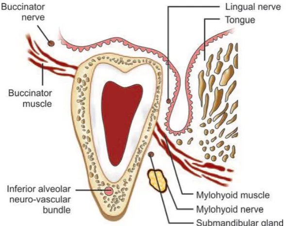
Figure 1: Schematic diagram showing coronal section through the third molar
region and the relationship of important anatomical structures to impacted
mandibular third molar
Neurovascular Bundle
Below or alongside the roots of the third molar is the mandibular canal. The
canal is usually positioned apically and slightly buccal to the third molar roots. The
canal encloses the neurovascular bundle. The neurovascular bundle contains the
inferior alveolar artery, vein and nerve enclosed in a fascial sheath. Since the
calcification of the mandibular canal is completed before formation of the roots of
third molar, the growing roots may impinge on the canal causing its deflection.
Occasionally roots are indented by the mandibular canal, and rarely
penetration of the roots of the wisdom tooth by this structure may occur. In the
latter case, the neurovascular bundle will be torn during extraction of the tooth.
[image:33.595.143.438.69.301.2]Surgical Anatomy
21
From its start at the mandibular foramen, the canal and its contents are
surrounded by a thin layer of bone with a configuration similar to lamina dura and
this is radiographically detectable. In cases where the roots of the third molar are in
direct contact with the neurovascular bundle, the lamina dura may be partially or
totally absent.
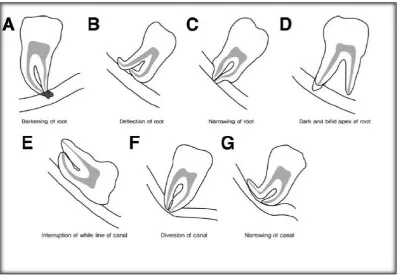
Figure 2: Rood’s Radiographic Predictors of Potential Tooth Proximity to the
Inferior Alveolar Canal (Rood JP, Shehab BA. The radiological prediction of
inferior alveolar nerve injury during third molar surgery. Br J Oral Maxillofac
Surg. 1990; 28:20-5)
Retromolar Triangle
Behind the third molar is a depressed roughened area which is bounded by
[image:34.595.109.507.244.522.2]Surgical Anatomy
22
to the retromolar triangle is a shallow depression, the retromolar fossa. Either in the
retromolar triangle or in the fossa an opening may be present through which
emerge branches of the mandibular vessel. This branch supplies the temporalis
tendon, buccinator muscle and adjacent alveolus.
The retromolar pad, which is the soft tissue covering the retromolar area is
predominantly made up of loose connective tissue.
The tendinous insertion of temporalis muscle terminates as two limiting
prongs on the borders of the retromolar triangle.
Facial Artery and Vein
The facial artery and anterior facial vein cross the inferior border of the
mandible just anterior to the masseter muscle and have a close relationship to the
second and third molar.
Lingual Nerve
The lingual nerve lies on the medial aspect of the third molar. Frequently
lingual nerve courses submucosally in contact with the periosteum covering the
lingual wall of the third molar socket or it may run below and behind the tooth. The
proximity of this important nerve to the third molar places it in danger during the
surgical removal of wisdom tooth. Injury to lingual nerve will lead to prolonged
anesthesia of the anterior two-thirds of the tongue.
Mylohyoid Nerve
This nerve leaves the inferior alveolar nerve just before the latter enters the
Surgical Anatomy
23
proceeds close to the mandible in the mylohyoid groove. In 16% of the cases the
nerve may be enclosed in a canal.
The nerve may be damaged during lingual approach for the removal of
impacted mandibular third molar.
Long Buccal Nerve
This nerve emerges through the buccinator muscle and then passes
anteriorly on its outer surface. When the mouth is wide open, the level at which the
nerve passes through the muscle corresponds to the upper part of the retromolar
fossa.
Rarely injury to the nerve can occur when the posterior part of the incision
is placed too laterally. This results in anesthesia of the lower part of the buccal
mucosa in the molar region.
Musculature
The various muscles surrounding the third molar region are:
• Buccinator - anteriorly
• Temporalis - distally
• Masseter - laterally
• Medial pterygoid and mylohyoid - medially
Buccinator muscle: This horseshoe-shaped muscle forms the musculature of the
Surgical Anatomy
24
pterygomandibular raphe. It is attached to the maxilla at the level of the apices of
molar roots.
During the surgical removal of deeply impacted third molar, the insertion of
attachment of buccinator on the external oblique ridge may have to be severed.
This predisposes to marked postoperative swelling, trismus and pain.
Temporalis muscle: This fan-shaped muscle is inserted on the coronoid process
and anterior border of mandible. Two tendons can be noticed where the muscle
attaches to the anterior border of mandible. The outer tendon is inserted to the
anterior border of coronoid process. The inner tendon is attached to the temporal
crest of mandible. The retromolar fossa is found in between these tendons.
During buccal approach for the removal of third molars, the outer tendon
has to be sectioned to enable reflection of the flap. This in turn will facilitate
adequate bone removal from the buccal and distal side.
Masseter: This muscle is inserted into the lateral side of the ramus from the
coronoid process up to the angle. The muscle is rarely involved in third molar
surgery.
Postoperative edema may extend posteriorly to involve the muscle leading
to trismus and pain. Additionally, preoperative or postoperative infection may lead
to submasseteric abscess formation.
Medial pterygoid muscle: This is inserted on the medial aspect of mandible in
the angle region.
Even though not directly involved in third molar surgery, while using a
lingual approach postoperative edema may result in trismus due to secondary
Surgical Anatomy
25
Mylohyoid muscle: This muscle is inserted on the mylohyoid line from canine to
the third molar region. In the lingual approach, the insertion of the muscle is partly
severed. This leads to transient swallowing difficulty. Moreover, postoperative
infection can spread to sublingual or submandibular space.
Figure 3: Winter’s ‘WAR’ lines for assessment of difficulty in removal of
Materials and Methods
26
MATERIALS AND METHODS
Selection of Patients
The present study was undertaken at the Department of Oral and
Maxillofacial Surgery, Tamil Nadu Government Dental College & Hospital;
Chennai, after obtaining approval from the Institutional Ethics Committee (IEC).
A total of 30 patients divided into 3 groups; both male and female, aged between
17 and 45 years, who had impacted mandibular third molars with similar
anatomical position were randomly selected for this study.
INCLUSION CRITERIA
1. Patients willing for voluntary participation and have signed informed
consent.
2. Patients aged between 17-45 years, either sex.
3. Patients with impacted mandibular third molar (38 or 48), with similar
anatomical positions, and similar surgical difficulty (Mesioangular
impaction).
4. Patients with platelet counts within physiological limits (1.5–4.5
lakhs/cmm).
EXCLUSION CRITERIA
1. Patients with systemic diseases.
2. Patients with possible compromised immune system.
3. Malignant and premalignant conditions.
Materials and Methods
27
5. Patients with documented allergy to any component of the bone substitute
or local anesthetics, drugs.
6. Patients unwilling to participate in the study.
SAMPLE SIZE:
GROUP I: a combination of platelet-rich fibrin and hydroxyapatite graft
was placed in the post-extraction sockets of 10 patients.
GROUP II: hydroxyapatite graft alone was placed in the post-extractionsockets of 10 patients
.
GROUP III: control group of 10 patients with their post-extraction
sockets left empty.
STUDY DESIGN
Ethics clearance was obtained from the Institutional ethics committee and the
ethical principles were followed throughout the course of the study. Subjects for
the study were selected randomly if they satisfied the inclusion criteria with no
discrimination on the basis of sex, caste, religion or socioeconomic status. After
explaining the study procedure written informed consent in the regional language
(Tamil) was obtained from all the subjects selected for the study. Examination
was preceded by a thorough medical and dental history of the patients.
STUDY PROTOCOL
Obtaining medical history and informed consent
Complete clinical examination by using diagnostic instrument set
Extra-oral and intra-oral examination
Materials and Methods
28
Pre surgical preparation
Surgical procedure
Post-operative review
Post-operative care
Clinical re-evaluation on 1st post-operative day, after 1 week and 2 weeks.
Radiographic re-evaluation on 1st post-operative day, at the end of 1, 3 and
6 months using OPG, and CT scan at the end of the sixth month.
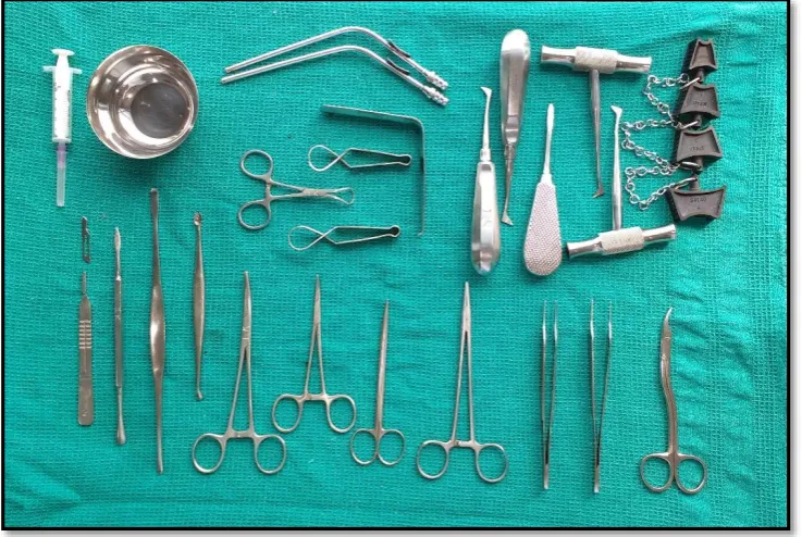
ARMAMENTARIUM
Diagnostic instrument set
Impaction kit
Micromotor
Straight handpiece and 703 bur
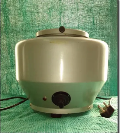
Blood collection armamentarium Centrifuge
10 ml glass test tubes Hydroxyapatite graft (G-bone)
Sterile bowl
Bone scoop
Suture material: 3-0 Black Braided Silk
Graft Material
0.5 mg of hydroxyapatite (HA) granules (G‑bone) with an average pore
Materials and Methods
29
Preparation of PRF
Requirements:
i. Centrifuge,
ii. 10-ml dry glass test tube (without anticoagulant),
iii. Blood collection armamentarium.
The steps involved are as follows:
a) Blood specimen is collected or drawn from the patient,
b) The blood specimen is placed in the centrifuge and is allowed to spin
immediately for 10 minutes at 3000 rpm,
c) Following this the blood sample settles into three layers.
Following layers are formed:
1) The lower fraction containing the RBCs,
2) The middle fraction containing the fibrin clot,
3) The upper fraction containing the straw-colored acellular plasma.
Materials and Methods
30
The upper portion of the test tube containing the acellular plasma is
removed.
The middle portion containing the fibrin clot is then removed and is
scrapped off from the lower part containing the red blood cells.
Surgical Procedure
Step 1: Transalveolar extraction of mandibular third molars
The procedure was performed with proper aseptic precautions. A single
operator carried out all the procedures.
All the patients were advised chlorhexidine mouthwash for oral rinsing
before the procedure. Standard scrubbing and painting procedures were done with
betadine. Standard draping procedures were followed.
Intra orally inferior alveolar nerve block was given along with lingual and
buccal nerve block using 2% Lignocaine with adrenaline 1:80,000. A standard
mandibular third molar incision (Ward’s incision) was placed distal to second
molar continued over the alveolar crest (if the tooth is completely embedded)/
along the buccal gingival sulcus of third molar, upto the distal aspect. Distal
releasing incision is started from the distal most point of the third molar across the
external oblique ridge into the buccal mucosa.
Anteriorly the incision was extended upto the distal of first molar if needed
for better exposure. A full thickness mucoperiosteal flap was raised and the crown
of third molar exposed. With the help of a micro motor, straight hand piece and
using 703 bur sufficient bone was removed forming a gutter on the mesial, buccal
Materials and Methods
31
some cases tooth was sectioned and retrieved. The socket was carefully examined
for remnants of tissue and then the follicular tissue if present was curetted out
from the socket. The socket was irrigated with saline and betadine.
Step 2:
A combination of PRF and hydroxyapatite graft (0.5 gm of porous
hydroxyapatite mixed with every 2 ml of PRF) was placed in post-extraction
socket of GROUP I patients.
Step 3:
Hydroxyapatite graft (0.5 gm of porous hydroxyapatite) alone was placed
in post-extraction socket of GROUP II patients.
Step 4:
Post-extraction socket of GROUP III patients was left empty and served as
control.
Step 5:
The wound was closed primarily with 3–0 black braided silk.
Step 6:
Patients were put on an antibiotic course commencing 1 day before
surgery to be continued post-operatively for 5 days.
Postoperative Instruction
All the patients were given routine post-operative instructions. They were
given Cap. Amoxicillin 500 mg QID, Tab. Metronidazole 400 mg TDS, Tab.
Prednisolone 10 mg BID, Tab. Diclofenac 50 mg BID and Tab. Ranitidine 150 mg
Materials and Methods
32
FOLLOW-UP AND OBSERVATION
All the patients were evaluated:
1. One day prior to the surgery
2. First postoperative day
3. One week postoperatively
4. Two weeks postoperatively
4. One month postoperatively
5. Three months postoperatively
6. Six months postoperatively
Mouth opening was measured preoperatively and postoperatively using Digital
Vernier Caliper.
The patients were asked to rate the pain intensity on a 10-point Visual
Analogue scale (VAS).
Osseous regeneration was evaluated with the help of standardized
orthopantomogram (OPG) taken at 1, 3 and 6 months postoperatively.
Criteria for radiographic evaluation:
1. Radiopacity of bone filling the socket.
2. Presence of trabecular bone formation.
Bone density was evaluated using computerized tomographic (CT) scan taken
at the end of 6 months.
CT scan technique and exposure:
Mandibles were scanned using 16 slice CT, specifics of which were as follows:
Materials and Methods
33 Scanner slice thickness: 0.75 mm;
Exposure time: 26.68 sec and
Field of view: 200 mm.
Images were taken in uncompressed DICOM 3.0 format.
Data analysis:
The data was analysed using SPSS (software package for social
sciences) version 20.
First part of the analysis was descriptive analysis of the parameters of age
and gender. Pre-op and post-op mouth opening and pain score in the three groups
were evaluated using one-way ANOVA analysis. Trabecular pattern assessment
was done using chi-square test. Bone density was evaluated after 6 months using
Materials and Methods
Figure 1: ARMAMENTARIUM
[image:47.595.123.492.131.378.2]
Materials and Methods
Materials and Methods
SURGICAL PROCEDURE
Step 1: INJECTION OF LOCAL ANESTHESIA
Materials and Methods
Step 3: MUCOPERIOSTEAL FLAP ELEVATION
Materials and Methods
Step 5: ELEVATION OF MANDIBULAR THIRD MOLAR USING
STRAIGHT ELEVATOR
Materials and Methods
GROUP I
Step 7: WITHDRAWAL OF BLOOD FROM BRACHIAL VEIN
Step 8: LAYERS FORMED AFTER CENTRIFUGATION OF BLOOD
Materials and Methods
Step 9: PLATELET-RICH FIBRIN AND HYDROXYAPATITE
GRANULES
Materials and Methods
Step 11: PLACEMENT OF PRF AND HA MIXTURE IN
POST-EXTRACTION SOCKET OF GROUP I PATIENT
Materials and Methods
GROUP II
Step 13: HYDROXYAPATITE (HA) GRANULES
Step 14: PLACEMENT OF HA GRANULES IN POST-EXTRACTION
Materials and Methods
Materials and Methods
GROUP III
Step 16: EMPTY POST-EXTRACTION SOCKET OF GROUP III
PATIENT
Case Report
34
CASE REPORTS
GROUP I
CASE REPORT 1
NAME : Mr. Lional Joshua
AGE/SEX : 22 years/ Male
CHIEF COMPLAINT : Pain in the left lower back tooth region
HISTORY OF PRESENTING ILLNESS : Intermittent pain present in left
lower back tooth for past six months
which increased in intensity in the
last one week
PAST MEDICAL HISTORY : Non contributory
PAST SURGICAL HISTORY : Non contributory
PAST DENTAL HISTORY : Non contributory
GENERAL EXAMINATION : 1) Patient is moderately built and nourished
2) Patient is conscious, alert, oriented
3) No signs of pallor, icterus, cyanosis,
clubbing, edema and regional
Case Report
35
LOCAL EXAMINATION
INTRA-ORAL EXAMINATION : 1) Mouth opening- 50 mm
2) Impacted- 38, 48
3) Dental caries- 37
4) Missing- 36
INVESTIGATION
OPG : Impacted 38, 48
DIAGNOSIS : Impaction 38, 48
TREATMENT PLAN : Transalveolar extraction of 38
under local anesthesia followed by
PRF with Hydroxyapatite graft
Case Report
GROUP I
CASE 1
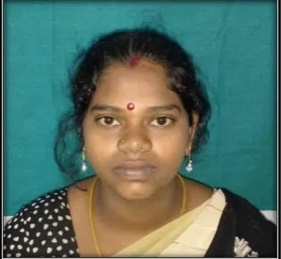
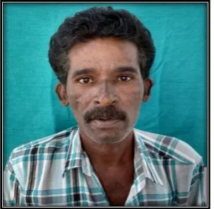
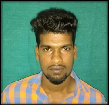
[image:60.595.216.400.412.551.2]Figure 1:PRE-OPERATIVE FRONTAL VIEW
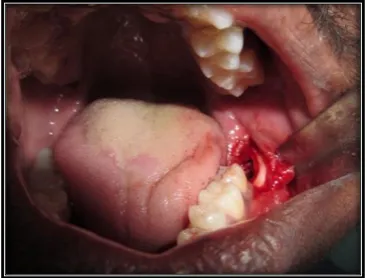
Figure 2: POST-EXTRACTION SOCKET
Case Report
Figure 4: LAYERS FORMED AFTER CENTRIFUGATION OF BLOOD SAMPLE
Figure 5: PLATELET-RICH FIBRIN AND HYDROXYAPATITE GRANULES
[image:61.595.222.393.376.535.2]Case Report
[image:62.595.208.407.161.306.2]Figure 7: PLACEMENT OF PRF AND HA MIXTURE IN POST-EXTRACTION SOCKET
Case Report
Figure 9: PRE-OPERATIVE OPG Figure 10: IMMEDIATE POST- OPERATIVE OPG
Figure 11: 6-month post-op OPG
Figure 12: CT SCAN AT THE END OF SIX MONTHS
Case Report
36
CASE REPORT 2
NAME : Mrs. Rooth
AGE/SEX : 32 years/ Female
CHIEF COMPLAINT : Pain in the left lower back tooth region
HISTORY OF PRESENTING ILLNESS : Pain present in left lower back tooth
for past three days
PAST MEDICAL HISTORY : Non contributory
PAST SURGICAL HISTORY : Non contributory
PAST DENTAL HISTORY : Non contributory
GENERAL EXAMINATION : 1) Patient is moderately built and nourished
2) Patient is conscious, alert, oriented
3) No signs of pallor, icterus, cyanosis,
clubbing, edema and regional
lymphadenopathy
LOCAL EXAMINATION
INTRA-ORAL EXAMINATION : 1) Mouth opening- 33 mm
2) Impacted- 38
Case Report
37
4) RCT treated- 11, 21, 22
5) Missing- 14, 16, 25, 46
INVESTIGATION
OPG : Impacted 38
DIAGNOSIS : Impaction 38
TREATMENT PLAN : Transalveolar extraction of 38
under local anesthesia followed by
PRF with Hydroxyapatite graft
Case Report
[image:66.595.229.387.140.285.2]CASE 2
Figure 1: PRE-OPERATIVE FRONTAL VIEW
Figure 2: PRE-OPERATIVE OPG Figure 3: IMMEDIATE POST- OPERATIVE OPG
Case Report
38
CASE REPORT 3
NAME : Mrs. Ananthi
AGE/SEX : 29 years/ Female
CHIEF COMPLAINT : Pain in the right lower back tooth region
HISTORY OF PRESENTING ILLNESS : Intermittent pain present in right
lower back tooth for past one month
which increased in intensity in the
last one week
PAST MEDICAL HISTORY : Non contributory
PAST SURGICAL HISTORY : Non contributory
PAST DENTAL HISTORY : Non contributory
GENERAL EXAMINATION : 1) Patient is moderately built and nourished
2) Patient is conscious, alert, oriented
3) No signs of pallor, icterus, cyanosis,
clubbing, edema and regional
lymphadenopathy
LOCAL EXAMINATION
INTRA-ORAL EXAMINATION : 1) Mouth opening- 26 mm
Case Report
39
INVESTIGATION
OPG : Impacted 48
DIAGNOSIS : Impaction 48
TREATMENT PLAN : Transalveolar extraction of 48
under local anesthesia followed by
PRF with Hydroxyapatite graft
placement
Case Report
CASE 3
Figure 1: PRE-OPERATIVE FRONTAL VIEW
Figure 2: PRE-OPERATIVE OPG Figure 3: IMMEDIATE POST- OPERATIVE OPG
Case Report
40
CASE REPORT 4
NAME : Mrs. Sangeetha
AGE/SEX : 26 years/ Female
CHIEF COMPLAINT : Pain in the right lower back tooth region
HISTORY OF PRESENTING ILLNESS : Intermittent pain present in right
lower back tooth for past one month
which increased in intensity in the
last two days
PAST MEDICAL HISTORY : Non contributory
PAST SURGICAL HISTORY : Non contributory
PAST DENTAL HISTORY : Non contributory
GENERAL EXAMINATION : 1) Patient is moderately built and nourished
2) Patient is conscious, alert, oriented
3) No signs of pallor, icterus, cyanosis,
clubbing, edema and regional
lymphadenopathy
LOCAL EXAMINATION
INTRA-ORAL EXAMINATION : 1) Mouth opening- 32 mm
Case Report
41
3) Impacted- 18, 38, 48
INVESTIGATION
OPG : Impacted 18, 38, 48
DIAGNOSIS : Impaction 18, 38, 48