| |49 © ARCHIVES OF BUDO | SCIENCE OF MARTIAL ARTS
The influence of different performance level of
fencers on muscular coordination and reaction
time during the fencing lunge
Štefan Balkó
1ABCDE, Zbigniew Borysiuk
2AD, Iva Balkó
1ABE, Daniel Špulák
3C1 Faculty of Education, University of J. E. Purkyně in Ústí nad Labem, Czech Republic 2 Faculty of Physical Education and Physiotherapy, Politechnika Opolska, Poland 3 Faculty of Electrical Engineering, Czech Technical University in Prague, Czech Republic
Received: 22 December 2015; Accepted: 29 January 2016; Published online: 24 February 2016
AoBID: 10970
Abstract
Background & Study Aim: The work is based on the assumption that there are differences in variables (time activation of selected mus-cles, reaction time) between fencers of different performance levels. This assumption is based on results and claims of previous studies and scientific literature, in which the authors draw attention to the fact that there are differences in the effectiveness of applied movement and speed of processing information from the envi-ronment between experienced and less experienced athletes. The aim of this work is information about the ac-tivation of selected muscles during the fencing lunge in different performance levels of fencers.
Material & Methods: The research sample consisted of 43 fencers (épéeist) aged 22.7 ±6.4 years. Based on the current performance of the fencers were divided into appropriate groups To determine the activation of selected muscles, surface elec-tromyography was used (ME6000). FitroSword system was used to identify the reaction time and total time needed to perform the lunge.
Results: Among the groups of fencers were no differences in the order of the selected muscle activation. The differences were reflected in monitoring the time activation of these muscles. For effective lunge can be considered a ma-jor priority of activation of m. deltoideus – pars anterior on the armed arm before m. rectus femoris on the front lower limb. Among the elite fencers and beginners was found a difference in reaction time during a lunge. Conclusions: Due to the fact that there was no difference in overall lunge time between the groups of fencers, muscular
co-ordination and reaction time level of lunge can be considered as important components of sports performance in fencing.
Key words: combat sports • defensive action • épée • muscle activation • offensive action • surface electromyography Copyright: © 2016 the Authors. Published by Archives of Budo
Conflict of interest: Authors have declared that no competing interest exists Ethical approval: The study was approved by the local Ethics Committee Provenance & peer review: Not commissioned; externally peer reviewed
Source of support: The research was realised and supported by SGS University of J.E. Purkyně in Ústí nad Labem.
Author’s address: Štefan Balkó, Department of Physical Education and sport, Faculty of Education, Jan Evangelista Purkyně in Ústí nad Labem, České mládeže 8, 400 96 Ústí nad Labem, Czech Republic, e-mail: [email protected]
Authors’ Contribution: A Study Design
B Data Collection
C Statistical Analysis
D Manuscript Preparation
IntroductIon
In the life of a man level of reaction time and muscle coordination play in his daily activities a major role. It is possible to meet with both of these areas for example in very confusing situ-ations at the busy intersection where the indi-vidual must respond quickly to changing traffic situation and adequately coordinate the move-ment of the arms and legs. In some situations (e.g. red light at the traffic light) can auto-mated responses and effective coordination of selected body segments affect human life and safety. These areas, or their level, also affect sports performance in a number of sports dis-ciplines (motoring, combat sports, ball games, etc.). Their importance is evident even during a fencing match, where in the variable terms of a struggle fencers must quickly respond to the movements of the opponent‘s arms with an appropriate defensiveor offensive action. Fencing is a combat sport, in which in direct conflict with an adversary it is sought to defeat him by a higher count of hits with an appro-priate weapon (épée, foil and the sabre). Quick response, which is associated with the occurrence of visual or tactile stimuli, efficient coordination of muscles during movement, technical and tac-tical skills or optimal psychological attunement represent areas that affect sports performance of fencers. During the match, the two adversaries are both trying to surprise their opponent at the right time with a quick attack out of the optimal distance. The speed abilities fencers in the context of genetic predisposition were also addressed. The authors [1] mention that in the majority of elite fencers was detected ACTN3 gene (R577X) which positively affects the speed capabilities.
According to Bottoms et al. [2] and Cheris [3] is lunge the most often used offensive action in fencing. Potential response in fencing match must be quick because any delay in reaction time may adversely affect the outcome of the match. Quickness of response then becomes an essen-tial component of performance in fencing. The importance of reaction time in sports such as fencing indicate several studies [4-7]. Differences in reaction time between judo athletes of dif-frent performance level also observed Cojocariu and Abalasei [8]. In their study, however, no difference was found neither simple nor choice reaction time between observed groups to visual stimuli.
Continuous repetition and correction of errors of movement implemented based on the occur-rence of visual stimuli is related to the creation of specific musculoskeletal program stored in memory, as stated in Véle [9] for instance. Due to the frequent furnishing of this exercise pro-gram occurs strengthening and improvement of its quality. At the same time according to [10] increasing experience (training) leads to better analysis of information coming from the envi-ronment and motion manifestation of more experienced athletes is more effective than in beginners. It is conected with conclusions focused on motor program stated in the work of Latash [11]. Motor program is a set of hypo-thetical variables stored in memory, which in the case of induction is converted to the desired motor model. Muscular coordination applied at different performance levels of fencers is well evaluable using surface electromyography. As opposed to the other study [12] focused on the intensity of the attack in a combat sport in our study was to determine the time of activation of selected muscles. Here it should be noted that the use of surface electromyography has a wide range of applications.This issue is discussed in fencing by Williams and Walmsley [13, 14]. But many authors [15-21] studied the lunge in terms of kinematic analysis. Compared to other sports (e.g. cycling, weightlifting) that depend largely on the level of a dominant factor (endurance, strength), the resulting performance in a fencing is affected by higher amount of factors. Only by identifying the key factors that determine perfor-mance in fencing, can be optimally implemented training process, which will lead to performance improvement.
The work is based on the assumption that there are differences in variables (time activation of selected muscles, reaction time) between fencers of different performance levels. This assumption is based on results and claims of previous stud-ies and scientific literature, in which the authors draw attention to the fact that there are differ-ences in the effectiveness of applied movement and speed of processing information from the environment between experienced and less expe-rienced athletes.
The aim of this work is information about the activation of selected muscles during the fencing lunge in different performance levels of fencers. Épée – is the heaviest of the three
modern fencing weapons (foil, épée, and sabre), each a separate event, épée is the only one in which the entire body is the valid target area. Épée is the heaviest of the three modern fencing weapons.
Reaction time – is the time from occurence of stimulus to first initiation of movement of the relevant segment of the body
Defensive action – is the appropriate movement of the armed arm with weapon with intent to prevent the attack of opponent
Offensive action – is the towards movement of the weapon or body of fencer towards the his opponent with intent to hit him
Reaction time (RT) – was determined by the time period, which started by lighting of LED and finished with the movement of epee goblet on a horizontal highly sensitive obstacle, which can was identified as motor response
Movement time (MT) – was characterized as the time from the moment of displacement of epee goblet over the horizontal obstacle to the moment of hitting of the target
Total lunge time (TLT) – formed the sum of reaction time and the movement time
MaterIalandMethods
As in previous studies, in this work, activation of selected muscles during a lunge will be investi-gated through surface electromyography (SEMG) in three different performance groups of fenc-ers (elite, subelite, beginnfenc-ers). Device ME6000 and software MegaWin was used. Reaction time is measured by Fitrosword device that generates visual stimuli and simultaneously with the soft-ware SWORD values recorded separately reac-tion time and movement time required to perform the movement. Based on the recommendations of the authors of previous studies and thanks to the coaching practice were for the detection of muscle activation during the lunge was measured these muscles: m. deltoideus, m. trapezius and m.
rectus femoris.
Participants
The research sample consisted of 43 fenc-ers (epeists) aged 22.7 ±6.4 years. Based on the current performance of the fencers were divided into appropriate groups. The first group, labeled as group A consisted of 14 elite fencers. These fencers participate in household domestic com-petitions and championships and even in interna-tional competitions and World Cup competitions. Group referred as B consisted of 15 so-called sub-elite fencers. Fencers from this group participate in domestic competitions and domestic cham-pionships. Group labeled C was represented by 14 beginners. Probands from this group did not participate in any competitions in the wepon of épée. Overview of the basic characteristics of the reviewed samples is given in Table 1 All tested persons were before the research investi-gation acquainted with the aim of the work and confirmed the agreement to participate in the research.
Standardization of measurement conditions
Based on the implementation of preliminary research and studied literature were taken into account ambient conditions (same pad, mini-mum ambient noise, always the same air temper-ature) and other variables (warm up, weight of the weapon, movement distance, clothing, laterality, training) that could affect the course of investi-gation and research results. Always was used flat substrate (linoleum) and the same air tempera-ture (22°C). The test subjects were not disturbed by audio stimuli from the environment. None proband indicated any physical limitations or injuries that could affect the course of the inves-tigation. Before measuring the probands thor-oughly warmed up according to a predetermined protocol made by recommendations from [22-25] and then were instructed to carry out all attempts at maximum speed.
Investigation of the reaction time
To eliminate the possible result influence of inter-individual differences in height of the test subjects, methodology of the study [13, 14] was used. As in the said study, the height of the tested person was multiplied by the coefficient of 15 for deter-mination of movement distance. Foot on the rear (more distant from the target) side (inner edge of the shoe) could not exceed the mark before the occurrence of the stimulus. The center of the tar-get (Figure 1) was located at a height of a center of the sternum of the tested person in an upright position. Stimulus for the lunge initiation, car-ried out from the guard position (Figure 2A), was always lighting up of one of three LEDs (the red LED diode in the middle) with the interval of stimulus incidence from 600 to 2000 ms. On the basis of the occurrence of the stimuli should the test persons perform a lunge as quickly as possi-ble from a predetermined movement distance and Table 1. Basic characteristics of the reviewed groups of fencers
Basic characteristics n Age(years) AT Height[cm] Weight[kg] BMI
Group
A 14 25.9 14.8 184.9 77.7 22.7
B 15 21.2 8.6 181.2 74.8 23.1
C 14 21.3 1.6 179.4 73.1 22.8
hit the target (Figure 2B). Height of the horizon-tal obstacle was placed individually according to the height of the tested person, so that the elbow joint angle was approximately 90°. The reaction time (RT), movement time (MT) and total lunge time (TLT) was measured.
Each test person had five test attempts that pre-ceded measurement itself. Between the experi-ments and actual measurement was determined three minute rest interval to eliminate the effects of fatigue. The measurement included 20 lunges (attempts) between which was set interval of rest up to 15-20 seconds.
Procedure in surface electromyography
Hairs on the legs and shoulders were removed. Sites for application of electrodes (localization is presented in Figure 3) were subsequently cleaned with an abrasive paste and degreasing by alco-hol-gasoline. Used electrodes had Ag/AgCl sen-sor covered by a moist gel. Measuring gel area was 154 mm2 with center distance of two
elec-trodes always 34 mm. Elecelec-trodes were applied to
m. trapezius (upper part of armed arm,
hereinaf-ter referred to as TR), m. deltoideus – pars anhereinaf-terior
(armed arm, hereinafter MDA), m. deltoideus –
pars medialis (unarmed arm, MDM), m. rectus femoris (for rear lower limb – MRFR and for front
lower limb – MRFF).
Data analysis
Based on the recommendations of Tanaka et al. [27] were from simple reaction excluded values that exceeded 1000 ms. Excluded were also values below 100 ms, which by reccomendations of [28] are indicate as anticipatory. Data from the area focused on the detection of selected muscle activation was recorded in the MegaWin software. The measured signal was in the recording device hardware filtered by a 15-500 Hz frequency filtration and converted to digital form with a sampling frequency of 1000 Hz. This digital signal was then rectified (converted to absolute values). To determine the activation time was chosen thresholding method relative to the local maximum EMG envelope in the studied phase [29]. The envelope of the EMG signal converted to abso-lute values was obtained using a low pass filter (FIR order 501 with a cutoff frequency of 3.6 Hz pass-band). Time muscle activation was detected using scripts in Matlab program (version 2012b R). Figure 1. Hitting of the target with three LEDs [3]
Figure 2. Fencer in on guard postition (A) and in the
lunge (B) [3]
Furthermore was calculated arithmetic average, median and standard deviation for the activation time at the threshold of 20% of the local maxi-mum EMG envelope in the studied phase. Even Williams and Walmsley [13, 14] used similar pro-cedure. Before the lunge, tested persons stayed in guard position when the muscles were active. Due to this fact was determined so-called „arti-ficial“ baseline that corresponded to the average value of the signal at interval of 550 ms before the lighting up of LED to initiation of lunge. At the moment when the signal amplitude exceeds 20% of the local maximum taken from this „arti-ficial“ baseline muscle was considered as activated. The muscle was also considered active if it was above the threshold for at least 50 milliseconds. A total of 20 trials were recorded. During the data analysis, only the first 15 correct trials (without anticipation or incorrect attempts) were processed because of the potential impact of fatigue on per-formance. Only that data was registered, which corresponded to the correct attempts recorded in the software SWORD and MegaWin.
To clarify the time relationship in activation of the five monitored muscles between the groups will this activation be related to the time aspect, which is defined by the appearance of the stimu-lus and the activation of the entire muscle at 20% level of the local maximum of the observed phase.
Statistic analysis
The resulting values were related to the time (ms) aspect.For the purposes of statistical pro-cessing of the results was used Statistica 6.1 and Microsoft Excel, 2010.On the basis of a normal-ity test (Shapiro Wilks W test) it was found that the resulting values of selected muscle activation time is not indicative of a normal frequency dis-tribution. For this reason, the data will be con-sidered non-parametric [30]. Due to this fact and due to the low number of test persons in stud-ied groups, the results were processed using non-parametric statistical procedures. For the equality test of medians of all studied groups was used Kruskal-Wallis test. Using theMann-Whitney U test were always measured differences between the two groups fencers. For statistically signifi-cant differences were considered the results when p value was less than 0.05.
results
Using the Kruskal-Wallis test statistically signif-icant differences were observed in all groups of fencers in activation time of MDA (p = 0.0002, η2 = 0.4), TR (p = 0.0309, η2 = 0.17) and MDM
(p = 0.0061, η2 = 0.24). In addition was found
a statistically significant difference in the values of RT (p = 0.0205, η2 = 0.19).
Due to visual stimulation was first activated MRFR in all groups. For all three groups is minimal difference in the activation of this muscle. Interestingly, in group A was activated MRFF before RT. In all other cases (groups) were monitored muscles always activated before RT (figure 4).
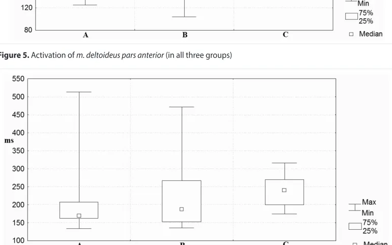
Between groups A and B was no statistically sig-nificant difference in activation time in any of the variables (Figures 5 to 8). Statistically sig-nificant difference in the time activation of the MDA (p = 0.0003, d = 0.69) and TR (p = 0.0062, d = 0.52) was observed between groups A and C. Statistically significant difference was also proved in RT (p = 0.0067, d = 0.51). Between groups B and C was statistically significant dif-ference in activation time of MDA (p = 0.0005,
d = 0,65) and MDM (p = 0.0009, d = 0,62).
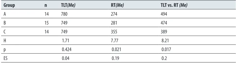
The last part of the study is focused on identify-ing the differences of TLT (total lunge time) and RT between the all groups of fencers (Table 2). The results will help to clarify the relationship Figure 3. Electrodes localization [26]
Figure 4. The muscle activation observed in groups A, B and C
MDA: m. deltoideus pars anterior; MRFR: m. rectus femoris – rear lower limb; MRFF: m. rectus femoris – front lower limb; TR: m. trapezius – upper part (armed arm); MDM: m. deltoideus pars medialis; RT: reaction time
Figure 5. Activation of m. deltoideus pars anterior (in all three groups)
Figure 7. Activation of m. deltoideus pars medialis (in all three groups)
Figure 8. Reaction time (all three groups)
Table 2. Differences in values of lunge duration (in all three groups)
Group n TLT(Me) RT(Me) TLT vs. RT (Me)
A 14 780 274 494
B 15 749 281 474
C 14 749 355 389
H 1.71 7.77 8.21
p 0.424 0.021 0.017
ES 0.04 0.19 0.2
H: value of Kruskal-Wallis test; p: the probability of error in rejecting H0;ES: effect size (substantive significance,η2);
between RT (RT) and TLT observed in groups of fencers.
Between monitored groups using the Kruskal-Wallis showed a significant difference (p = 0.021, η2 = 0.19) in the values of RT. There was no
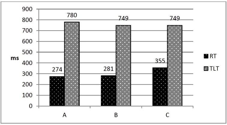
sig-nificant difference in TLT. A statistically signif-icant difference was found in relation TLT vs. RT (p = 0.017, η2 = 0.2). Balanced TLT values
in all groups of fencers are evident in Figure 9. We also investigated the relationships between groups using the Mann-Whitney U test eval-uation. A significant difference in the RT was observed only between groups A vs. C (p = 0.007, d = 0.51).
dIscussIon
The muscle coordination during rapid movements plays an important role in sports performance of fencers. Unveiling regularities of interaction between muscle activation fencers of different performance levels may be useful in focusing the training process and increasing of performance of athletes.
Due to the total lunge time (TLT) 95% fencers activated m. rectus femoris on the rear lower limb (MRFR) before m. deltoideus pars anterior on the front side (MDL). Similarly in study of Schmidt and Wrisberg [10] authors mention that deltoid muscle of extending arm was activated 80 ms
after activation of muscles of the lower limbs. Corresponding results are reported by Lee [31]. These findings are not inconsistent with the claim of Szilagyi [32] that the activity of rear lower limb launches lunge in fencing. From the above inves-tigation, we can infer the mechanical connection of the shoulder muscles and other parts of the body through functionally linked muscle chains, which describes [9]. It should also be noted that the largest physical response delay before starting the movement arises from the activation of the postural system (creation of the necessary pre-requisites for the subsequent motion), which is associated with maintaining balance. Earlier acti-vation of the muscle MRFR provides stabilization against adverse effects of the subsequent move-ment of the arm and is related to the recoil prep-aration necessary to perform a lunge.
Relationship of activation order of m. deltoideus
pars anterior on the front side (MDA) before m. deltoideus pars medialis on the rear side (MDM)
has been studied in relation to the results of the study [33] in which several fencers showed acti-vation order of MDM before MDA. In our work, however, all test persons of group A activated MDA before MDM. The sequence was found in a majority of the test subjects from group B (73%) and C (78.6%), there was no significant difference between the groups in the follow-ing order of activation. The result supports the claim that the non-dominant upper limb (on the Figure 9. Relationship of RT and TLT in groups A, B and C
rear side) during a lunge has a different function than the dominant upper limb (on front side). Dominant handles the weapon, while the non-dominant upper limb has only an auxiliary func-tion. Véle [9] in this regard states that both upper limbs work as a pair gripping organ and works as a functional closed chain. The dominant limb has a leading role and the other leg (non-dominant) rather supports the dominant limb. Since there were no statistically significant differences in the order of activation of these muscles included in any of the relationships between groups, suitabil-ity of determination of their expected sequence can be deduced.
After comparing groups A and B can be stated that there were no statistically significant differ-ences in time activation of any of the five mus-cles. Certain differences (p = 0.0701, d = 0.34) were observed in the activation time m.
rec-tus femoris on front lower limb (MRFF). In
group B occurred earlier activation of this mus-cle than in group A. The differences in a com-parison of these two groups with group C was found in time activation of m. deltoideus pars
ante-rior (MDA). Activation of this muscle was
iden-tified in the group A significantly earlier than in group C (p = 0.0003, d = 0.69). A similar differ-ence was also found between the groups B and C (p = 0.0005, d = 0.65). The above results clearly show that early (meaning at the time of occur-rence of the stimulus) activation of MDA on the armed arm at the beginning of the lunge may be an important factor for the successful implemen-tation of motion. On the basis of that muscle activation can be assumed the follow-up action of the armed arm.
Based on the results obtained in the muscle time activation can be supported by findings from [13, 14], that after the appearance of stimulus first activated during the lunge m. rectus femoris on the rear lower limb (MRFR) and subsequent activa-tion of m. deltoideus of the armed arm (MDA). The same was the case with all three groups stud-ied in this work. At the same time we can con-firm that m. rectus femoris on the front lower limb (MRFF) was activated later than MRFR. It can not, however, confirm their findings that elite fencers had faster activation of all the three mus-cles than beginners. MRFF was in the case of this work activated later in elite fencers group than in subelite fencers group and beginners.
At the same time with the muscle activation was in this work observed the reaction time level (RT), which some authors [34] defined as motor response (reaction). This variable was monitored during a lunge, for example, in a study [13, 14]. Between group A and C was statistically signifi-cant difference in the values of RT (p = 0.0067, d = 0.51). Between groups B and C, difference was noticeable, but did not prove to be statis-tically significant (p = 0.0636, d = 0.35). Based on the above findings, it should be emphasized that the time activation level of MDA and sub-sequent early motor response of the armed arm at the begininning of the lunge can be consid-ered as important variables affecting its perfor-mance. RT (reaction time) of the armed arm was in group A consistently identified with the vation moment of the MDM and then was acti-vated MRFF, while in groups B and C was MRFF activated before occurence of motor response of the armed arm. The difference between RT and MRFF however, showed statistically signifi-cant only between groups A and C (p = 0.0101, d = 0.49). However, between groups A and B was also a noticeable difference (p = 0.0668, d = 0.34). While watching the time difference between MRFR vs. MDA was found that 95.3% of all observed fencers activated MRFR before MDA (understood in the sense of order). Among the groups in this respect, however, there were differ-ences in activation time. When comparing groups A and B was this difference very balanced. Larger time difference between activation of MRFR and MDA occurred between groups A and C, and B and C. These results clearly show that the shorter time interval between activation of MRFF and MDA is desirable for the monitored movement. The last monitored relationship was between MDA and MRFF. Group A compared to group C activated MDA significantly earlier than MRFF. This results can also confirm conclusions of [4] that experienced fencers activate muscles of the armed arm first and after that they activate mus-cles on the front lower limb. In this work, this fact occurred in all three groups of fencers. At the same time it suggests the importance of a greater time difference between the activation of MDA and MRFF.
to the relation between the speed of a defined segment of the hand with a weapon, with other segments of the body. To shorten the total lunge time while maintaining optimal muscle coordi-nation found in group A comes into consider-ation practical influencing of MRFR activconsider-ation associated with subsequent recoil towards the tar-get through muscle activity of rear lower limbs. Shortening of MRFR latency is certainly an important predictor for the movement speed. Smaller time difference between activation of MRFR and MDA may also contribute to the suc-cessful execution of the movement, and thanks to that it creates time for optimal coordination with other muscles before hitting the target. It is obvious that the MRFF activity identified in Group A after a reaction time of the armed arm also holds significance during the lunge. Based on the results can be considered earlier activation of muscle undesirable. The results also clearly show that the time required to perform a lunge (under-stood since the occurrence of stimulus until the intervention) is in all three groups of fencers sim-ilar. Due to the fact that differences were found in the time activation of selected muscles, can be concluded the importance of muscular coor-dination and reaction time in the realization of the lunge.
conclusIons
Meaningful conclusions of the time relationship of muscles druing the lunge can be implemented
in the training process with regard to the „opti-mal“ model of movement initiation, which is implemented by a group of elite fencers. It is possible to believe that the use of this model for groups of fencers with lower performance lev-els will have a positive effect on improving their performance.
The results of this work can be used in stud-ies on biomechanics, sport training or physiol-ogy. In other researches may be findings of this work, especially those that identify relationships between the observed variables, used for the mea-surements, which may combine a variety of meth-ods (kinematic analysis, dynamometry, Kistler‘s board, etc.) And at other kinesiological analysis of movement. It can also monitor differences in bioelectric muscle tension through EMG before implementation of the lunge in different per-formance groups of fencers. In the case of the realization of another similarly oriented research would be useful to include in the investigation muscles belonging to the knee flexor muscles, gluteal muscles and mm. vasti on the thigh. It should be noted that the results observed in the present work should be approached with caution, mainly because the performance in fencing con-sists of a large number of factors that certainly can affect the performance.
references
1 Balkó I, Balkó Š, Týnková H et al. The occurence of R577X polymorphism of ACTN3 gene in selectted group of elite fencers. Acta Univ Carol Kin 2014; 50(2): 5-12
2. Bottoms L, Greenhalgh A, Sinclair J. Kinematic determinants of weapon velocity during the fenc-ing lunge in experienced épée fencers. Acta Bioeng Biomech 2013; 15(4): 109-113
3. Cheris E. Fencing. Step to Success. Champaign, IL: Human Kinetics; 2002
4. Harmenberg J, Ceci R, Barvestad et al. Comparison of different tests of fencing performance. Int J Sports Med 1991; 12: 573-576
5. Czajkowski Z. Understanding Fencing: the unity and practise. Staten Island, NY: SKA Swordplay Books; 2005
6. Borysiuk Z. Psychomotor reactions in fencing dependence of stimuli type. Rev Bras Cin Des Hum 2008; 10(3): 223-229
7. Borysiuk Z. The significance of sensorimotor response components and EMG signals depend-ing on stimuli type in fencdepend-ing. Acta Univiv Pal Olomuc Gymn 2008; 38(1): 43-54
8. Cojocariu A, Abalesei B. Does the reaction time to visual stimuli contribute to performance in judo? Arch Budo 2014; 10: 73-78
9. Véle F. Kineziologie, přehled klinické kineziol-ogie a patokineziolkineziol-ogie pro diagnostiku a tera-pii poruch pohybové soustavy. Praha: Triton; 2006 [in Czech]
10. Schmidt RA,Wrisberg CA. Motor learning and performance: A situation-based learning approach. 4th ed. Champaign IL: Human Kinetics; 2008 11. Latash ML. Neurophysiological basis of
move-ment. Urbana, IL: Human Kinetics; 2008 12. Estevan I, Falco C, Elvira JLL. et al. Trunk and
lower limb muscle activation in linear, circular and spin back kicks. Arch Budo 2015; 11: 243-250 13. Williams LRT, Walmsley A. Response
amend-ment in fencing: differences between elite and novice subjects. Perc Mot Skills 2000; 91: 131-142 14. Williams LRT, Walmsley A. Response timing
and muscular coordination in fencing: A com-parison of elite and novice fencers. J Sci Med Sport 2000; 3(4): 460-475
15. Geil DM. The role of footwear on kinematic and
plantar foot pressure in fencing. J Appl Biomech 2002; 18: 155-162
16. Stewart SI, Kopetka B. The kinematic determi-nants of speed in the fencing lunge. J Sports Sci 2005; 23(2): 105
17. Gholipour M, Tabrizi A, Farahmand F. Kinematics analysis of lunge fencing using stereophotogram-etry. World J Sport Sci 2008; 1(1): 32-37 18. Sillero M, Saucedo F, López E et al. Analysis of
the rear leg rotation movement during the fenc-ing lunge. Fencfenc-ing, Science & Technology. Paper session presented at 1st International congress on science and technology in fencing, Barcelona; 2008 19. Gutierrez-Davila M, Rojas FJ, Antonio R et al.
Response timing in the lunge and target change in elite versus medium-level fencers. Europ J Sport Sci 2013; 13(4): 364-371
20. Sinclair J, Bottoms L. Gender differences in the kinetics and lower extremity kinematics of the fencing lunge. Int J Perf Anal Sport 2013; 13(2): 440-451
kinematics of the hip and knee during the fenc-ing lunge. Human Mov 2013; 14(3): 229-237 22. Alter MJ. Science of flexibility. Champaing, IL:
Human Kinetics; 1996
23. Bartůňková S. Fyziologie člověka a tělesných cvičení. Učební texty pro studenty fyzioterapie a studia tělesná a pracovní výchova zdravotně postižených. Praha: Karolinum; 2007 [in Czech] 24. Ylinen J. Stretching therapy for sport and manual
therapies. New York: Elsevier; 2008
25. Anderson B, Anderson, JE. Stretching: 30th anni-versary edition. California: Shelter Publications; 2010
26. Cram JR, Kasman GS, Holtz J. Atlas for elec-trode placement. In E. Criswell (ed.), Cram´s
introduction to surface electromyography. Massachusetts: Jones and Bartlett Publishers; 2011: 247-283
27. Tanaka K, Hasegawa M, KataokaT. The effect of self-position andposture information on reac-tion time. Int J Comp Sci Sport 2010; 9(3): 4-14 28. Iida Y, Miyazaki M, Uchida S. Developmental
changes in cognitive reaction time of children aged 6-12 years. Europ J Sport Sci 2010; 10(3): 151-158 29. Špulák D, Čmejla R, Mikulíková et al. Muscle
activity detection using emg envelope thresholding– comparison of various approaches.Paper session pre-sented at the 20th Annual Conference Proceeding´s Technical Computing, Bratislava; 2012 30. Havel Z, Cihlář D. Vybrané neparametrické
statis-tické postupy v antropomotorice. Ústí nad Labem:
PF UJEP; 2011 [in Czech]
31 Lee WA. Anticipatory control of postural and task muscles during rapid arm Flexion. J Mot Beh 1980; 12: 185-196
32. Szilagyi T. Dynamic characterisation of fencing lunge. Paper session presented at the International Society of Biomechanics XIVth Congress, Paris; 1993
33. Balkó Š, Jelínek M, Kratochvílová I et al. Komparace timingu vybraných svalů participujících na výpadu u skupiny elitních šermířů a šermířů nižší výkonnostní úrovně. Studia Kin 2012; 13(3): 160-165 [in Czech]
34. Tu JH, Lin YF, Chin SCH. The influence of ball velocity on court illumination on reaction time for tennis volley. J Sports Sci Med 2010; 9: 56-61
![Figure 2. Fencer in on guard postition (A) and in the lunge (B) [3]](https://thumb-us.123doks.com/thumbv2/123dok_us/8592401.1721509/4.567.309.524.80.350/figure-fencer-guard-postition-lunge-b.webp)
![Figure 3. Electrodes localization [26]](https://thumb-us.123doks.com/thumbv2/123dok_us/8592401.1721509/5.567.41.233.87.307/figure-electrodes-localization.webp)