0022-538X/10/$12.00 doi:10.1128/JVI.00462-10
Copyright © 2010, American Society for Microbiology. All Rights Reserved.
Multiple ASF/SF2 Sites in the Human Papillomavirus Type 16
(HPV-16) E4-Coding Region Promote Splicing to the
Most Commonly Used 3
⬘
-Splice Site on
the HPV-16 Genome
䌤
Monika Somberg
1and Stefan Schwartz
1,2*
Department of Medical Biochemistry and Microbiology, BMC, Uppsala University, Sweden,1
and Dublin Institute of Technology, Dublin, Ireland2 Received 2 March 2010/Accepted 25 May 2010
Our results presented here demonstrate that the most abundant human papillomavirus type 16 (HPV-16) mRNAs expressing the viral oncogenes E6 and E7 are regulated by cellular ASF/SF2, itself defined as a proto-oncogene and overexpressed in cervical cancer cells. We show that the most frequently used 3ⴕ-splice site on the HPV-16 genome, site SA3358, which is used to produce primarily E4, E6, and E7 mRNAs, is regulated by ASF/SF2. Splice site SA3358 is immediately followed by 15 potential binding sites for the splicing factor ASF/SF2. Recombinant ASF/SF2 binds to the cluster of ASF/SF2 sites. Mutational inactivation of all 15 sites abolished splicing to SA3358 and redirected splicing to the downstream-located, late 3ⴕ-splice site SA5639. Overexpression of a mutant ASF/SF2 protein that lacks the RS domain, also totally inhibited the usage of SA3358 and redirected splicing to the late 3ⴕ-splice site SA5639. The 15 ASF/SF2 binding sites could be replaced by an ASF/SF2-dependent, HIV-1-derived splicing enhancer named GAR. This enhancer was also inhibited by the mutant ASF/SF2 protein that lacks the RS domain. Finally, silencer RNA (siRNA)-mediated knockdown of ASF/SF2 caused a reduction in spliced HPV-16 mRNA levels. Taken together, our results demonstrate that the major HPV-16 3ⴕ-splice site SA3358 is dependent on ASF/SF2. SA3358 is used by the most abundantly expressed HPV-16 mRNAs, including those encoding E6 and E7. High levels of ASF/SF2 may therefore be a requirement for progression to cervical cancer. This is supported by our earlier findings that ASF/SF2 is overexpressed in high-grade cervical lesions and cervical cancer.
Human papillomavirus type 16 (HPV-16) is the foremost cause of cervical cancer, which is one of the most common cancers in women globally (10, 37). Persistence of high-risk HPV types, such as HPV-16, is the highest risk factor for the development of cervical cancer. The majority of all DNA vi-ruses that establish persistence have evolved a highly organized gene expression program, often divided into clear early and late phases. The HPV-16 genome contains an early promoter that could potentially express mRNAs encoding all viral gene products, and a late differentiation-dependent promoter that specifically excludes expression of E6 and E7 (21). The switch from early to late gene expression includes a promoter switch as well as derepression and activation of the late poly(A) signal and late splice sites (16). To activate late splice sites and the late poly(A) signal, many early splice sites and the early poly(A) signal must be downregulated to allow for competition from mutually exclusive late splice sites and poly(A) signal (8, 26, 36). Other HPV-16 splice sites are used by both early and late mRNAs and should function well in both mitotic cells and terminally differentiated cells. One of the major splice sites used by both early and late mRNAs is SA3358 (Fig. 1A). This splice site is outstanding in that it is used to produce the majority of all HPV-16 mRNAs, including the mRNAs of the
oncogenes E6 and E7 and the E4, E5, L1, and perhaps L2 proteins. In contrast, efficient usage of SA3358 specifically prevents expression of HPV-16 E1 and E2.
Many, if not all, HPV types contain a 3⬘-splice site in the E4
open reading frame (orf) that is spliced to an upstream 5⬘
-splice site that joins the E1 AUG with the E4 orf. In HPV-16, these splice sites are named SA3358 and SD880 (Fig. 1A), whereas they are named SD847 and SA3325 in HPV-11 and SD877 and SA3295 in HPV-31 (1). Splicing between HPV-16 SD880 and SA3358 (6, 9, 27), or the corresponding sites in HPV-11 (5, 20, 23) and HPV-31 (11, 12), occurs on the most-common early mRNAs encoding E6 and E7, as well as on the most-abundant late mRNA encoding E4. In addition, the most-common L1 mRNA is also spliced between SD880 and SA3358 (17), or the corresponding sites in HPV-11 (23) and HPV-31 (12, 22). Analysis of HPV-16 splicing in cervical scrape samples revealed that splicing between SD880 and SA3358 was the most-common splicing event in both low- and
high-grade cervical lesions (25). In vitro transfection
experi-ments demonstrated that splicing to SA3358 was required for efficient expression of E6 and E7 (2). As a matter of fact, splicing between SD880 and SA3358 was required for produc-tion of E6 and E7 quantities that were needed for transforma-tion of cells by these HPV proteins. In HPV-31, SA3295 cor-responds to HPV-16 SA3358. Mutational inactivation of HPV-31 SA3295 in an infectious molecular clone of HPV-31
immediately caused splicing to a cryptic 3⬘-splice site located
three nucleotides further down (15). These results indicated
* Corresponding author. Mailing address: Department of Medical Biochemistry and Microbiology, Uppsala University, Biomedical Cen-tre, Husargatan 3, Box 582, 751 23 Uppsala, Sweden. Phone: 4618 471 4239. Fax: 4618 471 4673. E-mail: [email protected].
䌤Published ahead of print on 2 June 2010.
8219
on November 8, 2019 by guest
http://jvi.asm.org/
FIG. 1. (A) Schematic representation of the HPV-16 genome. Early and late viral promoters p97 and p670 are indicated. Numbers indicate nucleotide positions of 5⬘-splice sites (filled circles), 3⬘-splice sites (open circles), or early and late poly(A) signals pAE and pAL, respectively. LCR, long control region. A few selected early and late mRNAs are shown (1). Previously described splicing silencers and enhancers are indicated (24, 34, 35). (B) Diagram with potential ASF/SF2 sites upstream and downstream of SD3632 predicted by ESEfinder (4). Heights of the bars represent degrees of similarity to ASF/SF2 binding sites according to ESEfinder. HPV-16 splice sites SA3358 and SD3632 are indicated. Numbers indicate nucleotide positions in the HPV-16 genome. The position of a previously described enhancer is indicated (24). (C) ASF/SF2 sites in the mutant HPV-16 sequence in which the ASF/SF2 sites had been inactivated, as predicted by ESEfinder (4). (D) Exact sequences of the wt and mutant (mut) HPV-16 Predicted sequences between nucleotide positions 3407 and 3627 in the HPV-16 genome. Dots represent identical nucleotides.
8220 SOMBERG AND SCHWARTZ J. VIROL.
on November 8, 2019 by guest
http://jvi.asm.org/
that HPV-31 SA3295 is under the control of strong splicing enhancer elements and that there is a strong pressure on the
virus to maintain a 3⬘-splice site in that exact region.
We have previously reported that HPV-16 SA3358 has an
exceptionally poor 3⬘-splice site sequence compared to a
con-sensus 3⬘-splice site (24). This is due primarily to an almost
complete absence of an upstream row of uninterrupted
pyri-midines that normally characterize an efficiently utilized 3⬘
-splice site. However, SA3358 is one of the most efficiently used splice sites on the HPV-16 genome (24, 33). We have previ-ously shown that utilization of HPV-16 SA3358 is totally de-pendent on exonic sequences downstream of SA3358, and we concluded that a splicing enhancer was located downstream of SA3358 (24). Here, we have followed up these findings; we demonstrate that the enhancer elements downstream of HPV-16 SA3358 are binding sites for ASF/SF2, and we show that ASF/SF2 enhances splicing to SA3358.
MATERIALS AND METHODS
Plasmids.pMT1SD was constructed by cleaving pT1SD (24) with SalI and BssHII, filling in overhangs, and religation. pTEx4m was created by first PCR
amplifying an HPV-16 sequence with oligonucleotides exon4mutS (5⬘-GGCGC
GCCCAACTCGTGTGCCGCGACCCATACCATCGTGTGCGCCTTGGGT CGTGTGTCTAACATCGTGTGCTATCCAGCGACCAAGATCAGAGCCA
GATCGTGTGAACCCCTGCTCGTGTGCTAAGTTGTTGTCGTGTG-3⬘)
and exon4mutAS (5⬘-TTCTAGAAAATGTACTCACACGAGCACACGAACT
ATTACAGTTAATCACACGAACACGAAGCTGTTAAATGCACACGAGA TTGGAGCACACGACACACGATCACACGACAACAACTTAGCACACGA
GCAGGGGTTCACACGATCT-3⬘) (Scandinavian Gene Synthesis AB). The
PCR fragment was cleaved with XbaI and BssHII and subcloned into pT1SD (24), resulting in pTEx4m. pMTEx4m was constructed by cleaving pTEx4m with SalI and BssHII, filling in overhangs, and religation. Plasmid pT7wtexon4 was created by PCR amplifying a sequence from pBEL (35) with oligonucleotides
exon4wtSKpnI (5⬘-GGGTACCCCAACCACCCCGCCGCGACCCATAC-3⬘)
and exon4wtASXbaI (5⬘-TTCTAGAAAATGTACTATGGGTGTAGTGTTA
C-3⬘). The PCR fragment was cleaved with KpnI and XbaI and subcloned into
pUC19T7 (32), resulting in pT7wtexon4. pT7mutexon4 was created by PCR
amplifying a sequence from pTEx4m with oligonucleotides exon4mutSKpnI (5⬘
-CGGTACCCCAACTCGTGTGCCGCGACCCATAC-3⬘) and M3A (5⬘-GGGC
CCAGGCCTCGACACTGCAGTATACAATGTACAATGCT-3⬘). The PCR
fragment was cleaved with KpnI and XbaI and subcloned into pUC19T7 (32),
generating pT7mutexon4. GlutathioneS-transferase (GST), GST-ASF/SF2, and
GST-ASFDRS were generated using pGEX-GST (GE Healthcare).
The following plasmids have been described previously: pBEL (35), pBELM (35), pBEL-OPSA (35), pBSplice and pBSpliceM (30), pT1SD (24), pMT198
(29), pBearly97 (33), and pT1-5/GAR (A. Tranell, E. M. Fenyo¨, and S. Schwartz,
submitted). pASF/SF2, pASFDRS, and pAdE4orf4 are kind gifts from Go¨ran
Akusja¨rvi, IMBIM, Uppsala University, Sweden.
Transfection and cell culture.Maintenance and transfections of HeLa cells with Fugene 6 (Roche Molecular Biochemicals) have been described previously (30).
RNA extraction, Northern blotting and radiolabeled DNA probe synthesis, and RT-PCR.Cytoplasmic RNA was extracted using isoB/NP-40 buffer as de-scribed previously (32). Northern blot analysis was carried out as dede-scribed previously (30). Reverse transcription-PCR (RT-PCR) with cytoplasmic RNA was performed as previously described using oligonucleotides 757s (35), p97s
(33), and L1aM (35), E4A (35), or 135 (5⬘-CCTCGAGCTACTTATCGTCGTC
ATCCTTGTAATCTGGTTTCTGAGAACAGATGGGGCACAC-3⬘), as
indi-cated in the figures. Glyceraldehyde-3-phosphate dehydrogenase (GAPDH) cDNA was amplified as control using previously described primers (13).
siRNA-mediated knockdown of ASF/SF2.Transfections of HeLa cells with the ASF/SF2 siGenome Smartpool-designed silencer RNA (siRNA) (Dharmacon) was performed using Oligofectamine (Invitrogen) according to the manufactur-er’s protocol. The sequences of the sense and antisense strands of the four
siRNA duplexes were as follows: ASF/SF2-1 sense, 5⬘-GAAAGAAGAUAUG
ACCUAUUU-3⬘; ASF/SF2-1 antisense, 5⬘-AUAGGUCAUAUCUUCUUUCU
U-3⬘; ASF/SF2-2 sense, 5⬘-UAACUUACCUCCAGACAUCUU-3⬘; ASF/SF2-2
antisense, 5⬘-GAUGUCUGGAGGUAAGUUAUU-3⬘; ASF/SF2-3 sense, 5⬘-U
GAAGCAGGUGAUGUAUGUUU-3⬘; ASF/SF2-3 antisense, 5⬘-ACAUACAU
CACCUGCUUCAUU-3⬘; ASF/SF2-4 sense, 5⬘-CGACGGCUAUGAUUACG
AUUU-3⬘; ASF/SF2-4 antisense, 5⬘-AUCGUAAUCAUAGCCGUCGUU-3⬘.
Briefly, HeLa cells were plated in regular growth medium in a six-well tissue culture dish 24 h prior to transfection. The next day, cells were transfected with 133 nM siRNA mix in a total volume of 1.8 ml of serum- and antibiotic-free
Dulbecco’s modified Eagle’s medium (DMEM) with 4l Oligofectamine. After
incubation for 4 h, 0.5 ml DMEM containing three times the normal concentra-tion of antibiotics and fetal bovine serum was added to the transfected HeLa cells without removal of the transfection mixture. At 24 h after siRNA transfection,
cells were transfected with 0.5g of subgenomic HPV-16 plasmid pBELM (35).
At 72 h after the initial siRNA transfection, cytoplasmic RNA was extracted and RT-PCR was performed. A total cell extract was used for Western blotting with
antibody mab65 (a kind gift of Go¨ran Akusja¨rvi, Uppsala University, Sweden)
against ASF/SF2 to ascertain efficient ASF/SF2 knockdown.
Purification of recombinant GST proteins and UV cross-linking.Purification of recombinant GST fusion proteins using glutathione-Sepharose (GS) was per-formed as specified by the manufacturer (GE Healthcare). Plasmids pT7mutexon4
and pT7wtexon4 were individually linearized with XbaI and subjected toin vitro
transcription with T7 RNA polymerase in the presence of with [␥-32P]UTP. Each
RNA probe (105
cpm) was UV cross-linked to GST-ASF/SF2 or GST-ASFDRS as previously described (28, 31).
RESULTS
Multiple ASF/SF2 binding sites downstream of SA3358.
SA3358 is one of the major 3⬘-splice sites on the HPV-16
genome, and it is required for the production of all early mRNAs, except E1 and E2 mRNAs, as well as L1 and L2 mRNAs (Fig. 1A). We have previously shown that SA3358 is suboptimal and totally dependent on a downstream splicing enhancer (24). However, splicing factors that regulate SA3358 have not been identified. We have used the ESEfinder pro-gram (4) to search for potential SR-protein binding sites down-stream of SA3358. Interestingly we found 15 potential ASF/ SF2 binding sites in the 275-nucleotide exon located between SA3358 and SD3632, overlapping the previously identified splicing enhancer (Fig. 1A and B) (24). In contrast, analysis of the adjacent 275 nucleotides located downstream of SD3632 identified only three potential ASF/SF2 binding sites (Fig. 1B). This analysis suggested that ASF/SF2 may play a role in the regulation of SA3358. If these sites regulated SA3358, muta-tional inactivation of the predicted ASF/SF2 sites would impair splicing to SA3358. To test this idea, we mutationally inacti-vated all 15 ASF/SF2 sites located between SA3358 and SD3632. Analysis of the mutant sequence in ESEfinder (4) revealed that the predicted ASF/SF2 sites were no longer rec-ognized by ESEfinder (Fig. 1C). The exact nucleotide changes are shown in the alignment of wild-type (wt) and mutant HPV-16 sequence in Fig. 1D.
Mutational inactivation of all predicted ASF/SF2 sites downstream of SA3358 inhibits the use of SA3358 and redi-rects splicing to late splice acceptor SA5639.In order to in-vestigate if mutational inactivation of the predicted ASF/SF2 binding sites downstream of SA3358 affected HPV-16 mRNA splicing, we inserted all 15 mutations in subgenomic HPV-16 plasmid pBELM (Fig. 2A) (35), resulting in plasmid pTEx4m (Fig. 2A). The pBELM plasmid has been described by us previously (35). It produces high levels of E4 mRNA spliced from SD880 to SA3358 and low levels of the L1 mRNAs as a result of mutationally inactivated splicing silencers at the late
3⬘-splice site SA5639 (35). Analysis of mRNA produced from
pBELM and pTEx4m in transfected HeLa cells confirmed that pBELM produced high levels of E4 mRNA and low levels of
on November 8, 2019 by guest
http://jvi.asm.org/
FIG. 2. (A) Schematic representation of the HPV-16 genome and the subgenomic HPV-16 expression plasmids pBELM (35), pTEx4m, and pT1SD (24). Numbers indicate nucleotide positions of 5⬘-splice sites (filled circles) and 3⬘-splice sites (open circles) or poly(A) sites pAE and pAL. Structures of selected HPV-16 mRNAs produced by pBELM (35) are indicated below plasmids. Black bars represent E4 and L1 probes used for Northern blotting. RT-PCR primers 757s, E4A, and L1aM are indicated. (B) Left panels, Northern blots of cytoplasmic RNA extracted from HeLa cells transfected with pBELM, pT1SD (24), or pTEx4m. Blots were probed with L1 or E4 probe, as indicated. Gels were also probed for GAPDH. Right panels, RT-PCR on the same RNA analyzed by RT-PCR using primers 757s and L1aM or 757s and E4A (35). The E4A primer hybridizes to a sequence that is present in all three plasmids and that is unaffected by the nucleotide substitutions in pTEx4m.
8222 SOMBERG AND SCHWARTZ J. VIROL.
on November 8, 2019 by guest
http://jvi.asm.org/
L1 mRNAs, as expected (Fig. 2B). In contrast, pTEx4m did not produce detectable levels of E4 mRNAs (Fig. 2B), suggesting that usage of SA3358 was totally dependent on the ASF/SF2 sites. Interestingly, the decrease in E4 mRNA production for pTEx4m was paralleled by an increase in L1i mRNA produc-tion (Fig. 2A and B). Northern blot and RT-PCR analysis showed that mutational inactivation of the potential ASF/SF2 binding sites redirected splicing from SA3358 to late splice acceptor SA5639 (Fig. 2A and B). Similar results were ob-tained when the sequences downstream of SA3358 were de-leted as in plasmid pT1SD (Fig. 2A and B) (24). Endogenous HPV-18 sequences in HeLa cells do not interfere with the assays described here, as HPV-16 primers and probes are spe-cific for HPV-16. In addition, some of the experiments have been reproduced with HPV-negative cell lines, such as 293T cells, with similar results. Taken together, our results indicated that the potential ASF/SF2 sites were required for splicing to SA3358.
ASF/SF2 binds specifically to exonic sequences located be-tween SA3358 and SD3632. To determine if ASF/SF2 binds directly to the predicted ASF/SF2 sites in the sequence be-tween SA3358 and SD3632, GST-ASF was UV cross-linked to radiolabeled RNA representing the wt or mutant sequences shown in Fig. 1D. The results revealed that GST-ASF UV cross-linked efficiently to this HPV-16 sequence, whereas GST did not (Fig. 3A). GST-ASF did not UV cross-link to an RNA sequence derived from the HPV-1 late untranslated region (UTR) (data not shown). Cross-linking to the mutant RNA was less efficient, as expected (Fig. 3A). However, UV cross-linking of ASF/SF2 to the mutant RNA is probably overesti-mated by the gel, as the mutant RNA contains 72 radiolabeled U nucleotides, whereas the wt RNA contains only 36 (Fig. 1D). We concluded that ASF/SF2 binds directly to the sequence located between HPV-16 SA3358 and SD3632.
Overexpression of the ASF/SF2 RS mutant inhibits usage of SA3358.To confirm that ASF/SF2 acts via the ASF/SF2 sites in the enhancer region, we used an ASF/SF2 mutant that lacks the RS-domain (ASFDRS) and therefore binds RNA but fails
to activate splicing. The ASFDRS trans-dominant mutant
should compete with endogenous ASF/SF2 for the ASF/SF2 binding sites and inhibit splicing to SA3358. UV cross-linking of serially diluted GST-ASF or GST-ASFDRS to radiola-beled wt HPV-16 RNA, shown in Fig. 1D, revealed that the two proteins cross-linked to this RNA sequence with similar efficiency (Fig. 3B and C). In addition, overexpression of ASFDRS in transfected cells inhibited splicing to SA3358 in plasmid pBELM, thereby inhibiting production of E4 mRNA (Fig. 4B). Overexpression of ASFDRS also caused a redirection of splicing from SA3358 to SA5639, thereby inducing L1i mRNA production (Fig. 4C). Overexpression of ASFDRS therefore causes the same phenotype as muta-tional inactivation of ASF/SF2 sites at SA3358 in pTEX4m (compare Fig. 2B and 4C). This result is seen only when SA5639 is optimized either by mutational inactivation of splicing silencers located downstream of SA5639 or by direct optimization of the 5639 polypyrimidine tract as in pBEL-OPSA (Fig. 4C and D). Therefore, ASFDRS cannot induce splicing to wt SA5639 in pBEL since it is suppressed by previously identified splicing silencers (Fig. 4C). RT-PCR confirmed that ASFDRS induced L1i by exon skipping (Fig.
4E). This effect is clearly distinct from the induction of L1 mRNA production by the adenovirus E4orf4 protein, as previously described (Fig. 4E) (29).
Overexpression of ASFDRS did not induce L1 production from pT1SD or pTEx4m (Fig. 4F), in which the ASF/SF2 binding sites had been either deleted or mutationally inacti-vated. These results indicated that redirection of splicing to SA5639 by ASFDRS was dependent on the ASF/SF2 binding sites in the enhancer region downstream of SA3358. In con-trast, a small inhibitory effect on L1i mRNA levels was seen when ASFDRS was overexpressed (Fig. 4G). This result sug-gested that ASFDRS inhibited SA5639 and will be discussed below. We concluded that ASFDRS inhibits the ability of en-dogenous ASF/SF2 to enhance splicing to SA3358 and that this inhibition was dependent on intact ASF/SF2 sites in the en-hancer region downstream of SA3358.
Our results suggested that E6/E7 mRNAs are also regulated by ASF/SF2, as it has been shown previously that the most-common E6 and E7 mRNAs that are spliced between SD226 and SA409 or SA526 are also spliced between SD880 and SA3358. As such, they would be dependent on ASF/SF2. To investigate if this was the
FIG. 3. (A) UV cross-linking of GST or GSTASF to thein vitro synthesized, radiolabeled wt and mutant HPV-16 RNAs shown in Fig. 1D. UV cross-linking of serially diluted ASF (B) or GST-ASFDRS (C) to radiolabeled wt HPV-16 RNA shown in Fig. 1D. Molecular weights (MW; in thousands) are indicated on the left.
on November 8, 2019 by guest
http://jvi.asm.org/
[image:5.585.332.505.66.423.2]case, plasmid pBearly97 (Fig. 4F) was transfected into HeLa cells in the absence or presence of the mutant ASF protein named ASFDRS. If the E6/E7 mRNAs are regulated by ASF/SF2, over-expression of ASFDRS should inhibit the production of these mRNAs. As can be seen in Fig. 4F, E6/E7 mRNAs that are spliced between SD226 and SA409 or SA526 were negatively affected by ASFDRS but not by transfection with an empty ex-pression plasmid (Fig. 4F). We concluded that exex-pression of E6 and E7 mRNAs is dependent on ASF/SF2.
The ASF/SF2-dependent HIV-1 splicing enhancer termed GAR can substitute for the HPV-16 enhancer at SA3358.To provide further support for a role of ASF/SF2 in the regulation
of HPV-16 SA3358, we inserted the ASF/SF2-dependent GAR enhancer from HIV-1 (3), into the enhancerless HPV-16 plas-mid pT1SD (Fig. 5A) (24). As can be seen in Fig. 5B and C, pT1SD produced only L1i mRNA, as SA3358 was not used, whereas pT1-5/GAR, in which the ASF/SF2-dependent GAR enhancer had been inserted, produced only L1 mRNA as a result of efficient usage of SA3358 (Fig. 5B and C). We con-cluded that splicing to HPV-16 SA3358 could be restored by a heterologous but ASF/SF2-dependent enhancer, support-ing the conclusion that ASF/SF2 regulates HPV-16 SA3358. Overexpression of ASFDRS with pT1-5/GAR induced pro-duction of L1i mRNA (Fig. 5B and C), indicating that
FIG. 4. (A) Schematic representation of the HPV-16 genome and the subgenomic HPV-16 expression plasmids pBEL (35), pBELM (35), pBEL-OPSA (35), pT1SD (24), and pTEx4m. Numbers indicate nucleotide positions of 5⬘-splice sites (filled circles) and 3⬘-splice sites (open circles) or poly(A) sites pAE and pAL. Structures of selected HPV-16 mRNAs produced by pBELM (35) are indicated below plasmids. Black bars represent E4 and L1 probes used for Northern blotting. RT-PCR primers 757s and L1aM are indicated (35). CMV, cytomegalovirus. (B to D and G) Northern blots of cytoplasmic RNA extracted from HeLa cells transfected with the indicated plasmids in the absence or presence of ASFDRS-expressing plasmid. Blots are probed with E4 or L1 probe as indicated. Gels were also probed for GAPDH. Positions of the E4 and L1 or L1i mRNAs are indicated. (E) RT-PCR on cytoplasmic RNA from HeLa cells transfected with pBELM in the presence of ASFDRS expression plasmid or adenovirus E4orf4 expression plasmid. The RNA samples were analyzed by RT-PCR using primers 757s and L1aM (35). (F) Left panel, schematic drawing of plasmid pBeary97 (33). Right panel, RT-PCR on cytoplasmic RNA from HeLa cells transfected with pBeary97 in the absence or presence of ASFDRS expression plasmid. The RNA samples were analyzed by RT-PCR using primers p97s and 135 (35).
8224 SOMBERG AND SCHWARTZ J. VIROL.
on November 8, 2019 by guest
http://jvi.asm.org/
ASFDRS interferes with the enhancer function of GAR. In con-trast, ASFDRS did not alter splicing in pT1SD (Fig. 5C) as expected, since ASF/SF2 sites had been deleted. These data strengthened the conclusion that ASF/SF2 enhances splicing to SA3358.
ASF/SF2 contributes to the suppression of splice donor SD3632.To investigate if the ASF/SF2 binding sites located be-tween SA3358 and SD3632 contribute to suppression of SD3632, we constructed pMT1SD and pMTEx4m from pBSpliceM (Fig. 6A) (30). pBSpliceM does not produce detectable levels of E4 or L1 mRNA in the absence of transacting factors, e.g., PTB or AdE4orf4 (Fig. 6B) (29, 30). These plasmids contain a large deletion between nucleotide position 757 and 3395 in the early region of the HPV-16 genome which removes all HPV-16 splice sites except late splice sites SD3632 and SA5639 (Fig. 6A). In addition, the ASF/SF2 binding sites upstream of SD3632 were either deleted (pMT1SD) or mutationally inac-tivated (pMTEx4m) (Fig. 6B). Transfection of pMT1SD and pMTEx4m into HeLa cells and analysis of mRNA production showed that pMT1SD produced high L1 mRNA levels,
whereas L1 mRNA production from pMTEx4m was less ele-vated than that from pBSpliceM (Fig. 6B). These results indi-cated that sequences loindi-cated between nucleotide positions 3395 and SD3632 strongly suppressed SD3632. However, mu-tations of the potential ASF/SF2 binding sites upstream of SD3632 only partially alleviated inhibition, indicating that ASF/SF2 contributes to inhibition of SD3632, although addi-tional factors must be involved as well.
To investigate if ASF/SF2 indeed inhibits SD3632, we mon-itored the effect of ASFDRS on HPV-16 plasmids pBSplice and pBSpliceM (Fig. 6A) (30). Transfection of pBSplice or pBSpliceM in the absence or presence of ASFDRS showed that ASFDRS induced L1 mRNA from pBSpliceM but not from pBSplice (Fig. 6C). These results suggested that either wt ASF/SF2 inhibits SD3632 or ASF/SF2 can activate SD3632 in an RS domain-independent manner. To differentiate between these two possibilities, we overexpressed a similar level of ASF/ SF2 or ASFDRS with pBSpliceM. The results revealed that ASFDRS induced high levels of L1, whereas L1 mRNA was undetectable in the presence of ASF/SF2 (Fig. 6D). ASFDRS
FIG. 5. (A) Schematic representation of the HPV-16 genome and the subgenomic HPV-16 expression plasmids pBELM (35), pT1SD (24), and pT1-5/GAR. Numbers indicate nucleotide positions of 5⬘-splice sites (filled circles) and 3⬘-splice sites (open circles) or poly(A) sites pAE and pAL. The location of a previously identified splicing enhancer in the HPV-16 genome is indicated (24). The inserted HIV-1 sequence encompassing GAR, a previously identified splicing enhancer in the HIV-1 genome (3), is indicated. Structures of selected HPV-16 mRNAs produced by pBELM are indicated below plasmids. Black bar represents the L1 probe used for Northern blotting. RT-PCR primers 757s and L1aM are indicated (35). (B) Northern blot of cytoplasmic RNA extracted from HeLa cells transfected with pT1-5/GAR or pT1SD (24) in the absence or presence of ASFDRS-expressing plasmid. Blot was probed with L1 probe. (C) RT-PCR on the same RNA analyzed by RT-PCR using primers 757s and L1aM. L1 and L1i mRNAs are indicated.
on November 8, 2019 by guest
http://jvi.asm.org/
also caused an increase in L1 expression from pMTEx4m (Fig. 6E). Therefore, ASFDRS either inhibited independently of the ASF/SF2 binding sites upstream of SD3632, or ASFDRS could still bind to the sequences upstream of SD3632. Nevertheless, ASFDRS induced splicing from SD3632, indicating that ASF/SF2 therefore inhibits SD3632. To investigate if inhibition of SD3632 was mediated by the potential ASF/SF2 sites located downstream of SD3632 (Fig. 1B), two of the three sites were deleted in pM198 (Fig. 6A). This plasmid did not produce detectable levels of L1, and overexpression of ASFDRS resulted in induction of L1 (Fig. 6F). We concluded that sequences between SD3632 and pAE did not inhibit SD3632, nor were they targeted by ASF/SF2. We
concluded that ASF/SF2 exerts an inhibitory effect on SD3632, but other factors are major inhibitors of SD3632.
ASF/SF2 can induce splicing to SA5639 but less efficiently than to SA3358. As we described previously, mutations or deletions of the ASF/SF2 binding sites downstream of SA3358 resulted in redirection of splicing to SA5639 (Fig. 2B and 4G). However, these plasmids were not indifferent to ASFDRS overexpression, although the ASF/SF2 sites at SA3358 had been inactivated (Fig. 4G and 5B). Careful quantitations of L1 mRNA levels produced by these mutants in the absence or presence of ASFDRS revealed that ASFDRS actually reduced L1 mRNA production from pTEx4m (Fig. 4F) and pT1SD
FIG. 6. (A) Schematic representation of the HPV-16 genome and the subgenomic HPV-16 expression plasmids pBSplice (30), pBSpliceM (30), pMT1SD, pMTEx4m, and pMT198. Numbers indicate nucleotide positions of 5⬘-splice sites (filled circles) and 3⬘-splice sites (open circles) or poly(A) sites pAE and pAL. Structures of selected HPV-16 mRNAs produced by pBELM (35) are indicated below plasmids. Black bar represents L1 probe used for Northern blotting. (B to F) Northern blots of cytoplasmic RNA extracted from HeLa cells transfected with the indicated plasmids in the absence or presence of ASFDRS-expressing plasmid. Blots are probed with L1 probe. Gels were also probed for GAPDH. Position of the L1 mRNA is indicated.
8226 SOMBERG AND SCHWARTZ J. VIROL.
on November 8, 2019 by guest
http://jvi.asm.org/
(Fig. 4G and 5B). We concluded that splicing to SA5639 was inhibited by ASFDRS, suggesting that SA5639 also was af-fected by ASF/SF2.
If ASFDRS inhibits splicing from SD880 to both SA3358 and SA5639, the net effect of ASFDRS should be dependent on the concentration of ASFDRS. pBELM (35) was trans-fected with a serial dilution of ASFDRS plasmid. The results revealed that induction of L1 expression was dependent upon the amount of ASFDRS expression plasmid transfected (Fig. 7A). Optimal induction by inhibition of SA3358 occurred at a
concentration of 0.5g of ASFDRS plasmid, whereas higher
concentrations of ASFDRS reduced L1 mRNA levels, presum-ably by inhibiting SA5639 (Fig. 7A). Similar results were seen with two different concentrations of ASFDRS (Fig. 6D). We concluded that ASFDRS inhibits both SA3358 and SA5639. As a consequence, ASF/SF2 should have a positive effect on splic-ing to SA5639. To investigate if ASF/SF2 could induce L1 mRNA production, we overexpressed ASF/SF2 with plasmids pBEL and pBELM. Transfection of a 2-fold serial dilution of
ASF/SF2 plasmid, ranging from 4g to 0.25g, induced L1
expression from neither pBELM nor pBEL (data not shown). In contrast, submicrogram levels of ASF/SF2 plasmid induced L1 mRNA from both pBELM and pBEL (Fig. 7B and C). The induced L1 mRNA levels were higher from pBELM (Fig. 7B) than from pBEL (Fig. 7C), demonstrating that ASF/SF2 could not overcome suppression of SA5639 very efficiently. The fail-ure of high levels of ASF/SF2 to induce L1 mRNA production was expected, since ASF/SF2 also stimulates splicing to the
competing 3⬘-splice site SA3358 and has an inhibitory effect on
SD3632. We concluded that ASF/SF2 could stimulate splicing to SA5639, albeit with low efficiency.
Knockdown of ASF/SF2 reduces splicing to SA3358 and SA5639.We also wished to determine if ASF/SF2 was needed for splicing to SA3358 and SA5639. We therefore knocked down endogenous ASF/SF2 using siRNAs against ASF/SF2. We reasoned that siRNA-mediated knockdown of ASF/SF2 should inhibit the production of E4, L1, and L1i mRNAs. As can be seen in Fig. 8A, a Western blot shows successful knock-down of ASF/SF2 in cells transfected with pBELM. As ex-pected, E4, L1, and L1i mRNA levels were reduced when ASF/SF2 was knocked down (Fig. 8B). The E4 mRNA is sup-pressed to a lower extent, probably because E4 mRNAs are more abundantly expressed from pBELM than is the L1 mRNA (Fig. 8B). These results confirmed that splicing to both SA3358 and SA5639 is enhanced by ASF/SF2.
DISCUSSION
It has been shown previously that ASF/SF2 is a
proto-onco-gene that can transform rodent cells in vitro and that it is
overexpressed in various cancers (14). We have shown that a number of RNA-binding proteins, including splicing factor
FIG. 7. (A) Northern blot of cytoplasmic RNA extracted from HeLa cells transfected with pBELM (35) in the absence or presence of serially diluted ASFDRS-expressing plasmid (0.25 to 4g). Blot was probed with L1 probe. (B) Northern blot of cytoplasmic RNA extracted from HeLa cells transfected with pBELM (35) in the absence or presence of serially diluted ASF/SF2-expressing plasmid (10 to 160 ng). Blot was probed with L1 probe. Numbers indicate nanograms of ASF/SF2 plasmid used in the transfection. Blot was probed with L1 probe. Positions of the L1 and L1i mRNAs are indicated. Lower panel, quantitation of the L1 bands in the gel using phosphorimager. (C) Northern blot of cytoplasmic RNA
extracted from HeLa cells transfected with pBEL (35) in the absence or presence of serially diluted ASF/SF2-expressing plasmid (10 to 160 ng). Blot was probed with L1 probe. Numbers indicate nanograms of ASF/SF2 plasmid used in the transfection. Blot was probed with L1 probe. Positions of the L1 mRNA are indicated.
on November 8, 2019 by guest
http://jvi.asm.org/
[image:9.585.77.248.75.631.2]ASF/SF2, are overexpressed in cervical cancers (7). As a mat-ter of fact, ASF/SF2 expression increased with the grade of severity of the cervical lesion induced by HPV, as did expres-sion of most RNA binding proteins (7). Relatively few cells expressed ASF/SF2 in normal HPV-negative cervical epithe-lium (7). These cells were found in the basal cell layers as well as in suprabasal layers but not in the superficial layers of the epithelium. In contrast, ASF/SF2 was abundantly expressed in the entire epithelium for high-grade, CIN-III cervical intraepi-thelial lesions and all cervical cancers (7). Mole et al. also observed higher levels of expression of ASF/SF2 in high-grade lesions compared to those in low-grade lesions (18). Taken together, these results suggest that cancer cells, as well as some HPV-16 genes, benefit from a high expression level of ASF/ SF2. As ASF/SF2 is a key regulator of RNA processing, high expression in cancer cells may reflect the high metabolic activ-ity of these cells and a need for high and efficient expression of
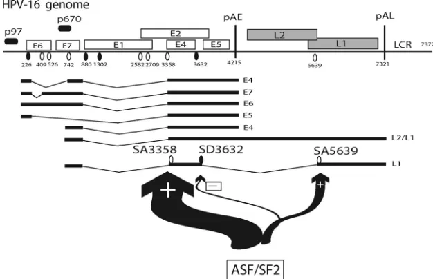
many genes. In case of HPV-16, ASF/SF2 is required primarily for splicing to SA3358. This splicing event removes the intron between SD880 and SA3358 or that between SD226 and SA3358, resulting in the production of HPV-16 mRNAs that are efficiently expressed in HPV-16-infected cells (Fig. 1). HPV-16 E6 and E7 mRNAs that are spliced between SD880 and SA3358 are vastly more common than those that retain the intron between SD880 and SA3358 (9, 17, 24, 35). In general, intron-containing mRNAs are retained and degraded in the nucleus to a higher degree than spliced mRNAs, which may explain the low levels of HPV-16 mRNAs that contain se-quences between SD880 and SA3358. One may therefore con-clude that mRNAs that are spliced between SD880 and SA3358 benefit from high levels of ASF/SF2 (Fig. 9). Such mRNAs encode E6, E7, or E4 proteins, and the shorter forms of E6 named E6*-I and E6*-II, depending on the upstream splicing events on these mRNAs (Fig. 1A). Similarly, mRNAs that are spliced between SD226 and SA3358, and have the potential to produce E5 protein (Fig. 1A), would benefit from high levels of ASF/SF2. Of the early HPV-16 mRNAs that are selectively stimulated by high levels of ASF/SF2, there is an overrepresentation of mRNAs that express viral cell growth-stimulatory proteins, including E5, E6, and E7. This deduction fits well with the previously observed high expression levels of ASF/SF2 in high-grade cervical lesions and cervical cancers (7). It was previously reported that HPV-16 E2 enhances ASF/ SF2 expression by transcriptional activation of the ASF/SF2 promoter (19). Here, we report that ASF/SF2 induces splicing to SA3358 on the HPV-16 genome. Interestingly, splicing to SA2709, the splicing event that generates E2 mRNAs, and splicing to SA3358 are two mutually exclusive events (Fig. 1A). High levels of ASF/SF2 may therefore downregulate expres-sion of E2, presumably in a negative feedback manner, perhaps marking the entry into the late phase of the viral life cycle with L1 and L2 expression.
As HPV-16 enters the late stage of the viral life cycle and the infected cell differentiates terminally, suppression of the exclu-sively late splice sites SD3632 and SA5639 is released and efficiency of early polyadenylation reduced. These events cause induction of L1 and L2 mRNA production. However, SA3358 must be efficiently used also in these cells, as it is used by late L1 mRNAs. This would require the presence of ASF/SF2 also in terminally differentiated cells. Alternatively, the role of ASF/SF2 is taken over by another, yet-unidentified splicing factor that is present in terminally differentiated cells. As ASF/ SF2 is normally not detected in the upper layers of uninfected cervical epithelium, this may indeed be the case (7). However, one may speculate that HPV-16 selectively enhances produc-tion of ASF/SF2, even in the terminally differentiated cells in which L1 and L2 protein can be detected. This could be per-formed by E2 or indirectly by another HPV protein. It would be interesting to compare expression of ASF/SF2 and HPV-16 L1 and L2 expression in individual cells in HPV-16-infected cervical epithelium to determine if ASF/SF2 is present in the cells that express L1 and/or L2 protein. In addition, it would be of interest to determine if the dependence on ASF/SF2 is conserved among different HPV types. We have found that the ESEfinder (4) predicts the presence of multiple ASF/SF2 sites in the corresponding position of both low-risk and high-risk HPV types and in mucosal and cutaneous HPV types. We
FIG. 8. (A) Western blot of cell extracts from HeLa cells trans-fected with pBELM in the absence or presence of siRNAs against ASF/SF2, as described in Materials and Methods. (B and C) RT-PCR on cytoplasmic RNA extracted from HeLa cells transfected with pBELM (35) in the absence or presence of siRNA against ASF/SF2. RT-PCR was performed with primers 757s and L1aM (B) or 757s and E4A (C) (35). RT-PCR was also performed with GAPDH mRNA (C) (13). Structures of the L1 and L1i mRNAs, or the E4 mRNA, are shown to the right of the gels. Numbers indicate positions of splice sites on the mRNAs. M, molecular size marker.
8228 SOMBERG AND SCHWARTZ J. VIROL.
on November 8, 2019 by guest
http://jvi.asm.org/
[image:10.585.59.262.73.407.2]therefore believe that ASF/SF2 regulates splicing of both low-and high-risk HPV types.
ACKNOWLEDGMENTS
We thank all the members in the S. Schwartz group as well as the groups of Go¨ran Magnusson, Go¨ran Akusja¨rvi, and Catharina Svens-son for comments at laboratory meetings and for generously sharing reagents.
This research was sponsored by the Swedish Cancer Society, the Swedish Research Council/Medicine, Linneus Support from the Swed-ish Research Council to Uppsala RNA Research Center, Technolog-ical Sector Research Study III, the Dublin Institute of Technology, and Science Foundation Ireland.
REFERENCES
1.Baker, C., and C. Calef.1997. Maps of papillomavirus mRNA transcripts.
InS. R. Billakanti, C. E. Calef, A. D. Farmer, A. L. Halpern, and G. L.
Myers (ed.), Human papillomaviruses: a compilation and analysis of nucleic acid and amino acid sequences. Los Alamos National Laboratory, Los Alamos, NM.
2.Belaguli, N. S., M. M. Pater, and A. Pater.1992. Nucleotide 880 splice donor site required for efficient transformation and RNA accumulation by human
papillomavirus type 16 E7 gene. J. Virol.66:2724–2730.
3.Caputi, M., M. Freund, S. Kammler, C. Asang, and H. Schaal.2004. A bidirectional SF2/ASF- and SRp40-dependent splicing enhancer regulates
human immunodeficiency virus type 1rev,env,vpu, andnefgene expression.
J. Virol.78:6517–6526.
4.Cartegni, L., J. Wang, Z. Zhu, M. Q. Zhang, and A. R. Krainer.2003. ESEfinder: a web resource to identify exonic splicing enhancers. Nucleic
Acids Res.31:3568–3571.
5.Chow, L. T., M. Nasseri, S. M. Wolinsky, and T. R. Broker.1987. Human papillomavirus types 6 and 11 mRNAs from genital condylomata acuminata.
J. Virol.61:2581–2588.
6.Doorbar, J., A. Parton, K. Hartley, L. Banks, T. Crook, M. Stanley, and L. Crawford.1990. Detection of novel splicing patterns in a HPV16-containing
keratinocyte cell line. Virology178:254–262.
7.Fay, J., P. Kelehan, H. Lambkin, and S. Schwartz.2009. Increased expres-sion of cellular RNA-binding proteins in HPV-induced neoplasia and
cervi-cal cancer. J. Med. Virol.81:897–907.
8.Graham, S. V.2008. Papillomavirus 3⬘ UTR regulatory elements. Front.
Biosci.13:5646–5663.
9.Grassmann, K., B. Rapp, H. Maschek, K. U. Petry, and T. Iftner.1996. Identification of a differentiation-inducible promoter in the E7 open reading frame of human papillomavirus type 16 (HPV-16) in raft cultures of a new
cell line containing high copy numbers of episomal HPV-16 DNA. J. Virol.
70:2339–2349.
10.Howley, P. M., and D. R. Lowy.2001.Papillomaviridaeand their replication,
p. 2197–2229.InKnipe, D. M., P. M. Howley, D. E. Griffin, R. A. Lamb,
M. A. Martin, B. Roizman, and S. E. Straus (ed.), Fields virology, 4th ed., vol. 2. Lippincott Williams & Wilkins, Philadelphia, PA.
11.Hummel, M., J. B. Hudson, and L. A. Laimins.1992. Differentiation-induced and constitutive transcription of human papillomavirus type 31b in cell lines
containing viral episomes. J. Virol.66:6070–6080.
12.Hummel, M., H. B. Lim, and L. A. Laimins.1995. Human papillomavirus type 31b late gene expression is regulated through protein kinase
C-medi-ated changes in RNA processing. J. Virol.69:3381–3388.
13.Johansson, C., H. Zhao, E. Bajak, F. Granberg, U. Pettersson, and C. Svensson. 2005. Impact of the interaction between adenovirus E1A and
CtBP on host cell gene expression. Virus Res.113:51–63.
14.Karni, R., E. de Stanchina, S. W. Lowe, R. Sinha, D. Mu, and A. R. Krainer.
2007. The gene encoding the splicing factor SF2/ASF is a proto-oncogene.
Nat. Struct. Mol. Biol.14:185–193.
15.Klumpp, D., F. Stubenrauch, and L. A. Laimins.1997. Differential effects of the splice acceptor at nucleotide 3295 of human papillomavirus 31 on stable
and transient viral replication. J. Virol.71:8186–8194.
16.Longworth, M. S., and L. A. Laimins.2004. Pathogenesis of human
papil-lomaviruses in differentiating epithelia. Microbiol. Mol. Biol. Rev.68:362–
372.
17.Milligan, S. G., T. Veerapraditsin, B. Ahamet, S. Mole, and S. V. Graham.
2007. Analysis of novel human papillomavirus type 16 late mRNAs in
dif-ferentiated W12 cervical epithelial cells. Virology360:172–181.
18.Mole, S., M. McFarlane, T. Chuen-Im, S. G. Milligan, D. Millan, and S. V. Graham.2009. RNA splicing factors regulated by HPV16 during cervical
tumour progression. J. Pathol.219:383–391.
19.Mole, S., S. G. Milligan, and S. V. Graham.2009. Human papillomavirus type 16 E2 protein transcriptionally activates the promoter of a key cellular
splicing factor, SF2/ASF. J. Virol.83:357–367.
20.Nasseri, M., R. Hirochika, T. R. Broker, and L. Chow.1987. A human papillomavirus type 11 transcript encoding and E1-E4 protein. Virology
159:433–439.
21.Norrild, B., C. H. Hansen, and J. A. Glahder.2007. Identification and activity
of promoters in the HPV genome, p. 1–26.InB. Norrild (ed.), Human
papillomavirus gene regulation and transformation. Transworld Research Network, Trivandrum, India.
22.Ozbun, M. A., and C. Meyers.1997. Characterization of late gene transcripts expressed during vegetative replication of human papillomavirus type 31b.
J. Virol.71:5161–5172.
23.Rotenberg, M. O., L. T. Chow, and T. R. Broker.1989. Characterisation of rare human papillomavirus type 11 mRNAs coding for regulatory and
struc-tural proteins, using the polymerase chain reaction. Virology172:489–497.
[image:11.585.134.447.70.272.2]24.Rush, M., X. Zhao, and S. Schwartz.2005. A splicing enhancer in the E4
FIG. 9. Effect of ASF/SF2 on HPV-16 mRNA splicing in HeLa cells. ASF/SF2 is primarily stimulating splicing to SA3358 and at the same time weakly inhibiting late splice site SD3632. However, SD3632 may be more efficiently suppressed by another factor(s). ASF/SF2 can also stimulate splicing to SA5639, but this effect is seen primarily when SA5639 is optimized by inactivation of silencers downstream of SA5639 or by optimization of the SA5639 polypyrimidine tract. Taken together, these results show that ASF/SF2 strongly favors production of mRNAs that are using SA3358 and are polyadenylated at pAE. These mRNAs include the E4, E5, E6, and E7 mRNAs indicated in the figure.
on November 8, 2019 by guest
http://jvi.asm.org/
coding region of human papillomavirus type 16 is required for early mRNA splicing and polyadenylation as well as inhibition of premature late gene
expression. J. Virol.79:12002–12015.
25.Schmitt, M., V. Dalstein, T. Waterboer, C. Clavel, L. Gissman, and M. Pawlita. 2010. Diagnosing cervical cancer and high-grade precursors by
HPV-16 transcription patterns Cancer Res.70:249–256.
26.Schwartz, S.2008. HPV-16 RNA processing. Front. Biosci.13:5880–5891. 27.Smotkin, D., and F. O. Wettstein.1986. Transcription of human papillomavirus
type 16 early genes in a cervical cancer and a cancer-derived cell line and
identification of the E7 protein. Proc. Natl. Acad. Sci. U. S. A.83:4680–4684.
28.Sokolowski, M., H. Furneaux, and S. Schwartz.1999. The inhibitory activity
of the AU-rich RNA element in the human papillomavirus type 1 late 3⬘
untranslated region correlates with its affinity for the elav-like HuR protein.
J. Virol.73:1080–1091.
29.Somberg, M., M. Rush, J. Fay, F. Ryan, H. Lambkin, G. Akusja¨rvi, and S. Schwartz.2009. Adenovirus E4orf4 induces HPV-16 late L1 mRNA
produc-tion. Virology383:279–290.
30.Somberg, M., X. Zhao, M. Fro¨hlich, M. Evander, and S. Schwartz.2008. PTB induces HPV-16 late gene expression by interfering with splicing
inhib-itory elements at the major late 5⬘-splice site SD3632. J. Virol.82:3665–3678.
31.Spångberg, K., L. Wiklund, and S. Schwartz.2000. HuR, a protein
impli-cated in oncogene and growth factor mRNA decay, binds to the 3⬘ends of
hepatitis C virus RNA of both polarities. Virology274:378–390.
32.Wiklund, L., M. Sokolowski, A. Carlsson, M. Rush, and S. Schwartz.2002. Inhibtion of translation by UAUUUAU and UAUUUUUAU motifs of the
AU-rich RNA instability in the HPV-1 late 3⬘untranslated region. J. Biol.
Chem.277:40462–40471.
33.Zhao, X., D. O¨ berg, M. Rush, J. Fay, H. Lambkin, and S. Schwartz.2005. A 57-nucleotide upstream early polyadenylation element in human papilloma-virus type 16 interacts with hFIP1, CstF-64, hnRNP C1/C2, and
polypyrimi-dine tract binding protein. J. Virol.79:4270–4288.
34.Zhao, X., M. Rush, A. Carlsson, and S. Schwartz.2007. The presence of
inhibitory RNA elements in the late 3⬘-untranslated region is a conserved
property of human papillomaviruses. Virus Res.125:135–144.
35.Zhao, X., M. Rush, and S. Schwartz.2004. Identification of an hnRNP A1-dependent splicing silencer in the human papillomavirus type 16 L1 coding region that prevents premature expression of the late L1 gene. J.
Vi-rol.78:10888–10905.
36.Zheng, Z. M., and C. C. Baker.2006. Papillomavirus genome structure,
expression, and posttranscriptional regulation. Front. Biosci.11:2286–2302.
37.zur Hausen, H.2002. Papillomaviruses and cancer: from basic studies to
clinical application. Nat. Rev. Cancer2:342–350.
8230 SOMBERG AND SCHWARTZ J. VIROL.