A Simple Cloning-free Method to Efficiently
Induce Gene Expression Using CRISPR/Cas9
Lyujie Fang,
1,2,3,7Sandy S.C. Hung,
1,7Jennifer Yek,
1,2Layal El Wazan,
1,2Tu Nguyen,
1,2Shahnaz Khan,
1Shiang Y. Lim,
4Alex W. Hewitt,
1,5and Raymond C.B. Wong
1,2,61Centre for Eye Research Australia, Royal Victorian Eye and Ear Hospital, East Melbourne, VIC, Australia;2Ophthalmology, Department of Surgery, University of Melbourne, East Melbourne, VIC, Australia;3Jinan University, Guangzhou, China;4O’Brien Institute Department, St Vincent’s Institute of Medical Research, Fitzroy, VIC, Australia;5Menzies Institute for Medical Research, School of Medicine, University of Tasmania, Hobart, TAS, Australia;6Shenzhen Eye Hospital, School of Medicine, Shenzhen University, Shenzhen, China
Gain-of-function studies often require the tedious cloning of transgene cDNA into vectors for overexpression beyond the physiological expression levels. The rapid development of CRISPR/Cas technology presents promising opportunities to address these issues. Here, we report a simple, cloning-free method to induce gene expression at an endogenous locus us-ing CRISPR/Cas9 activators. Our strategy utilizes synthesized sgRNA expression cassettes to direct a nuclease-null Cas9 com-plex fused with transcriptional activators (VP64, p65, and Rta) for site-specific induction of endogenous genes. This strategy allows rapid initiation of gain-of-function studies in the same day. Using this approach, we tested two CRISPR activation systems, dSpCas9VPR and dSaCas9VPR, for induction of mul-tiple genes in human and rat cells. Our results showed that both CRISPR activators allow efficient induction of six different neural development genes (CRX,RORB,RAX,OTX2,ASCL1, and NEUROD1) in human cells, whereas the rat cells exhibit more variable and less-efficient levels of gene induction, as observed in three different genes (Ascl1,Neurod1,Nrl). Alto-gether, this study provides a simple method to efficiently activate endogenous gene expression using CRISPR/Cas9 activators, which can be applied as a rapid workflow to initiate gain-of-function studies for a range of molecular- and cell-biology disciplines.
INTRODUCTION
Conventional gene overexpression studies require the need to clone the transgene cDNA into an expression vector and therefore involve DNA ligation, bacterial transformation, screening clones, plasmid purification, and quality check to confirm the vector sequences. This represents a tedious and costly procedure, especially for large-scale genome-wide overexpression studies. Further to this, the use of whole-transgene cDNA imposes a challenge for overexpressing multiple genes simultaneously in cells, due to the large burden of DNA required to be delivered into the cells.
The emergence of CRISPR/Cas technology has revolutionized the
field of molecular biology, providing a promising tool for precise gene editing with profound implications for development of gene
therapy.1CRISPR/Cas utilizes an RNA-guided mechanism for site-specific DNA cleavage, which has been used to knock in or knock out genesin vitro2,3andin vivo.4,5However, thefirst described Strep-tococcus pyogenes(Sp)Cas9 is large in size and presents a challenge for packaging into adeno-associated viruses (AAVs) for therapeutic de-livery. Subsequently, smaller Cas9 variants have been identified, including Staphylococcus aureus(Sa)Cas9,6,7Neisseria meningitidis
(Nm)Cas9,8and compylobacter Jejuni (Cj)Cas9,9which have higher therapeutic potential due to their smaller sizes. Also, further modifi -cations of Cas9 have expanded the ability of using CRISPR/Cas beyond genome editing, including control of gene regulation, epige-netics, and chromatin imaging.10For instance, a nuclease-null dead Cas9 can be fused to transcriptional activators to target the regulatory region of a gene to induce its expression.11Importantly, multiple sin-gle-guide RNAs (sgRNAs) targeting different genes can be utilized to induce multiplex gene expression. Since only a short sgRNA is needed to induce expression of a single gene, rather than a whole-transgene cDNA copy, the CRISPR activation approach can potentially reduce the number of viral vectors needed for overexpressing multiple genes. Another advantage of CRISPR activation is it activates gene expres-sion directly at endogenous locus with high specificity, which allows for expression of splice variants as well as endogenous gene regulation via UTR regions.12,13
Several reports have previously utilized a cloning-free CRISPR approach to knock in or knock out genes. Infission yeasts, Zhang et al.14 showed that the gap-repair mechanism can be used to
assemble PCR-amplified sgRNA fragments and linear Cas9 plasmids together. In mammalian cells, Arbab et al.15have reported a self-clon-ing CRISPR/Cas9 approach by usself-clon-ing palindromic sgRNA, either in expression cassette or short DNA sequences, to allow homologous recombination in target cells to yield a functional site-specific sgRNA
Received 20 August 2018; accepted 15 November 2018; https://doi.org/10.1016/j.omtn.2018.11.008.
7These authors contributed equally to this work.
Correspondence:Raymond C.B. Wong, Centre for Eye Research Australia, Level 6, 75 Commercial Road, Melbourne, VIC 3004, Australia.
plasmid. The authors also showed that co-transfection of the Cas9 expression plasmids and sgRNA expression cassettes synthesized as short 500-bp oligonucleotides allow efficient knockout of target gene in mouse embryonic stem cells. Others have described the use of CRISPR ribonucleoprotein complexes for generation of knockin mice,16,17as well as gene editing in mammalian cellsin vitrodelivered through various methods, such as transfection by cationic lipids, nanoparticles, or cell-penetrating peptides.18–22However, a cloning-free approach has not been systematically reviewed for gene activa-tion or repression using CRISPR/Cas9.
Here, we present a simple and rapid method to use CRISPR/Cas for gene activation. Our method utilizes commercially synthesized sgRNA expression cassettes, which bypass the need for molecular cloning of site-specific sgRNA plasmids. In this study, we compare the use of synthesized sgRNA expression cassettes with two CRISPR/Cas activators, dSpCas9VPR and dSaCas9VPR, to induce gene expression in human and rat cells. Our strategy vastly simplifies the initiation of gain-of-function studies and as such has implications across many disciplines in cell biology.
RESULTS
To utilize CRISPR/Cas to activate endogenous genes in mammalian cells, wefirst tested the dSpCas9VPR system.23This system utilizes a nuclease-null dead SpCas9 (dSpCas9) coupled with transcription factor activation domains VP64, p65, and Rta (VPR). The sgRNAs were designed using the synergistic activation mediator (SAM) sgRNA design tool. For gene activation, we chose six human genes that encoded for transcription factors that regulate neural and retinal development: RAX, OTX2, RORB, CRX, ASCL1, and NEUROD1
(Table 1). We synthesized sgRNA expression cassettes as linear oligo-nucleotide fragments containing an upstream U6 promoter, the sgRNA, and the corresponding sgRNA scaffold (Figure S1). The sgRNA expression cassettes and the dSpCas9VPR plasmids were
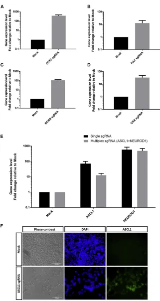
co-transfected into HEK293A cells. By day 3 post-transfection, we can detect robust gene activation in HEK293A cells using dSpCas9VPR, with upregulation ofOTX2(375-fold increase; Fig-ure 1A),RAX(13-fold increase;Figure 1B),RORB(110-fold in-crease; Figure 1C), andCRX(35-fold increase;Figure 1D). Simi-larly, the dSpCas9VPR system is also efficient in gene activation of
ASCL1and NEUROD1, with70-fold increase and 585-fold in-crease, respectively (Figure 1E). Immunocytochemistry analysis also confirmed efficient expression of ASCL1 protein in HEK293A following dSpCas9VPR gene activation (Figure 1F).
To determine the kinetics of the gene activation, we performed a time-course experiment followingASCL1gene activation. Our results showed that dSpCas9VPR upregulatedASCL1expression levels after 2 days and gradually decreased after 3 days, with persistent upregu-lated levels up to 6 days post-transfection (Figure S3). These results showed that dSpCas9VPR resulted in maximal gene activation level after 2–3 days following transfection.
Subsequently, we tested the feasibility of using dSpCas9VPR for multiplex gene activation. We co-transfected sgRNAs for ASCL1
andNEUROD1into HEK293A cells. Notably, both genes can be up-regulated simultaneously and efficiently using the dSpCas9VPR, with
13-fold induction ofASCL1expression and485-fold induction of
NEUROD1expression (Figure 1E). However, multiplexing resulted in a lower level of gene induction compared to using single sgRNA, an effect more prominent in induction of ASCL1(Figure 1E). Taken together, our results demonstrated that dSpCas9VPR can be used to efficiently activate gene expression in human cells.
[image:2.603.47.552.126.314.2]Furthermore, we assessed the feasibility of using dSpCas9VPR for gene activation in rat cells. As the SAM sgRNA design tool does not support design for the rat genome, we utilized the gene-acti-vator sgRNA design tool in Benchling (Table 1). We analyzed
Table 1. Information of sgRNAs Used for SpCas9
Species Name TSS Distance Strand Sequence PAM On-Target Score
Rat Ascl1 sgRNA 1 183 bp + 50-ACGCACTGCAACAACAAACC-30 CGG 46.1
Ascl1 sgRNA 2 351 bp 50-TCCTAGGTAGAAAGTCTGGA-30 GGG 73.4
Neurod1 sgRNA 1 258 bp + 50-TGCGGGTAAAAACAGGTCCG-30 CGG 56.1
Neurod1 sgRNA 2 164 bp + 50-ATACAAATAGGCAGGTCACG-30 TGG 84.1
Nrl sgRNA 1 573 bp 50-CTTTACCTCTCAAAGCCTTC-30 AGG 27.5
Nrl sgRNA 2 764 bp + 50-CCATCTGCTTAGACTCACCA-30 TGG 77.8
Human ASCL1 sgRNA 1 181 bp + 50-CGGGAGAAAGGAACGGGAGG-30 GGG 30.9
NEUROD1 sgRNA 1 33 bp + 50-AGGGGAGCGGTTGTCGGAGG-30 AGG 30.9
RAX sgRNA 99 bp + 50-GAGGGAGGGGCCGAGAGAAG-30 GGG 44.0
OTX2 sgRNA 170 bp + 50-AGATTGTAATTGCTTTCTTC-30 GGG 35.2
CRX sgRNA 114 bp 50-AGGGAGGCCCCAGCTCCTGC-30 CGG 51.6
RORB sgRNA 172 bp + 50-CCCGGCCACCTCGGACTCCC-30 TGG 41.8
Mouse Nkx2.5 sgRNA 171 bp + 50-GTATTTTCTTTGAGTGTGTC-30 TGG 36.1
Figure 1. Efficient Gene Activation Using dSpCas9VPR in HEK293A Cells
qPCR analysis of gene activation for (A)OTX2, (B)RAX, (C)
RORB, (D)CRX, (E)ASCL1,NEUROD1, or multiplex in-duction ofASCL1andNEUROD1. Results are displayed as the mean of three independent biological repeats±
dSpCas9VPR-mediated activation of three genes,Ascl1,Neurod1, and
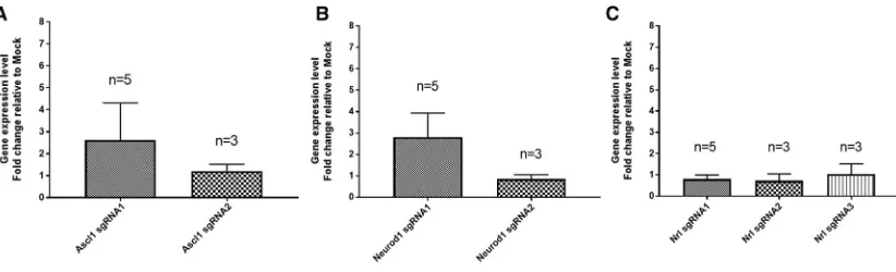
Nrl, in a rat Müller glial cell line rMC1, which can be transfected effi -ciently (Figure S5). Our results showed thatAscl1can be activated to modest levels using two sgRNAs, resulting in an3- and2-fold in-crease, respectively (Figure 2A). Similarly, two sgRNAs were tested forNeurod1gene activation. While one sgRNA resulted in a modest increase in Neurod1 expression levels (3-fold increase), another sgRNA failed to activateNeurod1(Figure 2B). ForNrl, the two de-signed sgRNAs resulted in an3- and1.6-fold induction in gene expression, respectively (Figure 2C). We also tested gene activation with dSpCas9VPR in another ratfibroblast cell line R12. However, we did not observe significant gene activation inAscl1, Neurod1, norNrl(Figure S4). This is unlikely due to problems with transfec-tion, as R12 can be efficiently transfected in this context (Figure S5). Moreover, we showed that the dSpCas9VPR can efficiently activate gene expression in mouse embryonicfibroblasts (19-fold increase inNkx2.5;Figure S6), suggesting that the inefficient CRISPR activa-tion seen is specific to rat cells and not other rodent cells. Collectively, these results demonstrated that dSpCas9VPR can be used to activate genes in rat cells, albeit only limited to modest levels of upregulation in certain rat cells and is not as efficient as in human cells.
Next, we assessed the efficiency of gene activation using another CRISPR/Cas activation system, dSaCas9VPR, which utilizes a nuclease-null SaCas9 coupled with VPR. Notably, the dSaCas9VPR is much smaller in size than dSpCas9VPR, which has important impli-cations for packaging into viral vectors for gene therapy. We designed sgRNAs using Benchling for both human and rat genes and selected two to three sgRNAs against each gene for evaluation (Table 2). The sgRNA expression cassette for dSaCas9VPR contained a 50 Myc tag, upstream U6 promoter, sgRNA, sgRNA scaffold, and a 30 HA tag (Figure S2). In HEK293A cells, we showed that dSaCas9VPR can activate the expression levels of endogenous ASCL1 efficiently (760-fold increase; Figure 3A). Also, we detected upregulation of theASCL1protein levels following dSaCas9VPR gene activation ( Fig-ure 3B). However, dSaCas9VPR exhibited variable efficiency in gene activation in rMC1 cells (Figure 4), a result similar to those observed in dSpCas9VPR. Two sgRNAs were tested for activation ofAscl1or
Neurod1, with one sgRNA inducing modest levels of gene upregulation and the other failing to do so (Ascl1,2.6,1 fold changes;Neurod1,
2.8,1 fold changes;Figures 4A and 4B). However, forNrl activa-tion, all three sgRNAs tested failed to upregulateNrlexpression levels (Figure 4C). In summary, we showed that dSaCas9VPR can be used to efficiently activate gene expression in human cells; however, its effect in rat cells is variable and remained inefficient.
DISCUSSION
A key limiting factor in the conventional design of overexpression studies is the requirement of tedious plasmid construction for each transgene, which involves molecular cloning steps that take more than 1 week. Here, we present a simplified method for gene activation using CRISPR/Cas9 activators in mammalian cells that is feasible in 1 day. Notably, our strategy to transfect sgRNA as a synthesized linear oligonucleotide fragment with U6 promoter allows sgRNA expression in the cells and eliminates the need to clone sgRNA into a designated vector prior to transfection. Our strategy provides a rapid way to initiate gene-overexpression studies using the CRISPR activation system.
This study compared two CRISPR activation systems, dSpCas9VPR and dSaCas9VPR, in human and rat cells. Our results demonstrated that both dSpCas9VPR and dSaCas9VPR can efficiently induce gene expression in human cells. We showed the use of dSpCas9VPR for multiplex activation of two genes in human cells, as well as its high efficiency for gene activation in mouse cells. Furthermore, in rat cells we have assessed activation of three genes using six sgRNAs for dSpCas9VPR and seven sgRNAs for dSaCas9VPR in two different rat cell lines. However, our results showed only modest levels and, in some cases, negligible levels of gene activation in rat Müller glial cells andfibroblasts. This is unlikely due to issues with transfection efficiency, as both rat cell types can be efficiently transfected with robust GFP expression. We speculate that this could be due to inad-equate support of the current sgRNA design algorithm for the rat genome. For instance, many of the sgRNAs used for rat genes have high ranking of on-target scores (Table 2), but they are mostly ineffi -cient in inducing expression of rat genes using the CRISPR activation
Figure 2. Assessment of Multiple sgRNAs for dSpCas9VPR to Induce Gene Activation in Rat Mu¨ller Glial Cell rMC1
systems. This highlights the need to improve the design and accuracy of predicting functional sgRNAs for the rat genome. Furthermore, it is also possible that the differences in gene activation we observed are due to variations in chromatic state and accessibility in different rat and human cells. Therefore, alternative CRISPR activation systems may be more effective at enhancing gene activation in rat cells. In addition to the dCas9-VPR system from the Church laboratory, other CRISPR activation systems have been described, such as the SAM sys-tem, the SunTag activator syssys-tem, and dCas9-VP128 system.24–26
These alternative CRISPR activation systems utilize distinct tran-scriptional activators to enhance gene activity. Chavez et al.11 under-took a comprehensive head-to-head comparison of various CRISPR activation systems and found that different systems showed varying activities at different gene loci as well as in different cell types tested.
Notably, the simplified method for gene activation described here can also be adopted for other CRISPR systems for gene editing or repres-sion, which allows rapid testing of effective sgRNA sequences that can be selected for further experiments in different delivery systems. Also, our sgRNA expression cassette design can be applied to different Cas9 activator systems, since the same sgRNA design can be used for different Cas9 activator systems (such as SpCas9VPR, SpCas9-Sun-tag, SpCas9-VP64).
A potential challenge of using CRISPR/Cas systems is that not all sgRNAs are efficient in Cas9 targeting; thus, multiple sgRNAs are often screened to identify the functional sgRNAs. In our experience, the sgRNA design tools for predicting functional sgRNAs are gener-ally very accurate for human cells. For dSpCas9VPR, thefirst sgRNA designed is functional for gene activation in 13 out of 15 genes tested (87%;Table S1). Similarly, although we only tested gene activation for a single gene using dSaCas9VPR, the first sgRNA designed is highly efficient for gene activation (Table S1). Therefore, we recom-mend designing two sgRNAs within 300 bp upstream of the tran-scription start site to be tested for gene activation in human cells. It should be noted that sgRNAs that target different proximities of the transcriptional start site can result in different efficiencies of gene acti-vation,18and this can be further optimized for individual target genes of interest. Additionally, it is also possible to combine several sgRNAs
targeting different promoter regions to improve the levels of gene activation using CRISPRa.
In summary, this study outlines a simple and robust workflow to effi -ciently activate endogenous gene expression in mammalian cells us-ing CRISPR/Cas activators, which can be applied as a rapid workflow to initiate gain-of-function studies for a range of molecular- and cell-biology subjects.
MATERIALS AND METHODS
Institutional Approval
This study was approved by the Institutional Biosafety Committee at Monash University (#10969) in compliance with the regulations by the Office of the Gene Technology Regulator in Australia.
sgRNA Design and Preparation of Expression Cassette
For SpCas9, sgRNAs with NGG protospacer adjacent motifs (PAMs) were designed using the SAM sgRNA design tool (http://sam. genome-engineering.org/database/) for human genes and Benchling (https://benchling.com/) for rat genes. The SpCas9 sgRNA expression cassette contains an upstream U6 promoter, sgRNA, and sgRNA scaf-fold with stem extension and stem loop (Figure S1).
For SaCas9, sgRNAs with NNGRRT PAMs were designed using Benchling (https://benchling.com/). Predicted sgRNAs with long stretches of repeating nucleotides are excluded from selection. The SaCas9 sgRNA expression cassette contains a Myc tag, U6 promoter, sgRNA, sgRNA scaffold, and HA tag (Figure S2).
[image:5.603.51.550.126.246.2]Both SpCas9 and SaCas9 sgRNA expression cassettes (<500 bp) were synthesized as gBLOCK gene fragments (Integrated DNA Technolo-gies). The sgRNA expression cassettes were amplified by PCR using the following primers: SpCas9 forward primer, 50-TGAGTAT TACGGCATGTGAGGGC-30; SpCas9 reverse primer, 50-TCAATG TATCTTATCATGTCTGCTCGA-30; SaCas9 forward primer, 50-GA ACAAAAACTCATCTCAGAAGAGGATCTG-30; SaCas9 reverse primer, 50-TACCCATACGATGTTCCAGATTACGCT-30. PCR was performed using KOD Hot Start DNA polymerase (Merck Millipore) with the following thermal profile: 95C for 2 min; 30 cycles of 95C
Table 2. Information of sgRNAs Used for SaCas9
Species Name TSS Distance Strand Sequence PAM On-Target Score On-Target Score Ranking
Rat Ascl1 sgRNA 1 180 bp + 50-GCACTGCAACAACAAACCCGG-30 CTGAAT 70.9 1
Ascl1 sgRNA 2 223 bp 50-TGGCGCGTGCCGGACTCCCGG-30 CTGAAT 64.2 5
Neurod1 sgRNA 1 258 bp + 50-CTGCGGGTAAAAACAGGTCCG-30 CGGAGT 56.1 2
Neurod1 sgRNA 2 205 bp + 50-TTCTTCTGGCCACAAAGGGGC-30 CGGAAT 38.5 5
Nrl sgRNA 1 575 bp 50-TTTACCTCTCAAAGCCTTCAG-30 GAGAGT 79.3 1
Nrl sgRNA 2 145 bp + 50-TTCAGGGCTGCTTCATTACTC-30 CGGAAT 45.1 9
Nrl sgRNA 3 760 bp 50-TTTAACTTAGCACCTGCCATG-30 GTGAGT 72.7 2
Human ASCL1 sgRNA 239 bp + 50-GCACTGCAACAACAAACCCAG-30 CTGAAT 74.2 2
On-target score is an optimized score for 20-bp sgRNA based on Doench et al.28. The on-target scores are ranked for sgRNAs targeting 2,000-bp upstream of transcription start site of
for 20 s, 66C (SpCas9 sgRNA) or 64C (SaCas9 sgRNA) for 10 s, 70C for 8 s; 70C for 5 min. The PCR amplicons were separated by gel electrophoresis, and the sgRNA expression cassettes were ex-tracted using the Wizard SV gel and PCR cleanup kit (Promega). The amplified sgRNA expression cassettes were checked with Nano-drop to confirm good DNA quality.
Cell Culture
Rat Müller glial cells rMC1, rat fibroblasts R12, mouse embryonic
fibroblasts, and HEK293A cells were maintained in DMEM high-glucose media supplemented with 10% fetal calf serum, 2 mM L-glutamine, and 0.5% penicillin/streptomycin (all from Thermo Fisher). All cells were passaged using 0.25% trypsin before the culture become confluent and maintained in incubators at 37C with 5% CO2
level.
Transfection Efficiency Assay
rMC1 and R12 cells were transfected with the pmaxGFP construct (Lonza) using Lipofectamine 3000 overnight, following the manufac-turer’s instruction. GFP expression is determined 1 day after transfec-tion using afluorescence microscope (Olympus CKX53).
Gene Activation Using CRISPR/Cas Activation
dSaCas9-VPR and dSpCas9-VPR plasmids were gifts from George Church (Addgene #68495 and #63798, respectively). rMC1, R12, mouse embryonicfibroblasts and HEK293A cells were transfected us-ing Lipofectamine 3000. In brief, cells were plated down on a 12-well
plate at day 0 (6104/well). At day 1, the cells were transfected with 360 ng sgRNA expression cassettes and 800 ng dSaCas9-VPR or dSpCas9-VPR plasmids using Lipofectamine 3000 overnight. In some experiments, the cells and DNA were upscale proportionally to obtain more RNA. Mock control (no DNA transfected) was uti-lized as a negative control. At day 4, the samples were harvested for RNA to assess gene expression levels orfixed for immunocytochem-istry analysis. In some experiments, RNA was harvested at different time points (days 2, 3, 4, 5, and 6) to determine kinetics of CRISPR gene activation.
qPCR Analysis
Total RNA was extracted using the RNeasy kit (QIAGEN) or the Illus-tra RNAspin kit (GE Healthcare) followed by DNase treatment. For mouse embryonic fibroblasts, RNA was extracted from cells using TriReagent (Thermo Fisher) followed by RNA precipitation with chlo-roform and isopropanol (Sigma-Aldrich). cDNA synthesis was per-formed using the High Capacity cDNA Reverse Transcription Kit (Thermo Fisher) following the manufacturer’s instructions. Taqman gene-expression assay (Thermo Fisher) was performed using the Taq-man Fast Advanced Master Mix with the following probes: huTaq-man
RAX (Hs00429459_m1), human OTX2 (Hs00222238_m1), human
ASCL1 (Hs00269932_m1), human NEUROD1 (Hs00159598_m1), human CRX(Hs00230899_m1), human RORB (Hs00199445_m1), human ACTB (Hs99999903_m1), rat Ascl1 (Rn00574345_m1), rat
Neurod1 (Rn00824571_s1), rat Nrl (Rn01481925_m1), rat Gapdh
[image:6.603.52.369.109.222.2](Rn01775763_g1), mouse Nkx2.5 (Mm00657783_m1), and mouse
Figure 3. Efficient Gene Activation Using dSaCas9VPR in HEK293A Cells
(A) qPCR analysis of gene activation for ASCL1 in HEK293A cells. Results are displayed as mean of three independent biological repeats±SEM. (B) Immunocyto-chemistry results showed upregulated protein expression of ASCL1 (green) in HEK293A following gene activation with dSaCas9VPR. Scale bars, 100mm.
Figure 4. Comparison of Multiple sgRNAs to Induce Gene Activation in Rat Mu¨ller Glial Cell rMC1 Using dSaCas9VPR
[image:6.603.97.508.566.691.2]Gapdh(Mm99999915_g1). qPCR was processed using the ABI Step One Plus system, the ABI 7500 system, or the QuantStudio 6 Flex Real-Time PCR system (Applied Biosystems). The delta delta Ct method was used to calculate relative gene expression compared to control. Gene expression for each transfected condition was normal-ized to its corresponding mock control. The housekeeping genes
ACTBorGapdhwere used for normalizing gene expression in human and rat cells, respectively.
Immunocytochemistry Analysis
Standard immunocytochemistry procedures were carried out as pre-viously described.27In brief, samples werefixed in methanol, followed by blocking and permeabilization (0.1% Tween 20). Subsequently, the samples were immunostained with antibodies against ASCL1 (Ab-cam, #ab74065, 5mg/mL), the appropriate Alexa Fluor 488 secondary antibodies (Abcam), and nuclear counterstain with DAPI (Sigma). Samples were imaged using a Nikon Eclipse TE2000-Ufluorescent microscope.
SUPPLEMENTAL INFORMATION
Supplemental Information includes sixfigures and one table and can be found with this article online at https://doi.org/10.1016/j.omtn. 2018.11.008.
AUTHOR CONTRIBUTIONS
S.S.C.H., A.W.H., and R.C.B.W. designed the experiments; L.F., J.Y., L.E.W., T.N., S.K., S.Y.L., and S.S.C.H. performed the experiments; L.F., J.Y., L.E.W., S.S.C.H., S.Y.L., A.W.H., and R.C.B.W. analyzed the data; R.C.B.W. wrote the manuscript; all authors approved the manuscript.
CONFLICTS OF INTEREST
The authors declare no competing interests.
ACKNOWLEDGMENTS
We thank Chris Karelas for technical support. rMC1 is a generous gift from Mark Gillies’s laboratory, and R12 is a generous gift from Peter Van Wijngaarden. This work was supported by funding from the Ophthalmic Research Institute of Australia (R.C.B.W.), the Univer-sity of Melbourne De Brettville Trust (R.C.B.W.), and the Kel and Ro-sie Day Foundation (R.C.B.W.). The Centre for Eye Research Australia receives operational infrastructure support from the Victo-rian government.
REFERENCES
1.Hung, S.S.C., Chrysostomou, V., Li, F., Lim, J.K.H., Wang, J.-H., Powell, J.E., Tu, L., Daniszewski, M., Lo, C., Wong, R.C., et al. (2016). AAV-Mediated CRISPR/Cas Gene Editing of Retinal Cells In Vivo. Invest. Ophthalmol. Vis. Sci.57, 3470–3476.
2.Cong, L., Ran, F.A., Cox, D., Lin, S., Barretto, R., Habib, N., Hsu, P.D., Wu, X., Jiang, W., Marraffini, L.A., and Zhang, F. (2013). Multiplex genome engineering using CRISPR/Cas systems. Science339, 819–823.
3.Mali, P., Yang, L., Esvelt, K.M., Aach, J., Guell, M., DiCarlo, J.E., Norville, J.E., and Church, G.M. (2013). RNA-guided human genome engineering via Cas9. Science 339, 823–826.
4.Hung, S.S.C., McCaughey, T., Swann, O., Pébay, A., and Hewitt, A.W. (2016). Genome engineering in ophthalmology: Application of CRISPR/Cas to the treatment of eye disease. Prog. Retin. Eye Res.53, 1–20.
5.Platt, R.J., Chen, S., Zhou, Y., Yim, M.J., Swiech, L., Kempton, H.R., Dahlman, J.E., Parnas, O., Eisenhaure, T.M., Jovanovic, M., et al. (2014). CRISPR-Cas9 knockin mice for genome editing and cancer modeling. Cell159, 440–455.
6.Friedland, A.E., Baral, R., Singhal, P., Loveluck, K., Shen, S., Sanchez, M., Marco, E., Gotta, G.M., Maeder, M.L., Kennedy, E.M., et al. (2015). Characterization of Staphylococcus aureus Cas9: a smaller Cas9 for all-in-one adeno-associated virus de-livery and paired nickase applications. Genome Biol.16, 257.
7.Ran, F.A., Cong, L., Yan, W.X., Scott, D.A., Gootenberg, J.S., Kriz, A.J., Zetsche, B., Shalem, O., Wu, X., Makarova, K.S., et al. (2015). In vivo genome editing using Staphylococcus aureus Cas9. Nature520, 186–191.
8.Hou, Z., Zhang, Y., Propson, N.E., Howden, S.E., Chu, L.-F., Sontheimer, E.J., and Thomson, J.A. (2013). Efficient genome engineering in human pluripotent stem cells using Cas9 from Neisseria meningitidis. Proc. Natl. Acad. Sci. USA110, 15644– 15649.
9.Kim, E., Koo, T., Park, S.W., Kim, D., Kim, K., Cho, H.-Y., Song, D.W., Lee, K.J., Jung, M.H., Kim, S., et al. (2017). In vivo genome editing with a small Cas9 orthologue derived from Campylobacter jejuni. Nat. Commun.8, 14500.
10.Adli, M. (2018). The CRISPR tool kit for genome editing and beyond. Nat. Commun. 9, 1911.
11.Chavez, A., Tuttle, M., Pruitt, B.W., Ewen-Campen, B., Chari, R., Ter-Ovanesyan, D., Haque, S.J., Cecchi, R.J., Kowal, E.J.K., Buchthal, J., et al. (2016). Comparison of Cas9 activators in multiple species. Nat. Methods13, 563–567.
12.Wang, H., La Russa, M., and Qi, L.S. (2016). CRISPR/Cas9 in Genome Editing and Beyond. Annu. Rev. Biochem.85, 227–264.
13.La Russa, M.F., and Qi, L.S. (2015). The New State of the Art: Cas9 for Gene Activation and Repression. Mol. Cell. Biol.35, 3800–3809.
14.Zhang, X.-R., He, J.-B., Wang, Y.-Z., and Du, L.-L. (2018). A Cloning-Free Method for CRISPR/Cas9-Mediated Genome Editing in Fission Yeast. G3 (Bethesda)8, 2067–
2077.
15.Arbab, M., Srinivasan, S., Hashimoto, T., Geijsen, N., and Sherwood, R.I. (2015). Cloning-free CRISPR. Stem Cell Reports5, 908–917.
16.Aida, T., Chiyo, K., Usami, T., Ishikubo, H., Imahashi, R., Wada, Y., Tanaka, K.F., Sakuma, T., Yamamoto, T., and Tanaka, K. (2015). Cloning-free CRISPR/Cas system facilitates functional cassette knock-in in mice. Genome Biol.16, 87.
17.Ma, X., Chen, C., Veevers, J., Zhou, X., Ross, R.S., Feng, W., and Chen, J. (2017). CRISPR/Cas9-mediated gene manipulation to create single-amino-acid-substituted andfloxed mice with a cloning-free method. Sci. Rep.7, 42244.
18.Zuris, J.A., Thompson, D.B., Shu, Y., Guilinger, J.P., Bessen, J.L., Hu, J.H., Maeder, M.L., Joung, J.K., Chen, Z.Y., and Liu, D.R. (2015). Cationic lipid-mediated delivery of proteins enables efficient protein-based genome editing in vitro and in vivo. Nat. Biotechnol.33, 73–80.
19.Ramakrishna, S., Kwaku Dad, A.B., Beloor, J., Gopalappa, R., Lee, S.K., and Kim, H. (2014). Gene disruption by cell-penetrating peptide-mediated delivery of Cas9 pro-tein and guide RNA. Genome Res.24, 1020–1027.
20.Liang, X., Potter, J., Kumar, S., Zou, Y., Quintanilla, R., Sridharan, M., Carte, J., Chen, W., Roark, N., Ranganathan, S., et al. (2015). Rapid and highly efficient mammalian cell engineering via Cas9 protein transfection. J. Biotechnol.208, 44–53.
21.Kim, S., Kim, D., Cho, S.W., Kim, J., and Kim, J.-S. (2014). Highly efficient RNA-guided genome editing in human cells via delivery of purified Cas9 ribonucleopro-teins. Genome Res.24, 1012–1019.
22.Wang, M., Zuris, J.A., Meng, F., Rees, H., Sun, S., Deng, P., Han, Y., Gao, X., Pouli, D., Wu, Q., et al. (2016). Efficient delivery of genome-editing proteins using bioreducible lipid nanoparticles. Proc. Natl. Acad. Sci. USA113, 2868–2873.
23.Chavez, A., Scheiman, J., Vora, S., Pruitt, B.W., Tuttle, M., P R Iyer, E., Lin, S., Kiani, S., Guzman, C.D., Wiegand, D.J., et al. (2015). Highly efficient Cas9-mediated tran-scriptional programming. Nat. Methods12, 326–328.
transcriptional activation by an engineered CRISPR-Cas9 complex. Nature517, 583–588.
25.Li, Z., Zhang, D., Xiong, X., Yan, B., Xie, W., Sheen, J., and Li, J.F. (2017). A potent Cas9-derived gene activator for plant and mammalian cells. Nat. Plants3, 930–936.
26.Tanenbaum, M.E., Gilbert, L.A., Qi, L.S., Weissman, J.S., and Vale, R.D. (2014). A protein-tagging system for signal amplification in gene expression andfluorescence imaging. Cell159, 635–646.
27.Hung, S.S.C., Wong, R.C.B., Sharov, A.A., Nakatake, Y., Yu, H., and Ko, M.S.H. (2013). Repression of global protein synthesis by Eif1a-like genes that are expressed specifically in the two-cell embryos and the transient Zscan4-positive state of embry-onic stem cells. DNA Res.20, 391–402.