EFFECT OF THYROID DISORDERS ON NERVE CONDUCTION STUDIES IN FEMALES
Dissertation submitted to
THE TAMILNADU DR.M. G. R MEDICAL UNIVERSITY CHENNAI- 600032
In partial fulfillment of the requirement for the degree of Doctor of Medicine in Physiology (Branch V)
M. D. (PHYSIOLOGY) APRIL 2015
DEPARTMENT OF PHYSIOLOGY TIRUNELVELI MEDICAL COLLEGE
CERTIFICATE
This is to certify that the dissertation entitled, ‘‘EFFECT OF THYROID DISORDERS ON NERVE CONDUCTION STUDIES IN FEMALES’’ by DR.K.BALASUBRAMANIAM Post graduate in PHYSIOLOGY (2012-2015), is a bonafide research work carried out under our direct supervision and guidance and is submitted to The Tamilnadu Dr. M.G.R. Medical University, Chennai, for M.D. Degree Examination in Physiology (Branch V), to be held in April 2015.
Dr. L.D.THULASI RAM MS. (ortho) Dr. B. SUJATHA M.D .D.A.,
Dean Professor and Head
Tirunelveli Medical College Department of physiology Tirunelveli-11. Tirunelveli Medical College Tirunelveli- 11.
DECLARATION
I solemnly declare that the dissertation titled “EFFECT OF THYROID DISORDERS ON NERVE CONDUCTION STUDIES IN FEMALES’’ is done by me at Tirunelveli Medical College hospital, Tirunelveli.
The dissertation is submitted to The Tamilnadu Dr. M.G.R.Medical University towards the partial fulfilment of requirements for the award of M.D. Degree (Branch IV) in Physiology.
Place: Tirunelveli Dr. K. BALASUBRAMANIAM Date: Postgraduate Student,
M.D (Physiology)
ACKNOWLEDGEMENT
I sincerely express my heartful gratitude to the beloved Dean, Prof Dr. L.D.Thulasiram M.S.,(ortho) Tirunelveli Medical College,
Tirunelveli for all the facilities provided for the study.
I am thankful to Dr. N.Thirumani Pandian M.S., M.ch, Vice Principal, Tirunelveli medical college for his encouragement during the
study period.
I take this opportunity to express my profound gratitude to Dr. B . Sujatha M.D., D.A., Professor and Head, Department of Physiology,
Tirunelveli Medical College, whose kindness, guidance and constant encouragement and support enabled me to complete this study.
I am greatly indebted to Dr. R. Saravanan MD.DM Professor and Head, Department of Neurology and Dr. Radha MD.DM, Assistant professor, Department of Medical Neurology, Tirunelveli Medical College for their expert guidance in midst of their workload all through this work.
I thank Professors and Heads, Department of Medicine and Surgery, Tirunelveli Medical College for providing the subjects for the study.
corrective comments during the research period.
My special thanks are due to my postgraduate colleagues, Department of Physiology, Tirunelveli Medical College, for never hesitating to lend a helping hand throughout the Study. I am thankful to all other teaching and nonteaching staffs of Department of Physiology, Tirunelveli Medical College.
I thank Mr. Heber, senior clinical scientist, Dr Agarwal’s centre for translational research for helping me in statistical analysis of the study.
I sincerely deliver my thanks to all the subjects participated in the
study who are the backbone for the success of the study. Last but not the least I am indebted to my wife DR. C Meenakshi, my
children B.Subakrishna and B.Navaneeth Kumar , my parents and family members not only for their moral support but also for tolerating my dereliction of duty during the period of my study.
EFFECT OF THYROID
DISORDERS ON NERVE
CONDUCTION STUDIES
CONTENTS
S.No. Title Page No.
1. Introduction 1
2. Aim and Objectives 3
3. Review of literature 4
4. Materials and Methods 75
5. Result analysis 81
6. Discussion 99
7. Summary & Conclusion 103
8. Future study plan 105
9. Bibliography
ABBREVIATIONS USED TSH - Thyroid Stimulating Hormone TRH - Thyroid Releasing Hormone
T 4 - Thyroxine
T 3 - Triiodothyronine
THR - Thyroid Hormone Receptor
MIT - MonoiodoTyrosine
DIT - DiiodoTyrosine
Na+- K+ pump - Sodium potassium pump RMP - Resting Membrane Potential SNAP - Sensory Nerve Action Potential CMAP - Compound Muscle Action Potential
DL - Distal Latency
EFFECT OF THYROID DISORDERS ON NERVE CONDUCTION
STUDIES IN FEMALES
Balasubramaniam K, Sujatha B, Parvatharani R
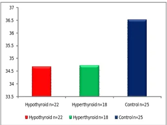
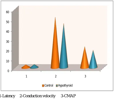
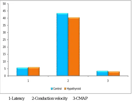
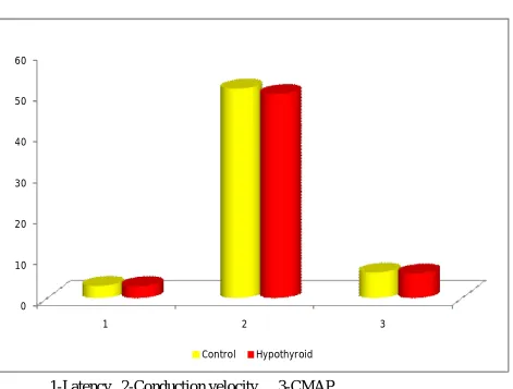
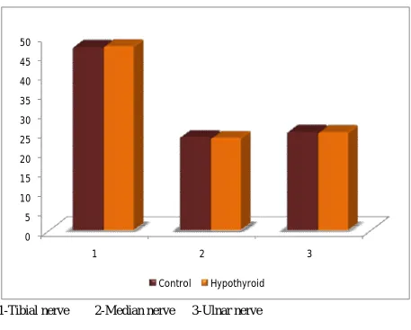
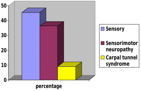
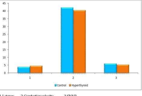
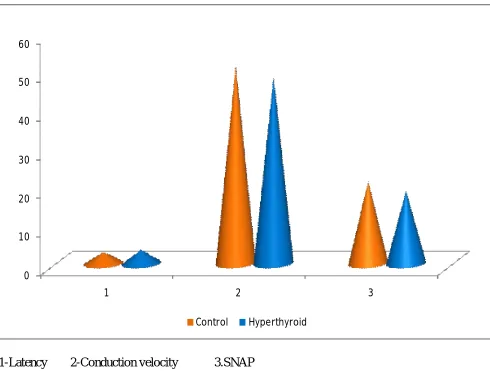
Introduction: The magnitude of thyroid disorders are increasing globally. In India also there is an increasing incidence even though India is in post iodination phase. Most commonly the thyroid disorder is due to autoimmune pathology. Thyroid disorders affect all systems of the body including nervous system. The affection of neuromuscular problems includes proximal myopathy and various peripheral neuropathies. The early detection of peripheral neuropathy before clinical manifestations using electrophysiological methods helps in prevention of morbidity associated with the complications. Aims and Objectives: This study was carried out to evaluate the changes in the abnormalities in nerve conduction parameters in thyroid disorders and to find out the magnitude of neuropathies. Materials and Methods: The study was conducted at Tirunelveli Medical college hospital after ethical clearance. Proforma was filled in to follow the inclusion criteria and exclusion criteria. 22 hypothyroid,18 hyperthyroid and 25 normal individuals were selected for the study and written informed consent was obtained. The sensory conduction was performed in sural, median and ulnar nerves and motor conduction in tibial, median and ulnar nerves of left side using RMS EMG EP MARG II equipment. Results and discussion: The study found out predominant sensory involvement in both groups. Sensory neuropathy was present in 45.4 % and 61 % of hypothyroid and hyperthyroid individuals respectively. Sensorimotor neuropathy was present in 9.1 % and 5.5 % of hypothyroid and hyperthyroid individuals respectively. The study has diagnosed carpel tunnel syndrome in 36 and 17 % of hypothyroid and hyperthyroid individuals. The involvement of nerves in hypothyroidism is due to less active sodium potassium pumps and hyperthyroidism due to hypermetabolism. Conclusion: The study has highlighted the involvement of peripheral nerves in thyroid disorders and the role of electrophysiological studies in early detection of neuropathies. The study also suggests to evaluate thyroid function in carpal tunnel syndrome.
EFFECT OF THYROID DISORDERS ON NERVE CONDUCTION STUDIES IN FEMALES
Introduction:
Thyroid disorders are one of the common endocrine disorders next to diabetes mellitus which has a multi factorial etiology. The prevalence of hypothyroidism in the world population ranges from 1-10% with female predominance and hyperthyroidism affects 2-5% of the women population in the world. In Indian scenario, hypothyroidism is found to affect 1 in 10 adult, and also hyperthyroidism to affect more people. In spite of the fact that India is in post iodination phase, increasing prevalence of thyroid disorders is due to poor awareness and cost factors.
As the thyroid hormone controls the activities of almost all cells in most tissues of the body, altered level of thyroid hormones leads to multi systemic disturbances. Cardiovascular complications like atrial fibrillation and cardiac failure, isolated systolic hypertension, increased appetite and unexplained weight loss are some of the common clinical manifestations of the hyperthyroidism. Poor appetite, weight gain, hypertension, cardiac failure and infertility are some of the common features of hypothyroidism.1
neuropathies like carpal tunnel syndromes and tarsal tunnel syndromes occurs in both thyroid disorders.
The magnitude of thyroid disorders among poor people of India leads to frequent absenteeism from duty because of the neurological manifestations. Moreover the proper treatment of hyperthyroidism results in complete resolution of neuropathy and late institution of adequate treatment of hypothyroidism does not offer complete cure2. Hence it is essential to study the prevalence of the same by doing neurophysiological studies in both symptomatic and asymptomatic patients of established thyroid disorders. And this type of study may necessitate the steps to be taken to increase the awareness among public.
AIMS AND OBJECTIVES
1. To study the pattern of the nerve conduction studies in hypothyroid individuals.
2. To study the changes in nerve conduction pattern in hyperthyroid individuals.
3. To perform nerve conduction study pattern in control groups.
4. To compare the changes in nerve conduction study between hypothyroid and controls.
5. To compare the changes in nerve conduction study between hyperthyroid and controls.
6. To estimate the magnitude of neuropathy in hypothyroid individuals. 7. To estimate the extent of neuropathy in hyperthyroid individuals
REVIEW OF LITERATURE HISTORICAL ASPECTS
The prescription by Emperor Shen during 2700 BC shows that seaweed can be used for treatment of goiter. This is the earliest evidence related to thyroid gland mentioned in Pen Tsao (1596) which is the herbal of Chinese Pharmacopoeia. In 300 BC, the discussion of goiter is found in Ayurvedic medicine.
In the year 650 AD, use of powdered form of dried mollusc shells and chopped thyroid gland was practiced by a Chinese physician, Sun Ssu-Mo. It was 1500 AD when Leonardo da Vinci first recognized and drew thyroid gland. Andreas Vesalius gave the first anatomical description and illustration about thyroid gland in 1543 AD. Eustachius coined the term Isthmus in 1563 AD. In 1656, the naming of the gland as “thyroid” resembling the shape of an ancient Grecian shield was given by Thomas Wharton3.
said that the goiter is caused by deficiency of iodine and he used iodine for treating the goiter. It was observation by Alexander von Humboldt during 1825 that the gland size gets reduced when persons suffering from goiter change residence to non endemic area of goiter from endemic area called, Andes. A physician from Brazil, Francisco Freire-Allemao proposed iodine prophylaxis programme. Aqueous solution from Potassium Iodide was proposed by JGA Lugol for treatment of scrofula in 1829.
A syndrome consisting of palpitation, goiter and exophthalmos was described by Robert Grave in three women in the year 1834. In individual suffering from cretinism in Switzerland, B.Niepce found out the enlargement of sella turcica in 1857AD. Post operative tetany after total thyroidectomy was observed by Th.Billroth in 1883. E.T. Kocher was the Nobel Prize winner of the year 1909 for his finding of association of myxoedema after thyroidectomy during 1883. He also recommended “half a sheep’s thyroid fried lightly and to take it with a current jelly every week4.The typical tremor in hyperthyroidism was found out by Pierre Marie in 1888.F.D.Von Recklinghausen reported that osteoporosis can follow hyperthyroidism. The effect of thyroid on basal metabolic rate was reported by Aldolf Magnuu in 1895 AD. The term “iodothyrin” was coined by Eugen Baumann. Pendred found out goiter in deaf mutism in 1897.
by him. Thyroid involution and failure of metamorphosis in hypophysectomised tadpole was described by both Philip E. Smith and Bennet M Allen individually in 1916.Kendall’s thyroxine from animal extract of thyroid was introduced in market with high price in U.S.A in 1920. The treatment of Graves’ disease using x-rays was proposed by M. Seymour in the same year. Preoperative iodine treatment in Graves’ disease was reported by H. S. Plummer in 1924.Nobel prize in 1943 was given to George Harvey for his work on radioactive tracers during year 1924.The chemical structure of thyroxine was discovered by Harington in 19265.Harington and Barger revolutioned the treatment of hypothyroidism by synthesizing thyroxine in 1928.Goiter development in cabbage fed rabbits was demonstrated by A. Chesney, T. Clawson and B.Webster in 1930.TSH was extracted and purified from bovine pituitaries in the year 1931.Use of radioactive iodine by Saul Hertz and Arthur Roberts was adopted to study the physiology of thyroid gland and to treat hyperthyroidism between 1937-1943.Negative feedback on pituitary by thyroid was termed as “servo mechanism” by R.G.Hoskins in 1949.In the same year, J . Wolff and I.Chaikoff found out the effect (Wolff Chaikoff’s effect) of inorganic iodine on thyroid physiology.
Association of LATS with Graves’ disease was discovered by Adams, Purves,
and McKenzie in 1960. Demonstration of calcitonin was by D.H.Copp,
A.G.F.Davidson, and B.A.Cheney during the year 1963. Breakthrough in
estimating hormonal assays by Radioimmunoassay was achieved by S. Berson
and R .Yalow in the year 1965.For this contribution they were honoured with
Nobel prize during the year 1977.Subsequently L. Braverman S. Ingbar, and
Sterling found out the peripheral conversion of T4 to T3, in the same year.
A.Schally discovered TRH and he was awarded Nobel Prize for the year 1977.
Hetzel in 1971 described the effect of iodine deficiency during pregnancy on
fetus nervous system. During year 1972, the receptor for triiodothyronine was
found out6. Thyroid hormone resistance was demonstrated by S. Refetoff and
L. De Groot in 1974. T. H. Liao and J.Pierce were able to obtain ultrapure form
of TSH and also found out common alpha subunits in TSH, LH and FSH in
1979.Recombinant TSH was approved in clinical use from 1998.
3-iodothyronine was discovered by Thomas Scanlan in the year 2002.
PHYSIOLOGYOFTHYROIDGLAND
Thyroid gland, a small bow tie like structure, situated in front of the
trachea has got some unique features compared to other endocrine glands. It is
the only endocrine gland which is visible and palpable and needs a trace
element-iodine for the synthesis of active hormone. This gland synthesizes a
hormone which is stored extracellularly as colloid, and stored within the gland
DEVELOPMENTALANATOMY:
The thyroid gland develops from the epithelial proliferation in the pharyngeal floor between the tuberculum impar and the copula in the area of foramen cecum and descends down as a bilobed diverticulum. Throughout the period of migration, the thyroid gland is attached to the tongue through thyroglossal duct which disappears subsequently. Failure of disappearance of thyroglossal duct leads to congenital anomaly called as thyroglossal cyst. Up to seventh week of embryonic life, the thyroid gland descends downwards in the midline up to the front part of the trachea.The front of trachea is the postnatal position of the gland. Two lobes and the isthmus in between them develops around ninth week. The first follicle lined by epithelial cells can be seen near the end of third month. This marks the beginning of secretory activity of thyroid gland. The follicle secretes colloid that contains thyroxine and triiodothyronine. Ultimobranchial body gives rise to Parafollicular cells, also called as C cells which secrete calcitonin7.
FUNCTIONAL ANATOMY OF THYROID GLAND:
membrane. The size of the follicle, amount of the colloid and nature of the epithelial lining depends on the activity of the gland. Inactive gland consists of large sized follicle filled with abundant colloid lined by flat epithelial cells. The active gland has small sized follicles with less amount of colloids lined by either cuboidal or columnar cells with resorption lacunae. The resorption lacunae indicate the active resorption of colloid. Microvilli responsible for formation of resorption lacunae projects from the apex of the epithelial cells into the colloid. Like any glandular epithelial cells, it consists of prominent endoplasmic reticulum and secretory granules. It is the only organ in the body except adrenal cortex receiving blood supply five times more than the weight of the gland8.
HORMONES SYNTHESISED BY THYROID GLAND:
The hormones secreted by thyroid glands are thyroxine or T4 and triiodothyronine or T3 involved in basal metabolism and a calcium lowering hormone called calcitonin. Thyoxine and triiodothyronine are synthesized by follicular epithelial cells and calcitonin is secreted by parafollicular cells. T3 is more potent than T4. In addition to the above biologically active hormones namely T4 and T3, small amount of inactive hormone called as reverse triiodothyronine or rT3 is also produced by thyroid gland.
IODIDE METABOLISM:
The uniqueness of thyroid hormone is that it needs iodine in trace for its biological activities. The daily requirement of iodine is
Adult : 150 µg
Pregnant woman : 200 µg
Our daily intake of iodide is around 500 µg which is much more than the
minimum requirement of iodide. The food contains this raw material in the form
of iodine which in converted to iodide and absorbed in gastrointestinal tract. In
the equilibrium state the iodide intake equals iodide excretion. The principle
organ taking up most of the circulating iodide is the thyroid gland. In addition,
tissues like salivary gland, lacrimal apparatus, gastric gland, mammary glands
and choroid plexuses actively concentrate iodide.
The blood contains 250 to 750 µg of iodide. From circulation, thyroid
gland extracts 70 to 80 µg of iodide.Thyroid contains around 7500 µg of iodide.
The iodide in the thyroid gland is in the form of iodothyronines. Thyroid gland
releases around 70-80 µg of iodide i.e. around 1 % of the total iodide in thyroid.
Thyroid hormones forms 75 % of the released iodide and remaining 25 % is
released as free iodide. As the ratio between the stored iodide in hormonal form
and released iodide is more (100:1), human beings are protected from the effect
of iodide deficiency for at least 2 months. Kidney also contributes to iodide
homeostasis by reducing iodide excretion when there is a fall in serum
concentration of iodide9.
SYNTHESIS OF THYROID HORMONES:
Major hormone secreted by thyroid gland is in the form of thyroxine
triiodothyronine in peripheral tissues by deiodinases. The steps involved in
hormone synthesis consists of
Iodide trapping
Thyroglobulin synthesis
Oxidation of the iodide
Organification of thyroglobulin
Storage of thyroid hormones.
Precursors like iodide and thyroglobulin involved in thyroid hormone
synthesis are moved in basal to apical side of follicular cells. Thyroid hormone
after synthesis within the thyroglobulin is moved from apical to basal side for
secretion into the blood.
IODIDE TRAPPING:
The iodide entering the circulation from GIT, is actively concentrated into
thyroid follicular cells for about 70 times by 2Na+-1I- symporter (NIS). This
transporter is also called as Iodine Pump. This pump brings about influx of two
sodium ion by facilitated diffusion and influx of one iodide ion by active
transport. The favorable concentration gradient for sodium is maintained by
Na+-K+ pump .Both pumps are situated in basal aspect of cell membrane.
The activity of iodine pump is modified by altering gene expression. TSH
stimulates and iodide inhibits gene expression. Cytokines also inhibits
expression. In Iodide deficiency, expression of NIS is increased. Expression of
epithelium, the placental tissue, the ciliary bodies of the eye, the choroid plexus,
the mammary glands, and some cancers derived from these tissues. Drugs like
perchlorates and thiocyanates also inhibit the transporter10.
THYROGLOBULIN SYNTHESIS:
Thyroglobulin containing around 70 tyrosine molecules with molecular
weight of 3, 35,000 is synthesized in endoplasmic reticulum of the follicular cell.
This thyroglobulin after exocytosed into the lumen of the follicles will undergo
changes to synthesize thyroid hormones.
OXIDATION OF THE IODIDE:
Once the iodide is trapped inside the follicular epithelial cells, it is
transported into the lumen by a special transporter protein called Pendrin.
Deficiency of this protein leads to a disease called as Pendred's disease. Person
affected will develop cretinism and sensorineural deafness. After transport, the
iodide is oxidised to iodine by the enzyme called Peroxidase. Hydrogen
peroxide needed for this reaction is provided by the enzyme called as NADPH
oxidase. Both these enzymes are present in the luminal side of the membrane.
These peroxidases are inhibited by the antithyroid drugs11.
ORGANIFICATION AND COUPLING REACTIONS:
Once oxidized, the iodine combines with the tyrosine molecules to form
monoiodotyrosine (MIT) and diiodotyrosine (DIT). Coupling of two
diiodothyrosine molecules produces thyroxine and coupling of one
triiodothyronine. This organification and coupling is also catalyzed by
peroxidase. Synthesis of T3 is more during iodine deficiency and hyper
stimulation of the thyroid gland by TSH12.
SECRETION OF THE HORMONE:
The thyroid hormone after synthesis is stored in the colloid as a part of
thyroglobulin. Megalin, a transporter protein promotes endocytosis of this
protein and thyroglobulin is acted upon by lysosomal enzymes. It degrades
thyroglobulin into MIT, DIT, T4, T3, rT3 and uniodinated tyrosine molecules.
MIT and DIT are broken down into tyrosine and iodine by the microsomal
enzymes named intrathyroidal deiodinase13.
The iodine and amino acids are reused for thyroid hormone synthesis and
thyroglobulin synthesis respectively. Because of this recycling process, thyroid
hormone deficiency is not manifested up to two months. Deficiency of the
deiodinase enzyme can be confirmed by finding MIT and DIT in urine.
TRANSPORT IN PLASMA:
Once secreted into the plasma, 70 % of thyroid hormones bind to
Thyroxine binding globulin (TBG) .About 10% to 15 % binds to thyroid
binding prealbumin called transthyretin (TTR) .Another 15 % of thyroid
hormone is transported as a complex with albumin and 3% by lipoprotein.
Among these proteins, albumin due to its large size, possesses largest capacity to
bind to hormone as compared to thyroid binding globulin. But thyroid binding
circulates in plasma in unbound form.
Even though thyroid hormone binds to many proteins significant change in
the total plasma level of thyroid hormone occurs only when there is a change in
the level of thyroid binding globulin. Increase in the TBG level in situations like
pregnancy and estrogen therapy leads to increase in the level of total hormones
and normal level of free hormones. Drug therapy with glucocorticoids and
androgen decreases level of total hormone without altering the free hormone
level8.
The proteins by binding to thyroid hormone act as reservoir for the
hormone and also prevent the loss of the hormone by metabolism in tissues or
excretion in urine. As the bound form is not filtered at capillary level, it increases
half life of the hormone. The unbound form is responsible for hormonal actions.
Transthyretin is important for providing the hormones to central nervous system.
TRANSPORT INTO THE CELLS:
Once the hormone reaches the target tissues, it is transported into the cell
by specific transporters such as Sodium/taurocholate-cotransporting
polypeptides, organic anion-transporting polypeptides, L-type amino acid
transporters and the monocarboxylate transporters (MCT). The mutation
involving MCT 8 leads to deficiency of intrathyroidal hormone and elevated
level of serum T3 and psychomotor retardation. This is called as
Allan-Herndon-Dudley syndrome. In this syndrome there is defective transport
This leads to decreased myelination14.
METABOLIC TURNOVER OF THYROID HORMONE:
About 90 µg of T4, 35 µg of T3 and 35 µg of rT3 are produced daily by
thyroidgland.
Normal levels of total T4, T3 and rT3 are 8µg, 0.12µg and .04µg respectively.
The levels of freeT4, T3 and rT3 are 2ng/L, 0.28ng/L and 0.2ng/L
respectively.
Half life of T4, T3 and rT3 are 7, 1 and 0.8 days respectively.
Only 25 % of T3 and 5 % rT3 are formed by thyroid gland.
Peripheral conversion of T4 contributes to 75 % and 95 % of T3 and rT3
respectively by specific deiodinases.
DEIODINASES:
The three types of deiodinases are D1, D2 and D3. All deiodinases in
common contain Selenocysteine, an amino acid containing selenium instead of
sulphur. Selenium is important for its enzyme actions. D1, D2 converts T4 to T3.
• D1 is present in high amount in liver, pituitary, skeletal muscles, thyroid and kidneys where there is a high blood flow.D1 regulates the
formation of T3 in peripheral tissues.
• D2is present in high amount in brain tissues and pituitary and brown fat. The high level of deiodinases in brain provides brain tissue and
• D3 is present in brain and reproductive organ converting T4 to rT3.Hence D3 is also called as inactivating enzymes. D3 is elevated in
hyperthyroidism to increase the inactivation of active hormone15.
FATES OF ACTIVE HORMONES:
T4 and T3 after performing the physiological actions, they are either
deiodinated into deiodothyrosine or conjugated to their sulfate and glucuronide
metabolite. The conjugated metabolites are secreted into the bile. After reaching
the intestine they are hydrolysed and reabsorbed partly through enterohepatic
circulation and partly excreted into the stool. In addition to the above
mechanisms, iodide is also lost into the stool due to the direct entry of hormones
from circulation into intestinal lumen. The iodide loss in this route forms 4 % of
the total iodide loss16.
MECHANISM OF ACTION:
Most of the thyroxine hormone once transported inside the cell is
converted into T3 and T3 binds to thyroid hormone receptor present in the
nucleus.T4 also can bind to the receptors with less affinity. These receptors form
heterodimer with retinoic acid receptors. But this heterodimeric complex does
not bind to 9-cis retinoic acid though RXR binds to 9-cis retinoic acid. They can also form monomers and homodimers. These receptors are kept repressed by
corepressor molecules attached to the receptors are released and coactivators
gets attached. The corepressors9 are Alien, NCoR and Alien. The examples of
coactivators are the DRIP-TRAP complex, and SRC family (SRC-1/2/3).These
receptor - hormone complex binds to DNA through zinc fingers and bring about
the increase in the transcription of genes involved in the proteins. These
proteins are either structural or functional proteins. As thyroid receptors binds to
specific hormone responsive element of the DNA they can be designated as a
member of the superfamily of hormone-sensitive nuclear transcription factors.17
THYROID HORMONE RECEPTOR GENES:
There are two types of hormone receptor genes namely THRA,THRB.
THRA or TRα genes are present in chromosome 17 and THRB or TRβ genes are
present in chromosome 3. Alternative splicing of these genes leads to
transcription into two different types of mRNA .These m RNAs gets translated
into subcategories of receptors namely TRα1, TRα2, TRβ1 and TRβ2.TRβ2 is
exclusive to the brain. There is wide distribution of TRα1, TRα2, and TRβ1.
Among the four types of receptors, TRα2 does not bind to T3. T3 has more rapid
and potent action than T4 because of its less binding to plasma protein and also its
good affinity to TR. RT3 is inert in binding to the receptors.The mutation of
theTR β gene can present as three varieties of clinical problems.
1) Usually the resistance happens both in peripheral tissues and at
anterior pituitary level. As TRα is normal and there is high level
But there is an inappropriate increase in the TSH level even though T3
and T4 level is high. Exogenous thyroid hormone administration does
not suppress TSH level in these individuals.
2) In some patients, this mutation leads to resistance only at pituitary
level. This causes a hypermetabolic state and raised plasma T3 and T4
levels.TSH is normal and at nonsuppressible level.
3) In few patients it can lead to resistance at the level of peripheral
tissues and causes hypometabolic state and normal T4, T3 and TSH
level. These patients need large dose supplementation of hormones to
normalise the basal metabolic rate. The role of hTRβ on brain
development can be confirmed by a finding that Attention Deficit
Hyperactive Disorder (ADHD) is commonly present in individuals
with thyroid hormone resistance than its presence in general
population16.
NONGENOMIC ACTIONS OF THYROID HORMONES:
Predominant actions of thyroid hormone take place through genomic
influence. In tissues like heart, muscles, adipose tissues and pituitary, it exerts
nongenomic actions through the activation of cAMP or signalling cascade
mediated by protein kinase. Some examples of nongenomic actions of thyroid
hormones are ion channels regulation and oxidative phosporylation .These
PHYSIOLOGICAL ACTIONS OF THYROID HORMONES:
Thyroid hormone, by increasing the protein synthesis in almost all tissues
of the body, exerts all its physiological actions.
EFFECT ON BASAL METABOLISM OF THE CELLS:
Oxygen is the essential element involved in the oxidative phosporylation
reactions in generation of ATP from ADP. The rate of oxygen consumption
decides the rate of metabolism in the cells. By increasing the oxygen
consumption of the cells, thyroid hormone brings about the increase in the basal
metabolic rate. Normally about 250 ml of oxygen is consumed by the body per
minute at resting state. In hyperthyroidism it goes upto 400 ml/mt.
The enhanced activity of Na+- K+ pump and increase in the mitochondrial
enzymes also cause increase in the basal metabolic rate. Enhancement of the
metabolism in the cells increases the heat production also. This is known as
calorigenic action of the thyroid hormone. Thyroid hormones, by increasing the
synthesis of uncoupling of protein 1, increase the generation of heat. Gonads of
both sexes, spleen, brain, lymph nodes and uterus are the organs in which the
basal metabolism is not altered by this hormone. Thyroid hormone does not
bring about the enhancement of oxygen consumption during exercise and after
ACTION ON MITOCHONDRIA:
Thyroid hormone increases the size and number of the mitochondria.
Also the surface area of mitochondrial membrane is linearly increased with
the increase in rate of metabolism. These actions of the hormone increase the
production of Adenosine triphosphate (ATP) for increasing the metabolic
activity of the cells. But increase in the activity of mitochondria may also be
secondary to the increased basal metabolic rate.18
ION TRANSPORT:
Activity of the Na+- K+ pump is increased by the thyroid hormone. The
increased activity of the above pump increases the transport of sodium
potassium through cell membrane. The activity of this pump needs ATP. This is
the reason for the thyroid hormone to increase the basal metabolic rate. Thyroid
hormone also increases the leakiness of sodium channels. The increased activity
of the sodium channels in turn increases the activity of the sodium potassium
pump and also increases the heat generation.
EFFECT ON GROWTH:
Thyroid hormone promotes growth and maturation.Before the fetal
thyroid starts synthesising the hormone around mid gestation, thyroid hormone
from mother crosses placenta into the fetal circulation thereby bringing about
the growth activity and maturation of fetal organs. Normal nervous system
development also requires the thyroid hormones. The bone maturation and
enhanced growth rate in human being is observed mainly during fetal life and
childhood19.
The children born with the deficiency of thyroid hormone develop
cretinism in which the irreversible mental retardation and short stature are the
characteristic features. This necessitates the early diagnosis of the
hypothyroidism in children. The testing for hypothyroidism in cord blood is
nowadays practised in many centres.
The influence of thyroid hormone in growth and maturation is
experimentally proved by growing a tadpole of frog in thyroid deficient
situation. The effect of this is the arrest of metamorphosis into the frog. In
human being the growth rate is increased mainly during childhood
ACTIONS ON CARBOHYDRATE METABOLIM:
It increases the rate of glycolysis, gluconeogenesis by increasing the
enzymes involved in the metabolic pathways, absorption of glucose from GIT
and increases the secretion and action of insulin20.
ACTIONS ON FAT METABOLISM:
By increasing the mobilisation of fat stores, thyroid hormone reduces
storage of fat. This in turn increases the fatty acid level in plasma and oxidation
of fatty acids. By increasing the Low Density Lipoprotein receptors in the liver,
it increases the uptake of cholesterol from plasma. This rapid removal leads to
increased secretion of cholesterol in bile and then elimination of cholesterol in
reduces plasma phospholipids and triglycerides also.
THYROID HORMONES INCREASE VITAMIN REQUIREMENTS:
Most of the enzymes need coenzymes. Essential vitamins act as
coenzymes for enzymes. By increasing the activity of enzymes involved in the
metabolism, thyroid hormone increases the vitamin requirements. So in case of
thyroid hormone excess, there should be adequate supplementation of the
vitamins.
ACTIONS ON CARDIOVASCULAR SYSTEM:
As hormone increases the metabolic rate, it results in increased
consumption of oxygen and raised metabolic end products. These in turn lead to
vasodilatation and increased blood flow in all tissues. Cutaneous vasodilatation
leads to the elimination of heat. It also increases the cardiac output, heart rate
and cardiac contractility by its permissive actions on catecholamine.
Vasodilatation decreases diastolic blood pressure due to reduced peripheral
resistance and increased cardiac output increases systolic blood pressure and
thus leading to wide pulse pressure. But mean arterial pressure remains
unchanged21.
EFFECT ON OXYGEN CONSUMPTION AND RESPIRATION:
As basal metabolic rate is increased, basal oxygen consumption is also
increased. But it will not enhance the increased oxygen consumption met during
exercise or after a meal intake. This increased basal oxygen consumption and
rate and tidal volume. Both effects increase minute ventilation22.
Thyroid hormone stimulates erythropoietin synthesis. The increased
erythropoietin increases the rate of erythropoiesis thereby increasing the oxygen
carrying capacity of the blood.
ACTION ON DIGESTIVE TRACTS:
Thyroid hormone increases appetite as intestinal absorption is increased.
Intestinal absorption increases in order to increase the availability of substrate
for increased basal metabolic rate. Thyroid hormones increase the secretory rate
of gastrointestinal digestive juices and motility of the digestive tracts23.
ACTION ON OTHER ENDOCRINE GLANDS:
There are two mechanism by which thyroid hormone increases the
endocrine secretion. By its general action of increase in the basal metabolic rate
of all the cells, it increases the secretory activity of the cells of the endocrine
glands. Secondly increase in the tissue metabolism increases the requirement of
hormones by the tissues.
Increase in the metabolic rate of glucose increases the insulin secretion
from pancreas. As bone formation is increased by thyroid hormones, the
reduction in blood calcium increases the parathyroid secretion. As steroid
hormones which are derived from cholesterol are also inactivated by liver,
reduction in adrenocortical hormones by negative feedback increases
adrenocorticotrophic hormones (ACTH). The increase in the ACTH in turn
ACTION ON REPRODUCTIVE SYSTEM:
It regulates the activity of reproductive system in both sexes. In female it
promotes graffian follicle maturation and ovulation. So in female
hypothyroidism causes infertility and various menstrual disturbances. In males
it enhances spermatogenesis and Sertoli cell differentiation at puberty 24.
ACTION ON NERVOUS SYSTEM:
Thyroid hormone plays a vital role in the maturation and development of
nervous system in fetal life and in early part of the childhood. The maximal
growth of brain occurs between later half of fetal life and first six months after
the birth. In this part of human life, thyroid hormone is required for the
initiation and facilitation of the neural cell differentiation and maturation.
It stimulates the growth of both cerebral and cerebellar hemisphere. It also
promotes basal ganglia development. Axonal multiplication and branching
patterns of dendrites are controlled by thyroid hormone. It also promotes the
formation of synapse. Neurotransmitter synthesis is catalysed by enzymes.
Thyroid hormone stimulates the enzyme productions. It also enhances the
receptors formation for the attachment of neurotransmitters to perform their
physiological actions.
Thyroid hormone maintains the optimum amount of the enzyme galactosyl
sialyl transferase. This enzyme is very important for myelin formation around
neuronal processes. Myelination is an important factor responsible for the faster
of T4 in generation of nerve growth factors. It is essential for the migration of
the neuronal cells during the growth of brain. The velocity and amplitude of
stretch reflex is facilitated by thyroid hormones.
Thyroid hormone is important for the higher intellectual functions of the
central nervous system. It increases the alertness of the individual and response
to all stimuli. Thyroid hormone stimulates the synthesis of enzymes involved in
generation of energy for the neuronal cells. In spite of all the above influences,
the amount of blood in cerebral circulation is not altered by thyroid hormones.
Basal oxygen consumption and glucose metabolism in the nervous system is not
changed25.
REGULATION OF THYROID HORMONE SECRETION:
Thyroid glands are stimulated by thyroid stimulating hormone of anterior
pituitary. The thyroid stimulating hormone (TSH) is also called as thyrotropin.
Synthesis and secretion of thyrotropin is enhanced by the thyrotropin releasing
hormone (TRH) of hypothalamus. By its feedback on anterior pituitary and
hypothalamus, the thyroid hormones alter the synthesis and secretion of TRH
and TSH.
THYROTROPIN RELEASING HORMONE:
This hormone is produced by the arcuate nucleus and median eminence of
the hypothalamus.TRH is also present in other tissues like cerebral cortex,
digestive tracts and pancreatic beta cells. This tripeptide hormone has the amino
pyro-Glu. After its release from hypothalamic neurones, TRH is transported by
hypothalamic hypophyseal portal vessels.
Once TRH reaches the target cells (thyrotrophs) of the anterior pituitary,
it activates the TRH receptors present in the cell membrane. This receptor is a
G-Protein coupled receptor acting on Phospholipase-C to release second
messengers. The second messengers are diacyl glycerol (DAG) and inositol tri
phosphate.DAG activates protein kinase - C. Protein kinase - C upon activation
leads to phosporylation of proteins. Inositol triphosphate increases the calcium
release from the stores inside the cells. By the above mechanisms, TRH
increases the synthesis and secretion of the TSH from pituitary.
The TRH also activates Phospholipase-A2 leading to release of arachidonic
acid from cell membrane. Eicosanoids are synthesised from the arachidonic acid
and mediates some of the actions of the TRH.TRH also stimulates lactotrophs to
secrete prolactin26.
THYROID STIMULATING HORMONE:
TSH is a glycoprotein. Its molecular weight is 28-kDa.It is stored in the
secretory granules of the thyrotrophs like other protein hormones. It consists of
α, β subunits. α subunit resembles the α subunits of follicular stimulating
hormone, luteinizing hormone and human chorionic gonadotropin. β subunit is
specific for TSH. The receptor for TSH is present on the basolateral side of the
follicular cell membrane. TSH receptor is a membrane bound G-protein coupled
detached from the βγ subunits and stimulates adenylyl cyclase. Adenylyl
cyclase produces cAMP which acts as a second messenger. TSH receptor
stimulation produces following changes:
It simulates NIS to increase the iodine uptake and thereby concentrating
iodide inside the follicular cells 30 times more than in serum
Increases the iodination of thyroglobulin
Promotes the conjugation of the one monoiodo and one diiodothyrosine to
form T3 and of two diiodothyrosines to form T4
Increases endocytosis of thyroglobulin
Increases lysosomal enzyme activity to cleave the thyroglobulin
Enhances the secretion of T4 and T3 into systemic circulation
By its growth factor effect it brings about hyperplasia of thyroid gland27.
NEGATIVE FEEDBACK EFFECT OF THYROID HORMONES:
Like other endocrine hormones, alternation in the level of thyroid
hormones exerts their negative feedback effect through a long feedback loop
pathway. It acts on both thyrotrophs and neurons of the hypothalamus. TSH is
more sensitive to the changes in the level of free form of thyroid hormone.
When level of T4 gets reduced to 50 %, TSH increases 50 to 100 times. The
thyrotrophs as a sensor monitor the changes in the T3.The source of T3 for this
purpose is met either by the direct entry of T3 from the circulation or
deiodination of T4 to T3.TSH level is altered by direct inhibition of synthesis of
thyrotrophs. Genes concerned with the synthesis of α and β chain have an
inhibitory T3 response element in the promoter site.
Feedback effect of T3 and T4 is also regulated by somatostatin and
dopamine. These chemicals produced by hypothalamic neurons are carried to
anterior pituitary through hypophyseal portal system. By altering the set point of
thyrotrophs for T3, somatostatin and prolactin increase the sensitivity of
thyrotrophs to increased intracellular concentration of T3. Hence the secretion
of thyroid stimulating hormone is reduced counterbalancing the effect of
Thyrotropin Releasing Hormone. These effects have been demonstrated during
the infusion of somatostain and prolactin.
SPECIAL FEATURES OF REGULATION IN FETAL LIFE:
As T3 from mother crosses the placenta and fetal metabolic tissues are
immature, increase in the T3 level in the fetus reduces the level of TSH by
negative feedback.In immediate postnatal life, rapid metabolisation of T3 and
slow recovery of TSH suppression takes place. So if mother is hyperthyroid
newborn babies have the risk of developing transient hypothyroidism. The
reverse is true in case of babies born to hypothyroid mother.
THYROID FUNCTION TESTS:
It includes
1. Assay of thyroid hormonal levels and measuring TSH
level
3. TRH response tests
4. Identification thyroid antibodies
5. Plasma cholesterol measurements
6. Determination of basal metabolic rate28
THYROID HORMONES AND TSH ASSAY:
Thyroid hormone and TSH level in serum is estimated by
Radioimmunoassay or ELISA methods. TSH measurement is important to
differentiate primary thyroid pathology from pituitary pathology.
BINDING PROTEINS ESTIMATION:
It is carried out by resin uptake method. The radiolabeled T3 hormone when
added to the plasma binds to thyroid binding globulin in serum. After this, resin
is mixed with the plasma in the test tube and free radiolabeled T3 binds to resin.
There is an inverse relation between uptake and protein binding site and the
hormone level in serum is in direct proportion to resin uptake. If more binding
sites are free in TBG as in hypothyroidism the resin uptake is low. In
hyperthyroidism due to the reduced number of binding sites in proteins, resin
uptake is high.
TRH RESPONSE TEST:
In normal person, TRH administration increases the level of thyroid hormone
by increasing the secretion of TSH. In primary hyperthyroidism, uncontrolled
negative feedback by excessive T4 produces abnormal TRH response. Same
increased TRH response.
ESTIMATION OF ANTIBODIES:
In autoimmune diseases like Graves’ disease and Hashimoto’s thyroiditis,
specific antibodies are produced. Finding out the level of the antibodies in
serum may be necessary for establishing the etiology. These are not practised
routinely nowadays.
PLASMA CHOLESTEROL:
The concentration of cholesterol is not measured for routine diagnosis of the
thyroid disorders but used as prognostic markers in assessing the effectiveness
of treatment of thyroid disorders. There is inverse relation between cholesterol
level and serum concentration of thyroid hormone level.
MEASURMENT OF BASAL METABOLIC RATE:
It is increased in hyperthyroidism and decreased in hypothyroidism. This test
is not in routine practice for establishment of thyroid pathology.
PHYSIOLOGY OF NERVE CONDUCTION
HISTORICAL ASPECTS:
Invention of cathode ray oscilloscope in 1897 by Braun was the
breakthrough in the study of action potentials. In 1903 string galvanometer
was found out by Einthoven. Muscle action potential instead of the muscle
twitch was recorded in the measurement of conduction velocity in motor
nerves by Piper in year 1909 and Munnich in 1916.By stimulating tibial
was named as H reflex in 1918 .To amplify the action potentials, electron
tube was made use by Fobers and Thacker in 1920.String galvanometer was
used by them to record the action potentials. At Cambridge, United
Kingdom, Edger Douglas Adrian recorded action potential in a single nerve
fibre by amplifying the signal 5000 times. He observed the similarity of
impulses in sensory and motor impulses. He is famous for his contribution
of all -or- none law. Cortical representation of pain sensation localised to
thalamus was by Adrian. Routine use of acoustic properties of EMG signals
in present day clinical practice was also found out by Adrian in addition to
his contribution in the development of Electroencephalography. He was
honoured with Nobel Prize for his contribution on nerve transmission. In
1922, Joseph Erlanger and Herbert Spencer Gasser, a student of the former
found out that difference in conduction velocity of impulse in nerve fibres is
directly proportional to the diameter of the nerve fibres. Based on this
finding they grouped nerve fibres into three different groups. The large
nerve fibres were grouped as type A fibre with maximum velocity and
smaller fibres as type C with minimum velocity. In 1944 they received
Nobel Prize for the same29. John Eccles from Australia devised a
micropipette and demonstrated the existence of the potential difference
across cell membrane. He demonstrated the ability of the smaller ions in
causing greater change in the depolarisation. For this, he studied the effect
of postsynaptic inhibition was his yet another contribution. It was year 1929,
Denny Brown demonstrated motor unit potential. The publication of the
report of action potential in median and ulnar by stimulation was made by
Eichler in 1937 for the first time. The modern techniques for measuring
conduction in sensory nerves were devised a decade later. By stimulating the
motor nerves Harvey and Masland observed the decremental response in
myasthenia gravis in 1941. In 1957 Eaton and Lambert used the same
procedure in various neuromuscular disorders including myasthenic
syndrome. During second world war, the team of Seddon, Alexander GM
Waddell of Anatomy department at oxford, Feinstein of Canada and Richard
Pattle, an electrical engineer studied the effect of denervation. Pattle
volunteered to be the subject for this study. By methods to crush the nerve,
they performed hourly electromyography and demonstrated the fibrillation
potentials. Larger MUP in neuropathy and smaller MUP in myopathies were
the independent observations by Fritz Buchthal in 1941 and Kugelberg in
1945 respectively. Alan Lloyd Hodgkin and Andrew F. Huxley did study on
Squid and cattle fish having giant axons because it is easy to introduce many
electrodes and measure the resting membrane and action potential30. They
demonstrated the membrane permeability to sodium and potassium in
various phases of action potential and also refractoriness of nerve fibres for
about 1 ms. They were the pioneer in devising voltage clamp method. The
demonstrated by them. Huxley independently demonstrated saltatory
conduction in myelinated nerve fibres in 1949. He also found out the different bands in skeletal muscle fibre and proposed the sliding filament
theory of muscle contraction. In 1963 both Hodgkin and Huxley along with
John Eccles were awarded the Nobel Prize. Bernard Katz, while studying
the synaptic transmission demonstrated that miniature end plate potential is
due to the quantal release of acetyl choline. Role of calcium in releasing
neurotransmitters was his yet another finding in synaptic transmission. This
work on neuromuscular transmission conferred the Nobel Prize in the year
1970.
Changes in the nerve conduction parameters in peripheral neuropathies
were studied by Harvey and Kutfer by doing nerve conduction study. The
slowing effect of nerve impulse propagation in ischemia was the observation
by Kugelberg in 1944 and Cobb and Marshall in 195431. Conduction
velocity was calculated for the first time by Hodes, Laravee and German in
1948.They did it so by stimulating a nerve at its various levels. The
electromyography machine was designed by Goldseth in interaction with
Jasper and Fizell in 1948. The principle of photographic superimposition in
the calculation of nerve conduction velocity in sensory nerve was used by
Dawson and Scott in 194932. Dawson by devising digital nerve stimulation
technique in 1956 differentiated the sensory potential from antidromic
conduction is slowed in carpal tunnel syndrome. Use of nerve conduction
studies in the differentiation of demyelinating disease and axonal
neuropathy was adopted by Lambert and Kaeser. From the year 1960,
sensory nerve conduction study was made as an integral part of electro
diagnostic study in neurophysiology.
ORGANISATION OF NERVOUS SYSTEM
The nervous system is divided into central nervous system (CNS) and
peripheral nervous system (PNS). CNS is formed by brain and spinal cord.PNS
consists of nerves arising from or entering into the CNS. The peripheral nerves
are cranial nerves and spinal nerves.PNS is subdivided into somatic nervous
system and autonomic nervous system. The autonomic nervous system controls
the activity of the viscera and this control is purely involuntary.ANS maintains
the stability of the internal environment.ANS is again divided into sympathetic
(catabolic nervous system) and parasympathetic (anabolic nervous system)
components. Somatic nervous system consists of sensory nerve fibres entering
into CNS and motor fibres leaving the CNS. The sensory fibres otherwise called
as afferent fibre takes information from head and neck, body walls and
extremities into the CNS. The motor fibres or efferent fibres leave the CNS to
innervate the skeletal muscles33.
ORGANISATION OF PERIPHERAL NERVOUS SYSTEM:
Group of axons leaving the motor neurons of anterior horn of individual
dorsal root ganglion to reach the dorsal grey matter of the a spinal level forms
dorsal root. Hence ventral root fibers carry motor impulse (efferent) from the
spinal cord and dorsal root carries the sensory impulses (afferent) towards the
spinal cord. Ventral and dorsal root from a single level of the spinal cord unite
to form a mixed spinal nerve carrying both afferent and efferent impulse. Then a
mixed spinal nerves divide into ventral and dorsal rami. Dorsal rami innervate
the dorsal part of the body and ventral rami innervate ventral part of the body.
Vental rami or dorsal rami of one level of spinal cord combine with the
corresponding rami of the neighbouring segments to form the various named
plexuses. From these plexuses individually named peripheral nerves
arise.Cranial nerves arise from or end in their corresponding nuclei.
STRUCTURE OF PERIPHERAL NERVE:
A single nerve on its outer aspect has loose connective tissue sheath
called epineurium. Each nerve consists of group of fascicles surrounded by a
covering named perineurium. Each fascicle is made up of many axons. Each
axon inturn is covered by endoneurium.
The anastamosis of one fascicle with the neighbouring fascicles and shift
of axon from one fascicle to another fascicle increase the mechanical strength to
a peripheral nerve. The diameter of an individual axons ranges from less than
1 µm to 20 µm. As in peripheral nerves, a special glial cell called Schwann cells
forms myelin sheath around the axon. The ratio of unmyelinated axon to
VASCULARITY OF NERVES:
The nerves are supplied by arteries located in the epineurium arranged in longitudinal direction. Throughout the length of the nerve, blood vessels from
the adjacent tissues send anastamosing branches to the longitudinally oriented
vessels. This is analogous to vascular arrangements in the mesentery of the gut.
This preserves the blood supply to the nerve during injuries to the blood vessels.
From the epineurium, branches run obliquely to reach the deeper layers of the
nerve. The endoneurial capillaries form a tight blood nerve barrier. The blood
nerve barrier is impaired in some metabolic neuropathies. If this barrier is
broken down in nerve injury, there may be a problem during regeneration. Even
though the soma of the neurone supports the axonal metabolism, blood supply
to the endoneurium is important to maintain the functions of the axons. The
above message is evident from the appearance of clinical features of neuropathy
when there is a disruption in the endoneurial blood supply.
STRUCTURE OF NEURON:
The neurons vary in size and shape. But all neurons have three parts
namely a cell body, dendrites and an axon. The axon ends at axon terminal
which forms neuromuscular junction by giving branches to individual muscle
fiber.The relation of nerve fibres to neuron was demonstrated by Augustus
Volney Waller. He showed the changes in the neurons after the section of the
CELL BODY:
Perikaryon and soma are the synonyms for the cell body. Soma is
considered as the centre of a neuron. Like any other cells, the cell body is
composed of a nucleus and cytoplasm with its organelles. The important cell
organelles are Nissl granules, numerous mitochondria and Golgi apparatus and
lysosomes. Cytoskeletal proteins are neurofilaments, microtubules and actin
filaments. Nissl granules are also known as Nissl bodies. It consists of stacks of
rough endoplasmic reticulum.
In nucleus, there is one nucleolus and no centrioles. Some neurons have
more than one nucleolus. The presence of nucleolus and absence of centrioles
indicate that neurons can synthesize protein but are unable to divide. The
plasma membrane is called as plasmalemma28.
DENDRITES:
They are very short extensions of plamalemma of neurons. Presence of
dendrites increases the surface area of the neurons. The dendrites transmit the
signals from other cells towards the soma. They also synthesize proteins and
generate and conduct action potential in some parts of the brain. A single neuron
contains up to 10000 dendrites.
THE AXONS:
A peripheral nerve is a collection of many axons. Axon is also called as
axis cylinder. The axon is a long tubular structure which conducts the impulse
consists of mitochondria, Golgi apparatus and cytoskeletal proteins. The point
from where axon originates from the cell body is called as axon hillock. Initial
segment, the first portion of the axon, is the continuation of axon hillock. Axon
hillock and initial segment in combination are known as axon hillock-initial
segment portion. In motor neuron, the initial segment generates the action
potential. In sensory neuron the action potential is generated at the first node of
Ranvier which is a gap between two Schwann cells enveloping the axons.The
Schwann cells form the myelin sheath. Theodor Schwann found the presence of
myelin sheaths33.
Based on myelination, the axons are classified into myelinated and
unmyelinated axons. In CNS, myelination is contributed by oligodendrocyte
and in peripheral nervous system by Schwann cells. In peripheral nervous
system, myelinated nerve fibers are present in large somatic nerves and
preganglionic nerve fibers.
MYELIN FORMATION:
The Schwann cell wraps around 1mm length of a nerve fiber about 100
times in double layers. The protein P0 is an important protein involved in the
myelination by connecting extracellular portion of two opposing layers. The
mutation of this protein affects the myelination and causes peripheral
neuropathies. The gap between the membranes of two Schwann cells is called as
Node of Ranvier. At the node of Ranvier, the plasma membranes of nerve fibers
is around 0.5-1.0µm.The distance between two nodes is 1-2 mm.
Multiple sclerosis is an autoimmune condition affecting motor and
sensory nerve fibers causing reduced nerve conduction velocity. In this
condition there is a patchy destruction of myelin fibers. Myelination of dorsal
column sensory fibers occurs at 4th -5th month of gestation. The myelination of
corticospinal tracts starts at 2 months of postnatal life and completed at two
years of age. Injury to the nerve fibre leads to the degeneration of axons and
myelin but Schwann cells survive and increases in numbers. During the repair
of the injured fibres, the Schwann cells forms the myelin sheaths by reinvesting
the regenerating axons35
ADVANTAGES OF MYELINATION:
Myelination increases the speed of impulse conduction by a mechanism
called as salutatory conduction. It spends less energy for conducting the impulse.
In addition it forms a protective covering for the axon.
AXONAL TRANSPORT:
The substances in axoplasm can be transported in either direction of the
cell body. Centrifugal transport is also called as anterograde transport and it is
either slow in transporting the proteins involved in the growth and regeneration
of axon at a rate of 400 mm/day or fast in transporting enzymes involved in the
synthesis of neurotransmitter at the rate of 0.5 -2 mm/day. Fast transport is
brought about by kinensin.
fast at a rate of 200 mm/day. This is carried out by a microtubule associated protein called dynein. This route is used for the transport of nerve growth
factors taken up by presynaptic terminal towards soma and for reuptake of neurotransmitters like norepinephrine. The neurotransmitter at presynaptic nerve terminal is either inactivated or repacked into the vesicles. Some vesicles
reach the soma through this route to give feedback signal about the need of neurotransmitter synthesis.
Viruses like Varicella zoster and toxins like tetanus toxins also use this route for spread. This is an active transport. The centrifugal transport is mapped
by (3H)-leucin and centripetal by horse-radish peroxidase.
The other mode of transport is the transport of nerve growth factor between two neurons at synapse called transneuronal pathway 36.
SEQUENTIAL CHANGES IN THE MEMBRANE POTENTIALS:
At resting state or active state during signal transmission, potential difference exist between both side of the nerve fiber membrane due to the changes in the ionic composition brought about by specific ion channels. This
can be recorded either in the form of monophasic response by placing the recording electrode inside the axon and the reference electrode outside the cell membrane or biphasic response by keeping both electrodes extracellularly. The specific ion channels are located on the cell membrane. These may be leaky or
RESTING MEMBRANE POTENTIAL:
At rest, the cell membrane is at polarized state with a potential of -70 mV
inside the cell as compared to outside. This potential is called as resting membrane potential(RMP).This is brought about by presence of nondiffusible anions and leakiness of membrane more to potassium than other ions like
sodium and chloride through leaky channels at resting and RMP is maintained by Na+- K+ pump37.
GRADED POTENTIAL:
If a stimulus with less strength is applied, there is a generation of
nonpropagated local potential of low magnitude called as Graded potential. The graded potential can be either catelectrotonic (depolarizing) or anelectrotonic potential (hyperpolarizing). Amplitude of the graded potential increases by
increasing the strength of the stimulus .If a second stimulus is applied before the disappearance of the potential change produced by the first stimulus, the resultant potential has an increased amplitude. This property of the graded potential is called as summation. Graded potential decreases with time and
LOCAL RESPONSE:
When membrane potential is reduced by 7 mV, i.e. from -70 to - 63 mV
by a stimulus, with the strength more than for producing graded potential but less than the threshold stimulus, there is a change in the pattern of graded potential called as local response. The amplitude of the potential is larger than
the expected for the strength of applied stimulus. It is because of opening of voltage gated sodium channels. In contrast to the graded potential, the local response is always depolarizing in nature. Similar to graded potential, it decreases with the time and distance.
ACTION POTENTIAL:
When a threshold stimulus is applied, it raises the membrane potential to -55 mV. This potential causes instantaneous activation of more voltage gated
sodium channels. This potential at which instantaneous activation of sodium channel takes place is called as the threshold potential .The already opened voltage gated sodium opens up further voltage gated sodium channels. This positive feedback mechanism of opening of sodium channel is called as
Hodgkin’s cycle. Voltage gated sodium channels has the property of fast opening and fast closure. Now the membrane potential changes its polarity from RMP value to the peak through the stages of slow depolarization, threshold potential and rapid depolarization and overshoot. At this period, the membrane
is said to be depolarized.
opens the potassium channels which are slow to open and slow to close. Because of the potassium efflux, the membrane potential returns towards the
resting state. At this point of time the membrane is said to be repolarized. The repolarizing phase has a rapid falling and a slow terminal phase (after depolarization) 17.
Further efflux of potassium or influx of negative ions changes the membrane potential towards undershoot value than RMP. This state of membrane potential is called as hyperpolarized state. Sodium potassium pump at this time is activated to bring the membrane potential and ionic composition
towards the original resting state.
The duration of a