JOURNALOFVIROLOGY, May1990, p.1888-1896 Vol. 64,No.5 0022-538X/90/051888-09$02.00/0
CopyrightC) 1990, American Society forMicrobiology
Nuclear
Localization of
Semliki
Forest
Virus-Specific Nonstructural
Protein
nsP2
JOHAN PERANEN,1* MARJA RIKKONEN,1 PETER LILJESTROM,2 AND LEEVI KAARIAINEN'
Institute of Biotechnology, University of Helsinki, Valimotie 7, SF-00380 Helsinki,
Finland,'
and Departmentof MolecularBiology, theKarolinska Institute, Novum, S-14152 Huddinge, Sweden2Received 27September1989/Accepted4January 1990
About 50% of Semliki Forest virus-specificnonstructural protein nsP2 is associated with the nuclear fraction invirus-infected BHK cells. Transport into the nucleus must be specific, since only trace amounts of nsP3 and nsP4 and about 13% of nsPl, all derived from thesame polyprotein, were foundin the nucleus. Subfraction-ationof[35S]methionine-labeled Semliki Forestvirus-infected cellsshowedthat80to 90% ofthenuclearnsP2 wasassociated with the nuclear matrix. Indirect immunofluorescence, withanti-nsP2 antiserum, showed the mostintensive staining of structures which by Nomarski optics appearedtobenucleoli. In thepresence of1 to 5,ugof dactinomycin perml thenuclei werestained evenly andnonucleolicould be found.Transportof nsP2 into the nucleus occurred early in infection and was fairly rapid. AcDNA encoding the complete nsP2 was isolated by the polymerase chain reaction technique and ligated into a simian virus 40 expression vector derivative. WhenBHKcellswere transfected with thispSV-NS2vectorbythelipofection procedure,nsP2 was expressed in about 1 to 5% of the cells, as shown by indirectimmunofluorescence. Inpositively transfected cells theimmunofluorescence stain was most intensivein thenucleoli.Thus, Semliki Forestvirus-specificnsP2 must have information which directs it into the nuclear matrix and,morespecifically, into thenucleoli.
The alphaviruses code for four nonstructural proteins (nsPl to nsP4), which all are translated from a large poly-protein, P1234, previously designated as ns250. The func-tions of these proteins in RNA replication have been studied intensively during the last 10 years (24, 69). Temperature-sensitivemutantsofSindbis virus have been instrumental in these studies (24, 69). Oneof the functions (proteins) repre-sentedby complementation group F (ts6)wasdeduced to be theactualpolymerizing component (1, 27, 57). Another, tsll (group B), was implicated in the synthesis of RNA minus strands (57). Group A mutants (tslS, tsl7, ts2l, ts24, and
ts133)
weredefective in the synthesis of the subgenomic 26SRNA (27, 56). The same defect was reported for some representatives of complementation group G (27). A few group A mutants (ts24, tsl7, and
ts133)
also displayed a defect in theshutoff on minus-strand RNA synthesis (56, 59, 60).The thorough analysis by Strauss and collaborators has enabled researchers to establish the interrelation between thefourSindbis viruscomplementation groups (A, B, G, and F) and the four nonstructural proteins(nsPltonsP4) (19-21, 37, 51). Mutations in complementation group F mapped in the conserved regions of nsP4 (19), whereas the single representative ofgroup B (tsll) had a single mutation in nsPl.Mutantstsl7, ts2l,and ts24ofgroupA hadamino acid replacements in nsP2. Interestingly, mutants ts7 and tsl8 of groupGalso had amino acid changes in nsP2, suggesting that theregulation of 26S RNA is carried out by nsP2 only (20). ts7had, in addition to the amino acid change in nsP2, lethal replacement in nsP3. This is the only genetic indication of the involvement of nsP3 in RNA replication. These results for Sindbis virus must evidently be valid also for Semliki Forest virus (SFV), since their nonstructural proteins are highlyhomologous (24, 72).
Additional functions of the alphavirus nonstructural pro-teins are also emerging. The virus-specific, cytoplasmic,
*Correspondingauthor.
methyltransferase activity needed in the capping of viral RNAs (7, 8) has recently been suggested to be associated withnsPl in Sindbis virus-infected cells (41). Recentresults by Ding and Schlesinger (9) suggest that Sindbis virus-specific nsP2 has protease activity which cleaves the non-structural polyprotein between nsP1 and nsP2 as well as between nsP2 and nsP3.
In the workdescribed in this paper we have used
mono-specific antisera against the four nonstructural
proteins
(nsPl to nsP4) of SFV (46, 72) to demonstrate, by indirect immunofluorescence and cell fractionation, that nsP2 is specifically transportedinto the nuclei of virus-infected and transfected BHK cells. Inthesecells, about50% of nsP2 is associated with the nuclear matrix fraction. Light micros-copyshows that nsP2 is enriched in thenucleoli.
MATERIALS ANDMETHODS
Cells and viruses. The cultivation of BHK cells and the
propagationof the SFV prototypestrain and tslmutanthave beenpreviously described (26,55).
Radiolabeling and subcellular fractionation ofcells. BHK cells grownon two100-mmplasticdisheswereinfected with the wild-type virus or the tsl mutant (50 PFU per
cell).
These cellswerelabeled with
[35S]methionine
(100Ci/mmol; Amersham Corp.) at 200 iLCi per dish forvarious periods. After being labeled, the cells were washed twice with ice-coldphosphate-buffered saline, scrapedfrom thedishes, and pelleted. All manipulations were done on ice unless otherwise mentioned. Thepelletedcells werewashedtwice withisotonic bufferA(10mM N-2-hydroxyethylpiperazine-N'-2-ethanesulfonic acid [HEPES;pH7.4], 0.25 Msucrose, 0.1 mM phenylmethylsulfonyl fluoride [PMSF], 100 IU of Trasylol [BayerAG, Leverkusen, FederalRepublic of Ger-many] per ml) and resuspended in bufferA. The cells were disrupted byDouncehomogenization (25 strokes) in 1mlof buffer A supplemented with 2 mM EDTA and 0.1 mM dithiothreitol. The nucleiwereisolatedbylow-speed centrif-ugation(500 x gfor10min), and the supernatant(cytoplas-1888
on November 10, 2019 by guest
http://jvi.asm.org/
mic fraction) was removed. This step was repeated, and the nuclei were then suspended in buffer B (10 mM HEPES [pH
7.4],
0.25 M sucrose, 2mM
MgCl2, 0.1% Triton X-114, 0.1mM PMSF, 100 IU of Trasylol per ml) and incubated for 15
min
on ice. The nuclei were pelleted (500 x g for 10min) and suspended in an appropriate buffer.For subnuclear fractionation the nuclei were suspended in 500 ,ul of buffer C (10 mM HEPES [pH 7.4], 0.25 M sucrose, 100 mMNaCl, 0.5%Nonidet P-40 [NP-40], 0.1 mM PMSF, 100 IU of Trasolyl per ml) and kept for 30minon ice. After the NP-40 treatment the nuclei were pelleted (500 x g for 10 min) and reextracted once with buffer C. The supernatants represent the nucleoplasmic fraction. The final nuclear pellet was suspended in 250
RI
of buffer D (10 mM HEPES [pH7.4],
50 mM KCl, 100 mM NaCl, 5 mM CaCl2, 0.1 mMEDTA, 0.1 mM PMSF) supplemented with micrococcal nuclease (50 pLgIml) and incubated for 30 min at
37°C.
The nuclease digestion was stopped by adding 250,ul of buffer E (10 mM HEPES [pH 7.4], 4 M NaCl, 1% NP-40, 10 mM EDTA, 0.1 mM PMSF). After a 10-min incubation on ice the sample was layered on a100-,u
30% sucrose cushion con-taining 10 mM HEPES (pH 7.4) and centrifuged (12,000 x g for 15min)to separate the chromatin (supernatant) from the nuclear matrix (pellet). The nuclear matrix was solubilized in 1% sodium dodecyl sulfate (SDS) by boiling.Immunoprecipitation. Immunoprecipitation of SFV non-structural proteins with monospecific antibodies has been described previously (46). Before immunoprecipitation the volume of each subnuclear fraction was made equivalent.
Immunofluorescence microscopy. BHK cells, infected with wild-type SFV or the tsl mutant or transfected with plasmid, were processed for immunofluorescence microscopy as de-scribed by Kuismanen et al. (32). The antiserum directed against nsP2 was affinity purified by absorption to purified
nsP2-p-galactosidase
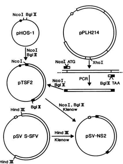
(46) immobilized onto nitrocellulose filter and eluted as described previously (36). Swine anti-rabbit immunoglobulin G conjugated to tetramethyl-rhodamine isothiocyanate (DAKO, Copenhagen, Denmark) was used as the second antibody.Plasmid constructs. To construct the nsP2 gene, plasmid pPLH214 (P.
LiUjestrom,
unpublished data), containing the entire nonstructural cDNA region of SFV 42S RNA (base pairs 1 to 7390), was cleaved with XhoI. The linearized plasmid was used to amplify a 2,400-base-pair fragment encoding the nsP2 protein by the polymerase chain reaction (PCR) method of Saiki et al. (54), with minor modifications. For this purpose, two oligonucleotides were synthesized. The upstream primer was a 30-nucleotide oligonucleotide(5'-CTC ACC ATG GGG GTC GTG GAA ACA CCT
CGC-3') corresponding to nucleotides 1697 to 1722 (sense strand) with additional nucleotides, which contained a trans-lation initiation codon and a NcoI site. The downstream primer was a 32-nucleotide oligonucleotide (5'-TC CAG ATC TTA ACA CCC GGC CGT GTG CAT GGC-3') corre-sponding to nucleotides 4068 to 4090 (antisense strand) with additional nucleotides, which contained a translation termi-nation codon and aBglIIsite. The PCR mix contained 50 ng of linearized pPLH214, 1,ugof each primer, 200FM each deoxynucleoside triphosphate, 0.1% gelatin, and lx PCR buffer (10 mM Tris hydrochloride [pH 8.35], 50 mM KCl, 1.5 mM MgCl2, 0.1 mg of gelatin per ml) in a total volume of 0.1 ml. To denaturate the DNA, the reaction mixture was boiled for 5 min in a water bath and 2 U of Taq DNApolymerase (New England BioLabs, Inc., Beverly, Mass.) and 1 pL. of 10% Triton X-100 were then added. Amplification was carried out for 15 cycles: denaturation for 75 s at 97°C,
Hind m
FIG. 1. Plasmid constructs. Plasmid pPLH214, which contains the complete coding region for SFV nonstructural proteins, was
used as atemplateforPCRtoobtainthecompletecodingregionfor nsP2 plusinitiation and terminationsignalsand theindicated restric-tion sites as described in Materials and Methods. The isolated NcoI-BgIII fragment was inserted into the transcription-translation
vector, pHOS-1, which is aderivative ofpGEM3.Forexpressionin mammalian cells the NcoI-BglII fragment containing nsP2 was
ligated to pSV-S-SFV vector from which the HindIII fragment
encoding SFV-specific proteins had been removed. This
construc-tionyielded thepSV-NS2vectorin which thensP2codingsequence wasunderthe controlof simian virus 40 early promoter.
annealing for 2 minat55°C, and extension for 20 minat
72°C.
After theseventhcycle, additional Taq DNA
polymerase
(2
U) wasadded to the reaction mixture.
DNA manipulations and cloning were done
by
standard methods (40). The amplified fragment wasdigested
with BglII and NcoI, run in an agarose gel, and eluted with the GeneClean kit (Bio 101). The pTSF2plasmid
wascon-structed by inserting the fragment into apGEM3
(Promega
Biotec, Madison,Wis.)-derived transcriptionvector,
pHOS-1 (J. Peranen, unpublished data), which had been cleaved withBglII andNcoI. Forexpression of nsP2 in animal
cells,
the vector pSV-NS2 was constructed by
ligating
theBglII-NcoI-cleaved and blunted fragment
encoding
nsP2 from pTSF2 into thepSV-S-SFV (a kindgiftfrom HenrikGaroff,
Department of MolecularBiology,The Karolinska
Institute,
Novum, Huddinge, Sweden) (31), which had been cleaved withHindIII andblunted and from whichtheSFVstructural cDNA fragment had been deleted (Fig. 1).
For in vitrotranscription the plasmid
pTSF2
was cleaved withBglII andthen transcribed asspecifiedbythe manufac-turer (Transprobe T; Pharmacia,Uppsala,
Sweden).
The transcripts were used for in vitrotranslation,
which was performed as recommended by the manufacturer(Promega
Biotec).
Cell transfection. BHK cellswere
plated
on coverslips
ata density of 0.5 x 10⁶ cells per 35-mm dish 1
day
prior
toon November 10, 2019 by guest
http://jvi.asm.org/
[image:2.612.326.539.65.349.2]1890 PERANEN ET AL.
transfection. Cells were transfected with 1
[ig
of pSV-NS2 bythelipofection transfection procedure (14) with the Lipo-fectin kit (Bethesda Research Laboratories, Inc., Gaithers-burg, Md.). The next day, the transfected cells were pre-paredfor immunofluorescence microscopy (32).RESULTS
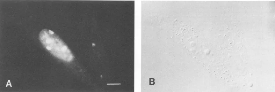
Immunofluorescence localization of nsP2.We have recently reported the production of monospecific antisera against selected amino acid sequences of the four nonstructural proteins of SFV (46). Only anti-nsP3 gave a satisfactory stainingin indirect immunofluorescence microscopy. When weaffinity purified the anti-nsP2 antiserum and used the tsl mutant of SFV, we could localize nsP2 by immunofluores-cenceinthe infected BHK cells (Fig. 2C). The rationale for using tsl wasbased onprevious studies, which have shown that in tsl-infected cells the nonstructural proteins are pro-duced in excess (33, 34, 58). Prominent fluorescence was
observed in the nuclei of both wild-type and tsl virus-infected BHK cells (Fig. 2A and C). In the nucleus an
intensive fluorescent staining in most cells was localized to spherical or irregular structures, which by Nomarski optics could be identified as nucleoli (Fig. 2B and D). A weaker diffuse nuclear staining gave the background for the bright nucleolar fluorescence. In the cytoplasm we occasionally foundstainedvesicularstructureson abackground ofafaint reticular staining (Fig. 2C and E). Only weak background fluorescence could be seen when uninfected cells were
treated with the affinity-purified anti-nsP2 antiserum (Fig. 2G) orwhen virus-infected cellswere treated with affinity-purified preimmuneserum(Fig. 2H).The nuclearstaining by anti-nsP2 antiserum couldalreadybedetectedat2h postin-fection (p.i.) (data notshown). When dactinomycin (1 or 5
[ig/ml)
wasadded at 1 hp.i.,nodistinct nucleolarstructurescould be seen at 6 h p.i. by Nomarski optics and the immunofluorescence was distributed all over the nucleus (Fig. 2E and F).
Isolation of nuclei and subnuclear compartments. We wanted toconfirm themorphological findingsby subcellular fractionation experiments. The conditions for isolation of intactnuclei andnuclear subcompartments wereoptimized. Wefound that isolation ofnucleiunder isotonicconditions gavethe best and most reproducible results (see Materials and Methods). Inspection of isolatednucleiby light micros-copy showed thatthey were intact and free ofcytoplasmic tags. Analysis by SDS-polyacrylamide gel electrophoresis (PAGE) showed a typicalprotein patternfor nuclei (Fig. 3, lane4; seeespecially the histones), whereas thecytoplasmic fractionwasdevoid oftheseproteins (Fig.3, lane 3). Further purification of nuclei by mild Triton X-114 treatment re-moved cytoplasmic contaminants and probably proteins from the outer nuclear membrane, since the number of protein species was clearly reduced (Fig. 3, lanes 5 and 6). Subnuclearfractionation was performed by first extracting the nucleitwice with an NP-40-containing buffer to release the nucleoplasmic proteins (Fig. 3, lanes 7 and 8). NP-40-extracted nuclei were treated withmicrococcal nuclease andhighsalttoobtainasoluble(chromatin) andaninsoluble (nuclearmatrix) fraction. The soluble fraction contained the histone proteinswhicharetypicalfor chromatin(Fig.3, lane 9), whereas the insoluble fraction contained the characteris-tic nuclear matrix proteinssuch aslamins (Fig. 3, lane 10). Subcellular location of SFV nonstructural proteins. The
distribution of nonstructural proteins in the cytoplasmand nucleiwasexaminedbyimmunoprecipitationwith
monospe-cific antibodies against all four nonstructural proteins of SFV. BHKcellswereinfected withwild-typeSFVorthe tsl mutant and
pulse-labeled
with [35S]methionine early during infection(1.5to 2.5h), when thenonstructuralproteinsaremaximally synthesizedand the firstreplicase complexesare formed. After the pulse the cellswere chased for 1 h in the presence of excess unlabeled methionine. The cells were
fractionated into cytoplasmic and nuclear fractions, which
weresubjectedtoimmunoprecipitations. Theresultsfor the wild-type-infectedcellsare shownin
Fig.
4A. MostofnsP3 (Fig. 4A,lanes 5and6) and nsP4(lanes7and8)werein the cytoplasm. In contrast, nsP2 was distributed aboutequally
between the
cytoplasmic
(Fig. 4A, lane3)andnuclear(lane 4)fractions. Quantitation bydensitometrictracings showed that about 13% of nsPl and about 5% of nsP3 and nsP4werefound in the nuclear fraction (Table 1). The distribution of the four nonstructural
proteins
was almost the same in tsl-infected cells(Fig. 4B).
The subnuclear distribution of nsP2 in
wild-type
SFV-infected cellswasinvestigated by fractionating
thenucleiasdescribed abovefor
Fig.
3.When eachfractionwasanalyzed
for the presence of nsP2by immunoprecipitation,it became clear that nsP2 was confined almost entirelyto the nuclear matrix fraction (Fig. 5, lane 10). A small amount of nsP2 could also be detected in the
nucleoplasm (Fig.
5,lanes 7 and8), but
virtually
none was found in the chromatin fraction(lane 9). The relativeamount ofnsP2 ineach fraction was
estimated
by
densitometrictracings.
Theproportion
ofma-trix-associated nsP2varied between70and90%in different experiments.
Tobe sure that the association of nsP2with the nuclear matrixwas not anartifact of the isolation
procedure (12, 25),
we mixed in vitro-translated nsP2 with unlabeled cells and fractionatedthem. After
fractionation, virtually
all the nsP2 was recovered in the cytoplasmic fraction (Fig. 6). This shows that the association ofnsP2with the nuclear matrixdid not take
place
during
the fractionation. It hasrecently
been shown that the formation of intermolecular disulfide bonds between
proteins during
fractionation can render nuclearproteinsnuclease and saltresistant(25).Toexaminethis,
weisolatednuclei andsubnuclearcompartmentsin thepresence of 10 mM iodoacetamide. lodoacetamide had no
influence onthedistribution of nsP2 in the nuclei: thesame
distributionwasalsoseenin
dactinomycin-treated
cells(data
not shown). We therefore conclude that the association of nsP2 to the nuclear matrix was not an artifact of the experimental conditions used.
Kinetics ofnuclear transport ofnsP2. To
investigate
the kinetics of nuclear transport of nsP2, SFV-infected cells were pulse-labeledfor 1 min, chasedfor3 and 7minat135 minp.i.,and thenfractionated intocytoplasm, nucleoplasm, chromatin, and nuclear matrixfractions. After1 min, nsP2 and its precursors, P12 and P123, were in thecytoplasmic
fraction. After the 3-min
chase,
nsP2 couldalready
beclearly
detected in thenucleus,
mainly
in the matrixfraction(Fig. 7, lanes 4). The precursor proteins remained in the cytoplasm
(Fig.
7, 4-and 8-minpanels,
lanes1).
After the 7-min chase a clearly detectable amount of the nsP2 wasfound in the nuclear matrix fraction.
Thus,
transportto the nucleus and association to the nuclearmatrix of nsP2 take placerapidly.
Longerpulses
with[35S]methionine
(5
to 15 min) followed by different chaseperiods
allowed us toestimate that half of the nsP2was
transported
tothenucleus within 15to20 min. The maximumproportion ofnsP2in the nucleus varied between 40 and60%indifferentexperiments.
J. VIROL.on November 10, 2019 by guest
http://jvi.asm.org/
.5.
'.4
J-
I
-B.
S
C
D
F
FIG. 2. Localizationof nsP2 in SFV wild-type-infected (A and B), tsl mutant-infected (C, D, E, F, and H), and mock-infected(G)BHK cells. Theinfected cells in panelsEand F were treated with dactinomycin (5jig/ml)from 0 to 6 hp.i. The cellswere stained for indirect immunofluorescence at 6h p.i. by using affinity-purified rabbit anti-nsP2 and preimmune serum (H) (46). Panels A, C, E, G, and Hare immunofluorescencemicrographs; panels A, E, G, and H were exposed identically. Panels B, D, and F are corresponding Nomarskiimages forpanels A, C, and E, respectively. Bars, 10 pum.
on November 10, 2019 by guest
http://jvi.asm.org/
[image:4.612.101.507.85.664.2]1892 PERANEN ET AL.
1
2 3
4 56
78 9 10
FIG. 3. Total proteins found in subnuclear fractionsfrom BHK cells.Proteins were separated on a linear 7.5 to15%gradient gel by the method of Laemmli (35) and stained with Coomassie blue. Lanes: 2, cell lysate; 3,postnuclear supernatant; 4, crude nuclear fraction; 5, Triton X-114 postnuclear supernatant; 6, Triton X-114-treatednuclearpellet; 7 and8,nucleoplasmic fractions released by treatment with 1% NP-40; 9, supernatant released from NP-40-extractednuclei after nucleaseandhigh-salttreatment(chromatin); 10, nuclease- andhigh salt-resistant matrix fraction after pelleting and boiling in 1% SDS. Lane 1 contains molecularmass markers (fromthe top, 94, 67, 43, 30, 20.1,and 14.4kilodaltons)(Pharmacia). The positionsof histones(Hi, H3, H2B, H2A, andH4) and lamins (A, B, C)have been indicated with dots and arrowheads, respec-tively.
A
M 1 2 3 4 5 6 7 8
Inhibition of protein synthesis during the chase period did
not affect thedistribution of nsP2 (Table 1).
nsP2 intransfectedcells. nsP2isproteolytically processed from a large polyprotein. Thus, the nsP2 gene has no
translation initiation or termination codons of its own. To
construct afunctional nsP2 gene we synthesized two
prim-ers, whichcorrespondedtothe5' and 3' ends ofthecDNA
sequence encoding nsP2. The primerswere supplied witha
translation initiation andatermination codon, respectively.
Usingtheseprimers,weamplifiedthe nsP2codingsequence
bythePCRmethod(54). Plasmid pPLH214, which contains
a cDNA fragment spanning the whole nonstructural region
of 42S RNA (see Materials and Methods), was used as a
template. The amplified nsP2 gene was cloned into a tran-scriptionvector. Whentranscripts madefrom this plasmid, pTSF2, were translated in vitro, a protein of the expected
sizewas synthesized, confirming that the reading framewas
open(Fig. 6). Toexpress the nsP2genein animal cells, we
inserted it into aeucaryotic expressionvector, creatingthe plasmid pSV-NS2 (Fig. 1). BHKcellsweretransfected with
pSV-NS2 by the lipofection method (14). After 16 h the transfected cells were processed for immunofluorescence
microscopy by using the affinity purified nsP2 antibody. Positive immunofluorescence was detected in transfected
cells, inwhich the nucleoliwereoften brightly stained (Fig.
8).IncontrasttotheSFV-infected BHK cells, the cytoplasm
was stained very weakly.
DISCUSSION
In the present study we could demonstrate, by indirect
immunofluorescence, thata substantialamountof nsP2was
transported to the nucleus in SFV-infected BHK cells. Morphological observationsweresupported by results from
cell fractionation, which revealed that
[35S]methionine-la-FIG. 4. Distribution of SFV nonstructural proteinsbetween
cy-toplasm and nucleus. BHK cells infected with wild-typeSFV(A)or
the tsl mutant (B) were labeled for 1 h at 1.5 h p.i. with [35S] methionine followed by a 1-h chase. At 3.5 h p.i. the cells were
harvested and fractionated intocytoplasm (includingTriton X-114 postnuclear supernatant) (lanes 1, 3, 5, and 7), andTriton X-114-treated nuclei(lanes 2, 4, 6,and8).Thefractionsweresubjectedto
immunoprecipitation with immuneseraagainstnsPl(lanes1and2), nsP2(lanes 3 and 4),nsP3(lanes5 and6),and nsP4(lanes7 and8). LaneMcontains14C-labeledmolecularmassmarkers(fromthetop, 200, 92.5, 46, and 30kDa [Amersham Corp., Arlington Heights, Ill.]). SDS-PAGE ina 7.5%acrylamide gel was performed bythe method of Laemmli(35).
beled nsP2wasfound in the nucleus. At the sametime,only
small amounts ofnsPl, nsP3, and nsP4, derived from the
same polyprotein precursoras nsP2, were transportedinto the nucleus. The specificity of the transport ofnsP2 was
supported by experimentsinwhichexogenouslysynthesized nsP2,added tothecells beforedisruption andfractionation,
was not found in the nuclear fraction (Fig. 6). Since the actualamountsof intracellular and invitro-synthesizednsP2
are not known, the value for this control alone can be
regardedas strongly suggestivebutnotabsolute.
Labeled nsP2 was already associated with the nuclear matrix 4 min after synthesis and reached a plateau after about 30 to 40 min of chase. Similar transport rates have beenreportedfor simian virus 40largeTantigen (53, 62)and
nucleoplasmin (52).The nuclear matrix hasanessential role
in organizing DNA into dormant and active transcription units (6), as well as in DNAreplication (17, 42, 43, 45, 73). The nuclear matrix also seems to be an assembly and
replicationsite ofmany DNA viruses(3, 28, 47, 48, 63,64, 68) and retrovirus oncoproteins (5, 11, 66). Why certain
am
B
em
O
--J. VIROL.
""'%
a= -- .-:,,.
4im .',
-zLiii, .,;,.
*oft
IW,ym-4m,NW1.20.
:-qmkk.
46 la w
4.WAI--
on November 10, 2019 by guest
http://jvi.asm.org/
[image:5.612.386.501.76.388.2] [image:5.612.129.249.77.236.2]TABLE 1. Distribution of SFV-specific nonstructural proteins betweencytoplasm and nucleus
% ofnonstructuralproteins'
Virus nsPl nsP2 nsP3 nsP4 C N C N C N C N
Expt 1l
Wild type 87 13 56 44 96 4 95 5
tsl 87 13 49 51 98 2 94 6
Expt2b
Wild type 42 58
tsl 40 60
Expt 3'
Wild type 46 54
Wild type plus 45 55
cycloheximide
"C,
Cytoplasmicfraction;N, nuclearfraction.bInfected BHK cells were labeled for 1 hwith[35S]methionineat 1.5 hp.i..
followed byachase of 1 h.
' Infected cells werelabeledfor 15 min with[35S]methionineat 225minp.i.. followed by a 90-min chase in the absence or presence of 100 ,ug of
cycloheximideperml. Afterthechase,the cells werescrapedfrom thedish andcytoplasmicandnuclear fractionswereisolated.Thecytoplasmicfraction includesthe Triton X-114 washfromthenuclear pellet. Thefractions were
immunoprecipitated with therespectiveantisera before SDS-PAGE analysis
(7.5%acrylamide).Thepercentagesarebasedondensitometric tracingsof the
exposedfilms.
1
2 34
as
n
0w
FIG. 6. Cell fractionation after addition of
[35S]methionine-la-beled in vitrotranslationalproduct ofnsP2 genetoBHK cellsbefore breakageasdescribed forFig.3. Lanes: 1,molecularmassmarkers (from the top, 200, 94, 67, 43,30,20, and 14.4 kilodaltons); 2, in vitro translationalproduct ofpTSF2-transcribedmRNAcodingfornsP2; 3, postnuclearsupernatant; 4, nuclearpellet.
proteins have affinity specifically for nuclear matrix, or how they interact with it, is so far unknown.
The immunofluorescence staining localized a substantial amount of nsP2 in the nucleoli of infected or transfected cells. It is known that in the presence of dactinomycin, the nucleoli disappear asmorphological entities and their protein components,suchasS1proteinand RNApolymerase I, may have different distributions in the nucleus (2, 44). In dacti-nomycin-treated SFV-infected cells no nucleoli could be distinguished. Immunofluorescence staining and cell
frac-M 1 2
3
4 5 6 7 8 910111213tionation revealed that nsP2 was also transported into the nucleus in the drug-treated cells. Similar results have been reported recently for dengue 4 virus core protein in mam-malian andmosquito cells (71)aswellasinlepidopteran cells (39).
We have previously shown that nsP2 can be found asso-ciated with ribosomes in SFV-infected cells. nsP2 could be cross-linkedtorRNAby UVirradiation, indicatingthat it is very close to therRNA (50). Thus, it would be temptingto assume that nsP2 recognizes rRNA orribosomal protein in the nucleolus during the biogenesis of ribosomes such as nucleolin, B23 (4), and ribocharin (22). Anotherpossibilityis that it binds to some nucleolus-specific proteins, which can beregardedasstructural components ofthenucleolus(RNA polymeraseI, fibrillarin, etc.)(45, 67,79). Since nsP2is also transportedto thenucleus inthe presenceofdactinomycin,
1'
_
4. 8
1 2 3 4 1 2 3 4 1 2 3 4
a
FIG. 5. Distribution of SFV nonstructuralproteinsin subnuclear fractions. BHK cells infected withwild-typeSFVwerelabeledasin Fig. 4, and the harvested cells were fractionated into subnuclear fractionsasperformed for Fig. 3. Lanes: 1, postnuclear supernatant; 2,Triton X-114 wash of nuclei; 3 to6, Triton X-114-treatednuclei: 7, nucleoplasmin first extraction with1% NP-40; 8, second NP-40 extraction;9, chromatin fraction; 10to 13, nuclear matrix. lmmu-noprecipitationswerecarriedoutwithantiseraagainst nsPl (lanes4 and 11), nsP2(lanes 1, 2, 3, 7,8, 9, and 10), nsP3 (lanes5and 12), and nsP4(lanes6and 13).SDS-PAGE wasperformedas in Fig.4.
FIG. 7. Kinetics of transport of nsP2 intonuclei. BHK cellswere
infected with wild-type SFV and labeled for1 minat 135 min p.i.
followed by chases of 3 and 7 min. The harvested cells were
fractionatedas follows: lanes 1, cytoplasm (excludingpostnuclear
Triton X-114 wash); lanes 2. nucleoplasm (first extract with 1% NP-40); lanes 3. chromatinfraction; lanes4, nuclearmatrix. SDS-PAGE wasperformed asin Fig.4.
on November 10, 2019 by guest
http://jvi.asm.org/
[image:6.612.391.466.75.288.2] [image:6.612.51.292.95.254.2] [image:6.612.75.269.484.635.2]1894 PERANEN ET AL.
B
FIG. 8. Immunofluorescence of BHKcells transfected with the plasmid pSV-NS2 containing cDNA encoding full-length nsP2. Cells exposed to the plasmid by the lipofectin method (14) were preparedforindirect immunofluorescence 24 hlaterbyusing affinity-purified anti-nsP2 antiserum. Immunofluorescence(A)and Nomarski image (B)from the samefield. Bar, 10p.m.
there must also be binding sites in the absence of visible nucleoli (Fig. 2E and F).
The factthat nsP2, expressed alone in transfected cells, also migrated into the nuclei, implies that the protein itself musthave information for its nuclear targeting. Only in few caseshas aspecific signal or signals for the nuclear location (NLS) been identified by using deletion mapping and/or site-directed mutagenesis (10, 17). The common feature for theidentifiedNLS seems to be a short stretch of basic amino acids, usually arginines and lysins. None of the identified viral or cellular nuclear targeting sequences can be found in the SFV nsP2.
There are someprevious reports on transport into nuclei ofproteinsencoded byRNAvirusesreplicatingexclusively in the cytoplasm. Lyles et al. (38) have recently shown evidence that some of the M protein of vesicular stomatitis virus was associated with the nucleus in the nucleoplasmic fraction.Similar observations have been made with Newcas-tle disease virus (13). Tobacco mosaic virus-specific 126-kilodalton protein, which is involved in viral RNA replica-tion, has been reported to be bound to the chromatin of infected tobacco cells (75-78). The coat protein of alfalfa mosaic virus has also been found in the nucleus late in infection (74).Rift Valley fevervirus-encoded nonstructural protein NS1 hasbeen shown to accumulate in the nuclei of infected monkey kidney cells (70). Most recent reports by Tadano et al. (71) and Makino et al. (39) describe that the coreprotein of dengue virus type 4 can be found in the nuclei andespecially in the nucleoli. Sofar, no nuclear targeting signalshave been identified for anyof these proteins.
Several functions in alphavirus-infected cells have been assigned to nsP2. First, it has a vital early function in RNA synthesis, since temperature-sensitive mutants, with muta-tions mapping in nsP2, display an RNA-negative phenotype at the restrictive temperature. Second, it is involved in the regulation of the synthesis of subgenomic 26S mRNA, typical for mutants of complementation group A (19, 20, 24, 27, 61, 69). Third, Sindbis virus nsP2 has an autoprotease activity, which is involved in the proteolytic processing of the nonstructural polyprotein (9). If this function is impaired, theprocessing of the polyprotein P1234 would be deficient, and this may explain the RNA-negative phenotype of nsP2 mutants. nsP2 can also bind to ribosomes, at least in SFV-infected cells (50). How this phenomenon is related to theobserved functions of nsP2 remains open. In this study we have shown that nsP2 is efficiently transported into
nucleoli relatively early in virus infection. Thismust be an additional function, most probably unrelated to the viral RNA synthesis and protease activities. Crude cell fraction-ation has shown that about half of the cytoplasmicnsP2 is located in themitochondrialpellet (P15) (46), which contains all the virus-specific RNA polymerase activity (18,
49).
Practically all of nsP4 and nsPl and about 80% of nsP3 are
located inP15, presumablyasanRNApolymerase
complex.
They are evidently associated with the cytoplasmic
vacu-oles,which havebeen showntobe the site of
virus-specific
RNA synthesis (16,
46).
Itcanbeestimated that about 25% ofsynthesizednsP2 is associated withthe membranous structuresinP15. Another 25% wouldbe free in thecytoplasm.This would be compat-ible with the immunofluorescence results of cytoplasmic staining, which reveals vesicular structures
(cytoplasmic
vacuoles)on aweakly stained cytoplasmic background
(Fig.
2). TherestofnsP2, about 50%, is transportedtothenuclei, where it associates with the nuclear/nucleolar matrix. What is the function, if any, of nsP2 in the nucleusornucleolus? So far, we cannot answer this question. Ishida et al.
(23)
have suggested that an 82-kilodalton protein in western equine encephalitis virus-infected cells is responsible for the virus-specific inhibition of host DNA synthesis (65). Thus, it is likely that nsP2 is responsible for the inhibition of host DNA and possibly also RNA synthesis. It remains to be shown whether nsP2 hasanucleoside triphosphatase activ-ity which has been claimed to beresponsible for the inhibi-tion of host DNA synthesis (29, 30). Since alphavirusescan
grow in enucleated cells (15), none of the nuclear functions are essential for the virus replication itself. This does not mean that the virus could not benefit from a nonvital function such as inhibition of host macromolecular synthe-ses.
ACKNOWLEDGMENTS
WethankAnnikki Kallio for excellent technicalassistance. Thiswork wassupported by theSigrid Juselius Foundationand Svenska Kulturfonden.
LITERATURECITED
1. Barton, D.J., S. G. Sawicki, and D. L. Sawicki. 1988. Demon-stration invitrooftemperature-sensitiveelongation ofRNAin Sindbis virusmutantts6. J. Virol. 62:3597-3602.
2. Benavente, R., K. M. Rose, G. Reimer, B. Hugle-Dorr,andU. Scheer. 1987. Inhibition ofnucleolarreformationafter microin-J. VIROL.
on November 10, 2019 by guest
http://jvi.asm.org/
[image:7.612.95.540.75.225.2]jection of antibodiesto RNA polymeraseIintomitotic cells. J. Cell Biol. 105:1483-1491.
3. Ben-Ze'ev A., R. Abulafia, and S. Bratosin. 1983. Herpes sim-plex virus and protein transport are associated with the cyto-skeletal framework and the nuclearmatrix in infected BSC-1 cells. Virology129:501-507.
4. Borer, R. A., C. F. Lechner, H. M. Eppenberger, and E.A.Nigg. 1989. Major nucleolar proteins shuttle between nucleus and cytoplasm. Cell56:379-390.
5. Boyle, W. J., M. A. Lampert, A. C.Li,and M.A.Baluda.1985. Nuclearcompartmentalization ofthe v-myb oncogne product. Mol. Cell. Biol. 5:3017-3023.
6. Ciejek, E. M.,M.-J. Tsai,and B. W. O'Malley. 1983. Actively transcribed genes are associated with the nuclear matrix. Na-ture(London) 306:607-609.
7. Cross, R. K. 1983. Identification ofaunique guanine-7-methyl-transferase inSemlikiForest virus(SFV) infectedcellextracts. Virology 130:452-463.
8. Cross, R. K., and P. J. Gomatos. 1981.Concomitantmethylation andsynthesis in vitro of SemlikiForestvirus(SFV)ssRNAsby
afraction from infectedcells. Virology 114:542-554.
9. Ding, M., and M. J. Schlesinger. 1989. Evidence that Sindbis virus nsP2 is an autoprotease which processes the virus non-structuralpolyprotein. Virology 171:280-284.
10. Dingwall, C., and R. A. Laskey. 1986. Protein import into the cell nucleus. Annu. Rev. CellBiol. 2:367-390.
11. Eisenmen, R. N., C. Y. Tachibana, H. D. Abrams, and S. R. Hann. 1985. v-myc-andc-myc-encodedproteinsareassociated withthe nuclear matrix. Mol. Cell. Biol. 5:114-126.
12. Evan,G.I.,and D.C. Hancock. 1985.Studiesontheinteraction ofhuman c-mycproteinwith cell nuclei: p62cmYcas amember ofadiscrete subset of nuclear proteins.Cell 43:253-261. 13. Faaberg, K. S., and M. E. Peebles. 1988. Strain variation and
nuclearassociation of Newcastle disease virus matrixprotein.J. Virol.62:586-593.
14. Feigner,P.L.,T. R.Gadek,M.Holm,R.Roman,H. W.Chan,
M. Wenz, J. P. Northrop, G. M. Ringold, and M. Danielsen. 1987.Lipofection:ahighly efficient,lipid-mediated DNA-trans-fectionprocedure. Proc. Natl. Acad. Sci. USA84:7413-7417. 15. Follet, E. A.C.,C. R.Pringle,and T. H.Pennington.1975. Virus
development in enucleate cells: echovirus, poliovirus, pseu-dorabiesvirus, reovirus, respiratorysyncytialvirus andSemliki Forestvirus. J. Gen. Virol. 26:183-196.
16. Froschauer,S., J. Kartenbeck, and A. Helenius. 1988.
Alphavi-rus RNA replication occurs on the cytoplasmic surface of endosomesandlysosomes.J. Cell Biol. 107:2075-2086. 17. Gerace,L., and B. Burke. 1988. Functionalorganizationofthe
nuclearenvelope. Annu. Rev.Cell Biol. 4:335-374.
18. Gomatos,P.J.,L.Kaariainen,S.Keranen,M.Ranki,and D. L. Sawicki. 1980. SemlikiForestvirusreplication complexcapable ofsynthesizing 42S and 26SnascentRNAchains.J. Gen.Virol. 49:61-69.
19. Hahn,Y. S.,A. Grakoui,C. M. Rice, E. G.Strauss, andJ. H. Strauss.1989.MappingofRNA-temperature-sensitivemutants
of Sindbis virus:complementationgroup Fmutantshave lesions in nsP4. J.Virol. 63:1194-1202.
20. Hahn,Y.S.,E.G.Strauss,andJ.H.Strauss. 1989. Mappingof RNA- temperature-sensitive mutants ofSindbis virus: assign-mentofcomplementation groupsA, B,and Gtononstructural proteins.J.Virol. 63:3142-3150.
21. Hardy,W.R.,andJ.H.Strauss. 1988.Processingthe nonstruc-turalpolyproteinsof Sindbis virus:studyofthekinetics in vivo byusingmonospecific antibodies.J. Virol. 62:998-1007. 22. Hugle, B., U.Scheer, and W. W. Franke. 1985. Ribocharin: a
nuclearMr40,000protein specificto precursorparticlesof the large ribosomal subunit. Cell 41:615-627.
23. Ishida, I.,B.Simizu,S.Koizumi,A.Oya,andM.Yamada.1981. Nucleoside triphosphate phosphohydrolase produced in BHK cells infected withwesternequineencephalitisvirus isprobably associated with the 82K dalton nonstructural polypeptide. Vi-rology108:13-20.
24. Kaariainen, L., K. Takkinen, S. Keranen, and H. Soderlund. 1987. Replication ofthe genome ofalphaviruses. J. Cell Sci.
Suppl. 7:231-250.
25. Kaufman, S.H., S. Okret, A.-C. Wikstrom,
J.-A.
Gustafsson,andJ.H.Shaper.1986.
Binding
of theglucocorticoid
receptortotherat livernuclear matrix. J.Biol. Chem. 261:11962-11967. 26. Keranen, S., and L. Kaariainen. 1974. Isolation and basic
characterization oftemperature-sensitivemutantsfrom Semliki Forest virus. Acta Pathol. Microbiol. Scand. Sect. B 82:810-820.
27. Keranen, S., and L. Kaariainen. 1979. Functional defects of
RNA-negative temperature-sensitive mutants of Sindbis and SemlikiForestvirus. J. Virol. 32:19-29.
28. Knipe, D.M., D. Senechek,S. A. Rice,andJ. L.Smith. 1987.
Stages in the nuclear association ofthe
herpes simplex
virustranscriptionalactivator
protein
ICP4. J. Virol.61:276-284. 29. Koizumi,S.,B.Simizu,K.Hashimoto,A.Oya,
and M.Yamada.1979. Inhibition ofDNA
synthesis
in BHKcells infectedwithwesternequineencephalitisvirus. 1. Inductionofan
inhibitory
factor of cellular DNA polymerase
activity. Virology
94:314-322.30. Koizumi, S.,B.Simizu,I.Ishida,A.Oya,and M. Yamada.1979. Inhibition ofDNAsynthesisinBHKcells infected withwestern
equine encephalitis virus. 2.
Properties
oftheinhibitory
factor of DNA polymerase induced in infected cells.Virology
98: 439-447.31. Kondor-Koch,C.,B.Burke,andH.Garoff.1983.
Expression
of Semliki Forest virusproteins
from clonedcomplementary
DNA. 1. Thefusion activity ofthespike
protein.
J. Cell Biol. 97:644-651.32. Kuismanen, E., K. Hedman, J. Saraste, and R. F. Pettersson. 1982. Uukuniemi virus maturation: accumulation of virus
parti-cles and viral antigens in the
Golgi complex.
Mol. Cell. Biol. 2:1444 1458.33. Lachmi, B.,and L. Kaariainen. 1976.
Sequential
translation of nonstructuralproteins
in cells infected with a Semliki Forest virus mutant. Proc. Natl. Acad. Sci. USA73:1936-1940. 34. Lachmi, B.,and L. Kaariainen.1977.Controlofprotein
synthe-sis in Semliki Forest virus-infected cells.J. Virol.22:142-149. 35. Laemmli,U. K. 1970.
Cleavage
of structuralproteins during
theassembly ofthe head of
bacteriophage
T4. Nature (London)227:680-685.
36. Last,R.L.,andJ.L.Woolford. 1986.Identificationandnuclear localization of yeast pre-messenger RNA
processing
compo-nents: RNA2 andRNA3
proteins.
J. Cell Biol. 103:2103-2112. 37. Li, G., and C. Rice. 1989.Mutagenesis
of the in-frameopal
termination codon
preceding
nsP4of Sindbis virus: studies of translationalreadthrough
andits effecton virusreplication.
J. Virol.63:1326-1337.38. Lyles, D. S., L. Puddington, and B. J.
McCreedy,
Jr. 1988. Vesicular stomatitis virus Mprotein
in the nuclei of infected cells.J. Virol. 62:4387-4392.39. Makino, Y.,M.Tadano,T.Anzai,S.-P.Ma, S.Yasuda,and T.
Fukunaga.1989.Detection of
dengue
4viruscoreprotein
in thenucleus.II.
Antibody against dengue
4coreprotein produced by
a recombinant baculovirus reacts with theantigen
in thenu-cleus.J.Gen. Virol. 70:1417-1425.
40. Maniatis, T.,E. F. Fritsch, andJ. Sambrook. 1982. Molecular
cloning: alaboratorymanual. Cold
Spring
HarborLaboratory,
Cold SpringHarbor, N.Y.
41. Mi, S., R. Durbin, H. V. Huang, C. M. Rice, and V. Stollar. 1989. Association ofthe Sindbis virus RNA
methyltransferase
activity with the nonstructural
protein
nsPl.Virology
170: 385-391.42. Nelson, W. G., K. J. Pienta, E. R. Barrack,and D. S.
Coffey.
1986. The role of the nuclear matrix in theorganization
and function of DNA. Annu. Rev. Biophys.Biophys.
Chem. 15: 457-475.43. Newport,J.W.,andD.J.Forbes.1987. Thenucleus: structure,
function,and
dynamics.
Annu. Rev. Biochem.56:535-565. 44. Nickerson,J.A., G.Krochmalnic,K. M.Wan,and S. Penman.1989. Chromatin architecture and nuclear RNA. Proc. Natl. Acad. Sci. USA 86:177-181.
45. Nigg,E. A. 1988. Nuclear function and
organization:
the poten-tialof immunochemicalapproaches.
Int. Rev.Cytol.
110:27-92.on November 10, 2019 by guest
http://jvi.asm.org/
1896 PERANEN ET AL.
46. Peranen, J., K. Takkinen, N. Kalkkinen, and L. Kaariainen. 1988. Semliki Forest virus-specific non-structural protein nsP3 is a phosphoprotein. J. Gen. Virol.69:2165-2178.
47. Pinard, M.-F., R. Simard, and V. Bibor-Hardy. 1987. DNA-bindingproteins of herpes simplex virus type-1 infected BHK cell nuclearmatrices. J. Gen. Virol. 68:727-735.
48. Quinlan, M. P., and D. M. Knipe. 1983. Nuclear localization of herpesvirus proteins: potential role for the cellular framework. Mol. Cell. Biol. 3:315-324.
49. Ranki, M., and L. Kaariainen. 1979. Solubilized RNA replica-tion complex from Semliki Forest virus-infected cells. Virology 98:298-307.
50. Ranki, M., I. Ulmanen, and L. Kaariainen. 1979. Semliki Forest virus-specific nonstructural protein is associated with ribo-somes. FEBS Lett. 108:299-302.
51. Rice, C. M., R. Levis, J. H. Strauss, and H. V. Huang. 1987. Production of infectious RNA transcripts from Sindbis virus cDNAclones: mapping of lethal mutations, rescue of a temper-ature-sensitive marker, and in vitro mutagenesis to generate definedmutants. J. Virol. 61:3809-3819.
52. Richardson, W. D., A. D. Mills, S. M.Dilworth, R. A. Laskey, and C. Dingwall. 1988. Nuclearprotein migration involves two steps:rapid bindingat the nuclearenvelopefollowedby slower translocation through nuclear pores.Cell52:655-664.
53. Rihs, H. P., and R. Peters. 1989. Nuclear transport kinetics depend on phosphorylation-site-containing sequences flanking the karyophilic signal ofthesimian virus40T-antigen. EMBO J. 8:1479-1484.
54. Saiki, R. K., D. H. Gelfand, S.Stoffel,S.J. Scharf, R. Higuchi, G. T. Korn, K. B. Mullis, and H. A. Erlich. 1988. Primer-directed enzymatic amplification of DNA with a thermostable DNA polymerase. Science 239:487-491.
55. Saraste, J., L.Kaariainen, H. Soderlund, and S. Keranen. 1977. RNA synthesis directed by a temperature-sensitive mutant of Semliki Forest virus. J. Gen. Virol.37:399-406.
56. Sawicki, D. L., and S. G. Sawicki. 1985.Functional analysis of the A complementation group mutants of Sindbis HR virus. Virology144:20-34.
57. Sawicki, D. L., S. G. Sawicki, S. Keranen,and L. Kiairiainen. 1981. Specific Sindbis virus-coded function for minus-strand RNAsynthesis. J. Virol. 39:348-358.
58. Sawicki, S. G., and D. L. Sawicki. 1986. Theeffect of overpro-ductionof nonstructuralproteinsonalphavirus plus-strandand minus-strandRNA synthesis. Virology 152:507-512.
59. Sawicki, S. G., and D. L. Sawicki. 1986. The effectofloss of regulation of minus-strand RNA synthesis on Sindbis virus replication. Virology 151:339-349.
60. Sawicki, S. G., D. L. Sawicki, L. Kaariainen, and S. Keranen. 1981. A Sindbis virus mutanttemperature-sensitive in the reg-ulation of minus-strand RNAsynthesis. Virology 115:161-172. 61. Scheele, C. M., and E. R. Pfefferkorn. 1969. Inhibition of
interjacentribonucleic acid (26S) synthesis in cells infected by Sindbis virus. J. Virol.4:117-122.
62. Schickedanz, J., K. H. Scheidtmann, and G. Walter. 1986. Kinetics of nuclear transport and oligomerization of simian virus 40 large T antigen. Virology 148:47-57.
63. Schirmbeck, R., and W. Deppert. 1987. Specific interaction of simian virus 40 large T antigen with cellular chromatin and
nuclear matrix during the course of infection. J. Virol. 61: 3561-3569.
64. Schirmbeck, R., and W. Deppert. 1989. Nuclear subcompart-mentalizationof simian virus40 large Tantigen: evidencefor in vivo regulationof biochemical activities. J. Virol. 63:2308-2316. 65. Simizu, B. 1984. Inhibition of host cell macromolecular
synthe-sisfollowing togavirus infection. Compr. Virol. 19:465-499. 66. Slamon, D. J., W. J. Boyle, D. E. Keith, M. F. Press, D. W.
Golde, and L. M. Souza. 1988. Subnuclear localization of the trans-activating proteinof human T-cell leukemia virustype I. J. Virol. 62:680-686.
67. Sommerville, J. 1986. Nucleolar structure and ribosome biogen-esis. Trends Biochem. Sci. 11:438-442.
68. Stamatos, N. M., S.Chakrabarti, B. Moss, and J. D. Hare. 1987. Expressionofpolyomavirusvirionproteinsby a vaccinia virus vector: association of VP1 and VP2 with the nuclearframework. J. Virol. 61:516-525.
69. Strauss, E., and J. H. Strauss. 1986.Structure andreplicationof the alphavirus genome, p. 35-90. In S. Schlesinger and M. J. Schlesinger (ed.), The Togaviridae and Flaviviridae. Plenum Publishing Corp.,NewYork.
70. Struthers, J. K., R. Swanepoel, and S. P. Shepherd. 1984. Proteinsynthesisin RiftValleyfever virus-infected cells. Virol-ogy134:118-124.
71. Tadano, M., Y. Makino, T. Fukanaga, Y. Okuno, and K. Fukai. 1989.Detection ofdengue4virus coreproteinin the nucleus. I. Amonoclonalantibodytodengue4virusreactswiththeantigen in the nucleus andcytoplasm. J.Gen. Virol. 70:1409-1415. 72. Takkinen, K. 1986. Complete nucleotide sequence ofthe
non-structuralprotein genesofSemliki Forestvirus.Nucleic Acids Res. 14:5667-5681.
73. Van derVelden, H. M. W., and F. Wanka. 1987. The nuclear matrix. Its role in the spatial organization and replication of eukaryoticDNA.Mol. Biol. Rep. 12:69-77.
74. Van Pelt-Heerschap, H., H. Verbeek, J. W. Slot, and L. Van Vloten-Doting. 1987. The location of coat protein and viral RNAs of alfalfa mosaic virus in infected tobacco leaves and protoplasts. Virology 160:297-300.
75. Van Telgen, H. J., R. W.Goldbach,and L.C. Van Loon. 1985. The126,000 molecularweight protein oftobaccomosaicvirus is associated with host chromatin in mosaic-diseased tobacco plants. Virology143:612-616.
76. VanTelgen, H. J., E. J. Van Der Zaal, and L. C. Van Loon. 1985.Evidence foranassociation betweenviralcoatproteinand host chromatin in mosaic-diseased tobacco leaves. Physiol. PlantPathol. 26:83-98.
77. VanTelgen, H. J., E. J. Van Der Zaal, andL. C. Van Loon. 1985. Some characteristics ofthe association ofthe 116 kD protein with host chromatin in tobacco leaves infected with tobacco mosaic virus.Physiol. PlantPathol. 26:99-109. 78. VanTelgen,H.J.,and L.C.VanLoon. 1985. Two-dimensional
gel electrophoretic analysis of chromatin-associated proteins from leaves ofdifferent speciesand varietiesof Nicotiana. J. PlantPhysiol. 118:285-296.
79. Verheijen, R., H. J. H. Kuijpers, R.0. Schlingemann,A. L. M. Boehmer, R. van Driel, G. J.Brakenhoff,andF.C. S. Ramaek-ers. 1989. Ki-67 detects a nuclearmatrix-associated prolifera-tion-related antigen. I. Intracellular localization during inter-phase. J. Cell Sci. 92:123-130.
J. VIROL.


![FIG. 6.translationalbeledbreakage(from3, postnuclear Cell fractionation after addition of [35S]methionine-la- in vitro translational product of nsP2 gene to BHK cells before as described for Fig](https://thumb-us.123doks.com/thumbv2/123dok_us/1321017.85769/6.612.75.269.484.635/translationalbeledbreakage-postnuclear-fractionation-addition-methionine-translational-product-described.webp)