Copyright C) 1989, American Society for Microbiology
Construction and Characterization of
a
Herpes Simplex Virus Type
1
Mutant
Unable To Transinduce Immediate-Early Gene Expression
CHRIS I. ACE,' TOM A. McKEE,' J. MICHAEL RYAN,2 JANET M. CAMERON,2AND CHRIS M.
PRESTON'*
Medical Research Council Virology Unit, Instituteof Virology, Church Street, GlasgowGIl 5JR, Scotland,' and GlaxoGroupResearchLimited, Greenford Road, Greenford, Middlesex UB6OHE, England2
Received 7 December1988/Accepted6February 1989
A herpes simplex virus mutant, in1814, possessing a 12-base-pair insertion in the gene encoding the
transinducing factorVmw65 has been constructed. Theinsertion abolishedtheabilityof Vmw65 totransinduce immediate-early (IE)geneexpressionandtoformaprotein-DNAcomplexwith cellproteinsandthe IE-specific
regulatory element TAATGAGAT. Accumulation of IE RNA 1 and 2 was reduced four- to fivefold in in1814-infected cells, but the levelof IERNA 4 wasreduced only by twofold, andIERNA3 wasunaffected. Mutantin1814hadahigh particle/PFU ratio,butmanyoftheparticles, althoughunabletoformplaques,were
capable ofnormal participationin theearlystagesof infectionathigh multiplicityof infection. The defect of inl814was overcomepartially by transfection ofaplasmid encodingtheIEproteinVmwllO intocellspriorto titration and by prior infection with ultraviolet light-inactivated herpes simplex virus. Mutant in1814 was
essentiallyavirulent when injected into mice. The results demonstrate that transinduction ofIEtranscription byVmw65 isimportantatlowmultiplicityof infection and in vivo but thatathigh multiplicityof infection the function is redundant.
Herpes simplex virustype1(HSV-1) encodes70predicted
genes which are expressed as three temporally regulated
classes (35, 70). The five immediate-early (IE or ao) genes are the first to be transcribed after infection, and their expression does not require de novo protein
synthe-sis, whereas early (L) and late(y)geneexpression is
depen-dent on the prior synthesis of IE polypeptides (8, 23).
The products of IE gene 1 (polypeptide VmwllO or ICPO)
and IEgene3 (Vmwl75 or ICP4)arepotenttransactivators of early- and late-gene promoters in transient expression
assays (12, 18, 40, 53). The IE gene 2 product (Vmw63
or ICP27) has also been implicated in the regulation of
viral promoters (13, 54, 60). Analysis of temperature-sensi-tive mutants indicates that both Vmwl75 and Vmw63 are
essential for productive infection; Vmwl75 is required for early- and late-gene expression (11, 47, 71), whereas Vmw63appears tobe required after the onsetof early-gene expression and DNA replication (56). Viable mutants
with deletions in VmwllO exhibit restricted growth in
cer-tain cell types at low multiplicity of infection (MOI) but
areapparently normal at high MOI(57, 65). Deletion
muta-tions in IE gene 4 (which specifies Vmw68 or ICP22)
also conferahostrangephenotypetothe virus(45, 59).The IE gene5 product (Vmwl2 or ICP47) appears to be unim-portant for virus replication in tissue culture cells since deletionswithinthegenehave little effectongrowth of HSV
(4, 29, 69).
Adistinguishing feature ofIE genesisthepresenceofthe cis-acting element TAATGARAT (where R is a purine
residue)intheir5'regulatory regions. This element responds
to the HSV-1 virion polypeptide Vmw65 (otherwise desig-nated VP16oroxTIF), resulting inastimulation of
transcrip-tionfrom IEpromoters(2, 5, 7, 9, 17, 25, 31-33, 41, 43, 44, 48). Although Vmw65 does notitself bind toDNA (34), the evidence currently available suggests that the polypeptide mediates transinduction of IE genes by associating with
cellularproteins, including nuclear factorIII,toformanIE
* Correspondingauthor.
complex (IEC) which is able to bind specifically to DNA
sequences that contain TAATGARAT (1, 19, 38, 39, 49). Mutation analysis of cloned DNA fragments encoding Vmw65 suggeststhat the polypeptide contains atleast two separable regions, both of which are necessaryfor
transin-duction ofIE transcription. The amino-terminal 411 amino acidsaresufficient forbindingtothecellularfactor(1;T. A. McKee, C. I. Ace, and C. M. Preston, manuscriptin
prep-aration),and the acidiccarboxy-terminaldomain definedby amino acids 411to490(the"acidtail,"afeaturecommonto many other eucaryotic and procaryotic transactivators [3, 20, 30, 52, 58, 66, 67]) isrequired forstimulating transcrip-tion and may interact with fundamental transcription
com-ponents, for example, the TATA binding factor TFIID or
RNA polymerase, orboth(24).
Because Vmw65 regulates the set ofgenes expressed at the earliest stages ofinfection, it isimportant to determine the role of thepolypeptide duringHSVgrowth. All informa-tion to date regarding the properties ofVmw65 has been obtainedby transfection, usingeitherstablytransformed cell lines or transient expression assays. Such systems are
clearly artificial,and the crucialbiological questionconcerns
thephenotypeof virusmutantswhichlack thetransinducing activity of Vmw65. A difficulty inherent in attempts to construct suchmutantsisthat, apartfrom its role in transin-ducing IEgenes, Vmw65 is alsoa majorstructural compo-nent which isrequired for virion assembly (1). To address this problem, functional domains of Vmw65 required for virion assembly and for transinduction were identified by
insertion mutagenesis of a cloned gene fragment (1). The construction and characterization ofaviable HSV-1mutant that containsaninsertion whichaffectsonlythe
transinduc-ing activity of Vmw65 are described here. The results
indicate that IE gene transinduction by Vmw65 is not essential for virusgrowth at high MOI but plays a critical role indeterminingwhetherinfection islyticor
nonproduc-tiveatlowMOI. Vmw65isalsoimportant forthe virulence of HSV-1 in mice.
2260
on November 10, 2019 by guest
http://jvi.asm.org/
MATERIALS AND METHODS
Cells and viruses. BHK cells (clone 13) were grown in Eagle mediumwith 10% newborn calf serum, 10% tryptose phosphate broth, and 100 U of penicillin and 100 ,ug of streptomycin per ml. Human fetal lung (HFL) cells (Flow Laboratories) were grown in Eagle medium with 10% fetal calf serum and 100 Uofpenicillin and 100 ,g of streptomycin perml.Thewild-type(wt)HSV-1virususedin these studies was Glasgow strain 17 syn+. Virus particle concentrations were determined by comparison ofvirus stocks with bead
preparationsof known concentration.
Plasmids. Plasmid pMC1, which contains the coding se-quencesforVmw65, has beendescribed previously (7). The construction of pMC1.inl4, which contains a 12-base-pair (bp) BamHI oligonucleotide linker inserted in the Vmw65 gene, has also been described (1). Plasmid pIE3CAT
con-tains the HSV-1 IE gene 3 promoter and regulatory se-quences linked to the chloramphenicol acetyltransferase
codingregion (64), and plll expresseswt HSV-1 VmwllO
(14).
Isolationofin1814 and Southern blot analysis. A BHK cell monolayer in a 35-mm-diameter petri dish was cotransfected with 0.5 ,ug of intact HSV-1 DNA, 0.5
jig
of anEcoRI-cleaved plasmidwith themutationof pMC1.inl4 in the larger
plasmidpGX158(whichcontains
BamHIf
[7]), and 2.0 ,ugof calfthymus carrierDNAby thecalciumphosphate precipi-tation method (51). After incubation for3 days at31°C, the progenyviruseswere harvested and titrated on BHK cells.Single plaques werepicked and used to infect BHK cells in multiwell plates containing 15-mm-diameter wells. After 2 days at 37°C, total DNA was prepared from infected cells (65) and the medium was retained as a viral stock. DNA samples were screened for the presence of viral genomes
containingaBamHI linkerinsertionwithin the Vmw65 gene. DNA was cleavedwithBamHI,andviral DNA analyzed by agarosegelelectrophoresisandSouthernblotting (61). Plas-mid pMC17, which contains the Vmw65 coding sequences cloned in pUC9 (1), was radiolabeled with 32P by nick translation (55) and used as aprobe. Hybridization,
mem-brane washing, and autoradiography conditions were as described previously (42). Progeny from a sample which
contained viral DNA with a linker insertion was plaque purified and screened by hybridization twice more, and a working stock of virus was prepared from BHK cells. The virus was named in1814.
Marker rescue of in1814. A BHK cell monolayer was
cotransfected with 0.1 ,ug of intact in1814 DNA, 0.5 p,g of
pMC1cleavedwithEcoRI, and 2.0
pig
ofcalfthymusDNA, as described above. After 5 days at 31°C, progeny were harvested and used to infect a BHK cell monolayer on a 90-mm-diameter petri dish at 0.0001 PFU per cell. After 3days, theprogenywereharvested andtitratedonBHK cells.
Single plaqueswerepickedand usedtoinfectBHKcellson
multiwell plates. Virusstockswerepreparedfrom wells,and
theirgenomes were analyzed as described above. Progeny fromasamplethatexhibited awtHSV-1 DNA structure was plaquepurified, and aworkingstock of virus was prepared in BHK cells. The viruswas named 1814R.
Quantitationof viralDNA in nuclei. BHKcellmonolayers in90-mm-diameterpetridisheswereinfected in the presence of 200 ,ug ofcycloheximide (CH)per ml. After incubation for 3 h at38.5°C, cell nuclei were isolated (46) and DNA was extracted. Virus DNAwasquantitatedby Southernblotting as described above but by using
32P-labeled
pTK1, whichcontains the cloned HSV-1 BamHI p fragment, in the
hybridization procedure.
Radiolabeling of viral polypeptides. For IE polypeptides,
BHK cell monolayers were infected in the presence of 200
Fgof CHper ml. After 4 hat38.5°C, CHwas washedfrom the plates and proteins were radiolabeled for 1 h with [35S]methionine in the presence of 1 ,ug ofactinomycin D per ml and analyzed by sodium dodecyl sulfate-polyacrylamide
gel electrophoresis (SDS-PAGE) (46). For early and late polypeptides, BHK cell monolayerswere infected and incu-batedat37°C for 8h, and proteinswereradiolabeled for 1 h with [35S]methionine and analyzed by SDS-PAGE.
Gel retardation analysis. Virion extracts were prepared (49), and proteins were analyzed by SDS-PAGE. The gel was stained with Coomassie brilliant blue. Virion extract wasadded to amixture containing HeLa cell nuclear extract and a 32P-labeled74-bp DNAfragmentcontaining HSV-1 IE gene 4/5 regulatory sequences (49). Reaction conditions for complex formation and analysis of protein-DNA complexes were as described previously (49).
Transinduction assay. BHK cell monolayers in 35-mm-diameter petri dishes were transfected with 3 ,ug ofpIE3CAT by the calcium phosphate precipitation method (7), except that thedimethyl sulfoxide boost was performed 1 h after the mediumoverlay. The cells were incubated at 38.5°C for 1 h and then superinfected. After incubation forafurther 3 h at 38.5°C, extracts were made from the cells and chloramphen-icolacetyltransferase assays wereperformed (21).
Quantitation of IE RNA. BHK cells were infected in the presence of200 ,ug of CH per ml. After incubation for 4 h at 38.5°C, cytoplasmic RNAwasextracted and quantitated by dot blotanalysis (72) by using DNA probes radiolabeled with 32P by primer extension (15). Gene-specific probes were prepared from DNAfragments that correspond to IE genes 1(a1,367-bp SaIl-NruI fragment from pJR3[12]), 2 (a
2,760-bpMluI-BamHIfragment from BamHI b [35]), 3 (a 3,210-bp HincII fragment from XhoI c [36]), and 4 (a 2,200-bp NruI-MluI fragment from BamHI n hybridizing predomi-nantlyto IE RNA 4 [37]).
TKassay. BHKcellswereinfected in the presence of 200 ,ugofphosphonoaceticacid per ml.After incubation for 15 h at 38.5°C, cytoplasmic extracts were made and thymidine kinase(TK) assayswereperformed (9).
Complementation assay. BHK cellmonolayers in 35-mm-diameterpetri disheswere transfected with 3 ,ug ofplll or
pUC9 bythecalciumphosphate precipitationmethod(7)and treated with dimethyl sulfoxide 1 h later. Afterafurther 1 h at37°C,viruswastitratedonthemonolayers. Alternatively,
monolayersweretreatedwith the HSV-1mutanttsK, which hadbeen UV irradiated toreduce its titer by5 x 105 (48). The MOIof UV-irradiated tsKcorresponded to 0.1 PFU of unirradiated virus per cell. Wt HSV-1orin1814wastitrated on the UV-irradiated-tsK-pretreated cells. After 2 days at
37°C, plates were stained andplaques werecounted. Virulence assay. Female Charles Rivermice,each weigh-ing approximately 15 g,wereinoculated eitherintracranially (ic) with 20
RI
orintraperitoneally (ip) with 200RI
of 10-folddilutions of virus stocks, as described previously (6). Ten micewereinoculated for each virusdilution,and thenumber of survivors after 21 days was recorded. The mean 50% lethal dose values from twoexperiments were calculated.
RESULTS
Isolation of a mutant containing an insertion within the Vmw65gene. The gene encodingVmw65 lies between map
on November 10, 2019 by guest
http://jvi.asm.org/
a
IE-1
-
3AV
b
x
cloned In pMC1
pmc
s X
0.669 0.685
a I
V
(-Vmw65(UL48) IE-2
A D P
CGCGGATCCG
BamHl
7
IE-4 IE-3
_
_-I I
,i
,.- -0
IE-1 IE-3 IE-5
R CG
/000,
i I
Vmw65 mRNA
FIG. 1. (a) Structure ofthe HSV-1genome showingthe positions ofthe five IE genes (without introns)and Vmw65. Theopen boxes represent repeated sequences. (b) Structures of the insertion mutation in Vmw65 encoded on a SaIl(S)-XhoI(X) fragment in plasmid pMC1.inl4.
coordinates 0.669 and 0.685 in the ULregionof the prototype HSV-1 genome and is contained withinplasmid pMC1 (Fig. 1A; 7, 10). The isolation of a number of plasmids with in-frame BamHI linker insertion mutations within the gene encoding Vmw65 has been described previously (1). In
particular, afour-amino-acid insertion at codon 397,
speci-fied on plasmid pMC1.inl4 (Fig. 1B), abolished the
transin-ducingactivity of thepolypeptideintransfection assays. The mutation disabled the binding of Vmw65 to the host-cell
factorsand thusdefinedaregion of the polypeptide involved in this interaction. The essential role of the polypeptide duringvirion assembly was notaffectedby the mutation, as
inferred from its ability to rescue an HSV-2 mutant with a
temperature-sensitive mutation in Vmw65 (1), suggesting
that a viable virus could be constructed that contained the
transinducing mutation specified by pMC1.inl4. To con-structsuchamutant virus,aplasmidconsistingofBamHIf containing the pMC1.inl4 mutation wascotransfected with
intactwt HSV-1 DNA into BHK cells, and the structure of progeny virus DNA was examined by restriction enzyme
analysis. One plaque from a total of 84 screened was
identifiedas arecombinant thatcontained the BamnHI linker
insertion. This mutant isolate, in1814, was plaque purified twice more, and a large-scale stock of virus was prepared. Torule out thepossibilityofasecond sitemutation in in1814 that might affect the phenotype of the mutant virus, a rescuedvirus was constructedby recombiningin1814 DNA withpMC1. If, as desired, the phenotype ofin1814depended on the insertion mutation, then a rescued virus should behaveas wtHSV-1. Initial observation ofthe properties of in1814 suggested that it grew poorly in comparison with wt HSV-1; thus, it was expected that rescued recombinants wouldoutgrowin1814duringsuccessive passages of a mixed population. This turned out to be the case, since after a
single passage of the progeny virus from the initial cotrans-fection of in1814 DNA and pMC1, 75% of the plaques screened had the wt DNA structure. These viruses were
unlikely to result from spontaneous reversion of in1814,
since no reversion was detected at any stage during the
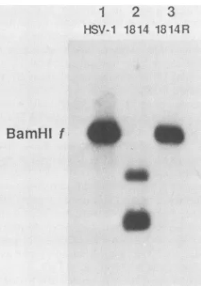
passaging and propagation of mutant virus. A stock of rescuedvirus,1814R,waspreparedafterplaque purification. Figure 2 shows a Southern blot ofwt HSV-1, in1814, and 1814R DNA which was digested with BamHI and probed with pMC17, a plasmid containing the Vmw65 coding se-quences. TheBamHIffragmentof 8 kilobasepairswasseen in both wt HSV-1 (lane 1) and 1814R (lane 3), whereas in in1814 (lane2), this fragmentwasreplaced bytwofragments of the sizes (5 and 3 kilobase pairs) expected from the presenceof theBamnHIlinkerinsertion.Overexposureofthe autoradiographrevealed no detectableBamHIf fragmentin
1
2
3
HSV-1 1814 1814R
BamHl
f
FIG. 2. Structure of the in1814 genome. Wt HSV-1 (lane 1), in1814(lane 2), or1814R(lane 3) DNA wascleaved withBamHI, and thefragmentswereseparatedon a1.5% agarosegel,transferred
tonitrocellulose,andhybridizedto32P-labeled pMC17.Theposition of HSV-1Baml?HI fis indicated.
on November 10, 2019 by guest
http://jvi.asm.org/
[image:3.612.156.470.78.288.2] [image:3.612.369.514.464.670.2]TABLE 1. Titration ofwtHSV-1,in1814,and 1814R onBHKandHFLcells
BHKtiter HFLtiter
Virus Particles/mi (PFU/mI)
(PFU/mI)
wtHSV-1 1.9 x 101l 5.0 x 10⁹ 1.7 x 10¹⁰
in1814 1.2 x 10¹¹ 1.3 x 10⁷ 7.0 x 10⁵
1814R 4.6x 10¹⁰ 4.0x 10⁹ NDa
aND,Notdetermined.
in1814 and therefore that the stock of mutant virus was
essentially pure.
The efficiency ofplaqueformation by in1814 is markedly reduced and dependentoncelltype.The successful isolation andpropagation of in1814 confirms that the insertion muta-tion is compatible with virus growth in BHK cells. When in1814 wastitratedon BHK cells, however, alow titerwas
obtained, and therefore virus particle concentrations were
determined (Table 1). Itwasfound that the particle
concen-trations ofwt HSV-1, in1814, and 1814R stocks were
com-parable but that the particle/PFU ratio was approximately
100 times greaterfor in1814 thanforwt HSV-1 and 1814R. The apparenttiter ofagiven preparation of in1814onBHK
cells varied by as much as 10-fold on different batches of
cells, whereas the titers ofwtHSV-1 and 1814Rweremuch
moreconsistent, suggesting that thecellularmetabolic state affects the efficiency of plaque formation by in1814. When titrations were performed on HFL cells, an even higher
particle/PFU ratio, 1.7 x 10⁵, was observed for in1814. In
view of the variation in titer of in1814 when expressed in terms of PFU, cell monolayers were infected with equal numbers of particles of wt HSV-1, in1814, or 1814R in subsequent experiments.
DNAmigrationtothe nucleus.Theearlystagesof infection by in1814 were examined, since it was possible that the
insertionmutation affected virus adsorption, penetration,or
uncoating.Ininitialexperiments, therateofadsorption ofwt HSV-1 or in1814 preparations, radiolabeled by incubation with [3H]thymidine during virus propagation, to BHK cell monolayers was investigated. The adsorption rates ofwt HSV-1 and in1814 particles were indistinguishable (results not shown). The efficiency of DNA migration to the cell nucleus was also determined. BHK cell monolayers were
infected in the presence of CH with 1,000, 100, or 10
particlesofwtHSV-1, in1814,or1814Rpercell,nucleiwere
prepared at 3 h postinfection, and nuclear DNA was
ana-lyzed by Southern blot hybridization (Fig. 3). Nosignificant differencesweredetectedinthe levelsof HSVDNA, show-ingthat the nuclear migration of in1814DNAisnotimpaired
at eitherhigh orlow MOI.
This result underlines the requirement to use particles
rather thanPFUas abasisfor thedesign of experimentswith
in1814; 1,000 particles ofwtHSV-1and in1814represent26 and0.1 PFU, respectively.
in1814 doesnotexhibit virion-mediatedtransinduction of IE
genes.TheabilityofVmw65,encodedby in1814,toform the protein-DNA complex IEC and to transinduce expression from transfected IE promoterswas investigated, since both of these properties weredisrupted in pMC1.inl4 (1).
Extracts of wt HSV-1, in1814, and
18i4R
virions werepreparedandanalyzed bySDS-PAGE(Fig. 4).Thelevels of Vmw65in theseextracts werevery similar, and theslightly
increasedmolecularweightof themutantpolypeptidedueto
the four-amino-acid insertion was apparent (lane 2). The virion extracts were incubated with HeLacell nuclear
ex-Particles
per cell
W14
R M
1000
100
*W
L
FIG. 3. DNAmigrationtothe nucleus. DNAisolated fromnuclei ofcells infected with wtHSV-1 (W), in1814 (14),or 1814R(R)or mock infected(M), in thepresence ofCH,wascleavedwithBamHI, and thefragmentswereseparatedon anagarosegel, transferredto
GeneScreen Plusmembrane, and probed with radiolabeled pTK1. The portionofthe blotrepresentingBamHl p is presented. Expo-suretimes were 0.4 h(1,000particlespercell),4h(100particlesper cell),or40h (10 particlespercell).
tract and a 74-bp DNA fragment containing the TAAT GAGAT sequence motif of IE gene 4/5. Asshown inFig. 5, the IEC was readily detected with extracts of wt HSV-1 (lane 2) and 1814R (lane 4) virions but notwith extracts of in1814 (lane 3)orwhen no virion extract waspresent (lane 1). This result demonstrates thatVmw65specified by in1814 is not capable of binding the cellular proteins required for IEC formation because of the mutationinthe viral polypep-tide.
The ability of in1814 to transinduce IE gene expression wasinvestigated bycomparing the level of activation froma transfected IE promoter in the presence or absence of superinfecting virus (Fig. 6). BHK cells were transfected with pIE3CAT and infected with 1,000 particles of wt
HSV-1, in1814, or1814R per cell. An increase of
approxi-mately sixfold inchloramphenicol acetyltransferase activity
wasobserved when cellswere superinfected withwtHSV-1
(lane2) or1814R(lane 4), but infection with in1814(lane 3)
1 2
3
..
Vmw65
pFIG. 4. Proteins extracted from virions ofwt HSV-1 (lane 1), in1814 (lane 2),and 1814R(lane 3)and usedasa sourceof Vmw65 for gel retardation analysis. The gel was stained with Coomassie
brilliant blue.
on November 10, 2019 by guest
http://jvi.asm.org/
[image:4.612.53.294.90.149.2] [image:4.612.378.489.479.681.2]1 2 3 4 -EXT HlSV-1 1814 1814R
IEC
H-C3
HCI
FIG. 5. IEC formation by Vmw65. A 74-bp DNA fragment
containingthe IE gene4/5 TAATGAGAT sequence was incubated
with HeLacell nuclearextract alone(lane 1) orwithvirionextract
(EXT) fromwt HSV-1 (lane 2), in1814(lane 3), or 1814R(lane 4),
andwas analyzed by gel electrophoresis. Thepositions of IECand
thecell-specific complexesHC1 and HC3 (49)are indicated.
gave no stimulation over the level in mock-infected cells
(lane 1).
Taken together, these results confirm that the properties
of themutantVmw65polypeptidein the viralcontextreflect the observations and expectations implicit in the initial characterization of the mutation in clonedcopiesof thegene;
thatis,themutation in in1814 disablestheabilityof the virus
todirect the formation of IEC andconsequentlyabolishes its
transducing activity.
Gene expression in in]1814-infected cells. It would be
ex-pectedthat theabolition oftransinduction by Vmw65 would
affecttheexpressionof viralgenes,especially Ie genes. The
accumulation ofIE RNA was quantitated by hybridization
byusingIEgene-specificprobes.BHK cellswereinfected in
thepresenceofCH with1,000particlesofwtHSV-1,in1814, or1814Rpercell for4h,andcytoplasmicRNAwas
applied
1
ml
2 HSV-1
3
1814
4
1814R
3-AC.
[image:5.612.131.231.71.320.2]CAM 41W
FIG. 6. Transinduction of IE transcription. Cells were trans-fectedwithpIE3CAT and mock infected (lane1)orinfected withwt HSV-1(lane 2), in1814 (lane3),or1814R (lane4).Chloramphenicol
acetyltransferase assays were carried out on cytoplasmic cell
ex-tracts.Thepositions of chloramphenicol(CAM) and 3-acetyl
chlor-amphenicol (3-AC. CAM)areshown.
HSV-1
F
*1814
1814R I*
0
[image:5.612.319.563.71.170.2]0*
FIG. 7. Production of IE RNA. Cellswere mock infected(MI; row1)orinfected withwtHSV-1 (row 2), in1814(row3),or1814R
(row4) in the presence of CH. RNA was prepared after 4 h and appliedtonitrocellulose filters infoursequential dilutions (3 ,ug, 1 ,ug, 0.3 ,g, and 0.1 ,ug). Filters were separately hybridized with 32P-labeled DNA probescorrespondingtoIEgenes1,2, 3, and4/5
(panelsa,b, c,andd, respectively).
tonitrocelluloseandseparately hybridized withradiolabeled DNA fragments corresponding to the IE-1, IE-2, IE-3, or
IE-4 genes. The levels of IE-1- and IE-2-specific RNA, as
determinedbydensitometric analysis, werereduced four-to fivefold in in1814-infected cells compared with wt HSV-1-and 1814R-infected cells (Fig. 7, a and b), whereas the
reductioninIE-4/5-specific RNAwasonly twofold (Fig.7d),
andnosignificanteffecton IE-3-specificRNAwasdetected
(Fig. 7c).
The expression of IEpolypeptideswas also investigated.
BHK cells were infected asdescribed above, but after 4 h
CH was washed from the cells and polypeptides were
radiolabeled in thepresenceofactinomycin D and separated by SDS-PAGE (Fig. 8). Densitometric analysiswas usedto
1 2 3 4
M I HSV-1 1814 1814R
Vmw175
Vmw136
Vmwllo
Vmw68
Vmw63
= t -
~~~~actin
FIG. 8. IEpolypeptide synthesis. Cellsweremock infected (MI;
lane1)orinfected withwtHSV-1 (lane 2), in1814 (lane 3),or1814R (lane 4) in the presence of CH. Proteins were labeled with [35S]methionine after removal of CH by washingat4hpostinfection. Thepositions of viral IE polypeptides and cellular actin are
indi-cated.
a
IE-1
b
IE-2
ml
c
IEE-3
d
IE-4f5
** 0
on November 10, 2019 by guest
http://jvi.asm.org/
[image:5.612.375.503.425.661.2] [image:5.612.60.305.570.663.2]Vmw65 MUTANT 2265
1 2 3 4
rI iSv-11t514 1814R
.. X
. i
i
t
t -Ss T*¢ * F X
* : *. * bF't
'big :' ! :* t #
# L
: B;
't
v.,*
F_
u
rtw
3_ilii
_w _
B.. Tss s
F:' :. ><
::* _
.e .:
Vmw65
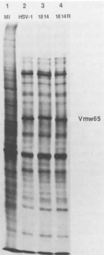
FIG. 9. Late polypeptide synthesis. Cells were mock infected (MI;lane 1)orinfected withwtHSV-1 (lane 2), in1814 (lane3),or
1814R(lane 4), and proteinswerelabeled with[35S]methionine at8 hpostinfection. The position of Vmw65 is indicated.
determine the relative rates of synthesis of individual IE polypeptides, and the values were normalized to that of actin. The rates of synthesis ofVmwllO and Vmw63, the products of IE genes 1 and 2, respectively, were reduced
four-tofivefoldinin1814-infected cells, whereas theratesof synthesis of Vmwl75, the product of IE gene 3, were
equivalent for the three viruses. It was not possible to
measure accurately the rate of synthesis of Vmw68, the
productofIEgene4, since thispolypeptideran as a diffuse band. It isclear, however, that the data on IE RNA levels and IE protein synthesis rates are in good agreement and show that theexpression ofIEgenes1and 2 is significantly reducedinin1814-infectedcellsbut that theexpression ofIE
gene3 is essentially unaffected.
Protein synthesis at 8 h postinfection, atime whenearly
and, especially, late polypeptides are synthesized, wasalso
examined (Fig. 9). The profiles ofwt HSV-1, in1814-, and 1814R-infectedcellswere verysimilar,theincreased
molec-ular weight of Vmw65 specified by in1814 being the only majordifference.
Thus, uponinfection ofBHK cells with1,000 particles of
in1814percell in thepresenceofCH,the levelofexpression
ofIEgenes 1, 2, 4, and presumably5 isreduced, but under normal conditions infection procedes to the late stage, suggesting thatthere is no overall consequence of reduced IE gene expression. From the high particle/PFU ratio, however, it appears that the growth ofin1814 is inefficient
when cells are infected with 1 virus particle per cell. To investigate whether the incapacity of in1814 at low MOI is reflected inreducedgeneexpression,thesynthesisofTK,an
earlyenzymethatcanbedetected withhigh sensitivity,was
examined. BHKcellswereinfected with1,000, 100, 10,or1
particleofwt HSV-1, in1814,or1814Rpercell, and incuba-tion was continued for 15 h in the presence of
phosphono-TABLE 2. TKproduction by wt HSV-1,in1814,and 1814R athigh and lowMOP'
Viral TKactivity (cpm/min of assay
MOI per ,ug ofprotein) Ratio of wt
(particles/cell) HSV-1/in1814
wtHSV-1 in1814 1814R
1,000 2,693 2,285 2,789 1.2
100 1,988 2,549 2,478 0.8
10 935 161 792 6.0
1 100 3 63 33.3
"BHK cells were infected at the multiplicities indicated. Cytoplasmic extracts werediluted as necessary to ensure that TKdeterminations were within thelinear responserangeoftheassay.Abackground of3 cpm permin of assay perp.gofproteinhasbeensubtractedfromall values.
aceticacidtopreventthesecondary spreadof virus. Table 2 shows the results of TK assays performed on the cell extracts. The level of TK after infection with 1,000or 100 particles per cell wasindistinguishableforwtHSV-1, in1814, and 1814R, reemphasizing that in1814 is not detectably impairedat high MOI. At 10particlespercell, the TKlevel inin1814-infected cells relative to that inwtHSV-1-infected cells was reduced by 6-fold, and at 1 particle per cell the decrease was33-fold. Therefore,theexpressionof TK(and
presumably ofother early and late genes) is more strictly
dependent onMOIfor in1814 thanfor wtHSV-1 or1814R, and it is likely that the observed reduction in expression is large enoughtoaccountfor theinefficiencyof plaque forma-tion by the mutant.
Complementation of in1814 by VmwllO and Vmw65. If in1814 fails to form plaques at low MOI because of the reduction in IE gene expression, then complementation of this stateshould increase theefficiency of plaque formation
and consequently the apparent titer of the mutant virus. In contrast, a compensating increase in IE gene expression would not complement in1814 growth if the mutant pheno-typeresulted fromadefectat astage beforetheonsetof IE transcription. Two experiments were carried out to test these possibilities.
BHKcellsweretransfected withplll (aplasmidencoding
the HSV-1 transactivator VmwllO) orpUC9andthen used
separatelyfor titration ofwtHSV-1, in1814,or1814R(Table
3).Althoughthetiters ofwtHSV-1 and 1814Rwereconstant in both cell samples, the apparent titer of in1814 increased
approximately 10-fold oncells transfected withplll. Since onlyaproportion ofcells(normallybetween 5 and50%)ina BHKmonolayerexpress VmwllOafter transfection ofplll, it is likely that a higher level of complementation could
potentially be obtained. Therefore, raising the level of VmwllO can, atleastpartially, rectifythedefect ofin1814 in BHKcells.
Complementationof in1814 in HFL cellswasachievedby infecting monolayers with UV-irradiated tsK, which
[image:6.612.121.225.71.326.2]sup-pliedfunctional Vmw65 in trans (48), priortotitrationofwt HSV-1orin1814. The apparenttiterof in1814increased from 5.7 x
10'
to1.5 x109
PFU/ml,whereas the titer of wt HSV-1TABLE 3. Titrationofwt HSV-1,in1814, and 1814RonBHK cellstransfected withpUC9orplll (encoding VmwllO)
PFU/ml Plasmid
wt HSV-1 in1814 1814R
+pUC9 1.2 x 10") 4.0 x 10⁶ 1.7 x 10⁹
+pill 9.5 x10⁹ 4.2 x10⁷ 2.2 x10⁹
VOL.63,1989
on November 10, 2019 by guest
http://jvi.asm.org/
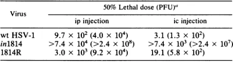
[image:6.612.308.550.93.171.2] [image:6.612.313.551.667.721.2]TABLE 4. Virulence of wtHSV-1,in1814, and1814Rin mice
50%Lethal dose(PFU)" Virus
ip injection icinjection
wtHSV-1 9.7 x 10² (4.0 x 10⁴) 3.1 (1.3 x 10²) in1814 >7.4 x 10⁴(>2.4 x 10⁸) >7.4 x 10³(>2.4 x 10⁷) 1814R 3.0 x 10³ (9.2 x 10⁴) 19.1 (5.8 x 10²)
a50%Lethal doseintermsofparticlesper mouseis shown in parentheses.
was 6.0 x
1010
on both cellmonolayers. No plaques wereobserved on monolayers treated only with UV-irradiated tsK. The titer of in1814on UV-irradiated-tsK-treated cells representedaparticle/PFU ratio of 74, and after account is taken of the fact that the MOI for UV-irradiated tsKwas
only0.1 PFU percell,it is clear that theefficiency of plaque formnation was similar to that of wt HSV-1. Thus, the
observedphenotype ofin1814onHFLcellswasreversedby provision of Vmw65 in trans, arguing against a cis-acting
defect, for example, inhibition ofuncoating by the mutant
protein.
in1814 has reduced virulence in mice. Anassessmentofthe invivo propertiesofin1814wasmadeby studying virulence after inoculation of miceeithericorip. The results(Table 4)
show thatin1814 was much less virulent thanwt HSV-1 or
1814R, regardless ofthemethod of inoculation. In fact, all mice challenged within1814 survived, withtheexceptionof three mice injected ic with undiluted virus. In these cases, death wasatypically rapid,occurringwithin 12 hasopposed
totheusual 3to5days, and it issuspectedthat theeffectwas due to thelarge number of virusparticles injected. The50%
lethal dose values intermsofparticles per mouse,the more relevantvalue, show that virulence ofin1814wasreducedby
a factor of at least 3 x
10'
for ip or 2.5 x 10⁴ for icinoculation, comparedwith wtHSV-1 or1814R. DISCUSSION
Theisolationofa mutantdefectiveintransinductionof IE
transcription is a crucial step in determining the biological
role of Vmw65. The 12-bp insertion mutation in in1814
appears tobe stable,since norevertants have beendetected
during passageand growth ofvirus stocks; reversionto the
phenotype ofwtHSV-1 would readily bedetected, asshown
bytheeasewith which 1814Rwasisolated. Twofeatures of in1814 areparticularly noteworthy. At MOI of100 or more
particles percell, no significant effect was observed on the overall pattern of virus gene expression, whereas at low
MOI, the efficiency ofplaque formation was severely re-duced inacell-dependent manner. The phenotype is similar to that exhibited by deletion mutants which do not express VmwllO (57, 65).
Although the results presented here suggest that
transin-duction by Vmw65 isnotessentialfor HSV geneexpression athighMOI, thisinterpretation mustbe takencautiously, as the assays available are of limitedsensitivity. The degree of impairment of transinduction is difficult to assess because thestimulation of expression from a transfected IE promoter is only 5- to10-fold, and thus, as argued previously (1), it is possible to stateonly that in1814 is reduced by at least 90%
in its ability to stimulate IE transcription. Analysis of the
ability to form IEC, as shown in Fig. 5, is more sensitive. and by this criterion in1814 is disabled by
99%
or more. Nevertheless, each HSV particle contains approximately 1,000 molecules of Vmw65 (22), and therefore a cumulativeeffectofalow residual activity might be sufficient to endow
in1814with the ability to formplaquesatthe observed low efficiency.
Intheabsence of transinduction by Vmw65, the IE genes would be expected tobe transcribed according to the inher-entstrengths of their promoters,afeaturethat ispresumably determined by interaction with cellular proteins. For IE genes 1 and 2, the 4-to 5-fold reduction in RNA accumula-tion and protein synthesis correlates well with the 5- to 10-foldstimulation oftranscription in BHK cells from trans-fected IE gene 1 and 2 promoters byVmw65 (C. M. Preston, unpublished results). The expression of IE genes 3 and 4, however,is greater than wouldbeanticipated from trahsfec-tion studies, since these promoters are also activated by more than fivefold (5, 48), and it is difficult to offer an obvious explanation for this apparent discrepancy. One possibility is that the enhancer-like sequence which lies between the promoters of IE genes 3 and 4 (28, 50), rather thanthe TAATGARAT elements, is the major requirement for transcription of IE gene 3 in the context of the viral genomeand that the strongproximal promotersufficesfor IE genes 4 and 5
(48).'
It is also noteworthy that the four upstream nucleotides of the TAATGARAT elements which control IE genes 1 and 2 confer a strong homology to the nuclearfactor III binding site, the octamer element ATGC AAAT, whereas this is not the case forthe TAATGARAT elements located between IE genes 3 and 4/5. A further consideration is that thetopology of the DNA template and the stoichiometric relationships between DNA and protein factors may vary considerably between transfected and infected cells. Clearly, the findings with virus-infected cells are the more relevant.Even though in1814lackstransinducing activity, the major polypeptides synthesized under IE conditions are the IE proteins. Activation by Vmw65 is therefore not adefinitive characteristic of IE genes, and other features must distin-guish them from early and late genes. It may be that the presence of strong promoters and enhancer-like sequences determines therelatively high efficiency of IE gene transcrip-tion in the absence ofIEproteins, but equally,the TAATG ARAT orotherIE-specificelementsmight beresponsible. It is known thatcellular proteins bind to various sequences in IE gene upstream regions (26, 27, 39, 49, 68), and these factors might increase the availability of IE promoters to transcription components in the absence of Vmw65. Thus,
IE-specific DNA sequences, rather thanVmw65, may be the primary determinants ofanIE gene.
Transinduction by Vmw65 is important for infection only
at low MOI. At a superficial level, it is straightforward to view this property as a reasonable adaptation, since the initial interaction of HSV with an organism is likely to involve a small number of virus particles. The inability to replicate at low MOI appears to result from the failure to produce IE proteins at levels sufficient to initiate infection,
andit is probable thatthe reductions in VmwllO and Vmw63 are crucial, since these polypeptides are required for gene expression (56,57, 65). Thus, it seems that threshold levels ofIEpolypeptides mustbe attained, and the role of Vmw65 is to ensure that such levels are reached, especially at low MOI. Itis not clear whether the few cells inwhich infection with in1814 results in the formation of a plaque represent a subpopulation in a particular metabolic state or simply randomvariation in response toinfection. Furthermore, the basis for the difference in behavior ofin1814 in BHK and HFLcells remains undefined. It may be that IEtranscription in the absence of Vmw65 is lessefficient in HFL cellsat low MOI or that HFL cells are less able to compensate for low
on November 10, 2019 by guest
http://jvi.asm.org/
levels of IE proteins. The fact that deletion mutants in VmwllOare alsoimpaired forgrowthatlow MOI and show
arelatively greater reduction in HFL cells than BHK cells (65; R. D. Everett, J. Gen. Virol., in press) supports the latter proposal, but further work is needed to clarify this important point.
Recently, Friedmanet al. have shown that atransformed
cell line which expresses the protein-binding portion of
Vmw65 supports virus growth poorly, presumably because theexpressedprotein sequesters thecell factors required to
mediate transinduction (16). In essence, transinduction by
Vmw65 is thought to be abrogated in the transformed cell line. The experiments dealt only with infectionat low MOI (0.1 or0.3PFU percell), but the results are similartothose found with in1814, namely, a significant reduction in the
efficiency of plaque formation, inefficient virus growth, anda
decrease (by 12-fold) in accumulation ofIE RNA 1. From the resultsreported here, it is predicted that virus replication inthetransformedcells should notbeas severelyaffectedat
high MOI.
The avirulence of in1814 in mice, even after injection of
highdoses, demonstrates that transinduction is importantfor infectionin vivo and emphasizesthe importance of host-cell factors in the replication of in1814. Furthermore, Vmw65
maybe agoodtarget for the design ofnew antiviral agents.
It is interestingtospeculateonthe roleof Vmw65 in HSV
latency in the light of the phenotype ofin1814, since the majority of genes, including IE genes, are silent during
latency (62, 63), suggesting that an early transcriptional
block may operate. One hypothesis is that Vmw65 may be
lostorrendered inactive duringtransport of the HSV nucle-ocapsid from the neuronal cell surface to the nucleus (27). From theanalysis of in1814 presented here it ispossible to
predict that under such circumstances, at low MOI, virus replication would not ensue, and thus latency might be
established. Support for this view comes from our recent
observation that noninfectious particles of in1814 can be
retained by tissue culture cells after infection at low MOI andcansubsequentlybereactivatedtoformplaques(C. Ace and C. M. Preston,unpublished results), asfoundin studies
withamutantlackingVmwllO (N. D. StowandE. C. Stow, J. Gen. Virol., in press). Therefore, the failure to transin-duce IE transcription byinterference with Vmw65 function isworthy ofseriousconsiderationasabasic precondition for
latency.
ACKNOWLEDGMENTS
We thank J.I. Daksis, R. D. Everett, N. D. Stow, and J. H.
Subak-Sharpe for helpfulcommentsonthe manuscriptand J. Aitken forperforming particlecounts.A.Collard providedtechnical assis-tancein the virulenceexperiments.
C.I.A. was supported bya Medical Research Council Research
Training Award, and T.A.M. was a Medical Research Council
Fellow.
LITERATURE CITED
1. Ace, C. I., M.A. Dalrymple,F. H. Ramsay,V. G. Preston, and C. M. Preston.1988. Mutationalanalysis of the herpes simplex
virus type 1 trans-inducing factor. Vmw65. J. Gen. Virol. 69:2595-2605.
2. Batterson, W., and B. Roizman. 1983. Characterization of the
herpes simplex virion-associated factor responsible for the
induction ofagenes.J. Virol. 46:371-377.
3. Brent,R., and M. Ptashne. 1985. A eukaryotic transcriptional
activator bearing the DNA specificity ofa prokaryotic
repres-sor.Cell 43:729-736.
4. Brown, S. M., and J. Harland. 1987. Three mutants of herpes simplex virus type 2: one lacking the genes US10, US11 and US12 and two in which Rs has been extended by 6 Kb to 0.91 map units with loss of Us sequences between 0.94 and the
U,/TRs
junction. J. Gen. Virol. 68:1-18.5. Bzik, D. J., and C. M. Preston. 1986. Analysis of sequences which regulate the transcription of herpes simplex virus imme-diate early gene 3: DNA sequences required for enhancer-like activity and response to trans-activation by a virion polypep-tide. Nucleic Acids Res. 14:929-943.
6. Cameron, J. M.,I. McDougall, H. S. Marsden, V. G. Preston, D. M. Ryan, and J. H. Subak-Sharpe. 1988. Ribonucleotide reductase encoded by herpes simplex virus is a determinant of thepathogenicity of the virus in mice and a valid antiviral target. J. Gen. Virol. 69:2607-2612.
7. Campbell, M. E. M., J. W. Palfreyman, and C. M. Preston. 1984. Identification of herpes simplex virus DNA sequences which encode a trans-acting polypeptide responsible for stimu-lation of immediate early transcription. J. Mol. Biol. 180:1-19. 8. Clements, J. B., R. J. Watson, and N. M. Wilkie. 1977. Tempo-ral regulation of herpes simplex virus type 1 transcription: location of transcripts on the viral genome. Cell 12:275-285. 9. Cordingley, M. G., M. E. M. Campbell, and C. M. Preston.
1983. Functional analysis of a herpes simplex virus type 1 promoter: identification of far-upstream regulatory sequences. Nucleic Acids Res. 11:2347-2365.
10. Dalrymple, M. A., D. J. McGeoch, A. J. Davison, and C. M. Preston. 1985. DNA sequence of the herpes simplex virus type 1 gene whose product is responsible for transcriptional activa-tion of immediate early promoters. Nucleic Acids Res. 13: 7865-7879.
11. Dixon, R. A. F., and P. A. Schaffer. 1980. Fine structure mapping and functional analysis of temperature-sensitive mu-tants in the gene encoding the herpes simplex virus type 1 immediate-early protein VP175. J. Virol. 36:189-203.
12. Everett, R.D. 1984. Trans activation of transcription by herpes virus products: requirement for two HSV-1 immediate-early polypeptides formaximum activity. EMBO J. 3:3135-3141. 13. Everett, R.D. 1986. The products of herpes simplex virus type
1(HSV-1)immediate-early genes 1,2 and 3 can activate HSV-1 gene expression intrans. J. Gen. Virol. 67:2507-2513. 14. Everett, R. D. 1987. Adetailed mutational analysis of VmwllO,
a trans-actingtranscriptional activator encoded by herpes sim-plex virus type1. EMBOJ. 6:2069-2076.
15. Feinberg, A. P., and B. Vogelstein. 1983. A technique for radiolabelling DNA restriction endonuclease fragments to high specificactivity. Anal. Biochem. 132:6-13.
16. Friedman, A. D., S. J. Triezenberg, and S. L. McKnight. 1988. Expression of a truncated viral trans-activator selectively im-pedes lytic infection by its cognate virus. Nature (London) 335:452-454.
17. Gaffney, D. F., J. McLauchlan, J. L. Whitton, and J. B. Clem-ents. 1985. A modular system for the assay of transcription regulatory signals: the sequence TAATGARAT is required for herpessimplex virus immediate early gene activation. Nucleic Acids Res. 13:7874-7862.
18. Gelman, I. H., and S. J. Silverstein. 1985. Identification of immediate earlygenes from herpes simplex virus that transac-tivate the virus thymidine kinase gene. Proc. Natl. Acad. Sci. USA 82:5265-5269.
19. Gerster, T., and R. G. Roeder. 1988. A herpesvirus trans-activating protein interacts with transcription factor OTF-1 and other cellular proteins. Proc. Natl. Acad. Sci. USA 85:6347-6351.
20. Gill, G., and M. Ptashne. 1987. Mutants of GAL4 protein altered in an activation function. Cell 51:121-126.
21. Gorman, C. M., L. F. Moffatt, and B. H. Howard. 1982. Recombinant genomes which express chloramphenicol acetyl-transferase in mammalian cells. Mol. Cell. Biol. 2:1044-1051. 22. Heine, J. W., R. W. Honess, E. Cassai, and B. Roizman. 1974.
Proteins specified by herpes simplex virus. XII. The virion polypeptides of type 1 strains. J. Virol. 14:640-651.
23. Honess, R. W., andB.Roizman.1974. Regulation of herpesvirus
on November 10, 2019 by guest
http://jvi.asm.org/
macromolecularsynthesis.I. Cascaderegulation ofthe
synthe-sis ofthreegroupsof viralproteins. J. Virol. 14:8-19. 24. Horikoshi, M., M. F. Carey, H. Kakidani, and R. G. Roeder.
1988. Mechanismofaction ofayeast activator: directeffect of GAL4 derivativesonmammalian TFIID-promoter interactions. Cell 54:665-669.
25. Kristie, T.M., andB.Roizman. 1984.Separation ofsequences definingbasal expressionfrom thoseconferring atgene recogni-tionwithin the regulatory domains of herpes simplex virus 1a
genes. Proc. Natl. Acad. Sci. USA 81:4065-4069.
26. Kristie,T. M., and B. Roizman. 1987. Hostcellproteins bindto
the cis-acting site required for virion-mediated induction of
herpes simplex virus 1 a genes. Proc. Natl. Acad. Sci. USA
84:71-75.
27. Kristie,T. M., and B.Roizman. 1988.Differentiation andDNA contact points of host proteins binding at the cis site for
virion-mediatedinductionofagenesof herpes simplexvirus1. J. Virol. 62:1145-1157.
28. Lang, J. C., D. A.Spandidos, and N. M. Wilkie. 1984.
Tran-scriptionalregulation ofaherpessimplex virusimmediateearly
geneismediated throughanenhancer-typesequence.EMBOJ.
3:389-395.
29. Longnecker,R., and B. Roizman.1986. Generation ofan invert-ing herpes simplex virus 1 mutant lacking the L-S junction a
sequences, an origin of DNA synthesis, and several genes
includingthose specifyingglycoprotein Eand the a47gene. J.
Virol. 58:583-591.
30. Ma, J., andM. Ptashne. 1987. A newclass ofyeast
transcrip-tionalactivators. Cell51:113-119.
31. Mackem, S., and B. Roizman. 1982. Differentiation between promoter and regulator regions ofherpes simplex virus 1: the
functional domains and sequences ofa movablea regulator.
Proc. Natl. Acad. Sci. USA 79:4917-4921.
32. Mackem, S.,and B. Roizman. 1982. Regulation ofa genes of herpessimplex virus: thea27genepromoter-thymidine kinase
chimera is positively regulated in converted L cells. J. Virol.
43:1015-1023.
33. Mackem, S.,and B. Roizman. 1982. Structural features ofthe
herpes simplex virus agene 4, 0, and 27 promoter-regulatory
sequences which confera regulation on chimeric thymidine
kinasegenes.J. Virol. 44:939-949.
34. Marsden,H. S., M. E. M. Campbell, L.Haarr, M. C. Frame,
D. S. Parris,M. Murphy,R. G.Hope,M. T.Muller, andC.M.
Preston. 1987. The 65,000 Mr DNA-binding and virion trans-inducing proteins of herpes simplex virus type 1. J. Virol.
61:2428-2437.
35. McGeoch, D. J., M. A. Dalrymple, A. J. Davidson, A. Dolan, M. C. Frame,D. McNab, L. J. Perry, J. E.Scott, and P. Taylor.
1988.The complete DNAsequenceofthelongunique regionin
the genome of herpes simplex virus type 1. J. Gen. Virol.
69:1531-1574.
36. McGeoch, D. J., A. Dolan, S. Donald, and D. Brauer. 1986.
Complete DNA sequence of the short repeat region in the
genome ofherpes simplex virus type 1. Nucleic Acids Res.
14:1727-1745.
37. McGeoch, D. J., A. Dolan, S. Donald, and F. J. Rixon. 1985.
Sequencedeterminationandgeneticcontentof the short unique
region in the genome of herpes simplex virus type 1. J. Mol.
Biol. 181:1-13.
38. McKnight, J. L. C., T. M. Kristie, and B. Roizman. 1987.
Binding of the virion protein mediating a gene induction in
herpes simplex 1-infected cells to its cis site requires cellular proteins. Proc. Natl. Acad. Sci. USA84:7061-7065.
39. O'Hare, P., and C. R. Goding. 1988. Herpes simplex virus regulatory elements and the immunoglobulin octamer domain
binda commonfactorandare bothtargets forvirion
transacti-vation. Cell52:435-445.
40. O'Hare, P., and G. S. Hayward. 1985. Three trans-acting regulatory proteins ofherpes simplex virus modulate immedi-ate-early geneexpression ina pathway involving positive and
negativefeedback regulation. J.Virol. 56:723-733.
41. O'Hare,P., and G. S.Hayward. 1987. Comparison ofupstream
sequencerequirementsfor positiveandnegative regulation ofa
herpes simplex virus immediate-early gene by three virus-encoded trans-acting factors. J. Virol.61:190-199.
42. Park, M., H. C. Kitchener, and J. C. M. Macnab. 1983. Detection of herpes simplex virus type 2 DNA restriction fragments in human cervical carcinoma tissue. EMBO J. 2: 1029-1034.
43. Pellett, P. E., J. L. C. McKnight, F. J. Jenkins, and B. Roizman. 1985. Nucleotide sequence and predicted amino acid sequence of a protein encoded in a small herpes simplex virus DNA fragment capable of trans-inducing a genes. Proc. Natl. Acad. Sci. USA 82:5870-5874.
44. Post, L. E., S. Mackem, and B. Roizman. 1981. Regulation ofa genes of herpes simplex virus: expression of chimeric genes produced by fusion of thymidine kinase withagene promoters. Cell 24:555-565.
45. Post, L. E., and B. Roizman. 1981. A generalized technique for deletion of specific genes in large genomes: a gene 22 of herpes simplex virus 1 is not essential for growth. Cell 25:227-232. 46. Preston, C. M. 1979. Abnormal properties of an immediate early
polypeptide in cells infected with the herpes simplex virus type 1 mutant tsK. J. Virol. 32:357-369.
47. Preston, C. M. 1979. Control of herpes simplex virus type 1 mRNA synthesis in cells infected with wild-type virus or the temperature-sensitive mutant tsK. J. Virol. 29:275-284. 48. Preston, C. M., M. G. Cordingley, and N. D. Stow. 1984.
Analysis of DNA sequences which regulate the transcription of a herpes simplex virus immediate early gene. J. Virol. 50: 708-716.
49. Preston, C. M., M. C. Frame, and M. E. M. Campbell. 1988. A complex formed between cell components and an HSV struc-tural polypeptide binds to a viral immediate early gene regula-tory DNA sequence. Cell 52:425-434.
50. Preston, C. M., and D. J. Tannahill. 1984. Effects of orientation and position on the activity of a herpes simplex virus immediate-early gene far-upstream region. Virology 137:439-444. 51. Preston, V. G. 1981. Fine-structure mapping of herpes simplex
virus type 1 temperature-sensitive mutations within the short repeat region of the genome.J. Virol. 39:150-161.
52. Ptashne, M. 1986. Gene regulation by proteins acting nearby and at a distance. Nature (London) 322:697-701.
53. Quinlan, M. P., and D. M. Knipe. 1985. Stimulation of expres-sion of a herpes simplex virus DNA-binding protein by two viral functions. Mol. Cell. Biol. 5:957-963.
54. Rice, S. A., and D. M. Knipe. 1988. Gene-specific transactiva-tion by herpes simplex virus type 1 alpha protein ICP27. J. Virol.62:3814-3823.
55. Rigby, P. W. J., M. Dieckmann, C. Rhodes, and P. Berg. 1977. Labelling deoxyribonucleic acid to high specificity in vitro by nick translation with DNA polymerase I. J. Mol. Biol. 113: 237-251.
56. Sacks, W. R., C. C. Greene, D. P. Aschman, and P. A. Schaffer. 1985. Herpes simplex virus type 1 ICP27is an essential regula-tory protein. J. Virol.55:796-805.
57. Sacks, W. R., and P. A. Schaffer. 1987. Deletion mutants in the gene encoding the herpes simplex virus type 1 immediate-early protein ICPOexhibit impaired growth in cell culture. J. Virol. 61:829-839.
58. Sadowski, I., J. Ma, S. Triezenberg, and M. Ptashne. 1988. GAL4-VP16 is an unusually potent transcriptional activator. Nature (London) 335:563-564.
59. Sears, A. E., I. W. Halliburton, B. Meignier, S. Silver, and B. Roizman. 1985. Herpes simplex virus 1 mutant deleted in the 22 gene: growth and gene expression in permissive and restrictive cells and establishment of latency in mice. J. Virol. 55:338-346. 60. Sekulovich, R. E., K. Leary, and R. M. Sandri-Goldin. 1988. The herpes simplex virus type 1 a protein ICP27 can act as a trans-repressor or a trans-activator in combination with ICP4 andICPO. J. Virol. 62:4510-4522.
61. Southern, E. M. 1975. Detection of specific sequences among DNA fragments separated by gel electrophoresis. J. Mol. Biol. 98:503-517.
62. Spivack, J. G., and N. W. Fraser. 1987. Detection of herpes simplex virus type 1 transcripts during latent infection in mice.
on November 10, 2019 by guest
http://jvi.asm.org/
J. Virol. 61:3841-3847.
63. Stevens, J. G., E. K. Wagner, G. B. Devi-Rao, M. L. Cook, and L. T. Feldman. 1987. RNA complementary to a herpesvirus
alpha gene mRNA is prominent in latently infected neurons.
Science 235:1056-1059.
64. Stow, N. D., M. D. Murray, andE. C. Stow. 1986. Cis-acting signals involved in the replication and packaging of herpes simplex virus type 1 DNA, p. 497-507. In M. Botchan, T.
Grodzicker, and P. A. Sharp (ed.), Cancer cells, vol. 4. Cold Spring Harbor Laboratory, Cold Spring Harbor, N.Y. 65. Stow, N. D., and E. C. Stow. 1986. Isolation and characterisation
ofa herpes simplex virustype 1 mutant containing a deletion
within the gene encoding the immediate early polypeptide VmwllO. J. Gen. Virol. 67:2571-2585.
66. Struhl, K. 1987. The DNA-binding domains of the jun
oncopro-tein and the yeast GCN4 transcriptional activator protein are
functionally homologous. Cell 50:841-846.
67. Triezenberg, S. J., R. C. Kingsbury, and S. L. McKnight. 1988. Functional dissection of VP16, the transactivator of herpes
simplexvirus immediateearlygeneexpression. Genes & Dev. 2:718-729.
68. Triezenberg,S. J.,K. L.LaMarco, andS.L.McKnight. 1988. Evidence of DNA:protein interactions that mediate HSV-1 immediate early gene activation by VP16. Genes & Dev. 2: 730-742.
69. Umene, K. 1986. Conversion ofa fraction of the unique
se-quencetopartof theinvertedrepeatsin the Scomponentof the
herpes simplex virus type 1 genome. J. Gen. Virol. 67:1035-1048.
70. Wagner, E. K. 1985. IndividualHSVtranscripts: characterisa-tion ofspecific genes, p. 45-104. In B. Roizman (ed.), The herpesviruses, vol. 3. Plenum Publishing Corp., New York. 71. Watson, R.J.,andJ.B. Clements. 1980. Aherpes simplexvirus
type 1 function continuously required for early and late virus RNA synthesis. Nature (London)285:329-330.
72. White, B. A., and F. C. Bancroft. 1982. Cytoplasmic dot hybridisation. Simple analysis of relative mRNA levels in mul-tiple small cellortissuesamples. J. Biol. Chem. 257:8569-8572.