Interneuron dysfunction in amyotrophic lateral sclerosis
by
Rosemary Maree Clark, BMedRes (Hons)
Submitted in fulfilment of the requirement for the
Degree of Doctor of Philosophy
Menzies Institute for Medical Research
University of Tasmania
i
COPYRIGHT STATEMENT
This thesis contains no material that has been accepted for a degree or diploma by the University or any other institution. All unoriginal works and background information are duly acknowledged in the thesis. To the best of my knowledge and belief no material previously published or written by another person is included in the text of this thesis, nor does the thesis contain any material that infringes copyright.
Rosemary Maree Clark
ii
STATEMENT OF AUTHORITY OF ACCESS
This thesis may be made available for loan and limited copying and communication in accordance with the Copyright Act 1968.
iii
STATEMENT OF CO-AUTHORSHIP
The following people and institutions contributed to the publication of work undertaken as part of this thesis:
Rosemary M Clark, Menzies Institute for Medical Research = Candidate Catherine A Blizzard, Menzies Institute for Medical Research = Author 1 Kaylene M Young, Menzies Institute for Medical Research = Author 2 Anna E King, Wicking Dementia Research and Education Centre = Author 3 Tracey C Dickson, Menzies Institute for Medical Research = Author 4
Paper 1, ‘Calretinin and Neuropeptide Y interneurons are differentially altered in the motor cortex of SOD1G93A mouse model of ALS’.
Located in Chapter 2
Candidate was first author. Authors 1, 2, 3 and 4 contributed to the idea, its formalisation and development.
Paper 2, ‘Inhibitory dysfunction in amyotrophic lateral sclerosis: future therapeutic opportunities’.
Located in Chapter 1
iv
We the undersigned agree with the above stated “proportion of work undertaken” for each of the above published (or submitted) peer-reviewed manuscripts contributing to this thesis:
Signed:
Candidate: _______________
Author 1: ________________
Author 2: ________________
Author 3: ________________
Author 4: ________________
v
This thesis contains work either published or submitted for publication as follows:
I. Rosemary Clark, Catherine Blizzard, Kaylene Young, Anna King and Tracey Dickson. 2017. Calretinin and Neuropeptide Y interneurons are differentially altered in the motor cortex of SOD1G93A mouse model of ALS. Scientific Reports. Accepted.
II. Rosemary Clark, Catherine Blizzard and Tracey Dickson. 2015. Inhibitory dysfunction in amyotrophic lateral sclerosis: future therapeutic opportunities. Neurodegenerative disease management 5:511-525.
III. Rosemary Clark, Mariana Brizuela, Catherine Blizzard and Tracey Dickson. 2017. Altered intrinsic electrical properties and morphological development of cortical interneurons in the SOD1G93A mouse model of ALS [manuscript in preparation].
Publications not included in the thesis:
I. Rosemary Clark*, Shiwei Wang*, Marta Bolos*, Carlie Cullen, Katherine Southam, Tracey Dickson and Kaylene Young. 2016. Amyloid beta precursor protein regulates neuron survival and maturation in the adult mouse brain. Molecular Cellular Neuroscience. 77, 21-33. *co-first authors.
vi
This thesis contained work presented at the following conferences:
I. Rosemary Clark, Mariana Brizuela, Catherine Blizzard, Anna King, Kaylene Young and Tracey Dickson. 2016. Subtype-specific alteration of inhibitory circuits in the primary motor cortex in motor neuron disease: a cellular basis for cortical pathophysiology. Australasian Neuroscience Society Meeting, Tasmania, Australia. Invited Talk, Symposia “Motor Cortex Excitability in Health and Disease.”
II. Rosemary Clark, Mariana Brizuela, Catherine Blizzard, Anna King, Kaylene Young and Tracey Dickson. 2015. Regional- and lamina-specific alterations in Calretinin and NPY interneuron populations in SOD1 mice and amyotrophic lateral sclerosis patients: A potential source of cortical hyperexcitability. Poster at the 26th International Symposium on
ALS/MND, Orlando, Florida, USA. Winner of the International ALS/MND Symposium Scientific Poster Prize.
III. Rosemary Clark, Mariana Brizuela, Catherine Blizzard and Tracey Dickson. 2015. Intrinsic interneuronal vulnerabilities in the GAD67.SOD1 mouse cortex. Poster at the Motor Neuron Disease Australia Research Meeting, Sydney, Australia. Runner up Scientific Poster Prize.
IV. Rosemary Clark, Anna King and Tracey Dickson. 2014. Interneuron loss and dysfunction in amyotrophic lateral sclerosis. Oral presentation at GABAergic Signaling in Health and Disease, 24th: Neuropharmacology Conference - Satellite to the 2014 Meeting of the Society for Neuroscience, Washington DC, USA.
V. Rosemary Clark, Timothy Fielder, Anna King and Tracey Dickson. 2013. Inhibitory loss or dysfunction: A primary mechanism in ALS? Poster at the International Symposium on ALS/MND, Milan, Italy.
VI. Rosemary Clark, Timothy Fielder, Anna King and Tracey Dickson. 2013. Inhibitory loss or dysfunction: A primary mechanism in ALS? Symposium Presented by T. Dickson at the International Symposium on ALS/MND, Milan, Italy.
vii SUMMARY
Despite more than a century of research, there is still no cure for amyotrophic lateral sclerosis (ALS) and the only available therapeutic extends survival by mere months. The most common motor neuron (MN) disease, ALS is traditionally characterised by selective degeneration of MNs and the systematic destruction of the motor system. However, in the last decade the classification of ALS is evolving from a pure MN disease to be considered instead a multi-system, non-cell autonomous and complex neurodegenerative disease. With new insights into the pathological mechanisms underlying ALS there is increased interest in the regulatory mechanisms that may compromise MN function, in particular those that may contribute to an excitatory and inhibitory imbalance in the disease. Indeed, there is great interest in determining the biological basis for increased cortical hyperexcitability and impaired inhibition identified in the motor cortex of both familial and sporadic ALS patients. It is proposed that altered motor network excitability may be a central pathogenic mechanism in the disease, possibly initiating the final progressive decline of motor neuron function. While intrinsic regulation of the MN is likely implicated in this pathophysiology, loss of inhibitory network function is presumably mediated by intra-cortical inhibitory interneurons; however, the exact cell types responsible are yet to be identified. As such, the intent of this thesis was to examine the role of key inhibitory neuronal populations in the cortex, as they are crucial for normal brain functioning and the balance of excitatory neurotransmission. The current thesis is based upon the hypothesis that the “ALS pathogenesis involves cortical interneuron dysfunction”.
The current thesis examined the role of cortical interneurons in disease by first establishing a timeline of cortical interneuron involvement in the motor circuitry of the SOD1G93A mouse model of ALS. The intent of this initial study was to determine which interneurons are involved in disease, and the time frame of their alteration relative to symptom-onset and motor neuron deficits. Subsequently, the validity of interneuron pathology was established in post-mortem ALS cases. An additional aim of this secondary study was to determine the relationship between interneuron pathology, cortical pathology and clinical characteristics. The final study investigated the potential vulnerability of interneuron populations using an in vitro approach, with electrophysiological techniques designed to explore the innate susceptibility of interneurons in the presence of the SOD1G93A mutation.
viii
motor cortex from early symptom onset, and progressed to involve the entire motor cortex by end-stage. Interneurons were unaltered in the somatosensory cortex, nor other interneuron populations altered in either region, suggesting NPY and CR-interneurons represent a motor-specific inhibitory phenotype early in disease. Interestingly, pathology is found to change throughout disease, suggesting inhibitory involvement may not be a static phenomenon.
The validity of interneuron involvement was subsequently investigated using post-mortem ALS cases. Comparing ALS cases and controls revealed changes in CR and NPY interneurons that largely recapitulated the interneuron pathology observed in the SOD1G93A mouse model. NPY-pathology was clearly increased in all ALS cases examined, supporting a similar pathogenic process in the motor cortex of ALS patients and the SOD1G93A mouse model. However, CR-interneuron pathology was recapitulated in a proportion of ALS cases, suggesting innate differences in the extent of interneuron involvement in individual cases. While no clear link was observed between interneuron pathology and clinical case characteristics, a positive correlation was demonstrated between heterogeneous CR-interneuron pathology, NPY-pathology and pyramidal pathology, which may provide a novel perspective on the neuronal basis of circuit dysfunction in the ALS motor cortex. In support of a role for inhibitory dysfunction in ALS, the examination of cortical interneurons in vitro identified that populations were innately susceptible to the SOD1 mutation, as demonstrated by the alteration of intrinsic electrophysiological properties and morphological development.
Collectively, these studies provide strong evidence in support of the hypothesis that the “ALS pathogenesis involves cortical interneuron dysfunction.” Herein, evidence is provided for a dynamic and continual influence of interneurons throughout disease, which may include subtype specific dysfunction, initiated during early development, influencing disease-associated circuitry until the final stages of disease. These results highlight the non-cell autonomous nature of ALS, and suggest that further efforts should be made to understand the biological basis of inhibitory deficits in the disease. This will be essential for future efforts aimed at the restoration of normal excitability for the treatment and prevention of ALS, as the efficacy of treatment regimes will likely depend on the extent, type and timing of underlying dysfunction, and therefore pathophysiology, in the disease. Equally, it may also be of great therapeutic benefit to investigate the potential compensatory processes initiated, or contributed to, by these diverse cortical interneuron populations.
ix
ACKNOWLEDGEMENTS
I would first like to thank my primary supervisor Associate Professor Tracey Dickson for her continued support and guidance throughout my PhD. Your attitude to science and life has made the past few years incredibly meaningful and fun, who without, this thesis would not have been possible. To my other supervisors, Dr Catherine Blizzard your enthusiasm is catching, Dr Kaylene Young and Associate Professor Anna King, you know what your support has meant to me and I thank you for always having an open door whenever I needed help.
I would like to thank my fellow friends and colleagues of the Dickson Lab, in particular Jayden Clark, who really should be my unofficial twin and has stuck with me on this journey from day one class one, to day two-thousand nine-hundred and nineteen. Special thanks to past members of the Dickson group Dr Katherine Southam, Dr Kate Lewis, Dr Edgar Dawkins, Dr Stan Mitew and in particular Dr Mariana Brizuela for representing team interneuron. I would also like my fellow peers at the Menzies Institute for Medical Research for their continual support and friendly faces.
I would also like to thank our collaborators, Professor Catriona McLean for her technical expertise, and Professor John Bekkers for providing the transgenic mouse line with the Gad67-GFP expression. I would also like to acknowledge Ron and Jo Statham for their continual support and friendship over the last few years; you have both been great role models for an aspiring scientist.
Finally, I would like to thank my family, my brother Nic, parents Adrian and Jenny and my literal twin Melissa, for their understanding and constant support, cheeky cheerful Noah and Tess, and grandpa John for always encouraging me to aim high in life. I would also like to thank my fiancé William Borthwick, for his constant support, understanding and fine tea-brewing skills.
x
SUMMARY ... VII
ACKNOWLEDGEMENTS ... IX
ABBREVIATIONS ... XIII
1 INTRODUCTION ... 15
1.1 A GENETIC CONVERGENCE ON ALTERED EXCITABILITY ... 17
1.1.1 Temporal dynamics of excitability – protective or pathogenic? ... 18
1.1.2 Glutamate-mediated excitotoxicity ... 20
1.2 THE EXCITING PROSPECT OF INHIBITION IN ALS ... 21
1.2.1 Inhibitory control of excitability ... 21
1.2.2 The motor cortex and its inhibitory cells ... 22
1.3 FUNCTIONAL CORRELATES OF INHIBITORY DYSFUNCTION IN ALS ... 23
1.4 PATHOLOGICAL EVIDENCE FOR INTERNEURON DYSFUNCTION ... 24
1.4.1 The cortex ... 24
1.4.2 The spinal cord ... 25
1.5 SELECTIVE MOTOR NEURON VULNERABILITY TO INHIBITORY DISTURBANCES ... 25
THESIS HYPOTHESIS AND AIMS ... 27
2 A TIME COURSE OF DIFFERENTIAL INTERNEURON VULNERABILITY IN THE CORTEX OF THE SOD1G93A MOUSE MODEL OF ALS ... 29
2.1 INTRODUCTION ... 29
2.2 METHODS ... 31
2.2.1 Animals ... 31
2.2.2 Genotyping ... 31
2.2.3 Preparation of time-series cortical tissue from SOD1G93A and WT mice ... 31
2.2.4 Immunolabeling for interneuron markers in SOD1G93A and WT cortex ... 32
2.2.5 Imaging and quantification ... 33
2.2.6 Morphological analyses ... 33
2.2.7 Statistical analyses ... 34
2.3 RESULTS ... 35
2.3.1 A subtype-specific interneuron alteration after symptom onset in the SOD1G93A motor cortex ... 35
2.3.2 Contrasting and progressive alterations of NPY and CR populations throughout the SOD1G93A time course ... 36
2.3.3 Progressive CR-interneuron involvement in the supragranular SOD1G93A motor cortex ... 37
2.4 DISCUSSION ... 39
2.4.1 CR-interneurons and vulnerability to enhanced excitation ... 39
xi
2.4.3 An initial vulnerability of CR-inhibitory networks in the SOD1G93A mouse ... 41
2.4.4 The contrasting alteration of NPY ... 41
2.4.5 Conclusion ... 43
3 DIFFERENTIAL INTERNEURON PATHOLOGY IS A FEATURE OF THE HUMAN ALS MOTOR CORTEX ... 44
3.1 INTRODUCTION ... 44
3.2 METHODS ... 46
3.2.1 Human tissue ... 46
3.2.2 Histology and Immunoperoxidase labelling of postmortem human brain tissue ... 46
3.2.3 Double immunolabelling for calretinin/SMI32 ... 47
3.2.4 Imaging and Quantification ... 47
3.2.5 Statistical analysis ... 48
3.3 RESULTS ... 49
3.3.1 Optimisation of neuron labelling in post-mortem human tissue sections ... 49
3.3.2 Calretinin-immunoreactive neurons are reduced in the ALS patient motor cortex ... 49
3.3.3 NPY-immunoreactive neurons are increased in the ALS patient motor cortex ... 50
3.3.4 CB-expressing interneurons are unchanged in the ALS patient motor cortex ... 50
3.3.5 Interneuron density in the ALS patient motor cortex is associated with SMI32 pyramidal cortical neuron pathology in ALS patients. ... 51
3.4 DISCUSSION ... 53
3.4.1 The pattern of neuron loss in the ALS motor cortex ... 53
3.4.2 Relationship of heterogeneous interneuron pathology to pyramidal neuron loss ... 54
3.4.3 Conclusion ... 56
4 THE SOD1G93A MUTATION ALTERS CORTICAL INTERNEURON DEVELOPMENT AND INTRINSIC ELECTRICAL PROPERTIES IN VITRO IN MOUSE MODEL OF ALS . 58 4.1 INTRODUCTION ... 58
4.2 METHODS ... 60
4.2.1 Animals ... 60
4.2.2 Primary neuronal cortical culture ... 60
4.2.3 Genotyping ... 61
4.2.4 Electrophysiology ... 61
4.2.5 Immunocytochemistry of cortical cultures ... 62
4.2.6 Confocal microscopy of cortical cultures ... 62
4.2.7 Image analysis and cell tracing ... 63
4.2.8 Statistical analysis ... 63
xii
4.3.1 Characterisation of Gad67-GFP interneurons in vitro ... 65
4.3.2 The SOD1 mutation affects the electrophysiological profile of Gad67-GFP interneurons ... 66
4.3.3 The SOD1 mutation affects the morphological development of Gad67-GFP interneurons ... 67
4.4 DISCUSSION ... 69
4.4.1 Cortical interneurons are susceptible to SOD1G93A during development ... 69
4.4.2 A role for potassium channels? ... 70
4.4.3 The morphology of select cortical interneuron populations is affected during development in the presence of the SOD1G93A mutation ... 71
4.4.4 The maturation and composition of cortical cultures interneurons was similar in WT and SOD1G93A derived cultures. ... 72
4.4.5 Conclusion ... 74
5 GENERAL DISCUSSION ... 75
5.1 CHARACTERISATION OF CORTICAL INTERNEURON INVOLVEMENT IN THE SOD1G93A MOUSE ... 75
5.2 IDENTIFYING INTERNEURON PATHOLOGY IN THE MOTOR CORTEX OF ALS CASES ... 76
5.3 TESTING THE THEORY OF INTRINSIC INTERNEURON VULNERABILTY IN THE SOD1G93A MOUSE .. 78
5.4 FUTURE DIRECTIONS AND LIMITATIONS ... 79
5.5 CONCLUSIONS ... 82
xiii ABBREVIATIONS
ALS amyotrophic lateral sclerosis
ANOVA analysis of variance
AMPA α-amino-3-hydroxyl-5-methylisoxazole-4-propionic acid C9orf72 Chromosome 9 Open Reading Frame 72
CB calbindin
CNS central nervous system
CO2 carbon dioxide
CR calretinin
DIV days in vitro
FALS familial amyotrophic lateral sclerosis
FTD frontotemporal dementia
FUS fused in sarcoma
Gad67 Glutamic acid decarboxylase 67
GABA Gamma aminobutyric acid
GFP green fluorescent protein
Hz hertz
IgG Immunoglobulin G
L Litre
LMN lower motor neuron
M Molar
mg milligram
ml milli litre
mM milli molar
mm milli metre
mV milli volts
MN motor neuron disease
MND motor neuron
ms milli seconds
xiv
nA nano amps
nM nano molar
nm nano metre
NPY neuropeptide y
PBS phosphate buffered saline
pA pico amps
PCR polymerase chain reaction
pF pico farad
PV parvalbumin
PSD-95 post-synaptic density 95
s seconds
SALS sporadic amyotrophic lateral sclerosis
SD standard deviation
SEM standard error of the mean
SMI32 neurofilament h non phosphorylated
SOD1 superoxide dismutase 1
SOM somatostatin
TDP-43 transactive response DNA binding protein 43
UMN upper motor neuron
VGAT vesicular GABA transporter VIP vasoactive intestinal peptide VGLUT-1 vesicular glutamate transporter 1 qPCR quantitative polymerase chain reaction
µL micro litre
µm micro metre
µM micro molar
oC degrees Celsius
15
1
I
NTRODUCTIONFirst described by Jean-Martin Charcot in 1869 (Charcot & Joffroy 1869), amyotrophic lateral sclerosis is a progressive neurodegenerative disorder that has become synonymous with the systematic destruction of the motor system. Characterised by the loss of upper motor neurons within the brain and lower motor neurons within the spinal cord (Figure 1.1), it is the most common form of motor neuron disease (Cleveland & Rothstein 2001, Bruijn et al 2004, Talbot 2014). An incurable and ultimately fatal disease, it has an annual incidence of 2/100,000 individuals and a mean onset of 55-60 years (Chio et al 2013). The loss of motor neurons rapidly destroys the motor system and the ability to control voluntary muscles required for walking, talking, swallowing and breathing (Hardiman et al 2011).
Clinically heterogeneous, ALS often begins focally with patients typically experiencing fatigue, cramp, muscle weakness and wasting of one or more limbs (defined as limb onset), or fasciculation of the tongue (bulbar onset) (Turner & Swash 2015). Approximately 35% of cases begin in a lower limb, 30% in an upper limb and 30% in the muscles involved in speech and swallowing, while a smaller percentage is initiated in the respiratory muscles (Figure 1.1) (Chio et al 2011, Swinnen & Robberecht 2014). In classical ALS this is followed by a diffuse and ordered spread of pathology throughout motor networks that culminates in progressive muscle weakness, atrophy and eventual paralysis, resulting in respiratory failure and death within 2 – 5 years of symptom onset (Ravits & La Spada 2009, Verstraete et al 2014). The systematic spread of pathology from a specific anatomical site raises a number of fundamental questions - what causes the initial onset of disease in a particular region? How does it spread? And what underlies the vulnerability of both the network and neurons implicated in the disease?
Figure 1.1. Site of onset in ALS.
“The phenotypic variability of amyotrophic lateral sclerosis, Swinnen & Robberecht, 2014” Spinal
onset Bulbaronset muscular atrophyProgressive lateral sclerosisPrimary
Pseudopolyneuritic
ALS Hemiplegic ALS syndromeFlail arm syndromeFlail leg Tongue
Arm muscle
Spinal lower motor neurons
Leg muscle
Axons bundles (nerves)
Rib muscles involved in
breathing Brainstem (Bulbar motor
neurons) Upper motor neurons a
b
UMN
LMN
More Severe Involvement Less Severe Involvement LMN
16
presentation higher order cognitive functions such as memory, problem solving and spatial orientation are spared (Strong et al 2009). Likewise, not all motor neuron populations degenerate, as eye movement and bladder control are unaffected by disease progression (Bruijn et al 2004). Therefore, it is essential to understand the interaction between system level vulnerability and cellular degeneration in order to understand the pathogenesis of disease.
At the cellular level a number of molecular and biochemical pathways have been implicated in the dysfunction of motor neurons and allow for insight into the potential pathogenicity underlying the disease. These include oxidative and proteasome stress (Karademir et al 2015), excitotoxicity caused by aberrant glutamate signalling (Van Den Bosch et al 2006), mitochondrial dysfunction (Carrì et al 2016), cytoskeletal dysfunction and axonal transport deficits (Clark et al 2016b), neuroinflammation (Hooten et al 2015), endoplasmic reticulum and protein folding stress (Matus et al 2013). While it remains to be determined which are primary disease mechanisms involved in the initiation of the disease, it is quite likely that mechanisms are not mutually exclusive, but instead are interconnected pathological processes, reflected by the non-cell autonomous nature of the disease (Figure 1.3)(Ilieva et al 2009). This is supported by the incredible spectrum of genetic factors now associated with the disease, which implicate a range of factors including RNA toxicity, maintenance of protein homeostasis and axonal transport (Renton et al 2014). Furthermore, while the majority of ALS cases are sporadic, with only ten percent of cases implicating genetic mutations (Al-Chalabi et al 2012), similarities in the clinical presentation of both the genetic and non-genetic forms of the disease have led to the suggestion of a commonality in the final neurodegenerative pathway (Byrne et al 2012). While several cellular, genetic and molecular mechanisms are likely involved in these common pathways, as a system-wide disorder, it is increasingly apparent that overactivation and abnormal excitability of circuitry may be of central importance in the disease (Eisen et al 1993, Vucic et al 2011, Bae et al 2013, De Carvalho et al 2014).
Figure 1.2. ALS a multi-system disorder on a spectrum with FTD.
“Converging mechanisms in ALS and FTD: Disrupted RNA and protein homeostasis, Ling et. al. , 2013”
a
17
(Eisen et al 1992), suggesting that hyperexcitability drives MN degeneration through a mechanism of glutamate-mediated excitotoxicity, driven by excessive calcium overload, leading to cell death. Although several molecular mechanisms may be implicated in this pathophysiology, this introductory thesis chapter will focus on the regulation of MN excitability. In particular, the extrinsic control of MNs is considered in relation to motor neuron hyperexcitability, highlighting inhibitory control of excitability and the potential contribution of altered inhibition to ALS disease processes and vulnerabilities.
1.1 A GENETIC CONVERGENCE ON ALTERED EXCITABILITY
In ALS the majority of cases are deemed sporadic (SALS) with no known cause, however, ten percent of cases are familiarly inherited (FALS) and have led to the identification of over 16 different genes and genetic loci associated with the disease. The most common genes implicated include a hexanucleotide repeat expansion in the C9orf72 gene (39.3%) (DeJesus-Hernandez et al 2011, Renton et al 2011), and mutation of the genes Superoxide Dismutase (SOD1) (12-23.5%) (Rosen et al 1993), TAR DNA Binding Protein (TARDBP) (5%) (Sreedharan et al 2008, Vance et al 2009) and Fused in Sarcoma (FUS) (4.1%) [reviewed in (Renton et al 2014)]. With the identification of the C9orf72 expansion also in 8% of SALS (Majounie et al 2012), there is increased interest in pathogenic mechanisms that may represent a point of genic convergence in the disease, such as altered excitability.
Several clinical and experimental studies suggest a genetic convergence on altered motor neuron excitability in both sporadic ALS and familial forms of the disease with mutations in C9orf72, SOD1, TDP-43 and FUS (Delestree et al 2014, Wainger et al 2014, Devlin et al 2015). Altered excitability has predominantly been associated with spinal pathology in ALS, due to the suggested relationship of fasciculation with LMN populations (De Carvalho & Swash 1998, Kleine et al 2008). However, more recently hyperexcitability has been shown to develop in both UMN (Eisen et al 1993, Vucic & Kiernan 2006b, Vucic et al 2013, Fogarty et al 2015) and LMN compartments (Kuo et al 2004, Kanai et al 2006, Vucic & Kiernan 2006a, Pambo-Pambo et al 2009).
Figure 1.3. ALS as a non-cell autonomous disease
18
identified in sporadic and familial forms of disease and precedes both the onset of clinical symptoms and measurable LMN dysfunction (Vucic & Kiernan 2006b, Vucic et al 2008, Menon et al 2014). This indicates that imbalances in motor cortex excitation may be a pathological event occurring upstream of LMN dysfunction. In line with this, cortical hyperexcitability has been linked to the anterograde trans synaptic propagation of glutamate toxicity (Menon et al 2014), leading to LMN degeneration (Vucic et al 2013), an adaption of the dying forward theory of ALS pathogenesis first proposed by Eisen (Eisen et al 1992). While it still cannot be discounted that perhaps cortical hyperexcitability may occur independent (Ravits et al 2007, Ravits & La Spada 2009) of, or secondary to (Pamphlett et al 1995), LMN dysfunction and degeneration, a relative loss of corticomotoneurons will have a greater clinical effect than loss of other MNs (Eisen & Weber 2001). Despite this controversy, as this chapter will emphasise, many studies in cellular and transgenic models of ALS highlight a primary role for altered excitability in both UMN and LMNs (Sareen et al 2013, Wainger et al 2014, Devlin et al 2015).
1.1.1 Temporal dynamics of excitability – protective or pathogenic?
Measures of excitability in transgenic animal models have suggested that a continuum of dynamic alteration is present throughout disease, which may represent, not only responsive compensatory mechanisms, but also initiating factors. Complimenting clinical studies, hyperexcitability is established in the G93A-hSOD1 mouse at embryonic (Pieri et al 2003, Kuo et al 2004, Martin et al 2013), neonatal (van Zundert et al 2008, Fogarty et al 2015, Saba et al 2015) and adult stages (Carunchio et al 2010) in both spinal and cortical cultures and acute cortical slices. However, transient states of both hyperexcitability, and hypoexcitability, are present throughout the disease course. In G93A-hSOD1 mice between postnatal day 34 and 82 at a presymptomatic stage, a third of spinal MNs have been reported to transition to hypoexcitability (Delestree et al 2014) and at end-stage hyperexcitability is absent (Fuchs et al 2013). The importance of varying excitability states is further confirmed by recent studies that demonstrate excitability is a disease-modifying factor capable of determining the fate of MNs. Disease-resistant MN populations are hyperexcitable, while vulnerable populations are unaltered (Leroy et al 2014). Additionally, generation of excitability in a vulnerable MN population has been shown to be capable of reversing disease pathology (Saxena et al 2013).
19
expansion demonstrate a decreased capacity for action potential initiation (Sareen et al 2013), while hyperexcitability is also reported in neurons with the SOD1, C9orf72 and FUS mutations (Wainger et al 2014). These studies highlight a common pathway of altered excitability alterations, however they also highlight an apparent contradiction of hypoexcitability and hyperexcitability in iPSC neurons with the C9orf72 expansion. This conflict is explained by a recent study that followed both TDP-43 mutation and C9orf72 expansion iPSCs over a 10-week period, finding an initial phase of hyperexcitability, which was followed by a progressive loss of both action potential output and synaptic activity (Devlin et al 2015). This suggests key disease-related genes may be involved in a progressive alteration of excitability throughout the life of a neuron. Indeed, it is theorised that this transition from hyper to hypoexcitability may represent the pathogenic decline of the MN, whereby chronic over-excitation initiates more spikes in response to a synaptic input and subsequent intracellular calcium overload leads to neuronal death. This theory is further substantiated by in vivo recordings from G93A-hSOD1 mice that demonstrate a proportion of spinal MNs become hypoexcitable at a stage just prior to neuromuscular denervation (Delestree et al 2014). Thus, a progression from hyperexcitability to hypoexcitability may represent a common final pathway in vulnerable MN populations (Figure 1).
20
these findings indicate that maintaining balanced neuronal excitability throughout disease could be of therapeutic benefit.
1.1.2 Glutamate-mediated excitotoxicity
Excitotoxicity is caused by over-stimulation of neuronal glutamate receptors, leading to neuronal dysfunction and ultimately to cellular death through activation of Ca2+-dependent enzymatic pathways (Arundine & Tymianski 2003). A glutamate-mediated pathogenic process was first proposed in ALS due to elevated levels of glutamate observed in the cerebrospinal fluid (CSF) of patients (Rothstein et al 1990). A follow up study involving one of the largest investigations of patients with sporadic ALS, found increased CSF glutamate levels in nearly 40% of patients which correlated with disease severity (Spreux-Varoquaux et al 2002). Anti-glutamate drugs subsequently entered clinical trials, producing riluzole, the only drug currently approved for ALS treatment (Cheah et al 2010). With advances in clinical screening of patients, it is now apparent that enhanced glutamate release may also be a central pathophysiological process involved in hyperexcitability in both familial and sporadic forms of the disease (Vucic et al 2013).
The release of glutamate during normal neurotransmission is a tightly regulated process (Figure 1.4 a). Glutamate exerts its effects through both ligand-gated ionotropic receptors, including N-methyl-D-asparate (NMDA), ∝-amino-3-hydroxyl-5-methyl-4-isoxazoleproprionic acid (AMPA)
and kainate receptors, and G-protein-coupled (metabotropic) receptors, allowing calcium influx and action potential propagation (Seeburg 1993). The excitatory signal is terminated by active removal of glutamate via transporters (EAAT1 and EAAT2) predominately found on astrocytes surrounding the synapse (Vandenberg 1998). Astrocytes then convert glutamate to glutamine, which is recycled back to glutamate at presynaptic neurons via glutaminase and packaged into functional vesicles, ready to be released again (Danbolt 2001).
21
this, several studies have indicated that motor neurons are particularly vulnerable to excitotoxic insults (Bar-Peled et al 1999, Sun et al 2006, King et al 2007, Mitra et al 2013), with a most recent study supporting a die forward mechanism of excitotoxicity, chronic excitotoxin exposure of the lower motor neuron soma initiated neuromuscular denervation (Blizzard et al 2015). Motor neurons are known to have an innately low calcium-buffering capacity that may render them more susceptible to excitotoxic insults (Alexianu et al 1994). Adding to this, in the ALS disease setting patients have been found to have deficits in RNA-editing of the GluR2 glutamatergic AMPA receptor subunit (Kawahara et al 2004), which appears specific to motor neurons, rendering them highly permeable to calcium (Williams et al 1997).
In combination such factors may explain innate vulnerability to normal glutamate levels, however calcium enters motor neurons not only through glutamate receptors during neurotransmission, but also through voltage-gated Ca2+ channels during each action potential (Powers & Binder 2001). Thus, the more excitable a cell is, the greater the calcium influx it may experience and the greater the likelihood of vulnerability to excitotoxicity. In the context of hyperexcitability, excessive glutamate release may therefore prime susceptible motor neurons towards excitotoxic degeneration. Hence there is great interest in the underlying factors that may be contributing to hyperexcitability in ALS.
1.2 THE EXCITING PROSPECT OF INHIBITION IN ALS
While intrinsic regulation of the MN is likely implicated in the pathophysiological basis of excitability changes in ALS (Williams et al 1997, Zanette et al 2002b, Zanette et al 2002a, Kawahara et al 2004, Kuo et al 2005, Kanai et al 2006), recent advances highlight the many facets of the inhibitory circuitry that are associated with extrinsic control of this selectively vulnerable population and may also contribute to disease (Andersen et al 1996, Weber et al 2000, Lorenzo et al 2006, Vucic et al 2008, Brockington et al 2013, Menon et al 2014).
1.2.1 Inhibitory control of excitability
22
the interneurons, that allows for the fine-tuning of network excitability (Markram et al 2004). Typically classified by morphology, axonal targets, or by the expression of calcium binding proteins and neuropeptides (DeFelipe 2002, Markram et al 2004, Ascoli et al 2008, Suzuki & Bekkers 2010b), it is their differing intrinsic biophysical properties, connectivity and placement in networks that determines their functionality (Figure 1.5) (Buzsaki et al 2004, Wonders & Anderson 2006, Kubota 2014). In essence, as interneurons usually project locally they can modulate circuitry and affect output via either the presynaptic recruitment that drives the interneuron itself to fire, and/or by the post-synaptic targets of their inhibitory influence. These actions subsequently determine circuit activity and the output frequency of local pyramidal, or MN, populations (Kepecs & Fishell 2014). While interneurons receive both excitatory and inhibitory synapses themselves (Douglas et al 2004), it is the distinct axonal arborisation patterns exhibited by specific interneuron subsets that allow them to selectively control the input, integration and output of target cells (Buzsaki et al 2004). Interneurons can innervate the dendrite, soma or axon of neurons in different columns, or laminar, with great precision (DeFelipe 1997, Somogyi et al 1998). This elaborate diversity of connections can be thought of as divisive (axo-axonic innervation) or subtractive (axo-somatodendritic) in nature and reflects the overarching complexity with which interneurons function to correctly apply balanced inhibition (Holt & Koch 1997, Silver 2010).
1.2.2 The motor cortex and its inhibitory cells
Figure 1.5 Cortical interneuron heterogeneity and classification
chandelier FS basket FS cell Martinotti RS/BS cell FS non-basket wide arbor PV VVA PV VVA PV/VVA AAc CR VIP small basket double bouquet neurogliaform large basket LS cell CR CCK SOM/NPY NOS/SPR SOM NPY VIP CRF AAc NPY AAc
elongated neurogliaform single bouquet
L1
L2/3
L5
thalamus
Current Opinion in Neurobiology
“Untangling GABAergic wiring in the cortical microcircuit, Kubota, 2014”
MGE dLGE vCGE dCGE vLGE Cortex Olfactory bulb r o ir e t s o P r o ir e t n A Sagittal view
“The origin and specification of cortical interneurons, Wonders & Anderson, 2006”
a
23
interneurons mediating reciprocal inhibition of antagonistic muscles and other Renshaw cells (Alvarez & Fyffe 2007). Therefore, interneuronal control is important for many facets of motor system function, from the initiation of motor movements to the facilitation of firing in functional motor units.
1.3 FUNCTIONAL CORRELATES OF INHIBITORY DYSFUNCTION IN ALS
Over the last few decades a wealth of evidence has pointed towards an excitatory dysfunction in ALS motor circuitry (Caramia et al 1991, Eisen et al 1993, Prout & Eisen 1994, Mills & Nithi 1998, Desiato et al 2002, Vucic & Kiernan 2006b, Vucic et al 2008), however it has remained unclear to what extent inhibitory alterations, or indeed interneuronal alteration, may underlie this pathophysiology. With the advent of transcranial magnetic stimulation (TMS) studies examining intracortical circuitry, it is has become evident that multiple measures of inhibitory regulation are pre-symptomatically disrupted.
In ALS patients concurrent reductions in the short interval intracortical inhibition (SICI) (Hanajima et al 1996, Yokota et al 1996, Ziemann et al 1997, Sommer et al 1999, Zanette et al 2002b), predominantly seen in patients with limb-onset disease, the cortical silent period (CSP) duration, predominantly seen in patients with bulbar-onset disease (Desiato et al 2002) and the resting membrane potential are documented (Menon et al 2014). Indeed, many of these TMS measures are now identified prior to LMN dysfunction and are associated with excitability in the cortex (Menon et al 2014). While some debate exists regarding the varying cortical components that are represented by these measures (Roshan et al 2003), it is a commonly held view that SICI is mediated by GABA-secreting inhibitory cortical interneurons via GABAA receptors (Ziemann et al 1996, Ziemann 2004). Although, perturbations that move neurons closer to their action potential threshold, such as alterations to voltage gated sodium (Na+) and potassium (K) channels may also contribute to reductions of SICI in ALS (Kuo et al 2005, Kanai et al 2006, Stafstrom 2007). Nonetheless, CSP duration is thought to reflect both inhibition of anterior horn cells from the spinal cord and cortical influences via GABAB receptors (Cantello et al 1992, Inghilleri et al 1993, Chen et al 1999, Werhahn et al 1999, Sanger et al 2001). Thus, the reduced CSP duration observed in ALS patients is likely in keeping with previous studies that have documented both disinhibition of anterior horn cells (Raynor & Shefner 1994, Drory et al 2001) and dysfunction of cortical inhibitory interneurons acting via GABAB receptors (Zanette et al 2002b).
24
alpha-1 mRNA is reduced (Petri et al 2003) and Flumazenil, a GABA receptor ligand shows reduced binding (Lloyd et al 2000). In this key-disease affected region concurrent losses of GABAergic activity is observed alongside elevated levels of glutamate (Foerster et al 2013). Therefore, a loss of cortical inhibitory influence (via GABAA and GABAB receptors dysfunction) and occurrence of ion conductance alterations (via Na+ and K channels) may concurrently facilitate increased motor network excitability. Together, these measures suggest a wider interneuronal phenotype that may underlie alterations in excitability and ultimately confer motor system vulnerability.
1.4 PATHOLOGICAL EVIDENCE FOR INTERNEURON DYSFUNCTION
In ALS a progressive destruction of motor control is emphasised by loss of UMNs (Betz cells) in the motor cortices (Udaka et al 1986), alpha and gamma MN losses in the spinal cord (Swash & Fox 1974, Swash et al 1986) and corticospinal degeneration (Swash et al 1988, Ellis et al 2001, Yin et al 2008). The functional deficit in ALS has therefore been assumed to result from motoneuronal death. However, a complex neuronal network controls the motor system, including interneurons that pervade these neuronal compartments (Jara et al 2014), and several histological studies support clinical findings and also suggest that inhibitory populations may be equally involved in pathology.
1.4.1 The cortex
25
(DeFelipe 2002, DeFelipe 2011), it highlights the need for further histological studies aimed at determining interneuronal alterations.
1.4.2 The spinal cord
In the ALS spinal cord, a loss of anterior horn interneurons occurs in parallel, and to a similar extent, with MN losses in this region (Stephens et al 2006). Likewise, in the G86R-mSOD1 mouse model the temporal onset of degeneration in MNs and interneurons is reportedly the same (Morrison et al 1996, Morrison et al 1998). This suggests that the greater motor circuitry may be equally vulnerable in the ventral horn. In line with this theory, human spinal interneurons display similar pathology to degenerative MNs, with ubiquitinated cytoplasmic inclusions observed in some patients (Stephens et al 2001). However, also in the G93A-hSOD1 model, it has been reported that reduced glycinergic interneuron populations precede degeneration of MNs (Martin et al 2007, Chang & Martin 2009). Therefore, there may be differential vulnerability of glycinergic and GABAergic populations in this region.
1.5 SELECTIVE MOTOR NEURON VULNERABILITY TO INHIBITORY DISTURBANCES
Despite heterogeneity in the potential mechanisms of ALS (Bruijn et al 2004, Goodall & Morrison 2006, Rothstein 2009), current studies lend support to differences in excitatory and inhibitory receptor profiles as key determinants of MN vulnerability and potential for survival.
26
MNs (oculomotor, trochlear and abducens) compared with vulnerable MNs (facial, hypoglossal and trigeminal nerve) (Lorenzo et al 2006, Wijesekera & Leigh 2009). There are a number of reasons why these differing receptor profiles may produce differential vulnerability in the ALS disease setting, as discussed below.
A baseline increase in GluR2 subunits in resilient MNs may maintain vital AMPA receptor calcium impermeability. The loss of the GluR2 subunit (Burnashev et al 1992, Lomeli et al 1994), causes AMPA receptors to become permeable to calcium (Van Damme et al 2002, Isaac et al 2007), resulting in increased burden on downstream cell signaling cascades (Rothstein 2009). In ALS, defects in the RNA-editing of the GluR2 subunit are identified in sporadic patients (Kawahara et al 2004) and genetically engineered mice lacking ADAR, the enzyme responsible for normal GluR2 editing, develop symptoms suggestive of MN degeneration (Hideyama et al 2010). Interestingly, ADAR2 has been identified as a site of TDP-43 misprocessing in sporadic ALS patient MNs (Yamashita & Kwak 2014), which may suggest a pathological convergence of the proposed RNA-editing dysfunction in ALS on this GluR2 subunit.
27
THESIS HYPOTHESIS AND AIMS
Hyperexcitability and excitotoxicity are increasingly implicated in the pathogenesis of ALS, yet studies to investigate the pathological alterations to interneurons, a key regulator of excitability, are fragmentary. The great diversity of interneurons in the CNS has likely contributed to the failure to fully explore the role of this cell type in disease processes, with most studies focusing on pyramidal and motor neuron populations. There is accumulating evidence for a role of interneurons in ALS, however, this evidence is limited and a systematic investigation is required. This thesis aims to establish the role of cortical interneurons in ALS by firstly establishing a time course of interneuron pathology in the motor neuron circuitry of a well-established ALS mouse model. The validity of interneuron pathology is subsequently determined in human post-mortem ALS cases, examining neuronal pathology in key disease-associated cortical regions. Finally, the vulnerability of interneuronal populations is assessed in vitro in order to develop insight into the pathogenicity of interneurons in disease.
Aim 1: Determine cortical interneuron involvement in the SOD1G93A mouse model of ALS. Hypothesis 1: The inhibitory motor neuronal circuit is central to disease in ALS, evidenced by specific interneuronal pathology in the motor cortex.
Investigations focusing on interneuronal populations in the cortex are few and incomplete, despite increasing evidence of aberrant excitation in ALS. The first aim of this thesis will be to investigate if specific interneuron populations are altered in the cortex of the SOD1G93A mouse model of ALS. In addition, interneuron populations will be assessed at defined stages relative to disease progression, as less is known about the potential timing of inhibitory involvement in the disease. This will be completed by systematic assessment of specific interneuronal markers in cortical lamina utilising immunohistochemical and confocal microscopy techniques.
Aim 2: Establish if specific interneuron pathology is recapitulated in human post-mortem ALS cortical tissue. Hypothesis 2: Specific interneuron populations affected in the SOD1G93A
mouse are affected in ALS patients and are associated with the extent of cortical pathology.
28
pathology identified in Aim 1 is present in post-mortem ALS cortical tissue. The extent of interneuron pathology will be established in individual cases and compared to cortical pathology after optimisation of relevant immunohistochemical markers.
Aim 3: Identify the effect of the SOD1G93A mutation on interneuron development in vitro. Hypothesis 3: The SOD1 mutation will affect the intrinsic function of interneurons and their normal development in vitro.
29
2
A
TIME COURSE OF DIFFERENTIAL INTERNEURON VULNERABILITY IN THECORTEX OF THE
SOD1
G93A MOUSE MODEL OFALS
2.1 INTRODUCTION
Clinical TMS studies have given incredible insight into the time frame and regional involvement of inhibitory/excitatory disturbance in the cortex (Eisen et al 1993, Zanette et al 2002b, Vucic & Kiernan 2006b, Vucic et al 2008, Menon et al 2014, Geevasinga et al 2015). In ALS, TMS critically identifies concurrent hyperexcitability and reduced inhibitory function in the motor cortex of both familial and sporadic ALS patients (Menon et al 2014, Geevasinga et al 2015). This is thought to contribute to an imbalance between inhibitory and excitatory synaptic drive to UMNs, which occurs prior to measurable lower motor neuron involvement and is linked to anterograde transynaptic propagation of glutamatergic toxicity. TMS studies suggest inhibitory dysfunction is mediated by intra-cortical inhibitory interneurons, however the parameters of TMS are such that it is difficult to identify the specific neuronal populations associated with pathophysiology (Menon et al 2014). This is particularly evident when investigating complex inhibitory cortical circuits, as a diverse range of interneurons comprise these networks, making it difficult to differentiate between individual populations (Ziemann 2004). Hence, there is relatively little known about the underlying architecture that may initiate inhibitory dysfunction in ALS, in particular the specific inhibitory populations involved and therefore interneuronal networks implicated in disease. Adding to this, it is difficult to establish a time course of alteration in ALS with either functional imaging studies or post-mortem histopathological studies, as patients are typically followed from symptom onset due to a lack of early biomarkers (Turner et al 2013, Benatar et al 2016). Moreover, investigation of other neurodegenerative diseases and cognitive disorders suggest that not all interneuron populations are equally vulnerable in disease with specific inhibitory populations susceptible in key disease-affected regions and neuronal layers (Kowall et al 1987, Baglietto-Vargas et al 2010, Lewis et al 2012). Therefore, despite increasing evidence that interneurons are likely of central importance in ALS pathophysiology, it is poorly understood which interneuron populations are involved in disease, and the time frame of their alteration relative to symptom-onset and motor neuron deficits.
30
but begin to develop MN dysfunction at 8 weeks of age that progresses from hindlimbs to forelimbs presenting as weakness, tremors, reduced extension reflex, total paralysis and finally death (Azzouz et al 1997, Bendotti & Carri 2004, Wooley et al 2005).
31
2.2 METHODS
2.2.1 Animals
Male transgenic mice carrying a high copy number of the human SOD1G93A mutation on the human SOD1 promoter (Gurney et al 1994) [strain 004435 B6.Cg-Tg(SOD1G93A)1Gur.J - backcrossed for more than 10 generations on a C57BL/6 background] [Jackson Laboratory (USA) (http://www.jax.org/strain/004435)], and their wild-type littermates, were used for histological analyses. Animals were housed in individually ventilated cages at 20oC, on a 12 hour light-dark cycle, with access to food and water ad libitum. All procedures were approved by the Animal Ethics Committee of the University of Tasmania and conducted in accordance with the Australian Code of Practice for the Care and Use of Animals for Scientific Purposes, 2013.
2.2.2 Genotyping
Mice were genotyped at the time of weaning (28 days post-natal) using a clipping from the end portion of the tail, which was removed and stored at -20°C until DNA extraction was undertaken. DNA was extracted using an Extract-N-Amp Tissue PCR tissue kit (Sigma-Aldrich) according to the manufacturer’s instructions and stored at -4°C. A multiplexed quantitative polymerase chain reaction was used to assess all mice for the presence of the SOD1G93A transgene and copy number determined according to standard protocols (25 ± 2) (Leitner et al 2009).
Briefly, qPCR was performed as a 12.5µl reaction containing 50-100ng DNA (~1µl), 0.625µl ApoB F+R primer mix (500nmol, GeneWorks), 0.19µl SOD1 F+R primer mix (150nmol, GeneWorks), 1µl Tmol ApoB (0.15µmol, HEX TaqMan probe, GeneWorks), 1µl Tmol SOD1 (0.15µmol, 6-FAM TaqMan probe, GeneWorks), 6.25ul 2xSensiFAST SYBR no-ROX kit (Bioline, USA), 2.5µl milliQ water. The PCR amplification was carried out using the following primers: SOD1G93A transgene forward, 5’-GGG AAG CTG TTG TCC CAA G-3’; SOD1 transgene reverse, 5’-CAA GGG GAG GTA AAA GAG AGC-3’; ApoB forward, CAC GTG GGC TCC AGC AT-3’; ApoB reverse, 5’-TCA CCA GTC ATT TCT GCC TTT G-3’ under the following conditions: 95oC for 5min, followed by 45 cycles of 95oC for 15sec, 45 cycles of 60oC for 15sec, 90sec preconditioning, 60-95oC, 5sec per step, on a Rotor-Gene Q (Qiagen, Germany). The apolipoprotein B (ApoB) gene was used as an internal positive control.
32
SOD1G93A transgenic mice and age-matched wild-type controls were sacrificed from symptom onset in the SOD1G93A mouse (Ozdinler et al 2011) and assessed at defined time points until
end-stage in the disease course: 8 weeks (symptom-onset), 12 weeks, 16 weeks and 20 weeks of age (end-stage). The final time point was determined as an ethical stage preceding the 157-day typical life span in this SOD1G93A mouse model (Wooley et al 2005), and the earliest time point investigated when distal pathology has been previously described in our laboratory (Clark et al 2016a). Mice were terminally anaesthetised (sodium pentobarbitone, 140mg/kg, i.p) and transcardially perfused with 4% paraformaldehyde (PFA; w/v) [(in 0.01M phosphate buffered saline (PBS)]. For each of the above time points, the cortex was obtained from six animals per genotype per time point. The brains were post-fixed in 4% PFA overnight at 4oC, then stored at 4oC in 0.01M PBS containing 0.1% w/v sodium azide (Sigma Aldrich, Australia).
Tissue processing. The brain was cut at Bregma -4.00mm, and the anterior portion cryoprotected with increasing concentrations sucrose (4%, 16%, 30%) dissolved in 0.01M PBS. Serial coronal cryostat sections (40µm) were generated using a Leica CM 1850 cryostat (Biosystems, Australia)
and collected as free-floating sections into 24 well plates (Corning Life Sciences, USA) containing sodium azide, kept in sequential order and stored at 4oC until processed for immunohistochemistry.
2.2.4 Immunolabeling for interneuron markers in SOD1G93A and WT cortex
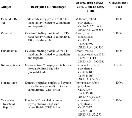
For analysis of cortical interneuron pathology, free-floating sections were processed using standard immunohistochemical methods (Blizzard et al 2016, Handley et al 2016). Cortical interneurons were identified by the expression of calcium binding proteins: calbindin (CB), calretinin (CR) and parvalbumin (PV), or by neuropeptides: neuropeptide Y (NPY), vasoactive intestinal peptide (VIP) and somatostatin (SOM)(Markram et al 2004). Every tenth serial sections (400µm apart) was incubated with antibodies recognizing cell-type specific interneuron markers
Table 2.1. Primary Antibodies used for immunohistochemistry in mouse cortex
Antigen Description of Immunogen
Source, Host Species, Cat#, Clone or Lot#,
RRID
Concentration Used
Calbindin D-28k
Calcium-binding protein of the EF-hand family related to calmodulin and troponin-C
Millipore, rabbit polyclonal, Cat#AB1778 Lot# RRID:AB_2068336
1:1000µl
Calretinin Calcium-binding protein of the
EF-hand family related to calbindin D-28k and calmodulin
Swant, mouse monoclonal, Cat#6B3 Lot#010399 RRID:AB_1000330
1:1000µl
Parvalbumin Calcium-binding protein of the
EF-hand family related to calmodulin and troponin-C
Swant, mouse
monoclonal, Cat#235 Lot#10-11 (F) RRID:AB_10000343
1:1000µl
Neuropeptide Y Neuropeptide Y conjugated to bovine
thyroglobulin (BTg) with glutaraldehyde Immunostar, rabbit polycloncal, Cat#22940 Lot#1112001 RRID:AB_572253
1:500µl
Somatostatin Synthetic peptide coupled to keyhole
limpet hemocyanin (KLH) with carbodiimide (CDI) linker
Immunostar, rabbit polyclonal, Cat#20067 Lot#216002 RRID:AB_572264
1:1000µl
Vasoactive Intestinal Peptide
Porcine VIP coupled to bovine thyroglobulin (BTg) with carbodiimide (CDI) linker
Immunostar, rabbit polyclonal, Cat#20077 Lot#1339001 RRID:AB_572270
1:1000µl
33
Specificity of all antibodies was verified by incubating sections with the corresponding secondary antibody without pre-incubation of primary antibody.
2.2.5 Imaging and quantification
Immunofluorescence was captured using a Zeiss LSM 510 DuoScan confocal microscope (Carl Zeiss Microscopy, Germany), running Zen software (V3.2, 2008) equipped with Ar488 and HeNe543 lasers. Cell bodies were quantified blind to genotype in the supragranular (layers I-IV) and infragranular lamina (layers V-VI) of the primary motor and secondary somatosensory cortices (Figure 2.1), comparable coronal sections were selected from 1.18mm to -0.58mm relative to bregma. Sections 21-36 according to the Paxinos and Franklin Mouse Brain Atlas (Paxinos & Franklin 2007). A plan-apochromat 20x objective (N.A. 0.8, Zeiss) was used to generate z-plane images with 2µm intervals through 16µm of tissue depth. Primary motor and
secondary somatosensory cortices were identified by anatomical landmarks referring to the appearance of the lateral ventricles, the shape of the third ventricle and the appearance of the anterior commissure and corpus callosum, as visualized with DAPI staining and according to the Allen Mouse Brain Atlas (© Allen Institute for Brain Science: http://mouse.brain-map.org) (Figure 2.1). Immunopositive neurons (cells with positive labeling in cell soma) were counted using Image J software (National Institutes of Health, USA) with the integrated Cell Counter plugin utilising Nissl staining to identify cortical layers. To compare densities of immunopositive neurons in SOD1G93A and wild-type mice, all neurons within the regions of interest (ROI) were manually marked, counted and the density calculated using the area of the ROI (values are given in cells/mm2). The densities were then averaged across animals with 4 sections per cortical region per mouse included in analyses.
2.2.6 Morphological analyses
For analysis of neurite labelling patterns, 40µm coronal tissue sections were used to generate
Z-stack images of neurons with 1µm intervals through 16µm of tissue depth within the motor cortex.
Figure 2.1. Representative images of sections used for quantitative analysis in WT and SOD1G93A mice.
a, Coronal sections, each 40µm thick, from bregma 1.18 to -0.58 were used for
34
length of primary, secondary, tertiary and quaternary order neurite processes, encompassing both axons and dendrites, of CR-labelled neurons.
2.2.7 Statistical analyses
Neuronal density was analysed using a two-way analysis of variance followed by Bonferroni post hoc tests (GraphPad Prism, Version 6.0) for group and regional comparisons. Overall group differences (main effects of genotype) were identified using non-parametric two-tailed t-tests. To assess neuronal densities across the disease course, three-way analysis of variance was used for comparisons of group and cortical regions between different time points (SPSS, Version 20). All variables were tested for statistical interaction, with any significant interactions included in the model. Statistical significance was set at P < 0.05. Average values were expressed as means ±
35
2.3 RESULTS
2.3.1 A subtype-specific interneuron alteration after symptom onset in the SOD1G93A motor cortex
Within the cortex, inhibitory microcircuits are comprised of a wide variety of interneuron populations that target specific neuronal domains to facilitate the fine-tuning of cortical neuronal activity (Holt & Koch 1997, Silver 2010). These cell types are arranged in well-ordered wiring patterns that maintain the complex functions of cortical regions by their unique placement, connections and firing properties (Kubota 2014). Changes in specific interneuron populations are therefore likely to affect synaptic transmission in the motor cortex and compromise the regulation of network excitability, including motor output from layer V corticomotoneurons. To determine if specific interneuron populations were altered in the SOD1 cortex, we used immunohistochemistry to assess the potential for changes in the density of interneuron populations in the motor and somatosensory cortex of late-symptomatic (20 week) SOD1 mice, and in age and litter-matched wild type (WT) controls. Interneuron density was quantified (cells per mm2) in both the supragranular and infragranular lamina of motor (Ms, Mi) and somatosensory cortices (Ss, Si) to determine if cell position in cortical regions influenced pathology (Figure 2.1 a-c). GABAergic interneuron subtypes were differentiated according to the selective expression of calcium binding proteins [calbindin (CB), calretinin (CR), parvalbumin (PV)] and neuropeptides [neuropeptide Y (NPY), somatostatin (SOM), vasoactive intestinal peptide (VIP)](Markram et al 2004) (Figure 2.2 a-f). Analysis revealed that of the interneuron populations expressing calcium-binding proteins, the density of CR-expressing neurons was significantly decreased in the supragranular lamina of the motor cortex (layers I-IV) (Figure 2.2 g-h). In this region, CR-neurons were reduced by up to 37% of WT controls (55 ± 6 p/mm2 WT Ms, 35 ± 6 p/mm2 SOD1G93A Ms) (P < 0.05, two-way ANOVA, Bonferroni post-hoc) (Figure 2.2 j), while the density of this population remained unaltered in the infragranular motor cortex (32 ± 5 p/mm2 WT Mi, 19 ± 1.9 p/mm2 SOD1G93A Mi), and unaltered in both lamina of the somatosensory cortex relative to WT controls(43 ± 6 p/mm2 WT Ss, 44 ± 4 p/mm2 SOD1G93A Ss; 12 ± 1 p/mm2 WT Si, 10 ± 1 p/mm2 SOD1G93A Si). No significant differences were detected in either of the other calcium binding populations, CB- or PV-expressing neurons in SOD1G93A mice and WT controls (Figure 2.2 i, k).
Figure 2.2. Calretinin and Neuropeptide Y interneuron subtypes are differentially altered in specific lamina of the SOD1G93A motor cortex.
a-f, Calcium binding proteins and neuropeptides (green) were used to visualise specific interneuron populations in the cortex, showing labelling patterns of calbindin (CB; a), calretinin (CR; b), parvalbumin (PV; c) and neuropeptide Y (NPY; d), somatostatin (SOM; e) and vasoactive intestinal peptide (VIP; f) populations in 20 week WT cortex stained with DAPI (blue) and Nissl (red). The boxed areas (a-f) in the high magnification images show co-localisation of interneuron labels with Nissl stain. g-h, At 20 weeks, analysis of motor cortex, reveals the normal distribution of CR-expressing interneurons in WT motor cortex (g), but a striking reduction in particular in layers I-IV of SOD1G93A motor cortex (h). Analysis of immunopositive neurons within the SOD1G93A motor (M) and somatosensory (S) cortex showed that the density of calretinin-expressing interneurons was significantly decreased specifically within the supragranular (Ms, layers I-IV) lamina of the motor cortex (j) and the density of Neuropeptide Y-expressing interneurons was significantly increased in both the supragranular (Ms, layers I-IV) and infragranular lamina (Mi, layers V-VI) of the motor cortex (l) (*P < 0.05, two-way ANOVA, Bonferonni’s multiple-comparison test). No other interneuron populations were significantly altered in either motor or somatosensory cortex (i, k, m, n). Values in graphs represent means ± SEM, n = 6 mice
36
± 2 p/mm2 SOD1G93A Mi) (P < 0.05, two-way ANOVA, Bonferroni post-hoc) (Figure 2.2 l), an unexpected finding as interneurons in the cortex are generally not thought to undergo adult neurogenesis (Ernst & Frisen 2015). This suggests increased NPY density may more likely reflect increased expression of the NPY peptide on neurons. In the somatosensory cortex the density of NPY-neurons remained unchanged in both lamina (45 ± 2 p/mm2 WT Ss, 48 ± 3 p/mm2 SOD1G93A Ss; 29 ± 1 p/mm2 WT Si, 32 ± 1 p/mm2 SOD1G93A Si). Analysis of other neuropeptide expressing populations, SOM- and VIP-expressing neurons, identified no further differences in motor or somatosensory lamina compared to WT (Figure 2.2 m, n). These investigations demonstrate that at end-stage in the SOD1G93A cortex, at a time of established cortical vulnerability in this ALS model (Jara et al 2012), distinct regions of the motor cortex undergo selective alteration involving the differential vulnerability of neurons expressing CR and NPY.
2.3.2 Contrasting and progressive alterations of NPY and CR populations throughout the SOD1G93A time course
The early alteration of interneuron populations in the motor cortex could initiate a cascade of events resulting in an inability to maintain excitability in the cortex. In the TDP-43 model of ALS, SOM-expressing interneurons has been shown to initiate hyperexcitability in the motor cortex at an early disease stage (Zhang et al 2016). Therefore in this study the density of CR- and NPY-expressing populations was examined at earlier stages in the SOD1G93A disease course: 8 weeks (from the earliest signs of symptoms in this model), 12 weeks and 16 weeks (Wooley et al 2005) (Figure 2.3 a). The mean density of CR-expressing neurons was decreased by 31% from (16 weeks) in the supragranular motor cortex of SOD1G93A mice compared with WT (88 ± 13 p/mm2 WT Ms, 60 ± 8 p/mm2 SOD1G93A Ms) (P < 0.05, two-way ANOVA, Bonferroni post-hoc) (Figure 2.3 b). This decrease was not significant in the infragranular lamina of the motor cortex (61 ± 8 p/mm2 WT Ms, 46 ± 6 p/mm2 SOD1G93A Ms), and was not present in the somatosensory cortex (71 ± 12 p/mm2 WT Ss, 48 ± 11 p/mm2 SOD1G93A Ss; 21 ± 9 p/mm2 WT Si, 17 ± 2 p/mm2 SOD1G93A Si). The density of CR-expressing neurons remained unchanged relative to controls at earlier time points in the SOD1G93A cortex at 8 and 12 weeks. This suggests either a loss of CR-expressing neurons, or a potential reduction in the expression levels of CR in this distinct interneuron population, occurs during the later symptomatic phase in this model and is restricted to the upper layers of the motor cortex.
Figure 2.3. Calretinin-expressing interneurons are progressively lost during the symptomatic phase in the SOD1G93A motor cortex.
a, CR-expressing interneurons were labelled throughout the SOD1G93A disease course, showing neurons were present at comparable levels in SOD1G93A and WT mice at 8 weeks (early symptom onset) and 12 weeks in motor (M) and somatosensory cortex (S). b, Analysis of 16 week symptomatic SOD1G93A mice showed that CR-expressing interneurons were significantly decreased in the supragranular lamina of motor cortex (Ms, layers I-IV) compared to WT mice. CR-expressing interneurons were progressively reduced in the supragranular lamina of motor cortex (Ms, layers I-IV) in 20 week end-stage SOD1G93A mice (arrow heads in a) (*P < 0.05, two-way ANOVA, Bonferonni’s multiple-comparison test). Values in graphs represent means ± SEM, n =