Copyrightq1997, American Society for Microbiology
Assembled Coronavirus from Complementation of Two
Defective Interfering RNAs
KYONGMIN HWANG KIM,† KRISHNA NARAYANAN,ANDSHINJI MAKINO*
Department of Microbiology, The University of Texas at Austin, Austin, Texas 78712
Received 11 December 1996/Accepted 14 February 1997
In the presence of an RNA2temperature-sensitive (ts) mutant helper virus, two coronavirus mouse hepatitis virus (MHV) defective interfering (DI) RNAs complemented each other, resulting in the assembly of MHV particles; we used this ability to complement as a means to study coronavirus assembly. One of the two DI RNAs was DIssA, a naturally occurring self-replicating DI RNA encoding N protein and the gene 1 proteins that encode RNA polymerase function; DIssA supports the replication and transcription of other non-self-replicating DI RNAs. The other DI was a genetically engineered DI RNA that encoded sM and M proteins. Coinfection of these two DIs at the nonpermissive temperature for the ts helper virus resulted in replication and transcription of both DI RNAs but not in synthesis of the helper virus RNAs. MHV particles containing DI RNAs, N protein, and M protein, all of which were exclusively derived from the two DI RNAs, were released from the coinfected cells; the amount of sM protein was below the limits of detection. Analyses of DI RNAs with mutations in the two envelope protein genes demonstrated that M and sM proteins appeared to be required for assembly and release of MHV particles that contained DI RNAs and N protein, while S protein was not required for assembly and release of MHV particles.
Coronavirus is a large enveloped virus with a single-stranded, positive-sense RNA genome. Coronaviruses gener-ally have three envelope proteins, M, S, and sM. M protein, a triple-spanning transmembrane protein (1), is the most abun-dant glycoprotein. Coronavirus S protein, which forms charac-teristic peplomers, binds to the coronavirus-specific receptor to initiate infection and induces cell fusion (6, 9, 40). A minute amount of sM protein associates with the coronavirus envelope (10, 23, 42, 48). Some coronaviruses contain a fourth envelope glycoprotein, HE protein (36), which is not essential for coro-navirus replication (36).
Recently, Risco et al. showed that within its envelope, the coronavirus transmissible gastroenteritis virus (TGEV) con-tains an internal core that harbors helical nucleocapsid (ribo-nucleoprotein consisting of viral RNA and N protein) (33). The authors claimed that the shell of the TGEV core structure that encloses the ribonucleoprotein consists of mostly M pro-tein and a lesser amount of N propro-tein (33). The core appears to be an icosahedral structure that is spatially separated from the lipid envelope that surrounds it. The internal core structure is probably a universal attribute of coronaviruses, because elec-tron microscopy also reveals an internal core structure in mouse hepatitis virus (MHV) (33).
MHV, a prototype coronavirus, contains a 31-kb genomic RNA that encodes seven to eight genes (18, 32). MHV genes are expressed through a genome-sized virus-specific mRNA and six or seven species of virus-specific subgenomic mRNAs
with a 39-coterminal nested set structure (17, 20). These
mRNAs are numbered 1 to 7, in order of decreasing size (17, 20), and have a common leader sequence of approximately 72
to 77 nucleotides at the 59 end (16, 38). Most of the MHV
mRNAs are functionally monocistronic; except for the sM protein, which is translated from the second open reading
frame (ORF) (gene 5b) of mRNA 5 (5, 19, 37), the 59-most
ORF on each mRNA is used for translation. The protein
product from the 59-most ORF (gene 5a) in mRNA 5 is not
identified in MHV-infected cells. The 59-most gene, gene 1, is
22 kb and probably encodes proteins for viral RNA synthesis, including RNA polymerase, proteinases, and helicase (18). Genes 3, 6, and 7 encode structural proteins S, M, and N, respectively (15). Other MHV genes, including genes 2, 2-1 (the gene for HE protein), 4, and 5a are not essential for MHV replication, at least in tissue culture (35, 47). The expression of genes 1 and 7 is sufficient for MHV RNA synthesis (13).
MHV assembly takes place at the budding compartment, the smooth membrane of the intermediate compartment between the endoplasmic reticulum and the Golgi complex (14, 41). M protein itself does not determine the budding site; when M protein is expressed in the absence of other viral proteins, it migrates beyond the budding compartment and localizes in the late Golgi complex (14). This indicates that an unidentified viral factor(s) restricts the migration of M protein at the bud-ding compartment. S protein interacts with M protein in the absence of other MHV proteins, and this interaction is prob-ably important for incorporation of S protein into the virion (31). Tunicamycin-treated MHV-infected cells release MHV particles lacking S protein (12, 34), indicating that S protein probably is not necessary for coronavirus assembly. Klumper-man et al. (14), however, discussed the possibility that MHV particles released from tunicamycin-treated cells may contain some part of the membrane anchors and the cytoplasmic tails of S proteins; in the presence of tunicamycin, MHV particles containing S protein may be produced initially, but later S proteins lacking N-linked oligosaccharides that directed the protein’s conformation may be subsequently degraded. All coronaviruses sequenced so far maintain the gene encoding sM protein (10, 23, 37, 42), indicating that this protein may be required for replication. The sM protein is not necessary for MHV RNA synthesis (13).
Coexpression of cloned MHV envelope proteins by using T7 polymerase-expressing recombinant vaccinia virus showed that both M and sM proteins, but not S protein, are needed for MHV envelope formation. Vennema et al. directly
demon-* Corresponding author. Phone: (512) 6876. Fax: (512) 471-7088. E-mail: [email protected].
† Present address: Division of Biology, California Institute of Tech-nology, Pasadena, CA 91125.
3922
on November 9, 2019 by guest
http://jvi.asm.org/
strated that virus-like particles (VLPs) are assembled and re-leased from MHV-uninfected cells that express both sM and M proteins (44). VLPs produced from the cells coexpressing N protein and other MHV envelope proteins do not contain N protein (44), indicating that neither the spherical core shell nor the helical nucleocapsid is incorporated into VLPs. Essentially the same procedure was used by Bos et al. (3) to explore the requirement of MHV envelope proteins for MHV assembly. Their data indirectly demonstrated that both sM protein and M protein, but not S protein, are necessary for MHV assembly. The mechanism of MHV RNA packaging into MHV parti-cles is poorly understood. Analyses of MHV defective inter-fering (DI) RNAs have identified a packaging signal (8, 43) that is necessary for the packaging of MHV DI RNA into virus particles. The packaging signal is sufficient for RNA packaging into MHV particles (45), yet how the packaging signal facili-tates specific packaging of viral RNA remains to be studied. Bos et al. (3) presented indirect evidence that infectious MHV DI particles can be produced by coexpressing a cloned MHV DI RNA, N, sM, M, and S proteins, suggesting that expression of MHV structural proteins and DI RNA are sufficient for MHV DI particle formation and DI RNA packaging.
The MHV envelope proteins are most likely involved in the mechanism of membrane envelopment of the viral core. Gra-dient-purified MHV nucleocapsid (ribonucleoprotein), but not free N protein, can interact with M protein in vitro (39), so M protein probably interacts specifically with nucleocapsid. This interaction may be important for incorporation of virus nu-cleocapsid into virus particles, yet a definite requirement of M protein for MHV nucleocapsid packaging has not been estab-lished. Whether S protein is required for envelopment of the viral core is not known. MHV particles released from tunica-mycin-treated cells lack S protein but contain N protein (12, 34), suggesting that the core, the core shell, or helical nucleo-capsid is present in these MHV particles. Some part of the S protein’s membrane anchors and cytoplasmic tails may be present in MHV particles that are released from the tunica-mycin-treated cells (14); therefore, S protein may be involved in packaging of the viral core.
We have been interested in a self-replicating MHV DI RNA, DIssA (13, 24, 27), for studying the mechanism of coro-navirus assembly. DIssA contains intact genes 1 and 7 and most likely contains gene 2 but lacks genes 3 to 6 and a part of gene 2-1 (Fig. 1A) (13). Coinfection of DIssA DI particles containing the JHM strain of MHV (MHV-JHM) with an
RNA2 MHV-A59 temperature-sensitive (ts) mutant, LA16,
and subsequent passage of the produced virus sample at the
permissive temperature (32.58C) eliminates virtually all
MHV-JHM (13). Helper viruses were replaced by LA16 while DIssA DI particles were maintained in the passaged virus sample (13). DIssA and DIssA-derived mRNA 7 but not helper virus mRNAs are synthesized in the cells infected with the passaged
virus sample at LA16’s nonpermissive temperature (39.58C)
(13). Importantly, DIssA supports replication and transcrip-tion of another DI RNA (13).
In the present study, we describe the establishment of a novel complementary DI system that enabled us to study coro-navirus assembly. We made two DI particle preparations, both of which used LA16 as a helper virus. One DI particle prepa-ration contained an engineered DI RNA encoding structural envelope proteins, and the other preparation contained DIssA
DI particles. After coinfection of both DI particles at 39.58C,
DIssA supported replication and transcription of the other DI RNA, which in turn provided envelope proteins for the coin-fected cells. All the necessary proteins for MHV replication accumulated, and MHV particles were produced. Using this
system, we determined which MHV envelope structural pro-teins are essential for MHV DI RNA packaging and MHV particle formation.
MATERIALS AND METHODS
Viruses and cells.The MHV-A59 ts mutant, LA16 (2, 13), plaque-cloned MHV-JHM, and a 19-fold undiluted serially passaged MHV-JHM (JHM19th) viral preparation (24) were used. Mouse DBT cells were used for RNA trans-fection and propagation of viruses (11). MHV was grown in 88% Eagle’s mini-mum essential medium (pH 6.5)–10% tryptose phosphate broth–2% heat-inac-tivated fetal bovine serum.
Plasmid construction.The 1.6-kb reverse transcription (RT)-PCR product that corresponds to the 390.1-kb region of gene 4, the entire gene 5 (both 5a and 5b), gene 6, and the 590.18-kb region of gene 7 was synthesized from MHV-JHM genomic RNA. This PCR product was inserted into the EcoRV site of DIssF-derived cDNA clone MC136-1 (25), yielding the parental clone, JW2 (Fig. 1B). JW2DsM and JW2DM were constructed by a recombinant PCR procedure using JW2 as a template. JW2DsM contained an insertion of a termination codon and a 2-nucleotide frameshift mutation at the 59region of gene 5b (Fig. 1C). JW2DM contained an insertion of a termination codon and a 2-nucleotide frameshift mutation at the 59end of gene 6 (Fig. 1C). JW2DsM and JW2DM had disrupted genes 5b and gene 6, respectively. JW2DsM1M was constructed by inserting the 5b region of JW2DsM into the corresponding region of JW2DM; JW2DsM1M had both genes 5b and 6 disrupted. For all constructs, we sequenced the inserts that were derived from PCR products to confirm the presence of specific muta-tions and the absence of extraneous mutamuta-tions.
RNA transcription and transfection.Plasmids were linearized by XmaI diges-tion, transcribed in vitro with T7 RNA polymerase (26), and transfected by lipofection, as described previously (7, 25).
Purification of viruses.The supernatant from virus-infected cells was har-vested at 12 h postinfection (hpi) and briefly centrifuged to remove cell debris. Released viruses were partially purified by ultracentrifugation on a discontinuous sucrose gradient consisting of 20 and 60% sucrose (27). After centrifugation at 26,000 rpm for 3 h at 48C in a Beckman SW28 rotor, virus particles at the interface between the 20 and 60% sucrose were collected and in some cases were further purified by the 20 to 60% continuous sucrose gradient centrifugation at 26,000 rpm for 18 to 24 h at 48C. Partially purified viruses or purified viruses were pelleted through a 20% sucrose cushion by centrifugation at 38,000 rpm for 3 h at 48C on a Beckman SW40 rotor.
Preparation of virus-specific RNA and Northern (RNA) blotting. Virus-spe-cific RNAs were extracted from virus-infected cells or purified virus particles as described previously (29). RNAs were denatured and electrophoresed through a 1% agarose gel containing formaldehyde and transferred onto nylon filters (25). Northern blot analysis was performed with a32P-labeled random-primed probe
as described previously (8).
Labeling of intracellular and virion proteins, immunoprecipitation, and SDS-PAGE.For labeling intracellular virus-specific proteins, actinomycin D (2.5mg/ ml) was added to virus-infected cells at 3 hpi and virus-specific proteins were labeled with [35S]methionine at 7.5 hpi for 20 min or for 2 h from 6.5 to 8.5 hpi
and cell lysates were prepared as described previously (25). For labeling of virion proteins, [35S]methionine was added to virus-infected cells at 7.5 hpi and culture
fluids were collected at 12 hpi (25). Virus proteins in the partially purified virion and intracellular virus-specific proteins were immunoprecipitated with anti-MHV-JHM serum (24). Immunoprecipitated proteins were incubated at 378C for 1 h in sample buffer to prevent M protein aggregation (39) and analyzed by SDS-PAGE.
Analysis of sM protein.To examine the synthesis of sM protein from engi-neered DI RNAs, MHV-specific cDNA was first synthesized by incubating in-tracellular RNA and oligonucleotide 1111 (59 -GAGCCAGAAGGCTGCGAT-39), which binds at 0.39 kb from the 39region of MHV mRNA 7, as described previously (28). MHV-specific cDNA was then incubated with oligonucleotide 1111 and oligonucleotide 10217 (59-TAATACGACTCACTATAGGGCATCTG GAGAGCCACGCAGCTCG-39), which contains the T7 promoter sequence at the 59region and hybridizes with negative-strand MHV genomic RNA at the 39
end of gene 5a, in PCR buffer (13) at 948C for 30 s, 428C for 45 s, and 728C for 100 s for 25 cycles. The 1.3 kb DI RNA-specific RT-PCR products were approx-imately 1 kb smaller than the helper virus-derived RT-PCR products. Capped transcripts were synthesized in vitro from the gel-purified DI-specific RT-PCR products (26), and proteins were synthesized in vitro with wheat germ extract (Promega). Protein products were immunoprecipitated by using anti-sM protein antibody (19, 48) and analyzed by SDS-PAGE.
RESULTS
Strategy for establishment of the complementary DI system.
We aimed to establish a complementary DI system in which two DI RNAs complemented each other to produce MHV particles. In this system, two MHV preparations, each of which contained a different type of DI particle, were used. One MHV
on November 9, 2019 by guest
http://jvi.asm.org/
sample contained the self-replicating DIssA DI particles (13, 24, 27) that provide polymerase function and N protein (pol/N supplier) (Fig. 1A). The other sample contained DI particles, each carrying an engineered DI RNA which subgenomically
expresses virus envelope proteins (env supplier). To exclude infectious helper MHV in the complementary DI system, an
RNA2 MHV-A59 ts mutant, LA16 (2, 13), was used as a
[image:3.612.76.526.76.588.2]helper virus for both the pol/N supplier and the env supplier.
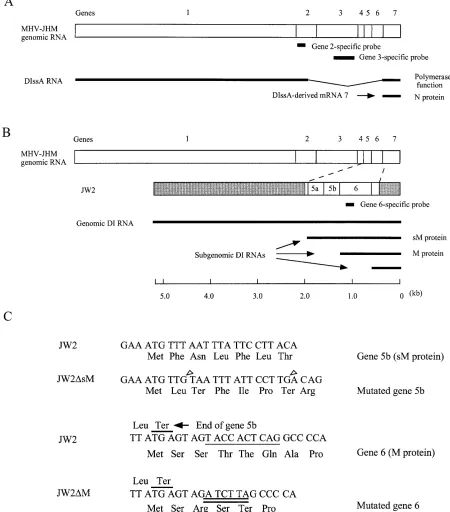
FIG. 1. Diagrams of the structures of DIssA (A), JW2 (B), and the nucleotide and deduced amino acid sequences of the 59regions of gene 5b and gene 6 (C). (A) Comparison of the structure of DIssA RNA and wild-type MHV-JHM genomic RNA. The thin line represents the deleted region in DIssA (13). The two bold lines represent two major mRNAs, DIssA, and N protein-encoding mRNA 7, which are synthesized in DIssA-replicating cells (13). The locations of a gene 2-specific probe and a gene 3-specific probe used for Northern blot analysis (see below) are also shown. (B) Structure of JW2 and its expected subgenomic DI RNAs. The location of the inserted 1.6 kb-long sequence in JW2 is shown. The shaded regions represent the parental MC136-1 sequence, which is almost identical to DIssF (25). The largest subgenomic DI RNA and the medium-sized subgenomic DI RNA encode sM protein and M protein, respectively. The location of a gene 6-specific probe used for Northern blot analysis (Fig. 2A and 3A) is also shown. (C) Mutations present in JW2-derived mutants. The two upper sequences and the two lower sequences represent the 59regions of gene 5b and gene 6, respectively. The two G nucleotide insertions in JW2DsM are shown by open arrowheads. The underlined JW2 gene 6 nucleotide sequence was mutated into the sequence shown by the double underlines in JW2DM. JW2DsM1M contained both the mutations introduced in JW2DsM and JW2DM.
on November 9, 2019 by guest
http://jvi.asm.org/
The pol/N supplier was prepared after coinfection of LA16 and JHM19th, which is an MHV-JHM sample containing DIssA
DI particles, at 32.58C and passaging them three times at
32.58C (13); most of the MHV-JHM virions were easily
re-moved and replaced by LA16, whereas the DIssA DI particles were maintained in this preparation (13). In agreement with a previous study (13), the ratio of viral infectivity in the pol/N
supplier preparations at 39.58C to that at 32.58C was on
aver-age 8.031023. Also, DIssA RNA and mRNA 7 but not DIssE
RNA, which is present in JHM19th, were efficiently
synthe-sized in the pol/N supplier-infected cells at 39.58C (data not
shown). For the preparation of the env supplier, in vitro-syn-thesized DI RNA transcripts were transfected into
LA16-in-fected cells and cultures were incubated at 32.58C. Released
viruses were passaged once at 32.58C and used as the env
supplier. For complementation experiments, cells were coin-fected with the pol/N supplier and the env supplier, and
incu-bated at 39.58C. We expected that both DI RNAs but not
LA16 would replicate, transcribe, and express all of the MHV proteins necessary for MHV replication. We assumed that MHV would be released from coinfected cells.
Preparation and characterization of the env suppliers.We constructed a DI cDNA that provided virus envelope proteins in the complementary DI system. Initially, DI RNA containing a region corresponding to all of MHV-JHM genes 3, 4, 5, and 6 was examined for its ability to replicate in the MHV-infected cells, but this DI RNA failed to replicate (data not shown). Since MHV particles lacking S protein are released from tu-nicamycin-treated cells (12, 34), we assumed that S protein is not necessary for MHV assembly. Therefore we constructed another DI cDNA clone, JW2, that contained genes 5 and 6
and the 59region of gene 7 (Fig. 1B). To examine replication
and transcription of JW2 DI RNA, in vitro-synthesized JW2 RNA was transfected into LA16-infected cells and incubated
for 20 h at 32.58C. The supernatant was collected and then
passaged once at 32.58C to prepare passage 1 virus sample.
This sample was named JW2 env supplier. The ratio of virus
infectivity at 39.58C to that at 32.58C of the JW2 env supplier
was 4.0 3 1024. DBT cells were infected with the JW2 env
supplier and incubated at 32.5 or 39.58C. Intracellular RNA
was extracted at 9 hpi, and JW2 RNA synthesis was examined by Northern blot analysis (Fig. 2A) using a gene 6-specific probe (Fig. 1B). JW2 replicated and transcribed two sub-genomic DI RNAs, each of which encoded genes 5 and 6 at
32.58C but not at 39.58C. As expected, Northern blot analysis
using a probe that hybridized at the 39end of the MHV
ge-nome revealed synthesis of the smallest subgenomic DI RNA
encoding the 59region of gene 7 (see Fig. 1B) at 32.58C but not
at 39.58C (data not shown).
To study the role of sM protein and M protein in MHV assembly, we constructed three JW2-derived mutant DI cDNAs.
JW2DsM and JW2DM had a mutation at the beginning of gene 5b
and gene 6, respectively (Fig. 1C); the ORF of gene 5b and the
ORF of gene 6 were disrupted in JW2DsM and JW2DM,
re-spectively. JW2DsM1M had both mutations which are present
in JW2DsM and JW2DM; accordingly, JW2DsM1M had
dis-rupted ORFs of genes 5b and 6. RNA transcripts were synthe-sized in vitro from these clones and transfected into LA16-infected cells. Passage 1 virus samples from cells transfected
with JW2DM, JW2DsM, and JW2DsM1M were named
JW2DM env supplier, JW2DsM env supplier, and JW2DsM1M
env supplier, respectively. Northern blot analysis of intracellu-lar RNA species in the passage 1 virus-infected cells showed that all three mutant DI RNAs replicated and transcribed subgenomic DI RNAs with efficiencies similar to those of JW2
(Fig. 2A). DI RNAs were synthesized at 32.58C but not at
39.58C. We also found that JW2 and its mutants replicated with
similar efficiency in the DI RNA-transfected, LA16-infected
cells at 32.58C (data not shown).
To know whether M protein encoded by subgenomic DI RNA was translated in DI RNA-replicating cells, infected
DBT cells were labeled for 20 min at 7.5 hpi with [35
S]methi-onine and cell extracts were prepared. MHV-specific proteins were immunoprecipitated with an anti-MHV serum (24) and analyzed by SDS-PAGE (Fig. 2B). As expected, virus-specific
proteins were synthesized at 32.58C but not at 39.58C in the
cells infected with env suppliers (Fig. 2B). Two M proteins with different electrophoretic mobilities were detected in the cells
infected with the JW2 env supplier or JW2DsM env supplier.
[image:4.612.324.555.71.406.2]Because MHV-JHM M protein migrated slightly faster than LA16 M protein (Fig. 2B, lanes 9 and 10), the faster-migrating
FIG. 2. Virus-specific RNA synthesis (A) and protein synthesis (B) in the cells infected with env suppliers. (A) RNA replication and transcription of JW2 and its mutants. DBT cells were infected with env suppliers at 39.58C (NP) or 32.58C (P). Intracellular RNAs were extracted at 9 hpi and analyzed by Northern blot analysis using a gene 6-specific probe (Fig. 1B). The numbers to the right of the gel indicate LA16-specific mRNAs. DI-g, DI-sub(sM), and DI-sub(M) rep-resent genomic DI RNA, sM protein-encoding subgenomic DI RNA, and M protein-encoding subgenomic DI RNA, respectively. (B) Analysis of the virus-specific proteins in env supplier-infected cells. DBT cells were infected with env suppliers at 39.58C (NP) or 32.58C (P). MHV-JHM-infected cells (lane 10) and LA16-infected cells (lane 9) were incubated at 37 and 32.58C, respectively. At 7.5 hpi, cultures were pulse-labeled with [35S]methionine, and cytoplasmic lysates
were prepared. Virus-specific proteins were immunoprecipitated with anti-MHV-JHM serum and electrophoresed on SDS–10% polyacrylamide gels. Po-sitions of MHV S, N, and M proteins are shown on the left. M (LA16) and M (JHM) represent LA16 M protein and MHV-JHM M protein, respectively. The lane labeled Marker contains a14C-labeled protein molecular mass marker (in
kilodaltons).
on November 9, 2019 by guest
http://jvi.asm.org/
M proteins must have been translated from the M protein-encoding subgenomic DI RNA. MHV-JHM-derived M protein
was not detected in the cells infected with the JW2DM env
supplier or the JW2DsM1M env supplier. These results were
consistent with the structure of these DI RNAs.
We examined production of intracellular sM protein by
im-munoprecipitation of [35S]methionine-labeled sM protein with
anti-sM protein antibody, and we failed to detect sM protein synthesis in the cells infected with env suppliers (data not shown). This was because only a minute amount of sM protein is synthesized (19, 48). Even if we had detected sM protein in the env supplier-infected cells, we would not have known whether the detected sM protein was derived from LA16 or from subgenomic DI RNA, because the molecular weights of the MHV-JHM sM protein and the LA16 sM protein are presumably very similar. Thus, we indirectly examined the syn-thesis of sM protein in env supplier-infected cells as described in Materials and Methods. The sM protein was synthesized in vitro from the RT-PCR products of the 5b gene of JW2 and
JW2DM but not from those of JW2DsM and JW2DsM1M
(data not shown). This result was consistent with the structure of DI RNAs and indicated that subgenomic DI RNA-derived sM protein was expressed only in the cells infected with the
JW2 env supplier and the JW2DM env supplier but not the
JW2DsM env supplier and the JW2DsM1M env supplier.
Characterization of intracellular RNAs and virion RNAs in the cells coinfected with the pol/N supplier and env supplier.
We examined whether coinfection of the pol/N supplier and
each of the env suppliers at 39.58C produced MHV particles.
To increase the infectivity of the env supplier, passage 1 virus sample was concentrated approximately 10 times by using Cen-triprep 100 (Amicon) and used as inoculum. DBT cells were coinfected with the pol/N supplier and each of the env
suppli-ers at 39.58C, and intracellular RNAs were extracted at 8 hpi.
For the analysis of virion RNAs, culture fluids were collected at 12 hpi and virus particles were partially purified by discon-tinuous sucrose gradient centrifugation. Virion RNAs from the partially purified virus sample and intracellular RNAs were examined by Northern blot analysis using a gene 6-specific probe (Fig. 3A); this probe hybridizes with env supplier genomic DI RNA and two larger subgenomic DI RNAs but not with DIssA RNA (13) (Fig. 1). All env suppliers replicated and transcribed with similar efficiencies (Fig. 3A, lanes 2 to 5). The small amount of mRNA 6 detected in the cells infected with the pol/N supplier (Fig. 3A, lane 1) probably was synthe-sized from LA16 revertant viruses and MHV-JHM that was present in the pol/N supplier preparation. A trace amount of RNA signal was detected in the cells infected with only the JW2 env. supplier (Fig. 3A, lane 6), demonstrating that LA16
did not support env suppliers’ replication at 39.58C and that
DIssA supported replication and transcription of env suppliers as demonstrated previously (13). Analysis of virion RNAs showed that JW2 genomic RNA was present in the partially purified virus particles (Fig. 3A), whereas only trace amounts of the genomic DI RNAs of other env suppliers were detected.
That there was a slightly larger amount of JW2DsM genomic
RNA than of JW2DM and JW2DsM1M genomic RNA was
due to the presence of slightly larger amounts of LA16
rever-tants and MHV-JHM in the JW2DsM supplier preparation
(see below).
Northern blot analysis using a gene 2-specific probe revealed that DIssA-containing virus particles were produced from the cells coinfected with the pol/N supplier and the JW2 env sup-plier (Fig. 3B, lane 5). A very limited amount of viral particles containing DIssA was produced from the cells infected with the pol/N supplier alone or from those coinfected with other
env suppliers and the pol/N supplier (Fig. 3B, lanes 1 to 6). To clarify that the majority of signals detected indeed represented DIssA RNA but not genomic RNAs of LA16 revertants and MHV-JHM, the same RNA samples were examined by North-ern blot analysis using a gene 3-specific probe (Fig. 3B, lanes 7 to 12). Because DIssA does not contain gene 3 (13) (Fig. 1A), the gene 3 probe should have hybridized only with the genomic RNAs of LA16 revertants and MHV-JHM and not with DIssA RNA. Only faint signals were detected in most of the RNA samples. The virion RNA sample from the cells coinfected with
the pol/N supplier and the JW2DsM env supplier showed a
slightly stronger RNA signal (Fig. 3B, lane 10), indicating the presence of a larger amount of genomic RNAs from possible LA16 revertants or from MHV-JHM contaminating this virus preparation. This may have contributed to the slightly stronger signal seen in Fig. 3B, lane 4; some of the signal probably represented helper virus genomic RNA. These data suggested that MHV particles containing DIssA RNA and env supplier’s DI RNA were efficiently produced only from the cells
coin-FIG. 3. Northern blot analyses of the env supplier-specific RNAs (A) and DIssA RNA (B). DBT cells were coinfected with the pol/N supplier and the env supplier and cultured at 39.58C. Intracellular RNA was extracted at 8 hpi, or the supernatant was collected at 12 hpi. Virion RNAs were extracted from the partially purified virus particles. The presence and absence of the pol/N supplier in the inoculum are represented by1and2, respectively. (A) Intracellular env supplier-specific RNAs (lanes 1 to 6) and virion env supplier-specific RNAs (lanes 7 to 12) were examined by Northern blot analysis using a gene 6-specific probe. Only the pol/N supplier was used as the inoculum for lanes 1 and 7. DI-g, DI-sub(sM), and DI-sub(M) represent genomic DI RNA, sM protein-encoding subgenomic DI RNA, and M protein-encoding subgenomic DI RNA, respec-tively. (B) DIssA and genomic RNAs of LA16 revertant viruses and MHV-JHM in the virion were examined by Northern blot analysis using a gene 2-specific probe (lanes 1 to 6) and a gene 3-specific probe (lanes 7 to 12), respectively (Fig. 1A). Only the pol/N supplier was used as the inoculum for lanes 6 and 12.
on November 9, 2019 by guest
http://jvi.asm.org/
[image:5.612.319.550.68.385.2]fected with the pol/N supplier and the JW2 env supplier. Thus, these data indicated that sM and M proteins, but not S protein, were necessary for packaging of DI RNAs as well as for MHV virion assembly and release.
To determine the buoyant density of MHV particles re-leased from cells coinfected with the pol/N supplier and JW2 env supplier, the partially purified virus particles were centri-fuged on a continuous 20 to 60% sucrose gradient at 26,000 rpm for 18 to 24 h in a Beckman SW28 rotor. Ten fractions were collected from the bottom of the gradient, and virion RNA was extracted from each fraction. Northern blot analysis of JW2 DI RNA in each fraction showed that a major peak of JW2 DI RNA signal was at a density of approximately 1.18
g/cm3, while the major peak of genomic RNA of MHV-JHM
and that of LA16 were at a density of approximately 1.19 g/cm3
(data not shown). MHV particles released from cells coin-fected with the pol/N supplier and JW2 env supplier were slightly lighter than those of infectious MHV.
Characterization of virus-specific intracellular proteins and virion proteins in the coinfected cells.Northern blot analysis of virion RNAs demonstrated that coinfection of the pol/N sup-plier and the JW2 env supsup-plier, but not other env supsup-pliers, produced virus particles containing DI RNAs. These data, however, did not exclude the possibility that empty virus par-ticles lacking virion RNAs were produced in certain combina-tions of the pol/N supplier and the env supplier. We charac-terized virus-specific intracellular proteins and virion proteins to examine whether MHV particles lacking S protein were being produced in the cells coinfected with the pol/N supplier and the JW2 env supplier and to rule out that empty MHVs were produced in the cells coinfected with other env suppliers and the pol/N supplier. Virus-infected cells were incubated at
39.58C and virus-specific proteins were labeled with [35
S]me-thionine for 20 min at 7.5 hpi or for 2 h from 6.5 to 8.5 hpi. As
a control, LA16-infected cells were maintained at 32.58C.
Vi-rus-specific proteins were immunoprecipitated with an anti-MHV serum and analyzed by SDS-PAGE. Lower-molecular-weight proteins and higher-molecular-Lower-molecular-weight proteins were separated on 10% gels (Fig. 4A and C) and 7.5% gels (Fig. 4B and D), respectively. DIssA-derived N protein was synthesized in all the pol/N supplier-infected cells. S protein was
synthe-sized in MHV-JHM-infected cells at 378C or in LA16-infected
cells at 32.58C but not in the pol/N supplier-infected cells, the
JW2 env supplier-infected cells, or the coinfected cells (Fig. 4B and D). We found three proteins (Fig. 4D) of unidentified origin in the 2-h labeling samples. Because one of these pro-teins appeared only in the cells infected with the pol/N sup-plier, this protein probably was a translation product of a naturally occurring DI RNA(s) present in the pol/N supplier (13). MHV-JHM-specific M protein was synthesized in the pol/N supplier- and JW2 env supplier-coinfected cells and in
the pol/N supplier- and JW2DsM env supplier-coinfected cells
(Fig. 4A, lanes 2 and 3; Fig. 4C, lanes 3 and 4), demonstrating expression of subgenomic DI RNA-encoding M protein in these coinfected cells. These data were consistent with intra-cellular viral RNA analysis (Fig. 3A) and with the structure of the DI RNAs. In some experiments, we detected a protein band that closely comigrated with LA16-specific M protein (Fig. 4C, lanes 2 to 7). This protein was most likely a host protein; the presence of this host protein in mock-infected cells was apparent after a long exposure of the autoradiogram (Fig. 4C, lane 11). To further confirm that LA16-specific proteins were not produced at the nonpermissive temperature, DBT cells were infected with LA16 virus samples that had been concentrated 10-fold. Virus-specific proteins were labeled with
[35S]methionine at 39.58C from 1 to 5 hpi or from 5 to 10 hpi.
Radiolabeled proteins were then immunoprecipitated by anti-MHV serum and analyzed by SDS-PAGE. We did not detect synthesis of any LA16-specific proteins (data not shown), dem-onstrating that LA16-specific protein synthesis did not occur at
any time during the course of virus infection at 39.58C.
To examine virion proteins, virus-infected cells were labeled
with [35S]methionine at 39.58C for 8 to 12 hpi. As a control,
[35S]methionine-labeled LA16 and MHV-JHM were grown at
32.5 and 378C, respectively. Released virus particles were
par-tially purified by sucrose gradient centrifugation, because we could not estimate the buoyant density of potential empty particles. In order to eliminate contaminated radiolabeled cel-lular proteins in the virus preparation, virus-specific proteins were immunoprecipitated with anti-MHV serum and analyzed by SDS-PAGE (Fig. 5A). Three major MHV-specific proteins, the S, N, and M proteins, were detected in partially purified MHV-JHM and LA16 (Fig. 5A, lanes 8 and 9). We detected N protein and MHV-JHM-specific M protein, but not S protein, in the MHV sample from cells coinfected with the pol/N sup-plier and the JW2 env supsup-plier (Fig. 5A, lane 2), demonstrating assembly and release of MHV particles containing DI RNAs (Fig. 3), N protein, and MHV-JHM-derived M protein. The sM protein is present in very small amounts in MHV (48). Under our experimental conditions, we could not demonstrate the presence of sM protein in the partially purified MHV from cells coinfected with the pol/N supplier and the JW2 env sup-plier even after immunoprecipitation using anti-sM protein antibody (data not shown). No MHV-specific proteins were detected in the supernatant from other coinfected cells, from the pol/N infected cells, or from the JW2 env supplier-infected cells (Fig. 5A), demonstrating that MHV particles containing N protein and M protein were not produced from these cells.
To directly examine the viral structural proteins, partially purified virus particles from cells coinfected with the pol/N supplier and the JW2 env supplier were further purified by continuous sucrose gradient centrifugation. The released virus particles showed a single radioactive peak at a buoyant density
of approximately 1.18 g/cm3, which was the same as the virus
buoyant density shown by the Northern blot analysis (see above). SDS-PAGE analysis of virus structural proteins showed that virus particles from the cells coinfected with pol/N supplier and JW2 env supplier contained N protein and M protein but not S protein (Fig. 5B); both N protein and M protein comigrated with MHV-JHM N protein and M protein, respectively. These data demonstrated that N protein was the product of DIssA-specific mRNA 7 and M protein was the product of the JW2-specific M protein encoding subgenomic DI RNA. We sometimes detected a high-molecular-weight protein of unknown origin (Fig. 5B, lane 2) in virus prepara-tions. This protein did not comigrate with MHV S protein and was not detected consistently (Fig. 5B, lane 6); this protein may be a protein encoded by some DI RNAs in the virus prepara-tion. Minor proteins found in one virus preparation from the cells coinfected with the pol/N supplier and the JW2 env sup-plier (Fig. 5B, lane 6) did not comigrate with MHV proteins and probably represent contaminating host proteins.
These observations and the viral RNA data (Fig. 3) showed that the M and sM proteins were needed jointly for assembly and release of virus particles that contained viral RNA bound to N protein. We could not rigorously exclude the possibility that empty MHV particles consisting of only sM proteins were synthesized in the cells coinfected with the pol/N supplier and
JW2DM supplier. If such empty particles were formed, they
did not package the virus core, which would contain DI RNA and N protein (Fig. 3 and 5). S protein was not synthesized in
on November 9, 2019 by guest
http://jvi.asm.org/
the pol/N supplier-, JW2-env supplier-coinfected cells at
39.58C, while MHV particles containing viral RNA were
re-leased. Thus, S protein was not required for assembly of the viral core, for envelopment of the viral core, or for viral re-lease. We have presented the first direct identification of a combined requirement for MHV envelope proteins M and sM in the envelopment of the viral core.
DISCUSSION
We established a complementary DI system that enabled us to study the effects of deleting entire individual envelope pro-tein genes on MHV assembly. Analysis of virion RNAs and proteins unambiguously demonstrated that MHV particles
[image:7.612.106.508.73.464.2]were assembled and released from the cells coinfected with the pol/N supplier and the JW2 env supplier. The released MHV particles contained M protein, N protein, and DI RNAs, while the presence of sM protein was not confirmed due to its small amount in the virus. MHV particles released from the cells coinfected with the pol/N supplier and JW2 env supplier had a slightly lighter buoyant density than wild-type MHV; the ab-sence of S protein probably affected the buoyant density of the virus particles. Studies of JW2-derived mutants suggested that both M protein and sM protein were necessary for incorpora-tion of the core (a spherical core shell around a filling of RNA and N protein) into enveloped, released viral particles. The S protein was not required for these processes. This conclusion is consistent with the results of Vennema et al. (44) and Bos et al.
FIG. 4. Analysis of the intracellular virus-specific proteins after coinfection of the pol/N supplier and env supplier. Virus-infected cells were labeled with [35S]methionine for 20 min at 7.5 hpi (A and B) or for 2 h from 6.5 to 8.5 hpi (C and D), and cytoplasmic lysates were prepared. Virus-specific proteins were
immunoprecipitated with anti-MHV-JHM serum and electrophoresed on an SDS–10% polyacrylamide gel (A and C) or an SDS–7.5% polyacrylamide gel (B and D). M (LA16), M (JHM), S (LA16), and S (JHM) represent LA16 M protein, MHV-JHM M protein, LA16 S protein, and MHV-JHM S protein, respectively. The lane labeled Marker contains14C-labeled protein molecular mass marker. For other lanes, DBT cells were infected as follows (numbers in parentheses are lane numbers
for panels A to D, respectively): mock infected at 39.58C (lanes labeled Mock), infected with the pol/N supplier only at 39.58C (1, 3, 2, and 2), JW2 env supplier only at 39.58C (6, 8, 7, and 7), infected with MHV-JHM only at 378C (9, 10, 9, and 8), infected with LA16 only at 32.58C (8, 9, 8, and 10), or coinfected with the env supplier and the pol/N supplier at 39.58C (2 to 5, 4 to 7, 3 to 6, and 3 to 6). (C) Lane 11 shows a longer exposure of the autoradiogram shown in lane 10. The asterisks show a host protein that migrated closely with LA16-specific M protein (see text). (D) Three proteins (marked by an open triangle and two asterisks) of unidentified origin were found in the 2-h labeling samples. Because the protein shown by the open triangle appeared only in the cells infected with the pol/N supplier, this protein probably was a translation product of a naturally occurring DI RNA(s) present in the pol/N supplier (13).
on November 9, 2019 by guest
http://jvi.asm.org/
(3); they showed the formation and release of empty VLPs from MHV-uninfected cells expressing M, S, and sM proteins, and concluded that both M and sM protein are required for VLP formation. We further identified viral envelope proteins
that were required for envelopment of the viral core. Our finding that S protein was not required for envelopment of the viral core excluded the possibility that some part of the S protein membrane anchors and/or cytoplasmic tails may play a role in core envelopment.
An electron microscopic study by Risco et al. (33) showed that coronavirus TGEV contains a spherical core structure inside of the viral envelope (33). The authors claimed that the spherical core shell contains mostly M protein and a lesser amount of N protein (33). If M protein is indeed present in the spherical core, then coronavirus M protein is present at two different sites in a virus particle, i.e., once in the envelope and again in the spherical core. They showed that MHV also con-tains the internal core. However, our biochemical study indi-cated that the MHV core contained N protein and MHV genomic RNA but not M protein (30). We also found that there was a stable interaction between envelope M protein and the core in the virus particles (30). It is possible that M protein detected in the TGEV spherical core represents envelope M protein that collapsed on the internal core. We speculate that the internal core is the structure that restricts the migration of M protein at the budding compartment (14) through a strong interaction between envelope M protein and the spherical core and that the interaction that leads to the envelopment of the core resulting in virus particles occurs between M protein and the internal core. Hopefully, future studies using the comple-mentary DI system will provide information on the mechanism of coronavirus spherical core formation and the mechanism of M protein restriction in the budding compartment of MHV-infected cells.
As shown in this study, S protein was not necessary for packaging of the internal core. Since expression of both M protein and sM protein is required for virus envelope forma-tion (3, 44), we could not identify whether envelopment of the internal core requires M protein alone, sM protein alone, or a combination of both M protein and sM protein. It is possible that sM may only be involved in the completion of envelope assembly and/or in the release of viral particles from the cells. Another question about coronavirus assembly is whether gene 1 proteins participate. Bos et al. showed that when helper MHV and the culture fluid from the cells expressing an MHV DI RNA and all MHV structural proteins are coinfected and passaged several times, MHV DI RNA accumulates (3); their data may indicate that gene 1 proteins are not required for MHV assembly. However, it appears that only a small amount of DI particles are produced in their system; no direct evidence was shown for the production of DI particles from the plasmid-transfected cells, and several passages of virus samples were required for DI RNA accumulation (3). The low level of DI particle production in the study of Bos et al. (3) raises ques-tions about whether the internal core was properly formed in the plasmid-transfected cells, and whether the DI RNAs were specifically packaged into VLPs. Comparing the efficiency of DI particle formation in the system used by Bos et al. (3) and that in the complementary DI system is difficult, yet it seems that a larger amount of DI particles were produced in the complementary DI system; without subsequent passage, we directly demonstrated formation of virus particles containing DI RNAs in the complementary DI system. One of the obvious major differences between the complementary DI system and the system used by Bos et al. (3) is involvement of DIssA, which expresses gene 1 proteins. If the amount of DI particles produced from the complementary DI system was indeed sig-nificantly higher than that in the system used by Bos et al., this would indicate that some gene 1 protein(s) were involved in viral assembly.
FIG. 5. Analysis of virion proteins after coinfection of the pol/N supplier and env supplier. MHV-JHM and LA16 were grown at 37 and 32.58C, respectively, while other virus samples (lanes 1 to 6 [A] or lanes 2 and 6 [B]) were grown at 39.58C. M (LA16) and M (JHM) represent LA16 M protein and MHV-JHM M protein, respectively. The lane labeled Marker contains a14C-labeled protein
molecular mass marker. (A) [35S]methionine-labeled virus was partially purified,
and virus-specific proteins were immunoprecipitated with anti-MHV-JHM se-rum and electrophoresed on an SDS–10% polyacrylamide gel. The presence and absence of the pol/N supplier in the inoculum are represented by1and2, respectively. Only the pol/N supplier was used as the inoculum for lane 1. (B) [35S]methionine-labeled virus was purified and electrophoresed on an SDS–10%
polyacrylamide gel. Two different preparations of virus samples from cells coin-fected with the pol/N supplier and JW2 env supplier are shown (lanes 2 and 6). The asterisk indicates a large protein of unknown origin (see text); this protein was only detected in some virus preparations from cells coinfected the pol/N supplier and JW2 env supplier. The virus sample applied to lane 2 was the progeny of 107cells, while virus samples from the cells infected with MHV-JHM
and LA16 (lanes 3 and 4) were progeny of 53105cells. S, HE, N, and M
represent S protein, HE protein, N protein, and M protein, respectively.
on November 9, 2019 by guest
http://jvi.asm.org/
Expression vectors based on RNA viruses other than retro-viruses have been developed previously (48). The MHV com-plementary DI system has a similarity with one of the Sindbis virus expression vector systems, in which two Sindbis tran-scripts complement each other to produce Sindbis virus carry-ing a non-Sindbis virus gene (4). One of the transcripts is a self-replicating Sindbis replicon that encodes Sindbis virus nonstructural proteins and subgenomically expresses a non-Sindbis virus foreign gene. Another transcript subgenomically expresses viral structural proteins. Cotransfection of these transcripts results in replication and assembly of Sindbis virus containing the foreign gene (4). Although Sindbis virus expres-sion vector systems are excellent in many aspects, they may be limited by the size of RNAs that they can package; very large RNAs may not be packaged well into alphavirus-based vectors. MHV DI RNAs containing an inserted MHV gene or a non-MHV sequence express these genes in DI RNA-replicating cells (21, 22, 25), indicating that coronaviruses themselves and/or coronavirus DI RNAs may be developed as expression vectors. Coronavirus carries the largest single-stranded RNA genome, and may be particularly suitable for delivery of large RNA molecules as an expression-vector. Coronavirus expres-sion vectors may express multiple genes through expresexpres-sion of multiple subgenomic mRNAs (25). The finding that S protein was not necessary for MHV assembly may potentially confer a unique property on coronavirus expression vector systems. De-velopment of a complementary DI system-based coronavirus vector in which the assembled viruses contain S protein derived from different coronaviruses, from an envelope protein(s) of other enveloped viruses, or even from a cellular membrane protein(s) is an attractive idea. Such vectors may prove to be invaluable for delivering a specific, large gene to a specific target cell. As we have shown in this study, insertion of S protein abolished DI RNA replication; currently we do not know what kind of inserted sequences DI RNA replication will tolerate.
ACKNOWLEDGMENTS
This work was supported by Public Health Service grants AI29984 and AI32591 from the National Institutes of Health.
REFERENCES
1. Armstrong, J., H. Niemann, S. Smeekens, P. Rottier, and G. Warren. 1984. Sequence and topology of a model intracellular membrane protein, E1 glycoprotein, from a coronavirus. Nature (London) 308:751–752. 2. Baric, R. S., K. Fu, M. C. Schaad, and S. A. Stohlman. 1990. Establishing a
genetic recombination map for murine coronavirus strain A59 complemen-tation groups. Virology 177:646–656.
3. Bos, E. C. W., W. Luytjes, H. van der Meulen, H. K. Koerten, and W. J. M.
Spaan.1996. The production of recombinant infectious DI-particles of a murine coronavirus in the absence of helper virus. Virology 218:52–60. 4. Bredenbeek, P. J., I. Frolov, C. M. Rice, and S. Schlesinger. 1993. Sindbis
virus expression vectors: packaging of RNA replicons by using defective helper RNAs. J. Virol. 67:6439–6446.
5. Budzilowicz, C. J., and S. R. Weiss. 1987. In vitro synthesis of two polypep-tides from a nonstructural gene of coronavirus, mouse hepatitis virus strain A59. Virology 157:509–515.
6. Collins, A. R., R. L. Knobler, H. Powell, and M. J. Buchmeier. 1982. Mono-clonal antibodies to murine hepatitis virus-4 (strain JHM) define the viral glycoprotein responsible for attachment and cell fusion. Virology 119:358– 371.
7. Felgner, P. L., T. R. Gadek, M. Holm, R. Roman, H. W. Chan, M. Wenz, J. P.
Northrop, G. M. Ringgold, and M. Danielson.1987. Lipofection: a high efficient, lipid-mediated DNA-transfection procedure. Proc. Natl. Acad. Sci. USA 84:7413–7417.
8. Fosmire, J. A., K. Hwang, and S. Makino. 1992. Identification and charac-terization of a coronavirus packaging signal. J. Virol. 66:3522–3530. 9. Frana, M. F., J. N. Behnke, S. Sturman, and K. V. Holmes. 1985. Proteolytic
cleavage of the E2 glycoprotein of murine coronavirus: host-dependent dif-ferences in proteolytic cleavage and cell fusion. J. Virol. 56:912–920. 10. Godet, M., R. L’haridon, J. Vautherot, and H. Laude. 1992. TGEV
corona-virus ORF4 encodes a membrane protein that is incorporated into virions. Virology 188:666–675.
11. Hirano, N., K. Fujiwara, S. Hino, and M. Matsumoto. 1974. Replication and plaque formation of mouse hepatitis virus (MHV-2) in mouse cell line DBT culture. Arch. Gesamte Virusforch. 44:298–302.
12. Holmes, K. V., E. W. Doller, and L. S. Sturman. 1981. Tunicamycin resistant glycosylation of coronavirus glycoprotein: demonstration of a novel type of viral glycoprotein. Virology 115:334–344.
13. Kim, K. H., and S. Makino. 1995. Two murine coronavirus genes suffice for viral RNA synthesis. J. Virol. 69:2313–2321.
14. Klumperman, J., J. Krinjnse Locker, A. Meijer, M. C. Horzinek, H. J. Geuze,
and P. J. M. Rottier.1994. Coronavirus M proteins accumulate in the Golgi complex beyond the site of virion budding. J. Virol. 68:6523–6534. 15. Lai, M. M. C. 1990. Coronavirus: organization, replication and expression of
genome. Annu. Rev. Microbiol. 44:303–333.
16. Lai, M. M. C., R. S. Baric, P. R. Brayton, and S. A. Stohlman. 1984. Characterization of leader RNA sequences on the virion and mRNAs of mouse hepatitis virus, a cytoplasmic RNA virus. Proc. Natl. Acad. Sci. USA
81:3626–3630.
17. Lai, M. M. C., P. R. Brayton, R. C. Armen, C. D. Patton, C. Pugh, and S. A.
Stohlman.1981. Mouse hepatitis virus A59: mRNA structure and genetic localization of the sequence divergence from hepatotropic strain MHV-3. J. Virol. 39:823–834.
18. Lee, H.-J., C.-K. Shieh, A. E. Gorbalenya, E. V. Eugene, N. La Monica, J.
Tuler, A. Bagdzhadzhyan, and M. M. C. Lai.1991. The complete sequence (22 kilobases) of murine coronavirus gene 1 encoding the putative proteases and RNA polymerase. Virology 180:567–582.
19. Leibowitz, J. L., S. Perlman, G. Weinstock, J. R. DeVries, C. J. Budzilowicz,
and S. R. Weiss.1988. Detection of murine coronavirus nonstructural pro-tein encoded in a downstream open reading frame. Virology 164:156–164. 20. Leibowitz, J. L., K. C. Wilhelmsen, and C. W. Bond. 1981. The virus-specific
intracellular RNA species of two murine coronaviruses: MHV-A59 and MHV-JHM. Virology 114:39–51.
21. Liao, C.-L., and M. M. C. Lai. 1994. Requirement of the 59-end genomic sequences as an upstream cis-acting element for coronavirus subgenomic mRNA transcription. J. Virol. 68:4727–4737.
22. Liao, C.-L., X. Zhang, and M. M. C. Lai. 1995. Coronavirus defective-interfering RNA as an expression vector: the generation of a pseudorecom-binant mouse hepatitis virus expressing hemagglutinin-esterase. Virology
208:319–327.
23. Liu, D. X., and S. C. Inglis. 1991. Association of the infectious bronchitis virus 3c protein with the virion envelope. Virology 185:911–917. 24. Makino, S., N. Fujioka, and K. Fujiwara. 1985. Structure of the intracellular
defective viral RNAs of defective interfering particles of mouse hepatitis virus. J. Virol. 54:329–336.
25. Makino, S., M. Joo, and J. K. Makino. 1991. A system for study of corona-virus mRNA synthesis: a regulated, expressed subgenomic defective inter-fering RNA results from intergenic site insertion. J. Virol. 65:6031–6041. 26. Makino, S., and M. M. C. Lai. 1989. High-frequency leader sequence
switch-ing durswitch-ing coronavirus defective interferswitch-ing RNA replication. J. Virol. 57: 729–737.
27. Makino, S., C. K. Shieh, J. G. Keck, and M. M. C. Lai. 1988. Defective-interfering particles of murine coronavirus: mechanism of synthesis of de-fective viral RNAs. Virology 163:104–111.
28. Makino, S., C.-K. Shieh, L. H. Soe, S. Baker, and M. M. C. Lai. 1988. Primary structure and translation of a defective interfering RNA of murine coronavirus. Virology 166:550–560.
29. Makino, S., F. Taguchi, N. Hirano, and K. Fujiwara. 1984. Analysis of genomic and intracellular viral RNAs of small plaque mutants of mouse hepatitis virus, JHM strain. Virology 139:138–151.
30. Narayanan, K., L. A. Brown, K. H. Kim, and S. Makino. Unpublished data. 31. Opstelten, D. J., M. C. Horzinek, and P. J. Rottier. 1993. Complex formation between the spike protein and membrane protein during mouse hepatitis virus assembly. Adv. Exp. Med. Biol. 342:189–195.
32. Pachuk, C. J., P. J. Bredenbeek, P. W. Zoltick, W. J. M. Spaan, and S. R.
Weiss.1989. Molecular cloning of the gene encoding the putative polymer-ase of mouse hepatitis virus, strain A59. Virology 171:141–148.
33. Risco, C., I. M. Anton, L. Enjuanes, and J. L. Carrascosa. 1996. The trans-missible gastroenteritis coronavirus contains a spherical core shell consisting of M and N proteins. J. Virol. 70:4773–4777.
34. Rottier, P. J. M., M. C. Horzinek, and B. A. M. van der Zeijst. 1981. Viral protein synthesis in mouse hepatitis strain A59-infected cells: effect of tuni-camycin. J. Virol. 40:350–357.
35. Schwartz, B., E. Routledge, and S. G. Siddell. 1990. Murine coronavirus nonstructural protein ns2 is not essential for viral replication in transformed cells. J. Virol. 64:4784–4791.
36. Shieh, C.-K., H.-J. Lee, K. Yokomori, N. La Monica, S. Makino, and
M. M. C. Lai.1989. Identification of a new transcriptional initiation site and the corresponding functional gene 2b in the murine coronavirus RNA ge-nome. J. Virol. 63:3729–3736.
37. Skinner, M. A., D. Ebner, and S. G. Siddell. 1985. Coronavirus MHV-JHM mRNA 5 has a sequence arrangement which potentially allows translation of
on November 9, 2019 by guest
http://jvi.asm.org/
a second, downstream open reading frame. J. Gen. Virol. 66:581–592. 38. Spaan, W., H. Delius, M. Skinner, J. Armstrong, P. Rottier, S. Smeekens,
B. A. M. van der Zeijst, and S. G. Siddell.1983. Coronavirus mRNA syn-thesis involves fusion of non-contiguous sequences. EMBO J. 2:1939–1944. 39. Sturman, L. S., K. V. Holmes, and J. Behnke. 1980. Isolation of coronavirus envelope glycoproteins and interaction with the viral nucleocapsid. J. Virol.
33:449–462.
40. Taguchi, F., T. Ikeda, and H. Shida. 1992. Molecular cloning and expression of a spike protein of neurovirulent murine coronavirus JHM variant cl-2. J. Gen. Virol. 73:1065–1072.
41. Tooze, J., S. Tooze, and G. Warren. 1984. Replication of coronavirus MHV-A59 in sac(2) cells: determination of the first site of budding of progeny virions. Eur. J. Cell Biol. 33:281–293.
42. Tung, F. Y. T., S. Abraham, M. Sethna, S. L. Hung, P. Sethna, B. G. Hogue,
and D. A. Brian.1992. The 9-kDa hydrophobic protein encoded at the 39-end of the porcine transmissible gastroenteritis coronavirus genome is mem-brane-associated. Virology 186:676–683.
43. van der Most, R. G., P. J. Bredenbeek, and W. J. M. Spaan. 1991. A domain
at the 39end of the polymerase gene is essential for encapsidation of coro-navirus defective interfering RNAs. J. Virol. 65:3219–3226.
44. Vennema, H., G.-J. Godeke, J. W. A. Rossen, W. F. Voorhout, M. C.
Hor-zinek, D.-J. E. Opstelten, and P. J. M. Rottier.1996. Nucleocapsid-indepen-dent assembly of coronavirus-like particles by co-expression of viral envelope protein genes. EMBO J. 15:2020–2028.
45. Woo, K., M. Joo, K. Narayanan, K. H. Kim, and S. Makino. 1997. Murine coronavirus packaging signal confers packaging to nonviral RNA. J. Virol.
71:824–827.
46. Xiong, C., R. Levis, P. Shen, S. Schlesinger, C. M. Rice, and H. V. Huang. 1989. Sindbis virus: an efficient, broad host range vector for gene expression in animal cells. Science 243:1188–1191.
47. Yokomori, K., and M. M. C. Lai. 1991. Mouse hepatitis virus S sequence reveals that nonstructural proteins ns4 and ns5a are not essential for murine coronavirus replication. J. Virol. 65:5605–5608.
48. Yu, X., B. Weizhen, S. R. Weiss, and J. L. Leibowitz. 1994. Mouse hepatitis virus gene 5b protein is a new virion protein. Virology 202:1018–1023.