cytoplasmic extracts from mock- and MHV-JHM-infected 17Cl-1 murine cells for the ability to form complexes with defined regions of the genomic RNA. We demonstrated the specific binding of host cell proteins to multiple sites within the 3*end of MHV-JHM genomic RNA. By using a set of RNA probes with deletions at either the 5*or 3*end or both ends, two distinct binding sites were located. The first protein-binding element was mapped in the 3*-most 42 nucleotides of the genomic RNA [3*(142) RNA], and the second element was mapped within an 86-nucleotide sequence encompassing nucleotides 171 to 85 from the 3*end of the genome (171-85 RNA). A single potential stem-loop structure is predicted for the 3* (1)42 RNA, and two stem-loop structures are predicted for the 171-85 RNA. Proteins interacting with these two elements were identified by UV-induced covalent cross-linking to labeled RNAs followed by sodium dodecyl sulfate-polyacrylamide gel electrophoresis analysis. The RNA-protein complex formed with the 3*-most 42 nucleotides contains approximately five host polypeptides, a highly labeled protein of 120 kDa and four minor species with sizes of 103, 81, 70, and 55 kDa. The second protein-binding element, contained within a probe representing nucleotides 487 to 85 from the 3* end of the genome, also appears to bind five host polypeptides, 142, 120, 100, 55, and 33 kDa in size, with the 120-kDa protein being the most abundant. The RNA-protein complexes observed with MHV-infected cells in both RNase protection/gel mobility shift and UV cross-linking assays were identical to those observed with uninfected cells. The possible involvement of the interaction of host proteins with the viral genome during MHV replication is discussed.
Mouse hepatitis virus (MHV), a coronavirus, contains a large, single-stranded, polyadenylated genomic RNA of posi-tive sense which is approximately 31 kb in length (12, 13, 29, 38). MHV utilizes a complex transcriptional strategy which is not completely understood. Several alternative models have been described to explain the mechanism of MHV RNA syn-thesis (9, 18, 34, 35). In all of these models, the initial step in MHV RNA replication is the synthesis of negative-strand RNA from a positive-strand genomic template.
One approach to studying MHV RNA synthesis is to identify
cis-acting signals for RNA replication and the proteins which
recognize these signals. Analysis of the structure of defective interfering (DI) RNAs (naturally occurring or constructed in vitro) indicated that approximately 470 nucleotides (nt) at the 59terminus, 436 nt at the 39terminus, and about 135 internal nt were required for DI RNA replication in MHV-infected cells and suggested that these sequences retain signals neces-sary for RNA replication (7, 21, 22). Studies to examine the effects of deletions at the 39 region on DI RNA replication demonstrated that any deletion within the 436 nt at the 39end of the genome was lethal for DI RNA replication (7). A recent study has shown that a 55-kDa host cell protein bound to positive-sense RNA corresponding to nt 56 to 112 at the 59end
of the genome but failed to demonstrate any protein binding to a 90-nt probe from the 39end of positive-strand genome (5). We have focused our attention on the 39 end of MHV genome. It is likely that the 39region contains sequences nec-essary for viral RNA polymerase binding and that recognition of the 39region of coronavirus RNA is carried out by viral and perhaps host cell proteins as well. To elucidate the molecular mechanism of MHV RNA replication mediated by the 39 re-gion of the genome, we have initiated studies to identify whether cellular and/or viral proteins form complexes with this region of genomic RNA, to localize protein-binding sites within this region, and to characterize binding proteins in RNA-protein complexes by RNase protection/gel mobility shift and UV cross-linking assays. MHV proteins binding to the 39end of virion RNA were sought by comparing the abil-ities of MHV-infected and uninfected cytoplasmic extracts to form complexes with synthetic RNA probes corresponding to 39-end sequences. Here, we report the specific binding of host cellular proteins to two regions within 487 bases of the 39end of genomic RNA of MHV and the identification of different host proteins contributing to RNA binding. The binding of host proteins to the 39 end of genomic RNA suggests the involvement of this interaction in the initial steps of MHV RNA replication.
MATERIALS AND METHODS
Virus and cells.Murine 17Cl-1 cells were cultured with Dulbecco’s minimal essential medium supplemented with 10% fetal bovine serum at 378C in a 3% * Corresponding author. Mailing address: Department of Pathology
and Laboratory Medicine, P.O. Box 20708, University of Texas Health Sciences Center, Houston, TX 77225. Phone: (713) 792-8360. Fax: (713) 794-4149. Electronic mail address: [email protected].
2016
on November 9, 2019 by guest
CO2atmosphere (16, 37). The origin and growth of the MHV-JHM strain used in this study have been described elsewhere (16).
Preparation of mock- and MHV-JHM-infected cytoplasmic extracts. Cytoplas-mic extracts from both uninfected and virus-infected cells were prepared accord-ing to a previously described protocol, with some minor modifications (3, 40). Virus-infected cytoplasmic extracts were prepared from 17Cl-1 cells inoculated with MHV-JHM at a multiplicity of infection of 3 and harvested after incubation at 378C for 6.5 to 7 h. Cells, either infected with MHV or uninfected, were washed twice with cold phosphate-buffered saline, scraped, and centrifuged at 5003g for 5 min. Cell pellets were resuspended in a hypotonic buffer (10 mM N-2-hydroxyethylpiperazine-N9-2-ethanesulfonic acid [HEPES; pH 7.6], 1.5 mM MgCl2, 10 mM KCl, 0.2 mM phenylmethysulfonyl fluoride) and held on ice for 5 min. Cells were lysed by adding Nonidet P-40 to a final concentration of 0.5%, vortexed, and checked for lysis by observation under a light microscope. Nuclei were removed by centrifugation at 4,0003g at 48C for 15 min. The supernatant was brought to 5% glycerol and stored at2808C. The protein concentration was determined with a Bio-Rad protein assay kit.
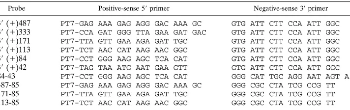
Design of primers and in vitro synthesis of RNA transcripts.The strategy used to synthesize cDNA fragments representing the 39end of the JHM genome and generate the RNA transcripts throughout this study is outlined in Fig. 1. A series of 59-end primers (positive sense) containing the T7 RNA polymerase promoter (TAA TAC GAC TCA CTA TAG GGC GAG) was synthesized. A series of 39-end negative sense primers was similarly synthesized. A double-stranded cDNA, p118-8a, representing 1,648 nt from the 39end of JHM genomic RNA (15) was used as a DNA template for PCR. PCR products of defined size were
synthesized by Taq DNA polymerase (Promega) and purified with either the Wizard PCR Prep kit (Promega) or the Mermaid kit (Bio 101) or by gel filtration through a Sephadex G-50 (Sigma) column. The primer pairs and the correspond-ing probes which were subsequently transcribed from the resultcorrespond-ing PCR products are described in Table 1.
In vitro transcription reactions of PCR product templates by T7 RNA poly-merase (Promega) were carried out as described by the manufacturer. The nucleotide concentrations were 0.5 mM for ATP, CTP, and GTP and 0 to 12.5 mM for UTP. Transcripts were uniformly labeled with 350 to 500 mCi of [32P]UTP (3,000 Ci/mmol; ICN) in the presence of RNasin (Promega). Tran-scription reaction mixtures were then treated with RNase-free DNase (Promega) and extracted twice with phenol-chloroform. Unincorporated nucleotides were removed by gel filtration through a Sephadex G-50 column. The specific activities of transcripts were approximately 13108to 73108cpm/mg. To examine the purity of RNA probes synthesized prior to their use in RNase protection/mobility shift assays, aliquots of several labeled transcripts, for example, 39(1)487, 39 (1)333, 39(1)113, and 39(1)84, in 13Tris-acetate-EDTA buffer were dena-tured by heating at 658C for 1 h, electrophoresed on 1% agarose gels, and autoradiographed. Each probe yielded a single homogeneous band (data not shown).
RNase T1protection and gel mobility shift electrophoresis assays.Binding
reactions were performed in a volume of 10ml as described previously (19, 40), with some modification. Cytoplasmic lysates containing 7mg of protein and 1 to 2 ng of32P-labeled RNA probes were incubated at 228C for 20 min in a buffer containing 50 mM KCl, 10 mM HEPES (pH 7.6), 2.5 mM MgCl2, 1 mM dithio-FIG. 1. Schematic diagram of the MHV genome, the locations of RNA probes, and summary of RNase T1protection/gel mobility shift assays. cDNAs representing various segments of the 39end of the MHV genome fused to the T7 promoter were synthesized as described in Materials and Methods.32
[image:2.612.130.491.71.306.2]P-labeled RNA probes were synthesized by in vitro transcription with T7 RNA polymerase. Each probe was named by nucleotide numbers or location within the genomic RNA sequence upstream from the 59-most A in the poly(A) tail (position 0). For example, probe 39(1)487 represents the 487 nt upstream from 0, and probe 171-85 represents an 86-nt sequence from nt 171 to 85 upstream from position 0. The size of each probe is indicated, as are the relative position and length.1, formation of an RNase-resistant complex; 2, no complex was detected in RNase protection/mobility shift assays.
TABLE 1. Probes and primers synthesized
Probe Positive-sense 59primer Negative-sense 39primer
39(1)487 PT7-GAG AAA GAG AGG GAC AAA GC GTG ATT CTT CCA ATT GGC 39(1)333 PT7-CCA GAT GGG TTA GAA GAT GAC GTG ATT CTT CCA ATT GGC 39(1)171 PT7-TTA GTT GAA AGA GAT TGC GTG ATT CTT CCA ATT GGC 39(1)113 PT7-TCT AAC CAT AAG AAC GGC GTG ATT CTT CCA ATT GGC 39(1)84 PT7-CCT GGG AAG AGC TCA CAT GTG ATT CTT CCA ATT GGC 39(1)42 PT7-TAG TAA ATG AAT GAA GTT GTG ATT CTT CCA ATT GGC 84-43 PT7-CCT GGG AAG AGC TCA CAT GGG CAT TGC AGG AAT AGT A 487-85 PT7-GAG AAA GAG AGG GAC AAA GC GGG CGC CTA TCG CCG TT 171-85 PT7-TTA GTT GAA AGA GAT TGC GGG CGC CTA TCG CCG TT 113-85 PT7-TCT AAC CAT AAG AAC GGC GGG CGC CTA TCG CCG TT
on November 9, 2019 by guest
http://jvi.asm.org/
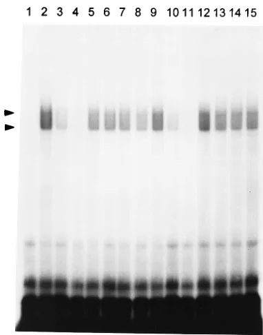
[image:2.612.130.486.614.726.2]genomic JHM RNA.Analysis of the structure of synthetic DI RNAs indicated that at least 436 nt corresponding to the 39 terminus of the genome were required for DI RNA replication in the presence of helper virus (21). The apparent requirement for as many as 436 nt at the 39end of MHV DI RNAs suggests that a sequence within this region would retain signals required to bind viral and/or host proteins necessary for negative-strand RNA synthesis (7, 21). To investigate this hypothesis, we have performed a series of RNase T1 protection/gel retardation assays. The first RNA probe selected for binding reactions corresponded to the 487 nt at the 39 end of the MHV-JHM genome and spans the minimal length (436 nt) of the 39region required for MHV DI replication (21). A cDNA containing the last 487 bases of the 39 end of the MHV-JHM genome (not including the polyadenylate tail) under the control of the T7 promoter was synthesized by PCR as shown in Fig. 1. 32 P-labeled RNA was transcribed from this template by T7 RNA polymerase. Cytoplasmic extracts prepared from both JHM-infected and unJHM-infected 17Cl-1 cells were incubated with 2 ng of labeled probe in the presence of heparin and poly(I)-poly(C) to decrease nonspecific binding. In a series of prelim-inary experiments, reaction mixtures containing this 487-nt RNA transcript were displayed by electrophoresis on nonde-naturing agarose or polyacrylamide gels. These experiments proved unsatisfactory because of poor resolution. Complexes formed with the 39(1)487 probe run on 4, 5, or 6% polyacryl-amide gels, or on agarose gels, yield a smear of RNA-protein complexes poorly resolved from free probe (data not shown). The addition of an RNase T1digestion step prior to electro-phoresis increases both the resolution and the sensitivity of gel shift assays by digesting unbound RNA and RNA nonspecifi-cally bound to protein and by decreasing the size and apparent heterogeneity of the RNA-protein complexes (14). Thus, after allowing formation of RNA-protein complexes, we subjected the reactions to limited digestion with RNase T1(0.05 U per reaction) prior to electrophoresis. RNase T1-resistant RNA-protein complexes were electrophoresed on 6% nondenaturing polyacrylamide gels and visualized by autoradiography. Incu-bation of the 39(1)487 probe with mock- or JHM-infected cell lysates protected a portion of the probe from T1 digestion, resulting in two stable, poorly resolved, RNase T1-resistant complexes, as shown in Fig. 2. The complexes formed by the 39 (1)487 probe with cytoplasmic extracts from either mock-in-fected (Fig. 2, lane 2) or JHM-inmock-in-fected (Fig. 2, lane 9) cells appeared to have identical electrophoretic mobilities.
To verify the specificity of the RNA-protein binding, com-petition experiments with 50- and 100-fold molar excesses of unlabeled specific and nonspecific competitor RNAs were
per-formed. The formation of the two T1-resistant complexes by the 39 (1)487 probe was greatly inhibited by a 50-fold molar excess of the unlabeled 39(1)487 RNA (Fig. 2, lanes 3 [unin-fected cells] and 10 [MHV-in[unin-fected cells]) and was completely blocked by a 100-fold molar excess of unlabeled 39 (1)487 RNA (Fig. 2, lanes 4 [uninfected cells] and 11 [MHV-infected cells]). Only a slight diminution of binding was observed with unlabeled nonspecific competitor RNAs, including a 50- to 100-fold molar excess of 75- to 90-nt tRNA (Fig. 2, lanes 5 and 6 [uninfected cells] and 12 and 13 [MHV-infected cells]) and a 294-nt synthetic transcript corresponding to the 59end of the MHV-A59 genome (Fig. 2, lanes 7 and 8 [uninfected cells] and 14 and 15 [MHV-infected cells]). Furthermore, no bands cor-responding to the complexes were observed when the 39 (1)487 probe alone was digested with RNase T1in the absence of cytoplasmic lysate (Fig. 2, lane 1).
[image:3.612.335.526.68.311.2]Localization of protein-binding sites at the 3*end of JHM genomic RNA.From the results shown in Fig. 2, it appears that at least one protein-binding element is contained within the 39 most 487 nt of the genome. Since two bands are visualized, it is possible that there are two different protein-binding ele-ments, or alternatively a single protein-binding site could form different complexes with multiple proteins. To localize the FIG. 2. Specific binding of host cellular proteins to the 39end of MHV-JHM genomic RNA. Two nanograms of32P-labeled 39(1)487 RNA was incubated with cytoplasmic extracts from either mock-infected (lanes 2 to 8) or MHV-JHM-infected (lanes 9 to 15) 17Cl-1 cells as described in Materials and Methods. Some samples contained competitor RNAs as indicated below. The binding mixtures were digested with RNase T1(0.05 U/10ml) and analyzed by electro-phoresis on a 6% nondenaturing polyacrylamide gel. Lane 1, free 39(1)487 RNA probe; lanes 2 and 9, 39 (1)487 probes incubated in the absence of competitor RNA with mock- and MHV-infected lysates, respectively; lanes 3 and 10, in the presence of a 50-fold molar excess of unlabeled specific 39(1)487 RNA; lanes 4 and 11, in the presence of a 100-fold molar excess of unlabeled 39 (1)487 RNA; lanes 5 and 12, in the presence of a 50-fold molar excess of nonspecific tRNA; lanes 6 and 13, in the presence of a 100-fold molar excess of tRNA; lanes 7 and 14, in the presence of a 50-fold molar excess of a 294-nt RNA from the 59end of the MHV-A59 genome; lanes 8 and 15, in the presence of a 100-fold molar excess of a 294-nt RNA from the 59 end of the MHV-A59 genome. Arrowheads denote the positions of RNA-protein complexes. The ma-terial migrating at the bottom of the gel represents the digestion products from the nonprotected portions of the probe.
on November 9, 2019 by guest
region(s) on the 39(1)487 RNA probe with which the cellular protein(s) may associate, a set of deleted, 32P-labeled RNA probes corresponding to various portions of the 39 (1)487 RNA was synthesized in vitro (Fig. 1) and tested for the ability to form RNA-protein complexes. Using 0.05 U of RNase T1 for limited digestion per 10-ml reaction, we demonstrated the specific formation of RNase T1-resistant complexes with probes representing the 39-most 333, 171, and 113 nt of the MHV genome (data not shown). These results are summarized in Fig. 1. These experiments mapped the binding site to within 113 nt from the 39end of the genomic RNA. However, variable results were observed when a shorter 39(1)84 probe was used; i.e., no complex formed in one experiment, and weak protein binding was observed in another (data not shown). To examine the possibility that shortening the probe to 84 nt lowered the binding affinity of proteins to the probe, either by altering the stability of the structure needed for protein binding or by deleting one of several elements involved in protein binding, we determined if digestion with lesser amounts of RNase T1 would allow us to visualize formation of less stable RNA-protein complexes. As shown in Fig. 3, RNA-RNA-protein com-plexes could be visualized with the 39 (1)84 probe when the concentration of T1 was less than 0.0125 U/10-ml reaction. Here we especially emphasize that titration of the T1in these digestions is critical to detect weak binding activity. Therefore, we have titered the T1concentration for each deleted probe to select optimal concentrations for digestion. To further define the sequence responsible for protein binding, two probes, 84-43 and 39 (1)42, representing each half of the 39 (1)84 RNA, were synthesized and examined for the ability to form complexes with cytoplasmic extracts. A single complex was observed with the32P-labeled 39(1)42 probe (Fig. 4B), but no complex could be detected with the 84-43 probe (Fig. 4A). Consistent with this result is the finding that only the unlabeled 39(1)84 probe, not the nonspecific tRNA, can compete with
32P-labeled 39 (1)42 probe for protein binding (Fig. 4B).
These results mapped a protein-binding element within the 42 nt at the 39end of JHM genomic RNA.
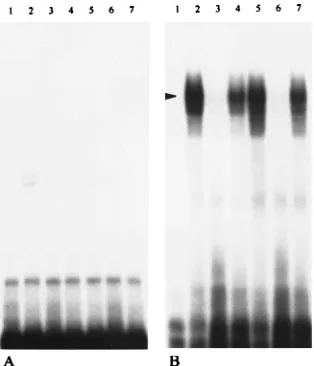
The fact that we observed two complexes in reactions con-taining the 39(1)487 RNA and host cell proteins, whereas a single complex formed with the 39(1)84 RNA, led us to search for the possible existence of other protein-binding elements at the 39 end of JHM genomic RNA. For this purpose, cross-competition assays were first performed with 32P-labeled 39 (1)84 and 39(1)487 probes to assay their relative abilities to bind host cell proteins in the presence of both unlabeled 39 (1)84 and 39(1)487 competitor RNAs (Fig. 5). No difference was observed when a 0.1- to 1-fold molar excess of either unlabeled 39 (1)84 or 39 (1)487 was used to compete with 32P-labeled 39(1)84 (Fig. 5A) and32P-labeled 39(1)487 (Fig.
[image:4.612.140.216.68.306.2]5B). With the 32P-labeled 39 (1)84 probe, the formation of RNase T1-resistant complexes was completely blocked in the presence of a 100- to 200-fold molar excess of either unlabeled 39(1)84 or 39(1)487 RNA but not in the presence of non-specific competitor tRNA (Fig. 5A). While the formation of complexes with32P-labeled 39(1)487 was completely blocked with unlabeled 100- to 400-fold-molar-excess 39(1)487 RNA, it was barely affected by adding the same molar excess of unlabeled 39(1)84 RNA and nonspecific tRNA (Fig. 5B). This result implied the possible presence of some other binding site(s) upstream of 84 nt from the 39end of the JHM genomic RNA. Protein-binding elements upstream of nt 84 were sought in an RNase protection/gel mobility shift assay using a probe spanning nt 487 to 85. Two complexes similar in pattern to those observed with 39(1)487 were formed. These complexes were demonstrated to represent specific protein binding to the FIG. 3. Titration of RNase T1concentration for 39(1)84 RNA-protein
[image:4.612.357.514.71.254.2]bind-ing.32P-labeled 39(1)84 RNA was incubated with uninfected-cell extracts at 228C for 20 min. Binding mixtures were then digested at 228C for 20 min in 10-ml reactions using decreasing amounts of RNase T1. Lane 1, 0.05 U; lane 2, 0.025 U; lane 3, 0.0125 U; lane 4, 0.00625 U; lane 5, 0.003125 U. The arrowhead indicates the RNA-protein complex.
FIG. 4. Localization of the protein-binding site with 39(1)42 and 84-43 RNA probes. RNA-protein binding reactions were carried out as described in the legend to Fig. 2 except that RNase T1digestion was with 0.00625 U. (A) Lane 1, free 84-43 probe; lanes 2 and 5, 84-43 probes incubated in the absence of competitor RNA with mock- and MHV-infected lysates, respectively; lanes 3 and 6, 84-43 probes incubated in the presence of a 75-fold molar excess of unlabeled specific 39(1)84 RNA with mock- and MHV-infected lysates, respectively; lanes 4 and 7, 84-43 probes incubated in the presence of a 75-fold molar excess of nonspecific tRNA with mock- and MHV-infected lysates, respectively. (B) Lane 1, free 39(1)42 RNA probe; lanes 2 and 5, 39(1)42 probes incubated in the absence of competitor RNA with mock- and MHV-infected lysates, respectively; lanes 3 and 6, 39(1)42 probes incubated in the presence of a 75-fold molar excess of unlabeled specific 39(1)84 RNA with mock- and MHV-infected ly-sates, respectively; lanes 4 and 7, 39(1)42 probes incubated in the presence of a 75-fold molar excess of nonspecific tRNA with mock- and MHV-infected lysates, respectively.
on November 9, 2019 by guest
http://jvi.asm.org/
RNA since a 150-fold molar excess of unlabeled tRNA did not block the binding, whereas the same molar excess of unlabeled specific competitor RNA (487-85) completely inhibited the formation of the RNase-resistant complexes (data not shown). Using a set of RNA probes containing deletions at the 59end (Fig. 1), we mapped the binding site to a sequence between nt 171 to 85 (data not shown). These experiments are summa-rized in Fig. 1. These results confirm that 487 bases within the 39 end of JHM genomic RNA contain two protein-binding elements, which are localized separately between nt 42 and 1 and nt 171 and 85 (Fig. 1).
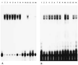
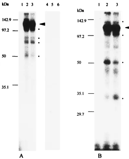
Characterization of binding proteins by UV-induced cross-linking.To characterize the protein(s) bound to the 39end of genomic RNA of MHV and determine their molecular weights, 32P label was transferred from RNA probes to pro-teins bound directly to these RNAs by UV irradiation. After RNase T1limited digestion, RNA binding reaction mixtures were irradiated with UV light for 30 min and then completely digested with RNase A. Proteins to which32P label was trans-ferred by cross-linking were analyzed by SDS-polyacrylamide gel electrophoresis (PAGE) through 12% polyacrylamide gels. No bands were observed after UV irradiation of RNA probes in the absence of cytoplasmic extracts (Fig. 6A, lanes 1 and 4; Fig. 6B, lane 1). As shown in Fig. 6A (lanes 2 and 3), the RNA-protein complex formed with 39 (1)42 RNA contains approximately five host polypeptides, a highly labeled protein of 120 kDa and four minor species with sizes of 103, 81, 70, and 55 kDa. No protein bands were visualized when the same cytoplasmic extracts was incubated with the 84-43 probe (Fig. 6A, lanes 5 and 6). This result is consistent with the results of
[image:5.612.147.466.65.330.2]the gel mobility shift assay (Fig. 4). The second protein-binding element, contained within a probe representing nt 487 to 85 from the 39end of the genome, also labeled five host polypep-tides of 142, 120, 100, 55, and 33 kDa in size, with the 120-kDa protein appearing to be the major protein component in the complex (Fig. 6B, lanes 2 and 3). However, some additional faint and indistinct bands were also sometimes observed. An example of such a band can be seen between the 50- and 35.1-kDa markers. We are unsure of the significance of these bands. When a 39 (1)487 probe was used, the pattern with protein bands was similar to that obtained with the 487-85 RNA. As the signal that we obtained was very weak for the 39 (1)487 RNA-protein complex after complete digestion of RNase A, we repeated this experiment using RNase T1 for complete digestion after UV cross-linking. The same protein profiles were obtained (data not shown). Since the efficiency of UV cross-linking for different proteins to the RNA is highly variable (33), it is possible that the weak transfer of32P label to the minor bands is due to particularly low efficiency of cross-linking, or it could reflect a relatively low quantity of these proteins in the complexes. Although the complexes formed with the 39 (1)42 RNA and the 487-85 RNA both contained 120- kDa and 55-kDa polypeptides, we are not sure whether these represent the same proteins contributing to complexes formed on separate sites. It is also possible that the 39(1)42 probe labeled a 142-kDa protein similar to that observed with the 487-85 RNA, but it was obscured by the heavy labeling and diffuseness of the 120-kDa protein in Fig. 6A. We have been unable to unequivocally resolve this question by repeated ex-periments and shorter exposures of the autoradiographs. In FIG. 5. Cross-competition assays of 39(1)84 RNA and 39(1)487 RNA. RNA-protein binding reactions were carried out as described in the legend to Fig. 2. (A) One nanogram of labeled 39(1)84 RNA was incubated with 7mg of mock-infected cell lysate in the presence of competitor RNA and subsequently digested with 0.00625 U of RNase T1. Lane 1, free probe; lane 2, probe with no competitor RNA; lanes 3 to 5, 0.1-fold molar excess of unlabeled 39(1)84, 39(1)487, and tRNAs, respectively; lanes 6 to 8, 1-fold molar excess of unlabeled 39(1)84, 39(1)487, and tRNAs, respectively; lanes 9 to 11, 100-fold molar excess of unlabeled 39(1)84, 39(1)487, and tRNAs, respectively; lanes 12 to 14, 200-fold molar excess of unlabeled 39(1)84, 39(1)487, and tRNAs, respectively. (B) One nanogram of labeled 39(1)487 RNA was incubated with 7mg of mock-infected cell lysate in the presence of competitor RNA and subsequently digested with 0.05 U of RNase T1. Lane 1, free probe; lane 2, probe with no competitor RNA; lanes 3 to 5, 0.1-fold molar excess of unlabeled 39(1)487, 39(1)84, and tRNAs, respectively; lanes 6 to 8, 1-fold molar excess of unlabeled 39(1)487, 39(1)84, and tRNAs, respectively; lanes 9 to 11, 100-fold molar excess of unlabeled 39(1)487, 39(1)84, and tRNAs, respectively;
lanes 12 to 14, 400-fold molar excess of unlabeled 39(1)487, 39(1)84, and tRNAs, respectively.
on November 9, 2019 by guest
addition, specific binding or complex formation was indeed mediated by protein in the cell lysates, since protein bands were no longer observed when the reaction mixture was incu-bated with proteinase K (1.5 mg/ml at 378C for 1 h) after UV cross-linking and just prior to SDS-PAGE (data not shown). UV irradiation was necessary to transfer 32P from RNA to protein; the identical procedure without UV irradiation did not result in any RNA-protein cross-linking (data not shown). The protein profiles obtained with JHM-infected cytoplasmic extracts were identical to those observed with uninfected cell extracts and the 39(1)42, 84-43, and 487-85 probes (Fig. 6).
DISCUSSION
Our approach to begin understanding the mechanism of MHV RNA replication is to precisely identify host and/or viral protein binding elements at the 39end of the genomic RNA, to characterize the RNA sequences and secondary structures re-sponsible for the RNA-protein interactions, and to identify the proteins which bind to these elements. In this study, we report several findings that may give some insight into the require-ments at the 39end of the genomic MHV RNA for replication. First, we have demonstrated that cytoplasmic proteins from host 17Cl-1 cells bind to the 39 region of the positive-strand MHV RNA. The RNA-protein binding is specific, since a
50-to 100-fold molar excess of unlabeled probes blocked the for-mation of complexes, while nonspecific competitors did not. Lysates prepared from uninfected and MHV-infected cells gave identical patterns of RNase T1-resistant complexes, and these complexes had identical protein profiles. Second, we mapped the RNA-binding elements to two regions of 42 and 86 nt within 39-most 436 nt of genomic RNA, which is reported as the minimal length of sequence required for DI RNA rep-lication (21). Finally, by using sequential and complete RNase digestion and UV cross-linking followed by SDS-PAGE, we were able to determine the apparent molecular masses of mul-tiple proteins cross-linked to RNA.
During MHV replication in infected cells, the RNA poly-merase complex starts from the positive-strand genomic 39end and transcribes a full-length copy of negative-strand RNA (1, 11, 36). It appears to transcribe subgenome-length negative-strand RNA as well (34). These negative-negative-strand RNAs are the templates from which seven or eight species of positive-strand virus-specific RNAs with a 39-coterminal nested-set structure are synthesized (9, 10, 17, 18, 35). Thus, synthesis of negative-strand RNA is a crucial early event in viral replication. If this event is blocked, virus replication will not proceed past the translation of input viral genomic RNA. Because of the large size of coronavirus genome RNA (about 31 kb) and unavail-ability of a full-length infectious cDNA clone, cDNA clones corresponding to replication-competent MHV DI RNAs have been created and extensively used to study the sequences re-quired for MHV replication (6–8, 21, 22). Such a strategy has also been applied to identify the cis-acting replication signals in other positive-strand viruses such as Sindbis virus (20, 39) and brome mosaic virus (4). Deletions constructed in vitro have allowed the identification of the minimal sequences necessary for MHV DI RNA replication in the presence of helper virus. At least 436 bases derived from the 39end of the genome are required for replication (7, 21).
A recent study has further characterized the cis-acting ele-ments required for MHV replication. In part on the basis of studies of other positive- and negative-strand viruses, protein-binding elements were sought within both the 59and 39termini of the MHV genome, and binding of a 55-kDa host cell protein to positive-strand RNA corresponding to nt 56 to 112 at the 59 end of the genome was detected (5). The biological role of this 55-kDa protein in viral replication is unknown. Experiments using a 90-base probe spanning the 39terminus of the genomic RNA of positive sense failed to demonstrate any protein bind-ing (5), which differs from our results reported here. This difference is probably due to the relatively low affinity binding of host proteins to the 39end of genomic RNA compared with the binding of host proteins to the 59end of the genome (41), combined with the different methodologies used by the two studies. The addition of an RNase T1digestion step increases the sensitivity and specificity of the assay that we used com-pared to simple mobility shift assays (14).
Host cell proteins binding to either the 39or the 59 end of genome RNA have been reported in positive-strand rubella virus (26), poliovirus (24, 25), and hepatitis A virus (28) as well as in negative-strand measles virus (19). Host proteins were also found to bind to multiple sites at the 59-end genome of positive-strand poliovirus (2) and at the 39 end of the full-length negative-sense Sindbis virus-specific RNA (30, 31). Al-though the definite biological role of these proteins in RNA replication has not been clarified, the interaction of host pro-teins with viral genomes has been suggested to be an important step in viral RNA replication (5, 19, 25, 26, 30, 31). At present little is known regarding host and virus-specific factors in the RNA-dependent RNA polymerase complex necessary for ini-FIG. 6. Identification of host cell proteins binding to the 39end of MHV
genomic RNA by UV cross-linking. RNA-protein binding reactions using labeled RNA were carried out as described in the legend to Fig. 2. Following incubation with RNase T1[0.00625 U for the 39(1)42 and 84-43 probes; 0.025 U for the 487-85 probe],32P-labeled RNA-protein complexes were UV cross-linked and digested with RNase A prior to SDS-PAGE (12% polyacrylamide gel). (A) Lane 1, free 39(1)42 probe; lane 2, 39(1)42 probe with mock-infected cell lysate; lane 3, 39(1)42 probe with MHV-infected cell lysate; lane 4, free 84-43 probe; lane 5, 84-43 probe with mock-infected lysate; lane 6, 84-43 probe with MHV-infected lysate. A highly labeled protein of 120 kDa is indicated by an arrowhead, and four minor proteins are indicated by asterisks. (B) Lane 1, free 487-85 probe; lane 2, 487-85 probe with mock-infected lysate; lane 3, 487-85 probe with MHV-infected lysate. A highly labeled protein of 120 kDa is indicated by an arrowhead; four minor proteins are indicated by asterisks. Prestained molecular weight standards (Bio-Rad) were used as markers.
on November 9, 2019 by guest
http://jvi.asm.org/
[image:6.612.66.284.68.336.2]tiation of negative-strand RNA synthesis during MHV RNA replication.
The data reported here suggest that host cell proteins prob-ably are involved in MHV RNA replication. Fine deletion analysis demonstrated that the replication of DI RNA could not occur if sequences from nt 107 to 16 or 267 to 90 from the 39end of the genome were deleted (7). Both sequences contain portions of protein-binding elements that we mapped, i.e., nt 42 to 1 and 171 to 85, and this finding further supports the possible role of binding of host cell proteins to the 39end of the genome in MHV RNA replication. All of our RNase protec-tion/gel retardation and UV cross-linking experiments to date have failed to demonstrate binding of a virus-specific protein to the 39 end of the MHV genome, yet the MHV RNA-depen-dent RNA polymerase must recognize this region of the ge-nome to initiate negative-strand RNA synthesis. This may be an indication that viral proteins do not bind directly to the
genomic RNA but associate with protein-binding elements within genomic RNA indirectly via protein-protein interac-tions and thus might not be detected by the UV cross-linking assay described here. Alternatively, it might be that the binding affinity of viral protein to RNA is so low that the RNA-protein complex formed with MHV polymerase proteins is not resis-tant to RNase T1digestion under any of the conditions that we used. The results of our UV cross-linking experiment indicate that the 42-1 and 487-85 protein-binding elements each bind proteins of 120 and 55 kDa. We have not yet determined if this represents binding of the same two proteins to both elements or if these proteins coincidently have similar electrophoretic mobilities.
RNA sequence elements with protein-binding activity often contain secondary structures (23, 27). Analysis of the 39-most 487 nt of the MHV-JHM genome by using the Fold program in the University of Wisconsin Genetics Computer Group se-FIG. 7. Secondary structure prediction and sequence comparison of protein-binding segments. (A) Secondary structure predictions of 39(1)42, 84-43, and 171-85 RNAs were made by using the RNAFOLD program. The free energy of the stem-loop structure at each site is given in kilocalories per mole (1 kcal5 4.184 kJ). The position of the sequence GAAGUU, which is present in both 39 (1)42 and 171-85 probes, is indicated with boldface letters. (B) The RNA sequences of nt 42 to 1 and nt 171 to 85 from the 39end of the JHM genome were aligned by using the GAP program. The sequence GAAGUU is present in both elements (marked with a box).
on November 9, 2019 by guest
quence analysis package indicates the potential formation of multiple stem-loop structures in this region. The sequence in this region is highly conserved among various strains of MHV (32). Predictions of secondary structure with RNA probes 39 (1)42, 171-85, and 84-43, of which the former two are sites for host cell protein binding and the latter are not, revealed that each RNA is predicted to contain potential stem-loop struc-tures, although these structures are not predicted to be partic-ularly stable (Fig. 7A). We compared the sequences of two protein-binding elements, 171-85 and 39(1)42, and about 42% sequence identity was found, with the sequence GAAGUU being present in both elements (Fig. 7B). This sequence is fully conserved among MHV strains. For both the 39(1)42 and the 171-85 elements, this sequence is predicted to be largely non-base paired, either as a bulge in a stem or a bulge in a loop (Fig. 7A). Future studies will be directed to determine the precise sequence and secondary structure required for host protein binding.
ACKNOWLEDGMENTS
We especially thank Ann-Bin Shyu of the Department of Biochem-istry for valuable advice and instruction on gel mobility shift and UV cross-linking techniques. We thank Brenda Hogue of Baylor College of Medicine for critical reading and helpful comments on the manuscript. We thank Robin Stalcup for preparing MHV-JHM virus stocks and all members of the Leibowitz laboratory for helpful discussion.
This work was supported by National Multiple Sclerosis Society research grant RG 2203-A-5 and by a grant from the Texas Advanced Research Program.
REFERENCES
1. Baric, R. S., S. A. Stohlman, and M. M. C. Lai. 1983. Characterization of replicative intermediate RNA of mouse hepatitis virus: presence of leader RNA sequences on nascent chains. J. Virol. 48:630–663.
2. Del Angel, R. M., A. G. Papavassiliou, C. Fernandez-Tomas, S. J. Silverstein, and V. R. Racaniello.1989. Cell proteins bind to multiple sites within the 59 untranslated region of poliovirus RNA. Proc. Natl. Acad. Sci. USA 86:8299– 8303.
3. Dignam, J. D., R. M. Lebovitz, and R. G. Roeder. 1983. Accurate transcrip-tion initiatranscrip-tion by RNA polymerase II in a soluble extract from isolated mammalian nuclei. Nucleic Acids Res. 11:1475–1489.
4. French, R., and P. Ahlquist. 1987. Intercistronic as well as terminal se-quences are required for efficient amplification of brome mosaic virus. J. Virol. 61:1457–1465.
5. Furuya, T., and M. M. C. Lai. 1993. Three different cellular proteins bind to complementary sites on the 59-end-positive and 39-end-negative strands of mouse hepatitis virus RNA. J. Virol. 67:7215–7222.
6. Furuya, T., T. B. Macnaughton, N. La Monica, and M. M. C. Lai. 1993. Natural evolution of coronavirus defective-interfering RNA involves RNA recombination. Virology 194:408–413.
7. Kim, Y. N., Y. S. Jeong, and S. Makino. 1993. Analysis of cis-acting se-quences essential for coronavirus defective interfering RNA replication. Virology 197:53–63.
8. Kim, Y. N., M. M. C. Lai, and S. Makino. 1993. Generation and selection of coronavirus defective interfering RNA with large open reading frame by RNA recombination and possible editing. Virology 194:244–253. 9. Lai, M. M. C. 1990. Coronavirus: organization, replication and expression of
genome. Annu. Rev. Microbiol. 44:303–333.
10. Lai, M. M. C., P. R. Brayton, R. C. Armen, C. D. Patton, C. Pugh, and S. A. Stohlman.1981. Mouse hepatitis virus A59: mRNA structure and genetic localization of the sequence divergence from hepatotropic strain MHV-3. J. Virol. 39:823–834.
11. Lai, M. M. C., C. D. Patton, and S. A. Stohlman. 1982. Replication of mouse hepatitis virus: negative-stranded RNA and replicative-form RNA are of genome length. J. Virol. 44:487–492.
12. Lai, M. M. C., and S. A. Stohlman. 1978. RNA of mouse hepatitis virus. J. Virol. 26:236–242.
13. Lee, H.-J., C.-K. Shieh, A. E. Gorbalenya, E. V. Koonin, N. LaMonica, J. Tuler, A. Bagdzhadzhyan, and M. M. C. Lai.1989. The complete sequence (22 kilobases) of murine coronavirus gene-1 encoding the putative proteases
and RNA polymerase. Virology 180:567–582.
14. Leibold, E. A., and H. N. Munro. 1988. Cytoplasmic protein binds in vitro to a highly conserved sequence in the 59untranslated region of ferritin heavy-and light-subunit mRNAs. Proc. Natl. Acad. Sci. USA 85:2171–2175. 15. Leibowitz, J. L., and J. R. DeVries. 1988. Synthesis of virus-specific RNA in
permeabilized murine coronavirus-infected cells. Virology 166:66–75. 16. Leibowitz, J. L., S. R. Weiss, E. Paavola, and C. W. Bond. 1982. Cell-free
translation of murine coronavirus RNA. J. Virol. 43:905–913.
17. Leibowitz, J. L., K. C. Wilhelmsen, and C. W. Bond. 1981. The virus-specific intracellular RNA species of two murine coronaviruses: MHV-A59 and MHV-JHM. Virology 114:39–51.
18. Leibowitz, J. L., P. W. Zoltick, K. V. Holmes, E. L. Oleszak, and S. R. Weiss. 1990. Murine coronavirus RNA synthesis, p. 67–74. In M. A. Brinton and F. X. Heinz (ed.), New aspects of positive-strand RNA viruses. American Society for Microbiology, Washington, D.C.
19. Leopardi, R., V. Hukkanen, R. Vainionpaa, and A. A. Salmi. 1993. Cell proteins bind to sites within the 39noncoding region and the positive-strand leader sequence of measles virus RNA. J. Virol. 67:785–790.
20. Levis, R., B. W. Weiss, M. Tsiang, H. Huang, and S. Schlesinger. 1986. Deletion mapping of Sindbis virus DI RNAs derived from cDNAs defines the sequences essential for replication and packaging. Cell 44:137–145. 21. Lin, Y. J., and M. M. C. Lai. 1993. Deletion mapping of a mouse hepatitis
virus defective interfering RNA reveals the requirement for an internal and discontiguous sequence for replication. J. Virol. 67:6110–6118.
22. Makino, S., C. K. Shieh, L. H. Soe, S. C. Baker, and M. M. C. Lai. 1988. Primary structure and translation of a defective interfering RNA of murine coronavirus. Virology 166:550–560.
23. Mattaj, I. W. 1993. RNA recognition: a family matter? Cell 73:837–840. 24. Meerovitch, K., J. Pelletier, and N. Sonenberg. 1989. A cellular protein that
binds to the 59-noncoding region of poliovirus RNA: implications for inter-nal translation initiation. Genes Dev. 3:1026–1034.
25. Najita, L., and P. Sarnow. 1990. Oxidation-reduction sensitive interaction of a cellular 50-KDa protein with an RNA hairpin in the 59noncoding region of the poliovirus genome. Proc. Natl. Acad. Sci. USA 87:5846–5850. 26. Nakhasi, H., X. Q. Cao, T. A. Rouault, and T. Y. Liu. 1991. Specific binding
of host cell proteins to the 39-terminal stem-loop structure of rubella virus negative-strand RNA. J. Virol. 65:5961–5967.
27. Nakhasi, H. L., T. A. Rouault, D. J. Haile, T. Y. Liu, and R. D. Klausner. 1990. Specific high-affinity binding of host cell proteins to the 39region of rubella virus RNA. New Biol. 2:255–264.
28. Nuesch, J. P. F., M. Weitz, and G. Siegl. 1993. Proteins specifically binding to the 39untranslated region of hepatitis A virus RNA in persistently in-fected cells. Arch. Virol. 128:65–79.
29. Pachuk, C. J., P. J. Breedenbeek, P. W. Zoltick, W. J. M. Spaan, and S. R. Weiss.1989. Molecular cloning of the gene encoding the putative poly-merase of mouse hepatitis coronavirus strain A59. Virology 171:141–148. 30. Pardigon, N., E. Lenches, and J. H. Strauss. 1993. Multiple binding sites for
cellular proteins in the 39end of Sindbis alphavirus minus-sense RNA. J. Virol. 67:5003–5011.
31. Pardigon, N., and J. H. Strauss. 1992. Cellular proteins bind to the 39end of Sindbis virus minus-strand RNA. J. Virol. 66:1007–1015.
32. Parker, M. M., and P. S. Masters. 1990. Sequence comparison of the N genes of five strains of the coronavirus mouse hepatitis virus suggests a three domain structure for the nucleocapsid protein. Virology 179:463–468. 33. Pino-Roma, S., S. A. Adam, Y. D. Choi, and G. Dreyfuss. 1989.
Ultraviolet-induced cross-linking of RNA to proteins in vivo. Methods Enzymol. 180: 410–418.
34. Sawicki, S. G., and D. L. Sawicki. 1990. Coronavirus transcription: sub-genomic mouse hepatitis virus replicative intermediates function in RNA synthesis. J. Virol. 64:1050–1056.
35. Spaan, W. J., D. Cavanagh, and M. C. Horzinek. 1988. Coronaviruses: structure and genome expression. J. Gen. Virol. 69:2939–2952.
36. Spaan, W. J., P. J. Rottier, M. C. Horzinek, and B. A. Vander Zeust. 1982. Sequence relationships between the genome and the intracellular RNA species 1, 3, 6, and 7 of mouse hepatitis virus strain A59. J. Virol. 42:432–439. 37. Sturman, L. S., and K. K. Takemoto. 1972. Enhanced growth of a murine
coronavirus in transformed mouse cells. Infect. Immun. 6:501–507. 38. Wege, H., A. Muller, and V. ter Meulen. 1978. Genomic RNA of the murine
coronavirus JHM. J. Gen. Virol. 41:217–227.
39. Weiss, B., H. Nitschko, I. Ghattas, R. Wright, and S. Schlesinger. 1989. Evidence for specificity in the encapsidation of Sindbis virus RNAs. J. Virol. 63:5310–5318.
40. You, Y., C. Y. A. Chen, and A. B. Shyu. 1992. U-rich sequence-binding proteins interacting with a 20-nucleotide U-rich sequence in the 39 untrans-lated region of c-fos mRNA may be involved in the first step of c-fos mRNA degradation. Mol. Cell. Biol. 12:2931–2940.
41. Yu, W., and J. L. Leibowitz. Unpublished data.