Activation and Inactivation of Primary
Human Immunodeficiency Virus
Envelope Glycoprotein Trimers by
CD4-Mimetic Compounds
Navid Madani,
a,bAmy M. Princiotto,
aConnie Zhao,
aFatemeh Jahanbakhshsefidi,
aMax Mertens,
aAlon Herschhorn,
a,bBruno Melillo,
cAmos B. Smith III,
cJoseph Sodroski
a,b,dDepartment of Cancer Immunology and Virology, Dana-Farber Cancer Institute, Boston, Massachusetts, USAa; Department of Microbiology and Immunobiology, Harvard Medical School, Boston, Massachusetts, USAb;
Department of Chemistry, University of Pennsylvania, Philadelphia, Pennsylvania, USAc; Department of
Immunology and Infectious Diseases, Harvard T.H. Chan School of Public Health, Boston, Massachusetts, USAd
ABSTRACT
Human immunodeficiency virus type 1 (HIV-1) entry into cells is
medi-ated by the viral envelope glycoproteins (Env), a trimer of three gp120 exterior
gly-coproteins, and three gp41 transmembrane glycoproteins. The metastable Env is
triggered to undergo entry-related conformational changes when gp120 binds
se-quentially to the receptors, CD4 and CCR5, on the target cell. Small-molecule
CD4-mimetic compounds (CD4mc) bind gp120 and act as competitive inhibitors of
gp120-CD4 engagement. Some CD4mc have been shown to trigger Env
prema-turely, initially activating Env function, followed by rapid and irreversible
inactiva-tion. Here, we study CD4mc with a wide range of anti-HIV-1 potencies and
demon-strate that all tested CD4mc are capable of activating as well as inactivating Env
function. Biphasic dose-response curves indicated that the occupancy of the
protom-ers in the Env trimer governs viral activation vprotom-ersus inactivation. One CD4mc bound
per Env trimer activated HIV-1 infection. Envs with two CD4mc bound were
acti-vated for infection of negative, CCR5-positive cells, but the infection of
CD4-positive, CCR5-positive cells was inhibited. Virus was inactivated when all three Env
protomers were occupied by the CD4mc, and gp120 shedding from the Env trimer
was increased in the presence of some CD4mc. Env reactivity and the on rates of
CD4mc binding to the Env trimer were found to be important determinants of the
potency of activation and entry inhibition. Cross-sensitization of Env protomers that
do not bind the CD4mc to neutralization by an anti-V3 antibody was not evident.
These insights into the mechanism of antiviral activity of CD4mc should assist efforts
to optimize their potency and utility.
IMPORTANCE
The trimeric envelope glycoproteins of human immunodeficiency
vi-rus type 1 (HIV-1) mediate vivi-rus entry into host cells. Binding to the host cell
recep-tors, CD4 and CCR5, triggers changes in the conformation of the HIV-1 envelope
gly-coprotein trimer important for virus entry. Small-molecule CD4-mimetic compounds
inhibit HIV-1 infection by multiple mechanisms: (i) direct blockade of the interaction
between the gp120 exterior envelope glycoprotein and CD4; (ii) premature
trigger-ing of conformational changes in the envelope glycoproteins, leadtrigger-ing to irreversible
inactivation; and (iii) exposure of cryptic epitopes to antibodies, allowing virus
neu-tralization. The consequences of the binding of the CD4-mimetic compound to the
HIV-1 envelope glycoproteins depends upon how many of the three subunits of the
trimer are bound and upon the propensity of the envelope glycoproteins to
un-dergo conformational changes. Understanding the mechanistic factors that influence
Received19 September 2016Accepted15 November 2016
Accepted manuscript posted online23 November 2016
CitationMadani N, Princiotto AM, Zhao C, Jahanbakhshsefidi F, Mertens M, Herschhorn A, Melillo B, Smith AB, III, Sodroski J. 2017. Activation and inactivation of primary human immunodeficiency virus envelope glycoprotein trimers by CD4-mimetic compounds. J Virol 91:e01880-16. https:// doi.org/10.1128/JVI.01880-16.
EditorGuido Silvestri, Emory University
Copyright© 2017 American Society for Microbiology. All Rights Reserved. Address correspondence to Joseph Sodroski, [email protected].
crossm
on November 7, 2019 by guest
http://jvi.asm.org/
the activity of CD4-mimetic compounds can help to improve their potency and
cov-erage of diverse HIV-1 strains.
KEYWORDS
antibody, conformation, envelope glycoprotein, inhibitor, neutralization,
sensitization, stoichiometry, virus entry
T
he entry of human immunodeficiency virus type 1 (HIV-1) into target cells is
mediated by the trimeric envelope glycoprotein (Env) complex, which consists of
three gp120 exterior envelope glycoproteins and three gp41 transmembrane envelope
glycoproteins (1). Binding of gp120 to the receptor, CD4, on the target cell surface
induces major conformational changes in the envelope glycoproteins (2–5). These
changes allow gp120 to bind the viral coreceptor, either CXCR4 or CCR5 (6–13). CD4
binding also induces the formation of a gp41 prehairpin intermediate, in which three
hydrophobic grooves on the surface of a coiled coil formed by the heptad repeat 1
(HR1) region of gp41 are exposed (14–17). These hydrophobic groves are subsequently
occupied by helices from the gp41 heptad repeat 2 (HR2) region during the formation
of an energetically stable six-helix bundle that is thought to drive the fusion of the viral
and target cell membranes (18–23).
Molecules that mimic CD4 have been developed as potential inhibitors of HIV-1
infection. They include soluble CD4 (sCD4), enhanced CD4 (eCD4), sCD4
minipro-teins, and small-molecule CD4-mimetic compounds (CD4mc) (24–41). All of these
inhibitors bind gp120 near the natural binding site for CD4, a well-conserved
surface element adjacent to a pocket located at the interface of the gp120 inner
domain, outer domain, and bridging sheet (32–39). A phenylalanine residue at
position 43 (Phe 43) of CD4 fills the opening of this gp120 pocket, and thus, the
pocket has been designated the Phe 43 cavity (42, 43). Small-molecule CD4mc bind
within the Phe 43 cavity of gp120, penetrating deeper into the gp120 interior than
CD4 (32–39). Soluble CD4 and CD4mc inhibit HIV-1 infection by multiple
mecha-nisms: (i) competitive inhibition of gp120 interaction with target cell CD4 and (ii)
premature activation of Env, followed by apparently irreversible inactivation (30, 32,
38, 44). Consistent with the latter mechanism, in some experimental systems, sCD4
and some CD4mc can enhance HIV-1 entry into CD4-negative cells expressing CCR5
(31, 32, 44). Under some circumstances, sCD4 binding leads to the detachment or
shedding of gp120 from the Env trimer (45, 46).
CD4mc were initially identified in a screen for small-molecule blockers of
gp120-CD4 binding (30). The hits from this screen, NBD-556 and the closely related
NBD-557, exhibited only weak antiviral activity against a very limited number of
natural HIV-1 variants (30–32). Iterative, structure-based modifications of these
prototypic CD4mc have led to significant improvements in the potency and breadth
of their anti-HIV-1 activity (32–41). Of particular importance was the replacement of
the chemically intractable tetramethylpiperidine ring of NBD-556 and NBD-557 with
an indane ring that allowed multiple substitutions (37, 38). The indane-containing
analogues (DMJ-II-121, JP-III-48, and BNM-III-170) exhibited progressive increases in
potency and breadth as various moieties were added to the indane ring at specific
locations (37–39).
The prototypic CD4mc, NBD-556 and NBD-557, efficiently activated HIV-1
infec-tion of CD4
⫺CCR5
⫹target cells (31, 32). Interestingly, as the potency of the CD4mc
in inhibiting HIV-1 infection of CD4
⫹CCR5
⫹cells increased, the activation of HIV-1
infection of CD4
⫺CCR5
⫹target cells diminished (35–39). In the assay used to detect
activation, the CD4mc was incubated at 37°C with a primary HIV-1 isolate, HIV-1
YU2,
for 30 min, and then the virus-compound mixture was added to CD4-negative
Cf2Th-CCR5 cells. Because the diffusion of virus to the target cells is a slow process
that can take several hours (47, 48), activation events, particularly those that
eventually lead to the decay of infectivity, can be missed by the assay. As an
understanding of the mechanism of inhibition is important to attempts to improve
and utilize the CD4mc, we adapted our assays to allow greater sensitivity in
on November 7, 2019 by guest
http://jvi.asm.org/
detecting transient activation of HIV-1 Env function. Here, we use these assays to
evaluate the antiviral mechanism of CD4mc exhibiting a range of potencies against
HIV-1 strains that differ in susceptibility to inhibition by CD4-mimetic molecules.
The results of these studies provide important insights into the interaction of
CD4mc with the functional HIV-1 Env trimer.
RESULTS
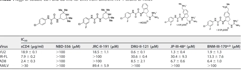
Susceptibility of selected HIV-1 variants to CD4mc.
For these studies, we selected
five CD4mc that exhibited a range of anti-HIV-1 potencies and three HIV-1 variants that
demonstrated a range of sensitivities to sCD4 and CD4-mimetic compounds (Table 1).
We tested the ability of the CD4mc to inhibit HIV-1 infection by incubating the
compound with recombinant, luciferase-expressing HIV-1 for 30 min at 37°C and then
adding the compound-virus mixture to Cf2Th-CD4/CCR5 target cells. Measurement of
the luciferase activity in the target cells 48 h later provided an indication of the level of
infection. Under the conditions of the assay, virus activation by the CD4mc is minimal
because of the short half-life of the activated state and the slow diffusion of the virus
to the target cell (42, 46, 49). As previously observed (39), BNM-III-170, a recently
developed analogue, exhibited dramatically greater antiviral activity against the three
HIV-1 strains than the prototypic CD4mc, NBD-556 (Table 1). Of the three HIV-1 strains
tested, HIV-1YU2
was the variant most sensitive and HIV-1JR-FL
was the variant least
sensitive to the CD4mc. The sCD4 sensitivities of the three HIV-1 strains did not
correlate with CD4mc sensitivity (Table 1). The antiviral activity of the CD4mc was
specific for viruses with HIV-1 Envs, as minimal inhibition of HIV-1 pseudotyped with the
amphotropic murine leukemia virus (AMLV) Env was observed.
[image:3.585.43.544.77.223.2]Activation of Env-mediated virus entry into CD4
ⴚCCR5
ⴙtarget cells.
To
investigate the activating/inactivating mechanism of Env inhibition by the CD4mc, we
utilized experimental systems in which competitive inhibition of CD4 binding does not
contribute to the antiviral effects of these compounds. One such experimental system
measures the infection of CD4-negative Cf2Th-CCR5 target cells by recombinant HIV-1
with different Envs in the absence or presence of increasing concentrations of CD4mc.
In these assays, after incubation of the CD4mc and virus at 37°C for different lengths of
time, the virus-compound mixtures were spinoculated onto the Cf2Th-CCR5 cells,
which were further cultivated and assessed for infection 48 h later. The spinoculation
step allows detection of activation events that might be missed in assays dependent on
the slow process of virus diffusion to the target cell (47, 48). In the absence of CD4mc,
the infection of the Cf2Th-CCR5 cells by the HIV-1 strains tested was near the assay
TABLE 1IC50s of soluble CD4 and CD4mc for Envs from different HIV-1 strains or AMLVVirus
IC50
sCD4 (g/ml) NBD-556 (M) JRC-II-191 (M) DMJ-II-121 (M) JP-III-48a(M) BNM-III-170a,b(M)
YU2 18.9⫾0.1 ⬎100 18.5⫾1.1 0.6⫾0.1 1.3⫾0.4 1.9⫾1.3 JR-FL 7.9⫾0.2 ⬎100 ⬎100 30.6⫾0.4 30.4⫾9.3 13.3⫾7.6 AD8 2.4⫾0.3 ⬎100 ⬎100 8.5⫾2.1 6.7⫾0.6 6.4⫾1.0
AMLV ⬎30 ⬎100 89.4⫾5.9 ⬎100 ⬎100 ⬎100
aThe cytotoxicity of JP-III-48 and BNM-III-170 for Cf2Th-CD4/CCR5 cells was tested with the CellTiter-Glo luminescent cell viability assay. No cytotoxicity was detected
at JP-III-48 and BNM-III-170 concentrations up to 300M.
bIn a pilot experiment, we varied the amount of the HIV-1
JR-FLEnv-expressing plasmid transfected into the 293T cells producing recombinant luciferase-expressing
HIV-1 up to 100-fold. No significant effect of the amount of Env on the IC50of BNM-III-170 was observed (data not shown). We determined that the IC50for
BNM-III-170 inhibition of a single-round infection of recombinant HIV-1 with the SF162-P3 Env ranged from 14.7 to 19.8M. These IC50s are relevant to the inhibition of
replication-competent immunodeficiency viruses. For example, we found that the replication of a simian-human immunodeficiency virus with the SF162-P3 Env in rhesus macaque peripheral blood mononuclear cells was completely inhibited at 50 to 100M concentrations of BNM-III-170 (data not shown).
on November 7, 2019 by guest
http://jvi.asm.org/
background, which was established by using recombinant HIV-1 lacking Env. The
prototypic CD4mc NBD-556 generally activated infection by all three HIV-1 strains,
with increased levels of activation at the highest tested concentration of the
compound (Fig. 1). This observation suggests that, at least for a low-affinity CD4mc
like NBD-556, the level of HIV-1 activation is usually limited by the concentration of
the compound. With prolonged incubation (4 h), the level of NBD-556 activation
diminished or even decreased for HIV-1JR-FL
and HIV-1AD8, respectively. As discussed
further below, at higher concentrations and after long incubation periods, NBD-556
may achieve sufficiently high stoichiometry of Env trimer binding to trigger some
inactivation.
In contrast to the NBD-556 dose-response curves, the dose-response curves for the
more potent CD4mc were biphasic (Fig. 1). In the lower concentration ranges of
DMJ-II-121, JP-III-48, and BNM-III-170, activation of infection of Cf2Th-CCR5 cells
in-creased with increasing concentrations of the CD4mc. At higher concentrations of
these compounds, less infection of the Cf2Th-CCR5 cells occurred as the concentration
of CD4mc increased. In several cases, particularly for longer incubation of the
com-pound with the virus, no infection was detected at the highest concentration of CD4mc
tested. These observations can be explained if saturated Env trimers (with all three
protomers occupied by a CD4mc) are much less able to support infection of the
Cf2Th-CCR5 cells than Envs with lower CD4mc occupancy.
Comparison of the dose-response curves for CD4mc activation of different viruses in
CD4
⫺CCR5
⫹target cells and virus inhibition in CD4
⫹CCR5
⫹target cells can provide
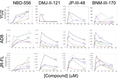
FIG 1Activation of HIV-1 infection of CD4⫺CCR5⫹target cells. Recombinant luciferase-expressing HIV-1 vectors with Envs from HIV-1
YU2, HIV-1AD8, and
HIV-1JR-FLwere incubated with increasing concentrations of the CD4mc at 37°C for the indicated times. The virus-CD4mc mixture was then spinoculated onto
Cf2Th-CCR5 cells at 500⫻gfor 1 h at 25°C. For the 0-min time point, the virus-CD4mc mixture was immediately spinoculated onto Cf2Th-CCR5 cells after mixing. The virus-cell mixtures were cultured for 48 h, after which the cells were lysed and luciferase activity was measured in the cell lysates. The level of viral infection is reported as a percentage of the level seen in the absence of added CD4mc. The results are reported as the means of triplicate measurements and standard deviations derived from 2 to 4 independent experiments.
on November 7, 2019 by guest
http://jvi.asm.org/
[image:4.585.46.545.68.400.2]insight into the stoichiometry of CD4mc-Env trimer binding required for these
pro-cesses. The concentration of the CD4mc and the affinity of the compound for the Env
trimer determine the CD4mc occupancy of the Env protomers on the trimer. At a given
CD4mc occupancy, the percentage of the Env trimers with a given number of bound
CD4mc can be estimated based on a binomial distribution (see Materials and Methods).
By postulating various stoichiometric requirements for virus activation and inhibition,
the hypothetical dose-response curves for these processes can be predicted (Fig. 2A
and B). Both the activation and inhibition curves for each virus variant are dictated by
Env trimer occupancy. If the same virus incubated with a given concentration of CD4mc
is used in both activation and inhibition assays, the Env trimer occupancy will be
identical. A comparison of the experimental dose-response curves for the activation
and inhibition assays, therefore, greatly constrains the possible models. For example,
for each model, the observed CD4mc concentration associated with optimal virus
activation and the 50% viral inhibitory concentration (IC50) are both functions of the
K
d(dissociation constant), which is a fixed property of a CD4mc for a given virus variant.
Thus, the underlying stoichiometric models of activation and inhibition by CD4mc can
be supported or refuted by analyzing the experimental dose-response curves.
We applied this analysis to the activation/inhibition data of the HIV-1
YU2, HIV-1
AD8,
and HIV-1
JR-FLvariants treated with the more potent CD4mc. In every case, the
concentration of CD4mc associated with the optimal level of activation in CD4
⫺CCR5
⫹target cells was similar to the IC
50of the compound for inhibition of infection by the
same virus in CD4
⫹CCR5
⫹cells. The correlation between the CD4mc concentration
resulting in maximal activation and the IC50
of inhibition is shown in Fig. 2C. We
confirmed this relationship when the same preparation of HIV-1
YU2, HIV-1
AD8, and
HIV-1
JR-FLwas incubated with JP-III-48 and then used for infection of CD4
⫺CCR5
⫹cells
and for infection of CD4
⫹CCR5
⫹cells (Fig. 2D). For all three viruses, the optimal
JP-III-48 concentration for activation of virus infection in CD4
⫺CCR5
⫹cells was closely
related to the IC
50for infection of CD4
⫹CCR5
⫹cells. These observations are compatible
with the following models: (i) under the conditions of our assay measuring inhibition of
infection of CD4
⫹CCR5
⫹cells, Envs with two and three CD4mc bound per trimer are
inactivated; (ii) under the conditions of our assay measuring the infection of CD4
⫺CCR5
⫹cells, Envs with one or two CD4mc bound can be activated.
Assuming that the total CD4mc concentration reflects the free CD4mc
concentra-tion, the
K
dvalues of JP-III-48 for the functional Env trimers of HIV-1
YU2, HIV-1
AD8, and
HIV-1JR-FL
are approximately 6 to 10
M, 9 to 10
M, and 40 to 50
M, respectively.
The stoichiometry of CD4mc inhibition of HIV-1 infection was investigated further by
studying recombinant viruses with differing proportions of Envs that are sensitive and
resistant to the CD4mc. The S375W change in HIV-1 Env fills the Phe 43 cavity and
renders HIV-1 resistant to the effects of CD4mc (32, 50). During the course of our
studies, we found that the I309A change in the gp120 V3 variable region resulted in
viruses that were more sensitive to BNM-III-170 than the wild-type HIV-1
JR-FL; the I309A
mutant virus infected Cf2Th-CD4/CCR5 cells with an efficiency similar to that of the
wild-type HIV-1
JR-FL(data not shown). Because the results of the mixing experiment are
interpreted at saturating concentrations of CD4mc, we used the I309A mutant to
facilitate achieving saturation with BNM-III-170. Recombinant HIV-1JR-FL
with differing
proportions of the S375W (CD4mc-resistant) Env and I309A (CD4mc-sensitive) Env were
incubated with increasing concentrations of BNM-III-170 and then used to infect
Cf2Th-CD4/CCR5 cells. The levels of infection observed at saturating concentrations
(typically 30
M and above) of BNM-III-170 are compared in Fig. 3 with the hypothetical
curves expected for the different models of BNM-III-170 inhibition stoichiometry. The
results support a model in which Envs with either two or three of the protomers
occupied by BNM-III-170 are neutralized.
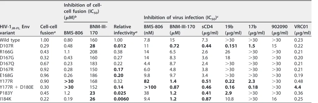
Env reactivity influences sensitivity to CD4mc.
In the experimental system
dis-cussed above, the CD4mc concentration associated with the peak level of activation in
CD4
⫺CCR5
⫹cells and with the IC
50
in CD4
⫹CCR5
⫹cells is closely related to the affinity
on November 7, 2019 by guest
http://jvi.asm.org/
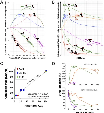
FIG 2Analysis of dose-response curves for CD4mc activation and inhibition. (A) Hypothetical relationship between the probability (P) of a CD4mc occupying an Env protomer and HIV-1 infection of CD4-negative, CCR5-expressing cells (top) and HIV-1 infection of CD4-positive, CCR5-positive cells (bottom) based on a binomial probability distribution. The curves shown are based on models in which the Env trimers with the indicated numbers of bound CD4mc (red ovals) are activated for infection (top) or neutralized (bottom). The protomers of the Env trimer bound by the CD4mc are colored black. (B) The hypothetical relationships in panel A are shown as a function of the free CD4mc concentration. The observation that CD4mc can sensitize HIV-1 to neutralization by antibodies (57–60) renders the model in which Env trimers with one, two, or three bound CD4mc are all neutralized improbable; nonetheless, the hypothetical curve is shown for comparison. (C) Relationship between the IC50associated with the inhibition of
infection of CD4-positive, CCR5-positive cells and the total [CD4mc] associated with maximal activation of infection of CD4-negative, CCR5-expressing cells. The Spearman rank correlation coefficient rS ⫽ 0.9074; two-tailed P ⫽ 0.000046. (D) The indicated
concentrations of JP-III-48 were added to recombinant luciferase-expressing HIV-1 with the HIV-1YU2, HIV-1AD8, and HIV-1JR-FLEnvs.
The virus-CD4mc mixture was immediately split into two portions, with one portion added to Cf2Th-CD4/CCR5 cells (bottom) and the other portion spinoculated onto Cf2Th-CCR5 cells at 500⫻gfor 1 h at 25°C (top). The virus-cell mixtures were returned to 37°C, and the level of infection was determined by measuring luciferase activity in the target cells 48 h later and is shown as a percentage of the level seen in the absence of added JP-III-48. The means and standard deviations of triplicate measurements are shown. Note the correspondence between the IC50associated with inhibition of HIV-1 infection of Cf2Th-CD4/CCR5 cells and the JP-III-48
concentration associated with maximal activation of infection of CD4-negative Cf2Th-CCR5 cells.
on November 7, 2019 by guest
http://jvi.asm.org/
[image:6.585.40.478.71.550.2]of the compound for the functional Env trimer of the particular HIV-1 strain. The CD4mc
apparently interact with the Env trimer in the rank order HIV-1
YU2⬎
HIV-1
AD8⬎
HIV-1
JR-FL. As the CD4mc binding site on the HIV-1 gp120 glycoprotein is well
con-served (32–34, 37–39), what accounts for the differences in sensitivity of these HIV-1
strains? CD4mc, like CD4, induce conformational changes in the Env trimer that
potentially contribute to binding and its consequences (44). Envs from different HIV-1
strains can vary in their propensities to undergo conformational changes, a property
that has been designated Env reactivity (51, 52). We hypothesized that differences in
Env reactivities among HIV-1 strains might contribute to variation in sensitivity to
CD4mc. Some changes in the HIV-1 gp120 V2 region have been shown to result in an
increase in Env reactivity (53–56). We introduced additional changes in the HIV-1
JR-FLgp120 V2 region to identify Env mutants with increased reactivity. Such mutants
typically exhibit increased sensitivity to soluble CD4, CD4mc, and antibodies against
CD4-induced gp120 epitopes; many HIV-1 Env mutants with increased reactivity exhibit
decreased sensitivity to blockers of conformational change, like BMS-806 (17, 51, 54,
56). We identified HIV-1
JR-FLV2 mutants that exhibited phenotypes consistent with
those associated with increased Env reactivity (Table 2) (56). Of note, both syncytium
formation and virus infection mediated by the Y177R, P183Y, and L193A Env mutants
were inhibited more efficiently by BNM-III-170 than the corresponding processes
mediated by wild-type HIV-1
JR-FLEnv. These results are consistent with the hypothesis
that changes in Env reactivity can alter HIV-1 susceptibility to inhibition by CD4mc.
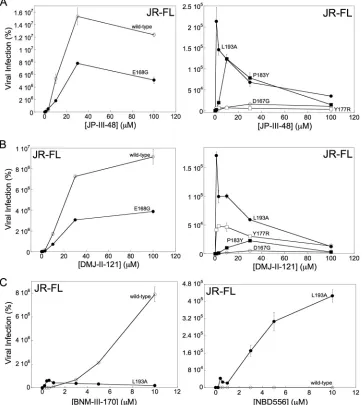
We also evaluated the effects of increased Env reactivity on CD4mc activation of
HIV-1 infection of CD4-negative cells expressing CCR5. We compared the abilities of the
wild-type HIV-1
JR-FLand three V2 mutants (Y177R, P183Y, and L193A) with increased
Env reactivity to infect Cf2Th-CCR5 cells in the absence and presence of the CD4mc
DMJ-II-121 and JP-III-48. We also included two control V2 mutants, D167G and E168G,
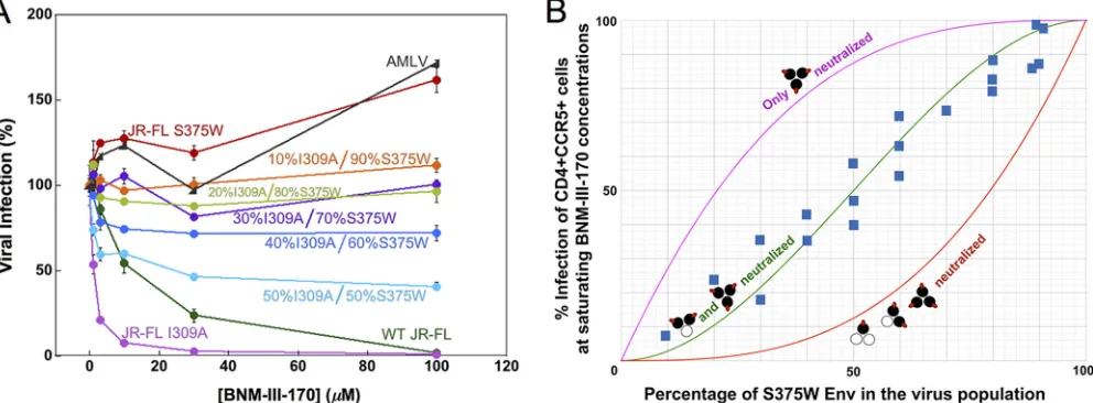
FIG 3Inhibition of viruses with mixed Env variants by a CD4mc. Recombinant HIV-1 containing different ratios of the I309A mutant Env and the S375W mutant Env were produced in 293T cells by transfecting the appropriate molar quantities of the Env expressor plasmids. The infectivities of the I309A and S375W mutant viruses for Cf2Th-CD4/CCR5 target cells were comparable to that of the wild-type HIV-1 (data not shown). The I309A change in the gp120 V3 region slightly increases HIV-1 sensitivity to BNM-III-170 inhibition. The S375W change fills the gp120 Phe 43 cavity, rendering HIV-1 resistant to the CD4mc BNM-III-170. (A) Recombinant luciferase-expressing HIV-1 strains with different ratios of I309A-S375W Envs were used to infect Cf2Th-CD4/CCR5 cells in the presence of increasing concentrations of BNM-III-170. The viruses were incubated with BNM-III-170 for 30 min at 37°C and then added to the Cf2Th-CD4/CCR5 cells. The level of infection 48 h later was determined by measuring luciferase activity in the target cells. The level of infection is reported as a percentage of the level of infection observed for a given virus in the absence of the CD4mc. Recombinant HIV-1 pseudotyped with the AMLV Env was included as a control virus. WT, wild type. The error bars indicate standard deviations. (B) Hypothetical inhibition curves showing the level of HIV-1 infection expected at saturating concentrations of the CD4mc BNM-III-170 as a function of the percentage of the CD4mc-resistant S375W Env in the virus population. The magenta curve represents the hypothetical levels of infection expected if only Envs with all three protomers bound by CD4mc are neutralized, the green curve depicts the hypothetical levels of infection expected if only Envs with two or three protomers bound by CD4mc are neutralized, and the red curve shows the levels expected if the Env is neutralized by any bound CD4mc. The blue squares represent the actual experimental data obtained for mixtures of HIV-1JR-FLS375W and I309A
Envs at saturating concentrations (typically 30M) of BNM-III-170. Each data point represents the results of a single experiment like that shown in panel A and indicates the percentage of the level of infection observed for a given virus at saturating BNM-III-170 concentrations relative to the level of infection seen in the absence of the CD4mc. The results of multiple independent experiments with different virus preparations are shown.
on November 7, 2019 by guest
http://jvi.asm.org/
[image:7.585.47.543.74.257.2]that exhibit phenotypes consistent with Env reactivities close to that of the wild-type
HIV-1
JR-FLEnv. The viruses were incubated with the CD4mc for 30 min at 37°C and then
spinoculated onto Cf2Th-CCR5 cells. As seen above, infection by the wild-type
HIV-1
JR-FLwas activated by JP-III-48, with maximal activation occurring at a compound
concentration of
⬃
30
M (Fig. 4A). Although the overall level of activation by JP-III-48
was less for the E168G mutant, and much lower for the D167G mutant, the maximal
activation of these mutants also occurred at a JP-III-48 concentration of 30
M. Thus,
the apparent affinities of JP-III-48 for the wild-type, D167G, and E168G Envs are
comparable. The levels of infection of Cf2Th-CCR5 cells by the Y177R, P183Y, and L193A
mutants following incubation with JP-III-48 were much lower than those of the
wild-type HIV-1
JR-FL. Importantly, the maximal level of activation of these mutants occurred
at concentrations of JP-III-48 significantly lower than that of the wild-type HIV-1
JR-FL.
Similar results were observed for this panel of HIV-1
JR-FLmutants with DMJ-II-121 (Fig.
4B). For both JP-III-48 and DMJ-II-121, the CD4mc concentration that resulted in
maximal activation of Cf2Th-CCR5 infection exhibited the rank order L193A
⬍
Y177R
⬍
P183Y
⬍
wild-type HIV-1
JR-FL, D167G, and E168G. Apparently, the Env mutants with
increased reactivity bind CD4mc and undergo activation/inactivation at much lower
concentrations of the compounds, likely reflecting a higher affinity of the CD4mc for
these Env trimers.
[image:8.585.42.554.82.248.2]Infection of CD4-negative target cells by the wild-type HIV-1
JR-FLis activated
inef-ficiently by the prototypic CD4mc NBD-556 (Fig. 1). To evaluate whether a more
reactive Env would be more susceptible to activation by NBD-556, we compared the
infection of Cf2Th-CCR5 cells by the wild-type HIV-1
JR-FLand L193A mutant in the
absence or presence of NBD-556. As a control, we also tested the more potent CD4mc,
BNM-III-170, in the assay. Infection of the CD4-negative cells by the wild-type HIV-1
JR-FLprogressively increased with increasing BNM-III-170 concentrations up to 10
M (Fig.
4C). As was seen for JP-III-48 and DMJ-II-121 (see above), the L193A mutant exhibited
a reduced level of infection that peaked at a very low BNM-III-170 concentration.
NBD-556 had minimal effect on the infection of the wild-type HIV-1
JR-FL. In striking
contrast, incubation with NBD-556 resulted in a dramatic increase in infection of the
Cf2Th-CCR5 cells by the L193A mutant. Thus, an increase in Env reactivity can lead to
TABLE 2Phenotypes of HIV-1JR-FLEnv mutantsHIV-1JR-FLEnv variant
Cell-cell fusiona
Inhibition of cell-cell fusion (IC50) (M)b
Relative infectivitya
Inhibition of virus infection (IC50)c
BMS-806
BNM-III-170
BMS-806 (nM)
BNM-III-170 (M)
sCD4 (g/ml)
19b (g/ml)
17b (g/ml)
902090 (g/ml)
VRC01 (g/ml)
Wild type 1.00 0.80 160 1.00 7.8 15 7.3 ⬎30 ⬎30 ⬎30 0.23 D107R 0.29 0.48 28 0.012 11 0.72 0.44 0.151 1.5 15 0.22
R166G 0.43 1.1 208 0.38 14 6.5 2.6 26 ⬎30 ⬎30 0.21
D167G 0.32 0.43 160 0.27 14 8.3 3.6 18 ⬎30 ⬎30 0.20 D167Q 0.67 0.23 183 0.22 4.4 8.7 2.4 ⬎30 ⬎30 ⬎30 0.21 D167R 0.92 0.28 150 0.17 6.0 4.8 3.8 ⬎30 ⬎30 ⬎30 0.21 E168G 0.96 0.26 186 0.20 9.8 9.7 3.4 ⬎30 ⬎30 ⬎30 0.19 Y177R 0.90 >30 168 0.32 82 1.4 0.51 0.22 2.3 ⬎30 0.48 Y177R⫹D180E 0.30 >30 152 0.14 >100 0.87 0.46 0.16 0.18 ⬎30 4.4
P183Y 0.45 1.2 23 0.025 38 1.2 0.41 2.9 ⬎30 ⬎30 0.36 I184K 0.22 0.19 26 0.0060 9.4 1.2 0.87 10.8 ⬎30 16 0.25
aCell-cell fusion and relative infectivity were measured as described in Materials and Methods, with the values normalized to those of the wild-type HIV-1
JR-FLEnv. The
values represent the means of those obtained in at least three independent experiments. Values that differ more than 5-fold from the value of the wild-type HIV-1JR-FLEnv are shown in boldface.
bCell-cell fusion mediated by the indicated HIV-1
JR-FLvariant was tested for inhibition by BMS-806, a blocker of conformational change (17, 56), and a CD4mc,
BNM-III-170. The values represent the means of those obtained in at least two independent experiments. Values in boldface differ from the value of the wild-type HIV-1JR-FL
by more than 5-fold.
cRecombinant HIV-1 pseudotyped with the indicated HIV-1
JR-FLEnv was tested for inhibition by the indicated Env ligands. The 19b antibody recognizes the gp120 V3
region, the 17b antibody recognizes a CD4-induced gp120 epitope, 902090 recognizes a gp120 V2 region epitope, and VRC01 recognizes the CD4-binding site of gp120. The values represent the means of those obtained in at least two independent experiments. Values that differ from the value of the wild-type HIV-1JR-FLEnv
by more than 5-fold are shown in boldface.
on November 7, 2019 by guest
http://jvi.asm.org/
enhanced susceptibility to activation of CD4-independent infection by a weak CD4mc.
These results are consistent with the concept that HIV-1 responsiveness to a CD4mc
depends upon the propensity of Env to undergo conformational changes.
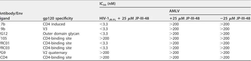
Sensitization of HIV-1 activated by CD4mc to neutralizing antibodies.
During
infection of CD4-positive cells, HIV-1 exposed to subneutralizing concentrations of
CD4mc becomes susceptible to neutralization by a number of antibodies that
recog-nize the CD4-bound Env conformation (57–60). We wished to determine if the virus
activated for infection of CD4
⫺CCR5
⫹cells by CD4mc is susceptible to neutralization
by these antibodies. For this purpose, we incubated HIV-1
JR-FLwith 25
M JP-III-48 for
15 min at 37°C and then incubated the virus-CD4mc mixture with anti-Env antibodies
or sCD4 for an additional 15 min at 37°C. The virus-CD4mc-antibody mixtures were then
spinoculated onto Cf2Th-CCR5 cells, and the efficiency of infection was measured
(Table 3).
In the absence of added JP-III-48, the infection of the Cf2Th-CCR5 cells by HIV-1
JR-FLwas very inefficient, as expected. This low basal level of CD4-independent HIV-1
JR-FLFIG 4Influence of Env reactivity on HIV-1 sensitivity to CD4mc. The HIV-1JR-FLvariants were incubated with the
indicated concentrations of CD4mc, and the virus-compound mixture was immediately spinoculated onto Cf2Th-CCR5 cells. After 48 h at 37°C, the cells were lysed, and the cell lysates were assayed for luciferase activity. The results represent the mean values from triplicate data points and standard deviations and are reported as the level of luciferase activity relative to that seen in the absence of the CD4mc. The results of a typical experiment are shown; the experiment was repeated with similar results.
on November 7, 2019 by guest
http://jvi.asm.org/
[image:9.585.42.404.76.481.2]infection was minimally affected by the addition of antibodies (data not shown). After
incubation with 25
M JP-III-48, HIV-1JR-FL
infection of Cf2Th-CCR5 cells was
dramati-cally enhanced, and this activated infection was efficiently inhibited by the 17b, 19b,
2G12, VRC01, and VRC03 antibodies (Table 3). JP-III-48-activated HIV-1JR-FL
infection was
not inhibited by the F105 antibody, a weakly neutralizing antibody directed against the
gp120 CD4-binding site. JP-III-48-activated infection of the Cf2Th-CCR5 cells was also
not inhibited by the PG9 antibody, which does not recognize HIV-1
JR-FL(61). These
results demonstrate that the CD4mc-activated infection of CD4
⫺CCR5
⫹cells by HIV-1
can be efficiently neutralized by broadly neutralizing antibodies like 2G12, VRC01, and
VRC03, as well as poorly neutralizing antibodies like 17b and 19b that recognize gp120
epitopes induced by CD4mc and CD4.
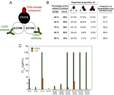
[image:10.585.44.544.83.205.2]To investigate whether the binding of a CD4mc to one Env protomer might expose
neutralizing epitopes on adjacent protomers, we tested the ability of the CH23
anti-body to inhibit the infection of Cf2Th-CCR5 cells by viruses with I309A-S375W and
F317A-S375W Env mixtures. The F317A virus has a change in the gp120 V3 region that
makes it resistant to neutralization by the CH23 antibody, which recognizes a V3
epitope (62). The I309A change in V3 does not affect sensitivity to the CH23 antibody,
so the I309A Env was included as a control in these experiments. Unlike the S375W
mutant, the I309A and F317A mutants can be activated by CD4mc. In the experiments,
we tested viruses with a high ratio of the S375W Env relative to the I309A or F317A Env.
Under these conditions, nearly all of the viruses capable of infecting CD4-negative
Cf2Th-CCR5 cells should contain mixed Envs with at least one CD4mc-activatable,
CH23-resistant Env protomer and at least one CD4mc-resistant, CH23-sensitive Env
protomer. Therefore, if the binding of a CD4mc to one Env protomer contributes to the
exposure of the CH23 epitope on an adjacent Env protomer, a substantial fraction of
the viruses should be neutralized by the CH23 antibody. The design of the experiment
is summarized in Fig. 5A and B, and the results are shown in Fig. 5C. The infection of
the Cf2Th-CCR5 cells by the S375W mutant was activated by sCD4, but not by the
CD4mc BNM-III-170, as expected (data not shown). Infection of the Cf2Th-CCR5 cells by
the BNM-III-170-activated I309A mutant was blocked by the CH23 antibody and by
another V3-directed antibody, 19b. In contrast, infection of the Cf2Th-CCR5 cells by the
BNM-III-170-activated F317A mutant virus was not inhibited by the CH23 antibody.
Notably, the CH23 antibody did not inhibit the BNM-III-170-activated infection of
Cf2Th-CCR5 cells by viruses with high proportions of the S375W mutant relative to the
F317A mutant Envs. In contrast, the CH23 antibody neutralized the infection of
Cf2Th-CCR5 cells by viruses with mixtures of the S375W and I309A Envs. Therefore, in this
experimental setting, we did not find evidence for the binding of BNM-III-170 to an Env
protomer sensitizing an adjacent Env protomer to CH23 binding and virus
neutraliza-tion.
TABLE 3Antibody neutralization of JP-III-48-activated HIV-1JR-FLinfection of CD4-negative Cf2Th-CCR5 cellsa
Antibody/Env
ligand gp120 specificity
IC50(nM)
HIV-1JR-FLⴙ25M JP-III-48
AMLV
ⴙ25M JP-III-48 ⴚ25M JP-III-48
17b CD4 induced ⬍3.3 ⬎200 ⬎200
19b V3 ⬍3.3 ⬎200 ⬎200
2G12 Outer domain glycan ⬍3.3 ⬎200 ⬎200
F105 CD4-binding site ⬎200 ⬎200 ⬎200
VRC01 CD4-binding site ⬍3.3 ⬎200 ⬎200
VRC03 CD4-binding site ⬍3.3 ⬎200 ⬎200
PG9 V2 quaternary ⬎200 ⬎200 ⬎200
sCD4 CD4-binding site ⬎200 ⬎200 ⬎200
aHIV-1 pseudotyped with the HIV-1
JR-FLor AMLV Env was incubated with 25M JP-III-48 for 5 min at 37°C and then for 15 min at 37°C with different concentrations
of antibodies or sCD4. The virus–JP-III-48 –antibody mixtures were then spinoculated onto Cf2Th-CCR5 cells. After 48 h at 37°C, the cells were lysed, and the cell lysates were assayed for luciferase activity. The mean values for luciferase activity were derived from triplicate samples. The level of luciferase activity relative to that seen in the absence of antibody or sCD4 was used to calculate the level of infection. The IC50s of the antibodies and sCD4 are reported.
on November 7, 2019 by guest
http://jvi.asm.org/
Activation of Env-mediated syncytium formation with CD4
ⴚCCR5
ⴙtarget
cells.
We investigated the activating/inactivating mechanism of Env inhibition by the
CD4mc in a second experimental system where competitive inhibition of CD4 binding
does not contribute to the antiviral effects of the compounds. In this experimental
system, HIV-1 Env-expressing cells are incubated with CD4mc and then assessed for the
ability to fuse with CD4-negative cells expressing CCR5. 293T cells transiently
express-ing different HIV-1 Envs were cocultivated with CD4-negative Cf2Th-CCR5 cells in the
absence or presence of increasing concentrations of various CD4mc. In the absence of
CD4mc, no syncytium formation was detected, consistent with the CD4 dependence of
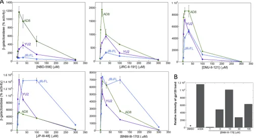
the HIV-1 Envs tested. All of the CD4mc tested exhibited some ability to activate Env
function in the assay (Fig. 6A). The indane analogues (DMJ-II-121, JP-III-48, and
BNM-III-170), which are more potent inhibitors of HIV-1 infection than the piperidine
ana-logues (NBD-556 and JRC-II-191), generally enhanced cell-cell fusion more efficiently.
The HIV-1
JR-FLEnv exhibited minimal levels of activation by the low-potency CD4mc,
NBD-556 and JRC-II-191, but was activated by the potent CD4mc, BNM-III-170 and
JP-III-48, even more efficiently than HIV-1
YU2and HIV-1
AD8Envs. The dose-response
FIG 5Test of CD4mc cross-protomer sensitization of recombinant HIV-1 to antibodies. (A) Env mixing strategy to test whether the binding of a CD4mc to one protomer of the Env trimer induces the exposure of the gp120 V3 region on an adjacent protomer. Mixed Env trimers were prepared from the S375W mutant, which does not bind the CD4mc BNM-III-170 but can be recognized by the CH23 anti-V3 antibody, and the F317A mutant, which can bind the CD4mc but is resistant to the CH23 antibody. (B) Mixed Envs with the indicated proportions of S375W and F317A mutant Envs are predicted to have the percentages of each Env mixture shown. The predicted percentage of the virus fraction that can potentially infect CD4-negative Cf2Th-CCR5 cells after BNM-III-170 treatment and that can potentially be neutralized by the CH23 antibody is shown in the right-hand column. This percentage is based on the assumption that binding of a single CH23 antibody neutralizes the HIV-1 Env trimer. (C) Recombinant viruses with the indicated ratio of I309A-S375W Envs or F317A-S375W Envs were incubated with 30M BNM-III-170 and increasing concentrations of the CH23 or 19b anti-V3 antibody for 30 min at 37°C and then added to Cf2Th-CCR5 cells. Recombinant viruses with the wild-type HIV-1JR-FLEnv or I309A, F317A, and S375W Envs were tested in
parallel. The level of infection after 48 h was determined by measuring luciferase activity in the target cells. The level of infection was normalized to that seen in the absence of antibody, and the IC50of the antibody was calculated.*, no infection
was detected above the assay background.
on November 7, 2019 by guest
http://jvi.asm.org/
[image:11.585.43.439.69.398.2]curves for activation of syncytium formation were biphasic; this implies that at lower
levels of occupancy of the Env trimer, activating events predominate, whereas at a
higher occupancy level, events detrimental to Env function occur.
One potential explanation for the biphasic activation curves is that, as Env
occu-pancy increases at higher concentrations, the CD4mc induce shedding of gp120 from
the Env trimer. To test this hypothesis, we incubated cells expressing the HIV-1JR-FL
Envs
with different concentrations of BNM-III-170 at 37°C for 24 h. As a positive control, the
cells were incubated with 20
g/ml sCD4 at 37°C for 24 h. The cell supernatants were
collected and precipitated with a polyclonal mixture of sera from HIV-1-infected
individuals. The precipitates were analyzed by SDS-PAGE, followed by autoradiography
(Fig. 6B). Compared with the dimethyl sulfoxide (DMSO) negative control, incubation
with sCD4 resulted in an increase in the gp120 concentration in the cell supernatants.
A dose-dependent increase in the amount of gp120 in the supernatant was observed
for BNM-III-170. These results suggest that BNM-III-170 increases gp120 shedding from
the Env trimer.
Kinetics of CD4mc interaction with Env.
In the experiments testing the activation
of HIV-1 infection of CD4-negative Cf2Th-CCR5 cells (Fig. 1), we also examined the effect
of the length of the 37°C incubation of the CD4mc with the virus. In general, longer
incubation times affected the infection efficiencies similarly to increasing the
concen-tration of CD4mc. In most cases, longer incubation of the CD4mc with virus resulted in
lower levels of infection (Fig. 1). However, in some cases (e.g., NBD-556 incubation with
HIV-1
JR-FL), longer incubation times were associated with increased levels of infection.
These results can be understood in light of the proposed biphasic relationship between
FIG 6Activation by CD4mc of Env-mediated syncytium formation with CD4⫺CCR5⫹cells. (A) 293T cells expressing the HIV-1 Env and the␣peptide of -galactosidase were cocultivated with CD4-negative, CCR5-positive Cf2Th-CCR5 cells expressing thepeptide of-galactosidase in medium containing the indicated concentrations of CD4mc. After 6 h at 37°C, the cells were lysed, and the lysates were assayed for-galactosidase activity. The results are reported as the percentage of-galactosidase activity compared with that seen in the absence of added CD4mc. The results of a single experiment, with each point done in duplicate/triplicate, are shown. The experiment was repeated with comparable results. The error bars indicate standard deviations. (B) 293T cells expressing the HIV-1JR-FLEnv were radiolabeled with [35S]cysteine and [35S]methionine in medium alone or in the presence of DMSO, 20g/ml sCD4, or different
concentrations of BNM-III-170 for 24 h at 37°C. The cell supernatants were then used for precipitation by a polyclonal antiserum from an HIV-1-infected individual. The precipitated gp120 was analyzed by SDS-PAGE, autoradiography, and densitometry. The background shedding of gp120 in the untreated control was subtracted from all of the other values.*, value less than or equal to the background of the assay.
on November 7, 2019 by guest
http://jvi.asm.org/
[image:12.585.42.549.75.348.2]the amount of CD4mc bound per Env trimer and the level of activation of infection of
the CD4-negative Cf2Th cells. When the affinity of the CD4mc for the Env trimer is low
(as in the cases of NBD-556 and HIV-1
JR-FL), longer incubation times would favor
increased occupancy of the Env trimer and greater activation of infection. When the
affinity of the CD4mc for the Env trimer is higher, the increased levels of trimer
occupancy resulting from longer incubation times can result in decreases in the level
of infection (e.g., DMJ-II-121 or JP-III-48 and HIV-1
AD8).
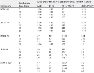
[image:13.585.40.371.94.350.2]The observed differences in the degree of activation for longer incubation periods
could reflect differences in the on rates of binding of the CD4mc to the Env trimer. We
used the activation of cell-cell fusion by the CD4mc to examine this possibility. Cells
expressing the wild-type HIV-1JR-FL, HIV-1JR-FL
Y177R, HIV-1JR-FL
P183Y, and AMLV Envs
were incubated at 37°C for various lengths of time with different CD4mc or the control
DMSO. The cells were then washed and cocultivated with Cf2Th-CCR5 cells, and
syncytia were scored. Thus, the assay requires the CD4mc to bind Env within the period
of incubation and to maintain Env binding through the wash. Under the conditions of
the assay, the cell-cell fusion activities of the wild-type HIV-1
JR-FLand HIV-1
JR-FLP183Y
Envs were minimally activated. In contrast, the more reactive HIV-1JR-FL
Y177R Env
exhibited marked activation of syncytium-forming ability by all of the CD4mc. The level
of activation of Y177R Env was greater for the more potent CD4mc, DMJ-II-121, JP-III-48,
and BNM-III-170, than for the less potent NBD-556 and JRC-II-191 (Table 4). Of note, the
level of activation increased with longer incubation periods of NBD-556 and JRC-II-191,
whereas the more potent CD4mc achieved high levels of activation within the 5-min
incubation period. These results are consistent with higher on rates for the more potent
CD4mc.
TABLE 4Activation of fusion between Env-expressing cells and CD4-negative, CCR5-positive cells
Compound
Incubation time (min)
Area under the curve (arbitrary units) for HIV-1 Enva:
AD8 JR-FL JR-FL Y177R JR-FL P183Y
NBD-556 5 ⬍10 ⬍10 36 ⬍10
10 ⬍10 ⬍10 23 ⬍10
30 ⬍10 ⬍10 106 ⬍10
60 ⬍10 ⬍10 105 ⬍10
JRC-II-191 5 ⬍10 ⬍10 140 ⬍10
10 ⬍10 ⬍10 272 ⬍10
30 ⬍10 ⬍10 411 ⬍10
60 ⬍10 ⬍14 285 ⬍10
DMJ-II-121 5 47 161 1,139 354
10 94 171 659 335
30 32 19 679 146
60 ⬍10 ⬍10 168 12
JP-III-48 5 59 59 877 12
10 69 12 884 29
30 25 30 1,175 19
60 20 29 746 15
BNM-III-170 5 47 74 1,567 76
10 37 77 1,095 90
30 66 27 1,334 58
60 14 27 872 60
aDifferent concentrations (0 to 100M) of the CD4mc were incubated with 293T cells expressing HIV-1 Env
variants and the␣peptide of-galactosidase at 37°C for the indicated times. The cells were then washed and cocultivated with CD4-negative CCR5-positive Cf2Th-CCR5 cells expressing thepeptide of -galactosidase at 37°C in medium without the CD4mc. After 6 h, the extent of cell-cell fusion was assessed by measuring-galactosidase activity in the cell lysates. The-galactosidase activity was plotted as a function of the CD4mc concentration, and the area under the curve was measured for each time point. The results of a typical experiment out of two independent experiments are shown.
on November 7, 2019 by guest
http://jvi.asm.org/
DISCUSSION
In this work, we investigated the mechanisms underlying the interactions of CD4mc
analogues of varying potencies with the functional Envs from different HIV-1 variants.
The rank order of potency of CD4mc and the rank order of susceptibility of HIV-1
variants were similar in three different contexts: activation of Env-mediated syncytium
formation, activation of virus entry into CD4
⫺CCR5
⫹cells, and inhibition of HIV-1 entry
into CD4
⫹CCR5
⫹cells. These observations are consistent with the affinity of the
CD4mc for the Env trimer being a major determinant of the efficiency of these
processes. The results also underscore the contribution of premature Env activation
leading to functional inactivation as an important antiviral mechanism of all CD4mc.
When competitive inhibition of CD4 binding is eliminated as a possible mechanism (for
example, in experimental systems where CD4
⫺CCR5
⫹target cells are used), the
biphasic nature of the relationship between the concentration of CD4mc and activation
of infection or syncytium formation becomes apparent. The experimental
dose-response curves correspond closely to theoretical predictions of CD4mc-Env trimer
binding based on a binomial probability distribution. The models suggest that
occu-pancy of one or two protomers of the Env trimer by a CD4mc can lead to entry
activation (Fig. 7). Note that in these models, Envs with two CD4mc bound per trimer
are either active or inactive, depending on the assay conditions. Envs with two
protomers occupied by a CD4mc can support infection if spinoculated onto CD4
⫺CCR5
⫹cells. This allows the activated Env trimer to engage CCR5 efficiently.
However, under conditions where the CD4mc-bound virus diffuses slowly to the
target cell, such viruses may undergo inactivation. Moreover, in target cells
ex-pressing CD4 and CCR5, viruses with two CD4mc bound per trimer infect
ineffi-ciently compared with untreated viruses. Even if these viruses remain infectious
upon reaching the target cell, blockade of the interaction with cellular CD4 inhibits
virus entry. Although the bound CD4mc can supplant host cell CD4 with respect to
Env activation, CD4mc do not anchor and orient the virus particle in the manner
expected for cellular CD4.
Occupancy of all three Env protomers by a CD4mc does not typically lead to
FIG 7Effects of stoichiometry of CD4mc binding to the HIV-1 Env trimer on activation and inactivation. The proposed effect of binding of the CD4mc (red oval) to the subunits of the HIV-1 Env trimer is depicted. In all cases, the binding of a single CD4mc to the Env trimer leads to activation, and the binding of three CD4mc leads to inactivation. The binding of two CD4mc to the Env trimer leads to virus activation of infection in CD4-negative, CCR5-positive cells but results in virus inactivation during infection of CD4-positive, CCR5-positive cells.
on November 7, 2019 by guest
http://jvi.asm.org/
[image:14.585.48.361.67.292.2]successful infection of CD4
⫺CCR5
⫹cells and apparently is inactivating under these
experimental conditions. A previous study has shown that, when HIV-1 is distant from
a potential target cell, Env inactivation by even modestly potent CD4mc occurs rapidly,
at a rate comparable to that which follows the binding of sCD4 (44). The transient
opening and spontaneous closure of the gp41 HR1 hydrophobic groove has been
shown to be correlated temporally with Env inactivation by CD4mc (44), supporting a
model in which Env conformations induced by CD4mc are functional only transiently.
Moreover, in some of the CD4mc-induced conformations, Env subunit association is
apparently weakened, resulting in gp120 shedding from the Env trimer. Because the
ability of CD4mc to both compete with CD4 and induce viral activation/inactivation is
a function of occupancy of the protomers of the Env trimer, the affinity of the CD4mc
for the Env trimer is a major determinant of the antiviral potency of these compounds.
The on rates of CD4mc binding are critical determinants of the binding affinity of
these compounds for the functional Env trimer. As might be expected for any
ligand-protein interaction, the enthalpic contributions of an increased number of bonds
between the CD4mc and gp120 result in higher affinity and increased potency (32–41).
However, because CD4mc induce conformational changes in Env in the process of
binding, the CD4mc-Env binding affinity is strongly influenced by the propensity of the
Env trimer to undergo conformational changes (i.e., Env reactivity) (51, 52). The HIV-1
Env trimer is maintained in its unliganded state (state 1) by multiple inter- and
intrasubunit interactions, thus averting premature triggering and inactivation. Changes
in particular gp120 and gp41 residues apparently release these restraints and lower the
energy barriers that separate state 1 from downstream conformations (56). For
exam-ple, alteration of several residues in the gp120 V2 region resulted in more reactive Envs
that exhibited increased sensitivity to both activation and inhibition by CD4mc. Of note,
changes in V2 residues 166 to 168, which are predicted by several available HIV-1 Env
structures to be involved in key interactions at the trimer apex (63–66), exerted
relatively little impact on HIV-1 phenotypes. These observations raise questions about
the relationship of these structures to functional Env states, particularly state 1. Further
work will be required to understand the structural restraints that maintain the HIV-1 Env
trimer in state 1 and resist the binding of CD4mc and consequent induction of
conformational changes. Such restraints operate much less efficiently in the context of
free monomeric gp120, which binds the potent CD4mc at least 2 orders of magnitude
better than our estimates for the interaction of CD4mc with the functional Env trimer
(31, 35–39).
Lastly, we established assays to evaluate whether the binding of a CD4mc to one
Env protomer can influence the conformation of the other Env protomers. This
phe-nomenon has been recently documented for sCD4 (X. Ma, A. Herschhorn, J. D. Ventura,
J. R. Grover, D. S. Terry, J. Gorman, X. Hong, Z. Zhou, H. Zhao, R. B. Altman, A. B. Smith
III, J. Arthos, P. D. Kwong, J. Sodroski, S. C. Blanchard, W. Mothes, and J. B. Munro,
unpublished data), but we did not find evidence that subneutralizing concentrations of
CD4-mimetic molecules sensitize adjacent Env protomers to neutralization by
antibod-ies (57–60). Apparently, the conformational changes induced in the HIV-1 Env trimer by
sCD4 and CD4mc are not identical. An understanding of these conformational changes
and how they contribute to virus entry or inhibition should help to optimize the
potency and utility of the CD4mc.
MATERIALS AND METHODS
Compounds.The CD4-mimetic compounds were synthesized and the chemical structures were characterized as described previously (31–39). The compounds were dissolved in dimethyl sulfoxide at a stock concentration of 10 to 20 mM, aliquoted, and stored at⫺20°C. Each compound was then diluted to 1 mM in serum-free Dulbecco’s modified Eagle medium (DMEM) and used for different assays.
HIV-1 Env variants.The wild-type HIV-1YU2, HIV-1AD8, and HIV-1JR-FLEnvs were expressed by the
pSVIIIenv plasmid (32). Mutations were introduced into the pSVIIIenv plasmid expressing the HIV-1JR-FL
Env using the QuikChange II site-directed mutagenesis protocol (Stratagene). The presence of the desired mutations was confirmed by DNA sequencing. All Env residues are numbered according to convention based on the HXBc2 prototypic sequence (67).
on November 7, 2019 by guest
http://jvi.asm.org/
Cell lines.293T human embryonic kidney and Cf2Th canine thymocytes (ATCC) were grown at 37°C and 5% CO2in DMEM (Invitrogen) containing 10% fetal bovine serum (Sigma) and 100g/ml
penicillin-streptomycin (Mediatech, Inc.). Cf2Th cells stably expressing human CCR5 and CD4 were grown in medium supplemented with 0.4 mg/ml G418 and 0.2 mg/ml hygromycin (Invitrogen). Cf2Th-CCR5 cells, which stably express human CCR5, were grown in medium supplemented with 0.4 mg/ml G418. The cytotoxicity of two of the more potent CD4-mimetic compounds, JP-III-48 and BNM-III-170, for Cf2Th-CD4/CCR5 cells was tested with the CellTiter-Glo luminescent cell viability assay (Promega).
Recombinant luciferase viruses. 293T human embryonic kidney cells were cotransfected with plasmids expressing the pCMVΔP1Δenv HIV-1 Gag-Pol packaging construct, the HIV-1 envelope glyco-proteins, or the envelope glycoprotein of the control AMLV and the firefly luciferase-expressing vector at a DNA ratio of 1:1:3g using the Effectene transfection reagent (Qiagen). For the production of the AMLV-pseudotyped viruses, a plasmid expressing HIV-1 Rev was added. Cotransfection produced re-combinant luciferase-expressing viruses capable of a single round of infection. The virus-containing supernatants were harvested 36 to 40 h after transfection, spun, aliquoted, and frozen at⫺80°C until further use. The reverse transcriptase (RT) levels of all virus stocks were measured as described previously (68).
Infection by single-round luciferase viruses.Cf2Th-CD4/CCR5 or Cf2Th-CCR5 target cells were seeded at a density of 6⫻ 103 cells/well in 96-well luminometer-compatible tissue culture plates
(PerkinElmer) 24 h before infection. On the day of infection, CD4mc (0 to 100M) were incubated with recombinant viruses (10,000 RT units) at 37°C for different lengths of time. In the case of sensitization assays, a constant concentration of compounds was incubated with virus at 37°C for the time indicated; then, antibodies (over a range of 0 to 200 nM) were added to the virus-compound mixture and incubated for an additional period at 37°C. The mixtures were then added to the target cells; for infections of Cf2Th-CCR5 target cells, the mixtures containing the recombinant viruses were spinoculated onto the cells at 500⫻gfor 1 h at 25°C. The target cells were then incubated at 37°C for 48 h. Afterward, the medium was removed from each well, and the cells were lysed by the addition of 30l passive lysis buffer (Promega) and three freeze-thaw cycles. An EG&G Berthold Microplate Luminometer LB 96V was used to measure the luciferase activity of each well after the addition of 100l of luciferin buffer (15 mM MgSO4, 15 mM KPO4, pH 7.8, 1 mM ATP, and 1 mM dithiothreitol) and 50l of 1 mM fireflyD-luciferin
free acid 99% (Prolume).
Cell-cell fusion (syncytium formation) assay.293T cells expressing the HIV-1 Env and the␣peptide of-galactosidase were cocultivated with either CD4-positive, CCR5-positive Cf2Th-CD4/CCR5 cells or CD4-negative, CCR5-positive Cf2Th-CCR5 cells expressing thepeptide of-galactosidase, as described previously (69). In some cases, CD4mc were added to the medium. After 6 h at 37°C, the cells were lysed, and the lysates were assayed for-galactosidase activity.
Modeling HIV-1 activation and neutralization by CD4mc.The dose-response curves for CD4mc activation of HIV-1 infection of CD4⫺CCR5⫹cells and neutralization of HIV-1 infection of CD4⫹CCR5⫹
target cells were analyzed as follows. The binding of CD4mc to the three protomers of the Env trimer was modeled using a binomial distribution:
b(x,P)⫽3Cx⫻Px(1⫺P)3⫺x (1)
wherebis the binomial probability of an Env trimer having the given number of bound CD4mc,xis the total number of protomers on the Env trimer bound by a CD4mc (x⫽0, 1, 2, or 3),Pis the probability of the CD4mc occupying an individual Env protomer, and3Cxis the 6/x!(3⫺x)! (3C0⫽1,3C1⫽3,3C2⫽3, and
3C3⫽1).
This binomial distribution model assumes that each binding event of the CD4mc to the Env trimer occurs independently of the others (i.e., the binding of one CD4mc does not change the probability that a second CD4mc binds to an adjacent protomer).
We considered four different models for activation of HIV-1 infection of CD4⫺CCR5⫹cells that differ inx. In this case,xrepresents the stoichiometry of the CD4mc with respect to the Env trimer that results in activation of infection of the CD4⫺CCR5⫹cells:
b(x⫽1)⫽3P(1⫺P)2 (2)
b(x⫽2)⫽3P2(1⫺P) (3)
b(x⫽1 or 2)⫽3P(1⫺P) (4)
b(x⫽3)⫽P3 (5)
Only the first three models are compatible with the biphasic dose-response curves observed, so the model in which only a fully occupied Env trimer is activated (x⫽3) (equation 5) was not considered further.
We also considered three models for pure neutralization of HIV-1 infection of CD4⫹CCR5⫹cells that
differ in x, the stoichiometry of the CD4mc required for neutralization of the Env trimer. Here, b represents the probability of infection in the presence of the CD4mc:
b(x⫽1, 2, or 3)⫽(1⫺P)3 (6)
b(x⫽2 or 3)⫽2P3⫺3P2⫹1 (7)
b(x⫽3)⫽1⫺P3 (8)
The binomial probabilities for each of the models of HIV-1 activation and neutralization are shown in Fig. 2A as a function ofP, the probability of the CD4mc occupying an individual Env protomer.
on November 7, 2019 by guest
http://jvi.asm.org/
Pis related to the free concentration of CD4mc, [CD4mc], and the dissociation constant of the CD4mc for the Env trimer,Kd, as follows:
P⫽ [CD4mc] [CD4mc]⫹Kd
(9)
Therefore, combining equation 9 with equations 2, 3, and 4, the probability of HIV-1 infection of CD4⫺CCR5⫹cells can be expressed as a function of the free concentration of CD4mc:
b(x⫽1)⫽ 3Kd 2
[CD4mc] ([CD4mc]⫹Kd)3
(10)
b(x⫽2)⫽ 3Kd[CD4mc]
2
([CD4mc]⫹Kd)3
(11)
b(x⫽1 or 2)⫽ 3Kd[CD4mc] ([CD4mc]⫹Kd)2
(12)
Likewise, combining equation 9 with equations 6, 7, and 8, the probability of HIV-1 infection of CD4⫹
CCR5⫹cells in the presence of the CD4mc can be expressed as a function of the free concentration of
CD4mc:
b(x⫽1, 2, or 3)⫽
冉
Kd [CD4mc]⫹Kd冊
3
(13)
b(x⫽2 or 3)⫽1⫺3
冉
[CD4mc] [CD4mc]⫹Kd冊
2
⫹2
冉
[CD4mc] [CD4mc]⫹Kd冊
3
(14)
b(x⫽3)⫽1⫺
冉
[CD4mc][CD4mc]⫹K d冊
3
(15)
The binomial probabilities for each of the models of HIV-1 activation and neutralization as a function of the free [CD4mc] are shown in Fig. 2B. Note that, depending on the model for virus activation, maximal activation occurs at different CD4mc concentrations that represent 0.5Kd,Kd, or 2Kd. Likewise, depending
on the model for virus neutralization, the IC50of the CD4mc is 0.26Kd,Kd, or 3.85Kd. Thus, for a given
virus and CD4mc, comparison of the CD4mc concentrations resulting in maximal activation of infection of CD4⫺CCR5⫹cells and in 50% neutralization of infection of CD4⫹CCR5⫹cells should indicate which
models best explain the experimental observations.
Should cooperative binding of CD4mc to the Env trimer occur, the dose-response curves associated with the binding of two or more CD4mc to the Env trimer would shift leftward, but the basic relationships among the curves predicted by the various models would remain the same.
Infections with recombinant-HIV-1-containing mixtures of Env mutants.The stoichiometry of CD4mc inhibition of infection was investigated further by testing viruses with differing proportions of sensitive and resistant Envs. Recombinant HIV-1JR-FLviruses with mixtures of an Env (S375W) that is
resistant to CD4mc and an Env (I309A) that is sensitive to CD4mc were produced as described above, varying the molar quantities of the plasmids expressing the Env mutants. The recombinant viruses were used to infect CD4-positive, CCR5-positive Cf2Th-CD4/CCR5 cells after incubation with saturating con-centrations of BNM-III-170. In this case, the probability (b) of infection in the presence of saturating concentrations of BNM-III-170 depends uponx, the stoichiometry of the CD4mc required to inhibit virus infection, and upon z, the proportion of the resistant Env in the population. Thus, at saturating concentrations of BNM-III-170:
b(x⫽1, 2, or 3)⫽z3 (16)
b(x⫽2 or 3)⫽3z2⫺2z3 (17)
b(x⫽3)⫽1⫺(1⫺z)3 (18)
The binomial probabilities for each of these three models of HIV-1 inhibition by CD4mc as a function ofz(the percentage of S375W Env in the virus population) are shown in Fig. 3B.
For studies of CD4mc-induced sensitization of HIV-1 to antibody neutralization, viruses with mixed Envs were generated as described above. Recombinant HIV-1JR-FLviruses with mixtures of an S375W Env
and an F317A Env were produced. The S375W Env is resistant to CD4mc but potentially sensitive to neutralization by the CH23 anti-V3 antibody (62); the F317A Env is resistant to the CH23 antibody but can be activated by CD4mc. Recombinant viruses with various proportions of mixed Envs were incubated with 30M BNM-III-170 and different concentrations of the CH23 antibody for 30 min at 37°C. The virus-CD4mc-antibody mixtures were then added to CD4-negative, CCR5-positive Cf2Th-CCR5 cells. After 48 h, the cells were lysed and the lysates were assayed for luciferase activity.