International Journal of Innovative Technology and Exploring Engineering (IJITEE) ISSN: 2278-3075, Volume-8 Issue-10, August 2019
Abstract: The integration of proper algorithms and computer graphics-based systems seems promising for the design of biomechanical models and the relative motion analysis. Thus, consequences on research fields as gait analysis are gathered, focusing on joints kinematics. Human motion patterns are indeed directly influenced from human model and associated joints parameters, such as centers and axes of rotation. These, as a matter of fact, determine the body segments coordinates systems. Joints parameters are estimated with several methods. The aim of this research is to evaluate the consistency of a functional approach versus a the predictive one. A validation of the algorithm used to estimate the lower limbs joints centers in gait analysis is provided with a proper subject-specific multibody model implemented in OpenSim space. Joints angles are estimated using a global optimization method and a comparison with the gold standard technique is also discussed. Overall the obtained results are consistent for the two different methodologies. The correlation of the curves is excellent in the sagittal plane, and very good in the coronal and transversal plane.
Index Terms: biomechanics, functional methods, gait analysis, motion analysis, global optimization method, joints.
I.INTRODUCTION
One of the research fields of applied biomechanics, such as the quantitative human motion analysis allows to compute joint kinematics, as well dynamics. Particularly, an accurate definition of body segments coordinates systems is required due to the emphasis set in gait analysis on the relative motion between body segments, hence on joint angles. Thus, joints parameters estimation like axes or centers of rotation (AoRs and CoRs) is a crucial issue, considering how these ones highly influence human motion patterns. As a consequence, articular joint model within its parameters are important to assess what is not directly measurable in vivo [1], [2], [3]. Moreover, an accurate estimation of joints parameters becomes necessary especially for people affecting by some pathologies or presenting physical deformities directly compromising motion tasks, as well kinematics outcomes [4], [5].
Joint parameters can be estimated according to different approaches [6], [7]. The laboratories of measurements and human movement analysis mostly adopt predictive methods, based on regression equations .[8],[9]. This approach
Revised Manuscript Received on August 05, 2019.
Giulia Lisco, Stefano Pastorelli Department of Mechanical and Aerospace Engineering, Politecnico di Torino, Torino, Italy.
Laura Gastaldi, Department of Mathematical Sciences, PolitoBIOMed Lab, Politecnico di Torino, Torino, Italy.
requires specific protocols for markers positioning on anatomical landmarks [10], resulting in a main drawback, due to the not often easily identification. Despite the easy-of-use, this method may lack of accuracy, resulting in several errors [2], [7], particularly with nonstandard cases. Thus, functional methods are proposed to remedy problems and limits affecting joints parameters estimation with the former approach. They do not refer to empirical relations and are just based on the relative motion between two body segments with respect to the considered joint. To this end, customized markers-based protocols are considered in order to define the relative segments position and orientation, not requiring the palpation of anatomical landmarks and by using just 3-markers set at least on each body segment. Is clear how these approaches may be suitable also with nonstandard cases, despite the analytical complexity. Several strategies are proposed in literature, providing a 2-categories classification of functional methods in fitting approaches and coordinates transformation techniques. The former are variants of the sphere-fitting method [11], where each marker can rotate around the same joint axes or center of rotation with a separate arc [12],[13], without a body rigid assumption. The second which considers instead each body segment as a rigid one, in order to determine a local coordinate system and rigid transformations into a global reference frame [14]
Functional methods are nevertheless based on several combinations of kinematical and geometrical constraints, as well optimization techniques, providing good results in certain conditions [15], [16], e.g. with a proper range of motion (RoM). Several studies reported in literature [11], [15] underline that functional methods provide more accurate results respect to the predictive approach, although both are influenced from the soft tissue artifact (STA), the main source of error which affects kinematics in human movement analysis [7]. Once three functional methods [12], [16] have been discussed, implemented and tested on a knee mechanical analog with a dummy of the lower limb[17], [18], authors focus on a follow-up work.
This research aims to evaluate the feasibility of gait kinematics trends computation using a functional method [15] for joints parameters estimation and a subject-specific human modeling technique. A first attempt to validate the proposed approach is analyzed and discussed, comparing the obtained gait trends with the gold standard ones.
II. MATERIALAND
METHODS
Functional Method for Joints Parameters
Assessment in Human Body Modeling
Understanding the real joint kinematics is important to comprehend how the considered joint may be modeled. The complexity of a biomechanical model is related to the large variety of joint types and body shapes and is ascertained that many assumptions have to be taken into account to model the human body as a multibody system. As a consequence, joint kinematics depends on many aspects including also the human body model and articular joints estimation. The position and the orientation of body segments, as well joints parameters computation derive from motion capture data provided in this study with a stereo-photogrammetric markers-based technique.
The methodology here considered can be summarized as in Fig.1, and later detailed.
A.Experimental MoCap sessions
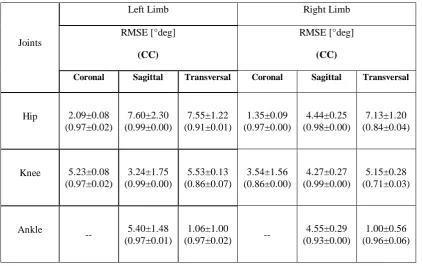
A stereo-photogrammetric Motion Capture technique (MoCap) Vicon® 460 system (Oxford Metrics, Oxford, UK), based on a set of 6 cameras working at 100Hz was used. One healthy subject (male, 28 years) walked at his self-selected walking speed performing one gait trial of 10 steps. A specific markers-based Helen Hayes protocol was considered by positioning markers (diameter 14mm) on anatomical landmarks for the predictive analysis approach. Moreover 3-markers sets (Fig. 2) were placed on each lower body segments to be used as input for the chosen functional algorithm [15], [16]. As aforementioned in the previous section these functional markers were placed according to a customize protocol, hence without anatomical landmarks constraints. Anthropometric measurements, like the leg length, knee and ankle width were collected in order to estimate the lower limbs kinematics according to the standard procedure [8], [19].
B.Functional joints CoRs estimation
The trajectories of each marker provided by the Vicon post-processing were used as input for the functional SCoRe
(Symmetrical Center of Rotation estimation)[15]. According to
this algorithm, local reference frames can be defined for each couple of adjacent body segments, connected by the considered joint and in relative motion.
Considering that the center of rotation with respect to body segment 1 (c1) and to body segment 2 (c2) is the same, is
possible to introduce an objective function fScore that can be
minimized with a linear least square method to evaluate c1
and c2. Given n as the number of the total acquired frames
during motion, is possible to define both orientation matrices (Ri, Si) and translation vectors (ti, di) for each considered i-frame and each segment respectively. Thus, equation (1) can be derived:
n
1 i
2 i 2 i i 1 i 2
1
Score c c R c t S c d
f
( , ) ( ) (1)C.Subject specific body modeling
Once the hip, the knee and the ankle CoRs have been estimated with the functional approach previously detailed, a multibody model implemented in open-source OpenSim [20] has been considered, in order to validate and assess the goodness of the solutions obtained with the functional algorithm. In OpenSim the musculoskeletal model is defined by bodies (bones), joints (articulations), actuators (muscles) and additive key points (markers), with the advantage that they can be customized. This allows to compare, as in this case study, the relative joints angles with respect to the standard ones, according to the orthopaedic angles [21]. Joints were modeled with appropriate DoFs, consistently defined according to the conditions imposed by the functional algorithm [15].
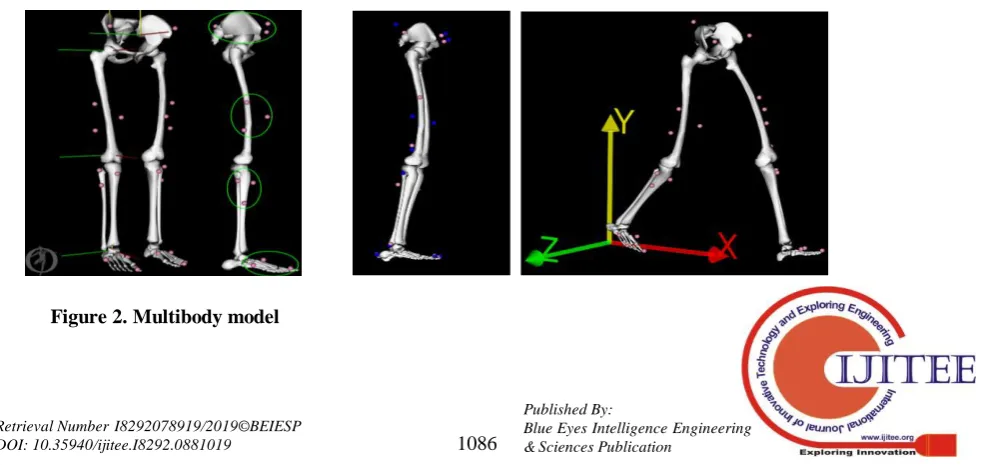
[image:2.595.110.489.55.112.2]Moreover, a subject-specific model was implemented by means of a scaling process that allows to achieve a subject-specific human model, due to the definition of a scale factor for each lower body single bone (Fig.3). To this end the functional joint CoRs previously estimated have been used, compared to those defined in the model, corresponding to the origins of local reference systems.
Fig. 1: Sketch of the methodology
[image:2.595.59.550.604.836.2]International Journal of Innovative Technology and Exploring Engineering (IJITEE) ISSN: 2278-3075, Volume-8 Issue-10, August 2019
D.Lower limb joints kinematics
Once MoCap acquisitions have been post-processed in order to obtain the three-dimensional experimental coordinates of each marker, an inverse kinematics problem is solved. Indeed the experimental data have been imposed into the model in order to simulate and replicate the acquired motion (Fig.3), exploiting the global optimization method [22]. Unknown joint angles have been extracted in order to minimize the squared error (SE) according to equation (2). Thus, for each i-marker is possible to compare the experimental position vector piE with the virtual one piV of the
model, with a proper set of weight wi, definedinversely
proportional to the amount of STA associated to each marker. The second part, related to the j-joint is instead considered if known initial joints constraints are available, always comparing the experimental configuration and the
virtual one. Thus, joints angles which best replicate the real configuration can be estimated:
j 21 j
V j S j j 2
m 1 i
V i S i
i p p w
w
SE min (2)
III. RESULTS
A comparison with respect to the gold standard kinematics curves (Vicon system) was carried out, to evaluate the feasibility of the functional approach based on direct measurements performed during a gait session. Results obtained using the functional method focus on 3D gait kinematics patterns for the lower limbs joints. Fig. 3 shows, for each joint, the resulting trends (red lines) compared to the Vicon ones (blue lines) and to the reference normative bands (mean-dotted black lines-, standard deviations-solid black lines) for the right leg. Errors (RMSE) and correlation coefficient (CC) between the two techniques are summarized in Table 1, for five averaged gait cycles and for each leg.
[image:3.595.61.540.320.657.2]Figure 2. Gait kinematics curves using a FM (blue) or PM (red) compared with normal bands (black) Figure 3. Scaling and walking trial after inverse
Table 1. RMSE And CC Data Analysis
Joints
Left Limb Right Limb
RMSE [°deg]
(CC)
RMSE [°deg]
(CC)
Coronal Sagittal Transversal Coronal Sagittal Transversal
Hip 2.09±0.08
(0.97±0.02)
7.60±2.30 (0.99±0.00)
7.55±1.22 (0.91±0.01)
1.35±0.09 (0.97±0.00)
4.44±0.25 (0.98±0.00)
7.13±1.20 (0.84±0.04)
Knee 5.23±0.08
(0.97±0.02)
3.24±1.75 (0.99±0.00)
5.53±0.13 (0.86±0.07)
3.54±1.56 (0.86±0.00)
4.27±0.27 (0.99±0.00)
5.15±0.28 (0.71±0.03)
Ankle -- 5.40±1.48
(0.97±0.01)
1.06±1.00
(0.97±0.02) --
4.55±0.29 (0.93±0.00)
1.00±0.56 (0.96±0.06)
IV. DISCUSSIONANDCONCLUSIONS
Joints parameters, like centers and axis of rotation (CoRs, AoRs), highly influence joints kinematics, thus higher is the accuracy of this estimation, as well of the following body segments frames, more reliable are the gait kinematics trends. This study mainly aims to assess the feasibility of a functional method [12] used to estimate lower limbs joints parameters, during a gait analysis session, in order to compare the following gait trends with the standard ones. The obtained results seem consistent for the declared purposes, as reported in Table 1. According to what is shown in Fig.4 there is an higher variability concerning the abduction/adduction and the internal/external rotation curves, particularly for the knee and the ankle joints. This may be due to both the accuracy of the human body model for the adopted joints models, but also to the markers residuals involving in the global optimization method, hence to the STA and the relative chosen weights. Moreover, considering that the major motion (RoM) during walking occurs on the sagittal plane, RoMs related to the other planes (transversal and frontal) are lower and maybe interfere with the limits concerning the functional methods, as reported in literature [15], [16].
Model modifications and additional motion tasks will be investigated in further studies; as well larger sample data will be considered in order to evaluate also the repeatability and the reliability of a functional approach, instead of the predictive one. Moreover the methodology can be applied also to people affecting pathologies, with walking devices or prosthesis.
ACKNOWLEDGMENT
The present research has been partially supported by
MIUR grant Dipartimenti di Eccellenza 2018-2022 (E11G18000350001). DISMA –Politecnico di Torino
REFERENCES
1. Cappozzo, A., Della Croce, U., Leardini, A., Chiari, L.: Human movement analysis using stereophotogrammetry. Part 1: theoretical background. Gait Posture. 21, 186–196 (2005).
2. Della Croce, U., Leardini, A., Chiari, L., Cappozzo, A.: Human movement analysis using stereophotogrammetry. Part 4: assessment of anatomical landmark misplacement and its effects on joint kinematics. Gait Posture. 21, 226–237 (2005).
3. Terzini, M., Zanetti, E.M., Audenino, A.L., Putame, G., Gastaldi, L., Pastorelli, S., Panero, E., Sard, A., Bignardi, C.: Multibody modelling of ligamentous and bony stabilizers in the human elbow. Muscles. Ligaments Tendons J. 7, 493–502 (2017).
4. Gastaldi, L., Pastorelli, S., Caramella, M., Dimanico, U.: Indoor motion analysis of a subject wearing prosthesis for adaptive snowboarding. In: WIT Transactions on Biomedicine and Health. pp. 361–372 (2011). 5. Agostini, V., Gastaldi, L., Rosso, V., Knaflitz, M., Tadano, S.: A
Wearable Magneto-Inertial System for Gait Analysis (H-Gait): Validation on Normal Weight and Overweight/Obese Young Healthy Adults. Sensors. 17, 2406 (2017).
6. Cappozzo, A., Catani, F., Leardini, A., Benedetti, M., Della Croce, U.: Position and orientation in space of bones during movement: experimental artefacts. Clin. Biomech. 11, 90–100 (1996).
7. Leardini, A., Chiari, L., Croce, U. Della, Cappozzo, A.: Human movement analysis using stereophotogrammetry. Part 3. Soft tissue artifact assessment and compensation. Gait Posture. 21, 212–225 (2005). 8. Kadaba, M.P., Ramakrishnan, H.K., Wootten, M.E.: Measurement of lower extremity kinematics during level walking. J. Orthop. Res. 8, 383–392 (1990).
9. Seidel, G.K., Marchinda, D.M., Dijkers, M., Soutas-Little, R.W.: Hip joint center location from palpable bony landmarks—A cadaver study. J. Biomech. 28, 995–998 (1995).
10. Rácz, K., Nagymáté, G., Kovács, T., Bodzay, T., Kiss, R.M.: Accuracy of anatomical landmark placement methods for gait analysis. Int. J. Mech.
International Journal of Innovative Technology and Exploring Engineering (IJITEE) ISSN: 2278-3075, Volume-8 Issue-10, August 2019
11. Piazza, S.J., Okita, N., Cavanagh, P.R.: Accuracy of the functional method of hip joint center location: effects of limited motion and varied implementation. J. Biomech. 34, 967–973 (2001).
12. Halvorsen, K., Lesser, M., Lundberg, A.: A new method for estimating the axis of rotation and the center of rotation. J. Biomech. 32, 1221–1227 (1999).
13. Gamage, S.S.H.U., Lasenby, J.: New least squares solutions for estimating the average centre of rotation and the axis of rotation. J. Biomech. 35, 87–93 (2002).
14. Woltring, H.J., Huiskes, R., de Lange, A., Veldpaus, F.E.: Finite centroid and helical axis estimation from noisy landmark measurements in the study of human joint kinematics. J. Biomech. 18, 379–89 (1985). 15. Ehrig, R.M., Taylor, W.R., Duda, G.N., Heller, M.O.: A survey of formal
methods for determining the centre of rotation of ball joints. J. Biomech. 39, 2798–2809 (2006).
16. Ehrig, R.M., Taylor, W.R., Duda, G.N., Heller, M.O.: A survey of formal methods for determining functional joint axes. J. Biomech. 40, 2150–2157 (2007).
17. Galetto, M., Gastaldi, L., Lisco, G., Mastrogiacomo, L., Pastorelli, S.: Accuracy evaluation of a new stereophotogrammetry-based functional method for joint kinematic analysis in biomechanics. Proc. Inst. Mech. Eng. Part H J. Eng. Med. 228, 1183–1192 (2014).
18. Gastaldi, L., Lisco, G., Pastorelli, S.: Evaluation of functional methods for human movement modelling. Acta Bioeng. Biomech. 17, 31–38 (2015).
19. Davis, R.B., Õunpuu, S., Tyburski, D., Gage, J.R.: A gait analysis data collection and reduction technique. Hum. Mov. Sci. 10, 575–587 (1991). 20. Delp, S.L., Anderson, F.C., Arnold, A.S., Loan, P., Habib, A., John, C.T., Guendelman, E., Thelen, D.G.: OpenSim: open-source software to create and analyze dynamic simulations of movement. IEEE Trans. Biomed. Eng. 54, 1940–50 (2007).
21. Lewis, J.L., Lew, W.D.: A note on the description of articulating joint motion. J. Biomech. 10, 675–678 (1977).
22. Lu, T.-W., O’Connor, J.J.: Bone position estimation from skin marker co-ordinates using global optimisation with joint constraints. J. Biomech. 32, 129–134 (1999).
AUTHORS PROFILE
Giulia Lisco is currently working in Italdesign -Passive Safety Development.
She received her Master degree in Biomedical Engineering and PhD degree in Mechanical Engineering from Politecnico di Torino. Her research interests focus on biomechanics, motion analysis and passive safety in automotive.
Stefano Pastorelli Stefano Pastorelli is associate professor of Applied Mechanics with the Department of Mechanical and Aerospace Engineering at Politecnico di Torino. He holds MS degree in Mechanical Engineering and PhD degree in Applied Mechanics. He teaches courses in the field of robotics and applied mechanics. His research interests include functional design of mechanical systems, kinematics and dynamics of multibody systems, biomechanics of human motion for human-machine interfaces and integration, wearable and collaborative robotics. Author of more than 140 papers published on international journals and conferences, and of three teaching books on Applied Mechanics.
Member of ASME, ISB and IFToMM Italy.
Laura Gastaldi is currently associate professor in Applied Mechanics at the Politecnico di Torino. She received her Master degree in Mechanical Engineering and PhD degree in Applied Mechanics from Politecnico di Torino. She teaches courses in the field of applied mechanics and biomechanics. Her research activity is mainly related to the study of mechanical, actuation and biomedical systems functional behavior.